Differential Gene Expression Profile in the Rat
Caudal Vestibular Nucleus is Associated with
Individual Differences in Motion Sickness
Susceptibility
Jun-Qin Wang☯, Rui-Rui Qi☯, Wei Zhou, Yi-Fan Tang, Lei-Lei Pan, Yi-Ling Cai*
Department of Nautical Injury Prevention, Faculty of Navy Medicine, Second Military Medical University, Shanghai, China
☯These authors contributed equally to this work.
Abstract
Objective
To identify differentially expressed genes associated with motion sickness (MS) susceptibil-ity in the rat caudal vestibular nucleus.
Methods
We identified MS susceptible (MSS) and insusceptible (inMSS) rats by quantifying rotation-induced MS symptoms: defecation and spontaneous locomotion activity. Microarray analy-sis was used to screen differentially expressed genes in the caudal vestibular nucleus (CVN) after rotation. Plasma stress hormones were identified by radioimmunoassay. Candi-date genes were selected by bioinformatics analysis and the microarray results were veri-fied by real-time quantitative-PCR (RT-qPCR) methods. By using Elvax implantation, receptor antagonists or recombinant adenovirus targeting the candidate genes were ap-plied to the CVN to evaluate their contribution to MS susceptibility variability. Validity of gene expression manipulation was verified by RT-qPCR and western blot analysis.
Results
A total of 304 transcripts were differentially expressed in the MSS group compared with the inMSS group. RT-qPCR analysis verified the expression pattern of candidate genes, includ-ing nicotinic cholinergic receptor (nAchR)α3 subunit, 5-hydroxytryptamine receptor 4 (5-HT4R), tachykinin neurokinin-1 (NK1R),γ-aminobutyric acid A receptor (GABAAR)α6
sub-unit, olfactory receptor 81 (Olr81) and homology 2 domain-containing transforming protein 1 (Shc1). In MSS animals, the nAchR antagonist mecamylamine significantly alleviated ro-tation-induced MS symptoms and the plasmaβ-endorphin response. The NK1R antagonist
CP99994 and Olr81 knock-down were effective for the defecation response, while the 5-HT4R antagonist RS39604 and Shc1 over-expression showed no therapeutic effect. In OPEN ACCESS
Citation:Wang J-Q, Qi R-R, Zhou W, Tang Y-F, Pan L-L, Cai Y-L (2015) Differential Gene Expression Profile in the Rat Caudal Vestibular Nucleus is Associated with Individual Differences in Motion Sickness Susceptibility. PLoS ONE 10(4): e0124203. doi:10.1371/journal.pone.0124203
Academic Editor:Jing Ai, Harbin Medical University, CHINA
Received:October 16, 2014
Accepted:March 10, 2015
Published:April 24, 2015
Copyright:© 2015 Wang et al. This is an open access article distributed under the terms of the
Creative Commons Attribution License, which permits unrestricted use, distribution, and reproduction in any medium, provided the original author and source are credited.
Data Availability Statement:All relevant data are within the paper and its Supporting Information files. All raw data files for microarray experiment are available from the ArrayExpress data base (accession number E-MTAB-3213).
Funding:This study is supported by grants from the National Natural Science Foundation of China (81272178) and the Natural Science Foundation of Shanghai (12ZR1437300).
inMSS animals, rotation-induced changes in spontaneous locomotion activity and the plas-maβ-endorphin level occurred in the presence of the GABAAR antagonist gabazine.
Conclusion
Our findings suggested that the variability of the CVN gene expression profile after motion stim-ulation might be a putative molecular basis for individual differences in MS susceptibility and provide information for the development of new therapeutic strategies for MSS individuals.
Introduction
Motion sickness (MS) is a syndrome of autonomic reactions, such as nausea, vomiting, pallor, sweating, increased salivation and stomach awareness, which are commonly provoked by ex-ternally imposed motion [1,2]. Currently, the etiology and precise neurobiological mechanism of MS has not been fully clarified and there are several theories interpreting different aspects of MS. The traditional‘sensory conflict hypothesis’and‘neuronal mismatch theory’suggested that motion sickness may be caused by conflicting auditory, visual and vestibular sensory in-puts leading to a mismatch between the actual and the anticipated internal model of the spatial environment [3,4]. According to the‘postural instability theory’, the occurrence of motion sickness may be associated with preceding unstable postural control at locomotive surround-ings on mobile devices [5–7]. However, these theories do not explain apparent individual dif-ferences in MS susceptibility, let alone provide detailed information on the underlying molecular bases and mechanisms [8].
As we know, an intact vestibular system is required for MS and serves as an integral compo-nent of motion signals in the central nervous system [1,2,9,10]. Vestibular nuclei receive not only vestibular inputs but also somatosensory, proprioceptive, visceral, and visual inputs and motor-related feedback signals [11]. Simultaneously applied vestibular and visual stimulation can reduce the behavioral gain of the vestibular-ocular reflex in mouse and monkey indicating that sensory conflict might be produced in the vestibular nucleus during MS [12,13]. Previous studies have demonstrated that the caudal vestibular nucleus (CVN), including the caudal medial vestibular nucleus (MVe) and the spinal (inferior) vestibular nucleus (SpVe), contribute to both cardiovascular control during head movements and autonomic manifestations of motion sick-ness through its strong connection with brain stem autonomic areas, such as the solitary tract nucleus and parabrachial nucleus, in a variety of species [14–18]. A variety of provocative envi-ronments, such as altered gravito-inertial force, off-axis rotation, centripetal acceleration and space flight, can induce intensive neuronal activation in the CVN as indicated by elevated Fos protein expression [19–22]. Using principal components analysis, a recent study confirmed that neurons in the CVN constitute principal parts of neural networks that contribute to autonomic manifestations, such as retching, excessive salivation, defecation and urination during galvanic vestibular stimulation in felines [23]. Through poly-synaptic connections with the hypothalamic paraventricular nucleus, CVN neurons may also mediate the stress hormone response after ves-tibular stimulation [24]. In addition, convergence of gastrointestinal afferent signals on CVN neurons can facilitate motion sickness susceptibility in cats exposed to rotation in vertical planes [25]. Based on the idea that neurons in the CVN participate in triggering motion sickness, it is conceivable that they might also contribute to the variability in MS susceptibility.
the consequent behavior responses. Altered gene expression patterns were also observed in VN neurons following motion stimulation in the rodents [26–29], yet these observations were not di-rectly connected with MS susceptibility. In the current study, to understand the underlying mo-lecular basis for individual variability in MS susceptibility, we sought to identify differentially expressed genes associated with motion sickness susceptibility in the CVN of male adult rats. Firstly, we established a MS susceptibility animal model by analyzing initial sensitivity to Ferris wheel-like rotation via quantifying two valid MS-related symptoms: defecation during rotation and spontaneous locomotion [30]. The rats’plasma stress hormone levels were also examined to identify MS susceptibility-related hormonal responses. Then, we identified differentially express-ed genes in the CVN between MS susceptible (MSS) and insusceptible (inMSS) animals using microarray analysis. Candidate genes were identified via bioinformatics analysis methods and microarray results were verified by real-time quantitative-PCR (RT-qPCR). Lastly, we examined the relative contribution of these genes to motion sickness susceptibility through functional an-tagonism or manipulation of gene expression level by using an in vivo Elvax implantation meth-od which is more efficient and convenient in sustained drug delivery over specific brain regions just underneath tissue surface than classical implantation of cannula [31–33].
Materials and Methods
1. Animals and general procedures
Adult male Sprague–Dawley rats weighing 250–300 g were purchased from Shanghai Labora-tory Animal Center. The animals were singly housed under a 12 h light: 12 h dark cycle (tem-perature: 22 ± 2°C and lighting: 8:00–20:00) with free access to food and water. A total of 540 animals were used in this study and all animals were acclimated to the lab environment for 2 weeks before initiation of the experiment and familiarized with the rotation device for 2 hours per day for 3 days prior to the beginning of rotation or static control treatment. The adaptation and rotation procedures were performed during 6:00–10:00 p.m. with the temperature main-tained at 22°C.
Ethics statement. All surgical procedures were performed under sodium pentobarbital (40 mg/kg, i.p.) anesthesia. All animal protocols and procedures complied with the Guide for the Care and Use of Laboratory Animals (US National Research Council, 1996) and were ap-proved by the Ethics Committee for Animal Experimentation of the Second Military Medical University (Shanghai, PR China). All animal experiments were reported in compliance with ARRIVE guidelines [34,35]. Efforts were made to minimize the number of animals used and the suffering for every animal in each experiment.
Rotation device and procedures. The rotation device and detailed rotation methods were described previously [36]. Briefly, the animals were placed in plexiglass containers with the long axis of the body perpendicular to the horizontal rotation rod. The device started to rotate in a clockwise direction at 16°/s2to reach an angular velocity of 120°/s and then began to decel-erate at 48°/s2to reach 0°/s. After a 1 s pause, the container continued to rotate in a counter-clockwise direction in the same manner as above. The counter-clockwise-pause-countercounter-clockwise cycle lasted approximately 21 s. All of the rats in the rotation (Rot) groups received 2 hours of rota-tion stimularota-tion in complete darkness, while the animals in the static control (Sta) groups were kept in the restrainer near the rotation device when Rot animals were being rotated.
stored at−80°C. In Elvax implatation exeriment, half sample from each animal was used for RT-qPCR analysis and another half was used for western blot test.
2. Experimental design and grouping
Establishment of MS animal model. Sixty rats were used and randomly divided into the following groups: two rotation (Rot) groups received saline (1 ml, 0.9% w/v) or scopolamine (0.1 mg/100 g body weight, i.p.) 30 min before rotation stimulation; two more Rot groups re-ceived intratympanic injection of saline (sham-lesioned) or 50–100μl sodium arsanilate
(15 mg in 50μl saline, chemical labyrinthectomy) 2 weeks before rotation stimulation; and
one Sta group (n = 12 in each group). Immediately after rotation or static control treatment, defecation response and spontaneous locomotion activity were evaluated for their validity to be used as indices for assessment of MS symptoms in the rats.
MS susceptibility evaluation for microarray experiment. Sixty eight rats were randomly divided into Rot or Sta groups (n = 34 in each group). MSS and inMSS animals were selected from the Rot group after 1st rotation stimulation by quantifying defecation and spontaneous locomotion responses (n = 5 in MSS-Rot and n = 6 in inMSS-Rot). Two weeks later, these ani-mals were re-exposed to rotation and plasma stress hormone levels were tested. Bilateral CVN collections satisfying tissue sampling criteria were further analyzed for differentially expressed genes in microarray experiment.
Verification experiment for microarray results. Additional 82 animals was used for screening MSS (n = 12) and inMSS (n = 12) subjects which were then randomly divided into a Rot or Sta group: MSS-Rot, MSS-Sta, inMSS-Rot, or inMSS-Sta (n = 6 in each group). Tran-scription levels of candidate genes identified in microarray experiment were examined by RT-qPCR test.
Pharmacological intervention experiment. Three hundred and thirty animals were used for screening MSS (n = 55) and inMSS subjects (n = 15) which were then divided into drug or adenovirus treatment groups or a sham operation control group (totally 11 MSS and 3 inMSS groups, n = 5 in each group). One week later, Elvax sheets loaded with either receptor antago-nists or recombinant adenovirus were implanted over the CVN in MSS or inMSS animals to in-vestigate their effects on motion sickness susceptibility. Animals in the sham operation control group were implanted with control Elvax loaded with solvent. After one week of surgery recov-ery, MS susceptibility in these animals was re-evaluated and their plasma stress hormone (β -endorphin) concentrations were tested. In adenovirus treated animals, the expression level of candidate genes (Olr81 and Shc1) was also examined by RT-qPCR and western blot test.
3. MS susceptibility evaluation
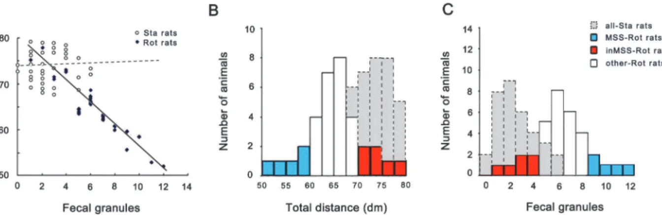
MS susceptibility evaluation criteria. MS susceptibility evaluation for microarray experi-ment revealed that there was a strong linear relationship between defecation level and total dis-tance traveled in the Rot group receiving rotation treatment (r = -0.935, F(1,33) = 171.081, p = 0.0001), but not in the Sta group (r = 0.059, F(1,33) = 0.111, p = 0.741) (Fig 1A). Shapiro-Wilk W test analysis showed a normal distribution pattern for the values of defecation level and total distance traveled in the Sta group (W = 0.928, P = 0.028; W = 0.956, P = 0.197) and in the Rot group (W = 0.951, P = 0.106; W = 0.966, P = 0.351) (Fig 1B and 1C). MS susceptibility evaluation criteria was then set as follows: animals in the Rot group with the value of fecal gran-ules distributed within the left 20% of the confidence interval and the value of total distance traveled within the right 20% interval simultaneously were chosen as MSS subjects (n = 5,Fig 1B and 1Cblue volume); those with the value for defecation distributed within the left 20% of the confidence interval and the value of total distance traveled within the right 20% of the con-fidence interval were chosen as inMSS subjects (n = 6,Fig 1B and 1Cred volume).
4. Molecular biological experiments
Microarray analysis. For Affymetrix microarray profiling, total RNA was extracted using an Rneasy Mini Kit following the manufactory’s instructions (Qiagen, German). The GeneChip WT cDNA Synthesis Kit, WT cDNA Amplification Kit, and the WT Terminal Labeling Kit (Affymetrix, Inc., Santa Clara, CA) were used for the cDNA preparation which was hybridized to Rat Exon1.0 ST GeneChip arrays (Affymetrix, America) according to the Users’Manuals. Affymetrix Expression Console Software (version 1.1.2) was used for microarray analysis. RVM t-test was applied to filter the differentially expressed genes between MSS-Rot and inMSS-Rot group. Fold-change was calculated as the ratio between the average values of gene expression in MSS-Rot relative to inMSS-Rot animals. Two-dimensional hierarchical cluster-ing of the expression data was performed uscluster-ing a Pearson correlation distance matrix and aver-age linkaver-age clustering. Gene ontology (GO) analysis was applied to analyze the main functions of differentially–expressed genes. Pathway analysis was used to identify the significant path-ways according to KEGG, Biocarta and Reatome databases via Fisher’s exact test and theχ2 test. The threshold of significance was defined by P-value at 0.05 and the screening condition was set as false discovery rate (FDR) under 5%. All microarray datasets were submitted to the ArrayExpress repository (http://www.ebi.ac.uk/arrayexpress/experiments/E-MTAB-3213). Fig 1. Data analysis for the defecation response and total distance traveled for MS susceptibility evaluation in microarray experiment.(A) Linear relationship between the number of fecal granules and total distance traveled in the Rot and Sta group (n = 34 in each group). The distribution of the number of fecal granules (B) and total distance traveled (C) in staic control (n = 34), MSS-Rot (n = 5), inMSS-Rot (n = 6), and other rotated animals (n = 23).
RT-qPCR test. Total RNA extraction procedure was the same as in the microarray experi-ment. The RT-qPCR reactions were conducted in a Rotor-Gene (RG-3000A, Corbett Research) PCR machine. The amount of cDNA per sample was determined using a SYBR Premix Ex Taq kit (Takara). Progression of the PCR reaction was assessed by changes of the SYBR Green dye fluorescence attached to double-stranded DNA. All values were normalized to the housekeep-ing gene glyceraldehyde phosphate dehydrogenase (GAPDH). The primers used for real-time PCR are shown inS1 Table.
Western blot test. Wetern blot analysis was performed as previously described [30]. The primary antibodies used in this study were anti-Olr81 (1:1000; Santa Cruz Biotechnology, Santa Cruz, CA, USA) and anti-Shc1 (1:1000; Cell signaling, Beverly, MA, USA). The second-ary antibodies used were peroxidase-labeled anti-goat IgG and anti-rabbit IgG (all from Jack-son, West Groove, PA, USA) at 1:5000 dilution. Signal intensities of Olr81 and Shc1 bloting bands were normalized against the internal control (GAPDH).
Plasma hormone measurements. Blood was collected immediately after decapitation and the plasma was separated and stored at−80°C for further analyses. Plasma epinephrine, norepi-nephrine, arginine-vasopressin (AVP), adrenocorticotropic hormone (ACTH) andβ -endor-phin levels were measured by radioimmunoassay following the instructions in the kits generously provided by Prof. Zhao XL at the Second Military Medical University or purchased from North Institute of Biological Technology Co (Beijing, China).
5. In vivo Elvax implantation technology
Recombinant adenovirus preparation. Recombinant adenovirus for the over-expression of Shc1 (pAd-Shc1) was generated as follows. The rat Shc1 gene was synthesized de novo by rapid polymerase chain assembly and cloned into the SpeI-SgsI site of pENTR-IRES-EGFP (Invitrogen). The adenoviral plasmid pAd-CMV-Shc1-IRES-EGFP was generated by LR clo-nase-mediated recombination using pAd-CMV-V5-DEST (Invitrogen) as the acceptor and the pENTR-Shc1-IRES-GFP (Invitrogen) as the donor. Recombinant adenoviruses were propagat-ed in HEK293 cells and purifipropagat-ed using the Adenovirus Purification Miniprep Kit (Biomiga V1160) following the manufacturer’s instructions.
The Olr81 targeting recombinant adenoviral pAd-miOlr81 was generated by cloning synthet-ic oligonucleotides encoding complimentary miRNAs for rat Olr81 mRNA into the pENTR-miR vector (Invitrogen), followed by homologous recombination with pAd-CMV-V5-DEST (Invitrogen). The sequence for the oligonucleotides was: 5’- TGCTGAGGAATGTGCTATTAC ATGAGGTTTTGGCCACTGACTGACCTCATGTAAGCACATTCCT -3’.
Elvax preparation and implantation. ELVAX has been successfully used to deliver water or dimethylsulfoxide (DMSO) soluble drug onto rodent’s brain surface [31,32]. In this experi-ment, Elvax sheets were prepared following the procedures described by Nodal FR [32]. Briefly, plastic beads of the ethylene–vinyl acetate copolymer Elvax 40-W (Elvax 40P; Du Pont) were washed in several changes of 95% and 100% alcohol for 24 h. After the beads were dried, they were dissolved in methylene chloride (0.15 g/1.5 ml solvent). Drugs dissolved in either 50μl of
distilled water (mecamylamine, CP99994 or gabazine) or 50μl of DMSO (0.03% in final
concen-tration, RS39604) were added to the Elvax mixture. The final embedded concentrations of the agents were as follows: mecamylamine, 1.25 mM or 2.50 mM; CP99994 and RS39604, 5 mM or 10 mM; and gabazine, 0.25 mM or 0.50 mM. Mecamylamine and gabazine were purchased from Sigma-Aldrich (St. Louis, MO, USA). CP99994 and RS39604 were purchased from Tocris (Tocris, UK). For in vivo adenovirus infection, 100 or 200μl of either pAd-miOlr81 or pAd-Shc1
sham control experiment, control Elvax sheets containing only the vehicle (water, DMSO or cul-ture medium) were also prepared. To visualize the implantation, 25μl of 5% Fast Green was also
added to the Elvax mixture. The mixture was then vortexed, frozen quickly in a dry ice/acetone bath, and transferred to a pre-chilled glass petri dish and freeze-dried overnight (−60°C, 10–4 atm). All dry Elvax mixtures (approximately 200μm thick) were cut into 4 mm×1.5
mm×200μm rectangular sheets. To avoid contaminating the cerebellum, two-layer Elvax sheets
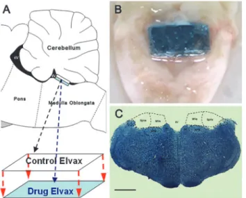
were prepared by covering an agent-loaded Elvax sheet (lower layer) with a solvent control Elvax sheet (upper layer) and stored on filter paper at 4°C prior to implantation (Fig 2A).
For the Elvax implantation surgery, animals were anesthetized and placed on a stereotaxic frame (Narishige, Japan). The skin and muscles of the neck were incised and dissected along the midline dorsally to expose the atlanto-occipital membrane. Then, a partial occipital craniotomy was performed and the cerebellum was pushed forward to expose the underlying fourth ventri-cle. A two-layer Elvax sheet was then placed over the CVN region, covering most of its surface (Fig 2B). After the cerebellum was repositioned completely, the opening in the skull was closed with dental cement and the overlying neck muscles and skin were sutured. Animals received postsurgery antibiotics penicillin (400000U/kg, i.p.) and analgesics ibuprofen (30mg/kg, in the drinking water) for 3 days.
6. Statistical analysis
All statistical analyses were conducted with the SPSS v13.0 statistical program and data are ex-pressed as the mean ± S.D. One-way ANOVA analysis was performed to examine the differ-ence among groups in the MS animal model establishment experiment and the differdiffer-ences following pharmacological intervention in theβ-endorphin concentration and gene expression (Olr81 and Shc1) test. Fisher’s LSD post hoc test was used to analyze the difference between each group when a significant main effect was obtained. Pearson correlation analysis was con-ducted to determine if there was a linear relationship between the defecation level and the total distance traveled for MS susceptibility evaluation in microarray experiment. Normal
Fig 2. Elvax preparation and CVN tissue sampling.(A) Illustration for Elvax sheet preparation and (B) photograph of a rat brain stem showing the location of Elvax sheet implantation. (C) Representative Nissl-staining image showing the CVN sampling region. 4V: fourth ventricle; MVe: medial vestibular nucleus; SpVe: spinal vestibular nucleus; NTS: nucleus of solitary tract; Pr: prepositus nucleus; X: nucleus X; Bar: 200μm.
distribution for these indices was constituted and evaluated by Shapiro-Wilk analysis. A t-test analysis was performed to examine the difference in MS symptoms and plasma hormone levels between MSS and inMSS animals in MS susceptibility evaluation for microarray experiment and the difference inβ-endorphin levels between the sham MSS and the sham inMSS group in Elvax experiment. Two-factorial analysis of variance (ANOVA) for repeated-measures was performed using the General Linear Protocol to examine the effect of susceptibility and rota-tion on the mRNA levels for candidate genes in the RT-qPCR verificarota-tion experiment and the effect of time, drug concentration (virus titer) or susceptibility on MS symptoms in the phar-macological intervention experiment. Boferoni post hoc test was used to analyze the difference between each group when a significant main effect or interaction effect was obtained. The level of significance was set at p<0.05.
Results
1. MS susceptibility evaluation
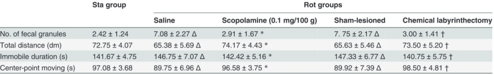
Establishment of MS animal model. Table 1show that rotation stimulation leads to an increase in defecation and a decrease in spontaneous locomotion activity (hypoactivity) in Rot animals receiving saline (i.p.) and sham lesion treatment compared with Sta controls. Defeca-tion response was significantly decreased in Rot animals receiving scopolamine administered prior to rotation stimulation compared to those receiving saline treatment. Bilateral labyr-inthectomy also significantly reduced defecation in Rot animals compared to the sham-le-sioned group (P<0.05). Both scopolamine administration and bilateral labyrinthectomy
significantly alleviated rotation-induced decreases in total distance traveled and center-point moving duration and the duration of immobility decreased in Rot animals compared to the sa-line and sham-lesioned group, respectively (P<0.05).
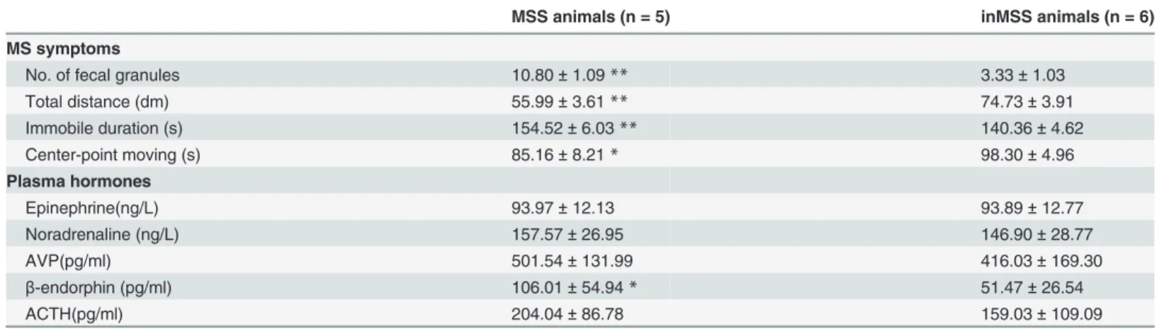
Behavioral and hormonal responses in MSS and inMSS animals. T-test analysis showed that defecation levels and immobility duration were significantly increased and the total dis-tance traveled and center-point moving duration were decreased in the MSS group compared to the inMSS group in MS susceptibility evaluation experiment prior to microarray analysis. There was also a significant increase in plasmaβ-endorphin levels and an insignificant trend of increased AVP and ACTH levels in MSS animals (Table 2).
2. MS susceptibility-associated genes
Candidate genes identified by microarray analysis. CVN tissue samples, which satisfied the sampling criteria (Fig 2C), were included in the following microarray experiment (n = 4 in
Table 1. MS symptoms observed in animals receiving static control or rotation treatment.
Sta group Rot groups
Saline Scopolamine (0.1 mg/100 g) Sham-lesioned Chemical labyrinthectomy
No. of fecal granules 2.42±1.24 7.08±2.27Δ 2.91±1.67* 7. 75±2.17Δ 3.00±1.41†
Total distance (dm) 72.75±4.07 65.38±5.69Δ 74.17±4.43* 65.63±5.46Δ 73.50±5.20†
Immobile duration (s) 141.67±4.75 146.75±7.07Δ 142.42±5.16* 147.33±6.77Δ 140.75±5.75†
Center-point moving (s) 97.08±3.68 89.75±6.96Δ 96.58±3.75* 89.92±7.39Δ 98.50±4.81†
ΔP<0.05 compared with Sta group
*P<0.05 compared with Rot-saline group
†P<0.05 compared with Rot-sham operation group.
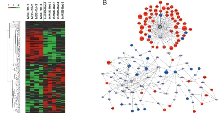
MSS-Rot and n = 5 in inMSS-Rot). Hierarchal clustering analysis revealed that gene differences can be successfully distinguished between the MSS-Rot and inMSS-Rot groups (Fig 3A). Mi-croarray analyses identified a total of 304 transcripts differentially expressed in the MSS-Rot group compared to the inMSS-Rot group: 207 transcripts were relatively up-regulated and 97 transcripts were down-regulated. The top 45 up-regulated genes (MSS-Rot/inMSS-Rot SS-Rot/ inMSS-Roted g7 down-regulated genes (MSS-Rot/inMSS-Rot SS-Rot/ are listed inS2andS3
Tables. Additionally, signal-net analysis integrated the up-regulated and down-regulated genes and delineated the signaling regulatory networks of their expression products (Fig 3B).
Gene ontology (GO) hierarchy analysis revealed significant functions, including the positive regulation of cholinergic synaptic transmissions (P = 0.0097, enrichment = 205.06), response to electrical stimulus (p = 0.00028, enrichment = 102.53), the G protein coupled receptor (GPCR) protein signaling pathway (P = 0.034, enrichment = 57.95), the response to nutrient levels (P = 0.00070, enrichment = 68.35), the response to calcium ions (P = 0.0049, enrich-ment = 11.19), the response to organic cyclic substances (P = 0.000054, enrichenrich-ment = 10.34), the response to peptide hormone stimuli (P = 0.0021, enrichment = 9.01) and GABAergic syn-aptic transmission (P = 0.029, enrichment = -67.61). Among these GO categories, we found four differentially expressed neurotransmitter receptor genes: Chrna3-nicotinic cholinergic re-ceptor (nAchR)α3 subunit; Htr4 -5-hydroxytryptamine (serotonin) receptor 4 (5-HT4R);
Tacr1-tachykinin neurokinin-1 receptor (NK1R) and Gabra6 (γ-aminobutyric acid A receptor
[GABAAR]α6 subunit). Gabra6 was also the most statistically significant down-regulated gene
in MSS animals.
Among the differentially expressed genes, we found 51 up-regulated genes and 4 down-reg-ulated genes belonging to the olfactory receptor (OR) superfamily. We then analyzed the evolu-tionary properties of the gene sequences and found 5“fish-like”(Class I) OR genes (Olr81, Olr82, Olr96, Olr128, Olr175), which are more conserved among species and more evolution-arily ancient than tetrapod (Class II) ORs (the remaining 50 ORs) [37,38]. Furthermore, by checking the Gene database on PubMed, we found that Olr81 is homologous to OR52J3 in hu-mans [www.ncbi.nlm.nih.gov/homologene/66215]. Additionally, pathway analysis revealed 3 up-regulated and 7 down-regulated pathways identified from the KEGG database (p<0.05).
The most significantly up-regulated pathway was the olfactory transduction pathway and the most down-regulated pathway was the insulin signaling pathway (S4 Table.). The most Table 2. MS symptoms and microarrayed plasma hormone levels of MSS-Rot and inMSS-Rot groups.
MSS animals (n = 5) inMSS animals (n = 6)
MS symptoms
No. of fecal granules 10.80±1.09** 3.33±1.03
Total distance (dm) 55.99±3.61** 74.73±3.91
Immobile duration (s) 154.52±6.03** 140.36±4.62
Center-point moving (s) 85.16±8.21* 98.30±4.96
Plasma hormones
Epinephrine(ng/L) 93.97±12.13 93.89±12.77
Noradrenaline (ng/L) 157.57±26.95 146.90±28.77
AVP(pg/ml) 501.54±131.99 416.03±169.30
β-endorphin (pg/ml) 106.01±54.94* 51.47±26.54
ACTH(pg/ml) 204.04±86.78 159.03±109.09
**P<0.01
*P<0.05 compared with inMSS-Rot group
statistically significant genes in these two pathways were Olr81 (P = 0.0035789) and homology 2 domain containing transforming protein 1 (Shc1) (P = 0.0366554). We finally selected Olr81, Shc1, Chrna3, Htr4, Tacr1 and Gabra6 as the candidate genes for the following experiment.
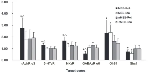
Verification of candidate genes by RT-qPCR. All tissue samples satisfied the sampling cri-teria and were further analyzed in this experiment. The MSS-Rot/ inMSS-Rot ratio of gene expres-sion for the nAchRα3 subunit, 5-HT4R, NK1R, the GABAARα6 subunit, Olr81 and Shc1 were
similar to the ratios obtained by microarray analysis (S5 Table.). A 2 (susceptibility difference) × 2 (rotation condition) factorial ANOVA analysis revealed significant effects of susceptibility [F (1, 20) = 4.616, P = 0.047] and rotation [F (1, 20) = 4.744, P = 0.045] and a susceptibility×rotation in-teraction [F (1, 20) = 6.915, P = 0.018] on nAchRα3 subunit expression; significant susceptibility [F (1, 20) = 8.237, P = 0.011] and rotation [F (1, 20) = 10.184, P = 0.006] effects and a susceptibili-ty×rotation interaction [F (1, 20) = 4.861, P = 0.042] on 5-HT4R expression; significant
suscepti-bility [F (1, 20) = 6.318, P = 0.023] and rotation [F (1, 20) = 8.995, P = 0.008] effects and a susceptibility×rotation interaction [F (1, 20) = 5.985, P = 0.026] on NK1R expression; significant
susceptibility [F (1, 20) = 6.096, P = 0.025] and rotation [F (1, 20) = 22.003, P = 0.0001] effects and a susceptibility×rotation interaction [F (1, 20) = 25.303, P = 0.0001] on GABAARα6
expres-sion. Boferoni post hoc analysis revealed that the mRNA levels of the nAchRα3 subunit, 5-HT4R,
and NK1R were increased, while the GABAARα6 subunit was decreased in MSS-Rot group
com-pared to the MSS-Sta and inMSS-Rot groups (P<0.05). Meanwhile, there was a significant
suscep-tibility effect on Olr81 [F (1, 20) = 5.139, P = 0.035] and Shc1 [F (1, 20) = 9.153, P = 0.007] expression. The Olr81 mRNA level was increased and the Shc1 mRNA level was decreased in the Fig 3. Hierarchical clustering of the signal value and signaling regulatory network of the differentially expressed genes.(A)Heat map of differential changes in gene expression between MSS-Rot and inMSS-Rot animals. The dendrogram was produced by hierarchical clustering of the signal value of differentially expressed genes in MSS-Rot rats (n = 4, left) and inMSS-Rot samples (n = 5, right). Colorimetric scaling of standardized gene expression values ranging from low (green) to high (red) is shown in the legend (upper left). (B) Signal-net of the differentially expressed genes. Red nodes and blue nodes represent up-regulated and down-regulated genes, respectively. Grey nodes represent intermediate genes functionally connecting the differentially expressed genes. Solid lines with arrow head indicate activation (a), dotted lines indicate inhibition (inh) and solid lines indicate binding (b) or expression regulation (ex). Node size represents the degrees of diiference in gene expression levels of MSS relative to inMSS.
MSS-Rot and MSS-Sta groups when compared with the corresponding inMSS animals (P<0.05,
Fig 4).
3. Effects of Elvax implantation on MS susceptibility
Validity of adenovirus delivery. One-way ANOVA analysis found a significant effect of pAd-miOlr81 delivery on Olr81 mRNA level (P = 0.047), and a significant effect of pAd-Shc1 treatment on Shc1 mRNA level (P = 0.002) in the CVN of MSS animals. LSD post hoc analysis showed that pAd-miOlr81, at the high final titer in Elvax (7.00 ×10⁷GFU/ml), significantly de-creased Olr81 mRNA level (P = 0.015), while pAd-Shc1, at both final titers (3.35 ×10⁸GFU/ml and 7.00 ×10⁸GFU/ml in Elvax), significantly enhanced Shc1 transcription (P = 0.011 and 0.001) in MSS animals compared to sham operation controls (S1 Fig). There was also a signifi-cant effect of pAd-miOlr81 treatment which decreased Olr81 protein level at 7.00 ×10⁷GFU/ml (P = 0.001), and a significant effect of pAd-Shc1 treatment which significantly increased Shc1 protein level at both final titers (P = 0.001,S1 Fig).
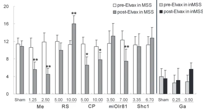
Defecation response. In sham operation control animals, two-way ANOVA analysis showed no effect of time [F (1, 19) = 1.005, P = 0.331], but a significant effect of susceptibility [F (1, 19) = 88.804, P = 0.001] and time×susceptibility interaction [F (1, 19) = 5.789, P = 0.029] on defecation levels. Boferoni post hoc analysis revealed that defecation levels remained un-changed after control-Elvax implantation in the MSS sham group and the inMSS sham group compared to their pre-Elvax implantation levels, but they were much higher in the MSS sham group than in the inMSS sham animals (P = 0.001,Fig 5).
In MSS animals, two-way ANOVA analysis found no effect of time [F (1, 29) = 2.251, P = 0.147], but a significant effect of concentration [F (2, 29) = 7.142, P = 0.004] and a time×con-centration interaction [F (2, 29) = 4.861, P = 0.042] on defecation levels after mecamylamine-Elvax implantation. Boferoni post hoc analysis found that mecamylamine treatment, at both concentrations (1.25 mM and 2.50 mM in Elvax), significantly deceased the rotation-induced defecation response compared with the corresponding pre-Elvax level, in a dose-dependent manner (P = 0.0019 and 0.0011). There was a significant effect of concentration [F (2, 29) = Fig 4. RT-qPCR verification of candidate genes mRNA levels, including the nAchRα3 subunit, 5-HT4R, NK1R, the GABAARα6 subunit, Olr81 and Shc1 in the CVN of MSS and inMSS rats receiving 2 hours of rotation or static treatment.Values in each group (n = 6) are expressed as the percentage of their corresponding GAPDH values (100%) and shown as the means (±S.E.).*P<0.05, compared with inMSS-Rot group;ΔP<0.05, compared with MSS-Sta group;†P<0.05 compared with inMSS-Sta group.
3.871, P = 0.0425] and a time×concentration interaction [F (2, 29) = 4.105,p =0.0363] on defe-cation levels after RS39604-Elvax implantation. CP99994-Elvax implantation resulted in a signif-icant effect of concentration [F (2, 29) = 8.735,p =0.00187] and a time×concentration
interaction [F (2, 29) = 5.648, P = 0.0113]. RS39604 treatment, at a concentration of 10 mM, sig-nificantly increased the rotation-induced defecation response compared with pre-treatment lev-els (P = 0.0037). CP99994 treatment, at both concentrations (5 mM and 10 mM in Elvax), significantly deceased the rotation-induced defecation response (P = 0.0016 and 0.049,Fig 5).
There is also a significant effect of time [F (1, 29) = 0.942, P = 0.342], titer [F (2, 29) = 8.426, P = 0.0019] and a time×titer [F (2, 29) = 9.976, P = 0.00082] interaction on the defecation re-sponse after pAd-miOlr81 infection, which at the high final titer in Elvax (7.00 ×10⁷GFU/ml) alleviated the defecation response to rotation in MSS animals when compared to their pre-Elvax treatment data (P = 0.0062); No significant effects were observed after pAd-Shc1 treatment.
In inMSS animals, no significant effect of time, concentration or time×concentration inter-action on the defecation response was observed after gabazine-Elvax implantation, while an in-significant trend towards increased defecation at a higher concentration (5.0 mM in Elvax) was found (P = 0.067).
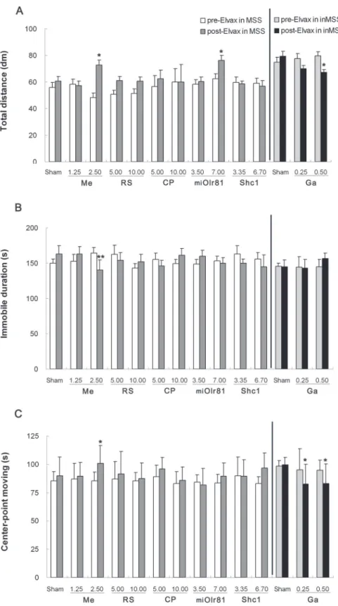
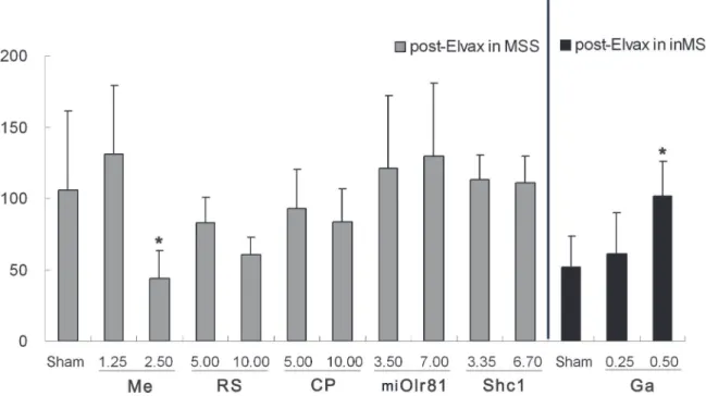
Spontaneous locomotion activity. Two-way ANOVA analysis found a significant effect of susceptibility on the total distance traveled [F (1, 19) = 20.756, P = 0.001], duration of immo-bile [F (1, 19) = 29.309, P = 0.001] and center-point moving duration [F (1, 19) = 27.995, P = 0.001] in sham operation control animals. Boferoni post hoc analysis revealed that immo-bility duration was significantly increased and the total distance traveled and center-point moving duration were decreased in the MSS sham group compared to the inMSS sham group Fig 5. Effects of Elvax implantation over the CVN on the defecation response induced by rotation stimulation.In MSS animals, Elvax was loaded with solvent (Sham); 1.25 or 2.50 mM mecamylamine (Me); 5.00 or 10.00 mM RS39604 (RS); 5.00 or 10.00 mM CP99994 (CP); 3.50×10⁷or 7.00 ×10⁷GFU/ml pAd-shOlr81 (shOlr81); or 3.35 ×10⁸or 6.70 ×10⁸GFU/ml pAd-Shc1 (Shc1) at final concentration/titer (n = 5 in each group). In inMSS animals, Elvax was loaded with solvent (Sham) or gabazine (Ga) to a final concentration of 0.25 or 0.5 mM (n = 5 in each group). All data presented are expressed as the means and the vertical bars represent SEM. Statistical significance:**P<0.01,*P<0.05 compared with the rotation-induced defecation response before
Elvax implantation.
(P<0.05), while there was no difference in these indices between pre-Elvax and post-Elvax
level in either the MSS sham or the inMSS sham animals.
There was a significant time×concentration interaction on the total distance traveled [F (2, 29) = 5.067, P = 0.035] and immobility duration [F (2, 29) = 10.715, P = 0.0004] and a significant effect of concentration [F (1, 29) = 4.085, P = 0.029] on center-point moving duration after mec-amylamine-Elvax implantation. Mecamylamine treatment, at a concentration of 2.50 mM in Elvax, increased the total distance traveled (P = 0.0233,Fig 6A) and the center-point moving du-ration (P = 0.0467,Fig 6C) and decreased immobile duration (P = 0.0054,Fig 6B) when com-pared with pre-Elvax treatment level. No significant effects of time, concentration or
time×concentration interaction on the total distance traveled, the duration of immobility and center-point moving duration were observed after RS39604, CP99994 or pAd-Shc1 treatment. There was a significant time×concentration interaction [F (2, 29) = 3.811, P = 0.0379], but no ef-fect of time [F (1, 29) = 0.506, P = 0.484] or titer [F (2, 29) = 1.956, P = 0.165] on the total dis-tance traveled after pAd-miOlr81 Elvax implantation, and no significant effect on the duration of immobility or center-point moving duration was observed. Boferoni post hoc analysis found that pAd-miOlr81 treatment at 7.00 ×10⁷GFU/ml significantly relieved the rotation-induced de-crease in total distance traveled (P = 0.025,Fig 6A).
In inMSS animals, a significant concentration effect [F (2, 29) = 4.308, P = 0.050] on the total distance traveled and a significant effect of time×concentration [F (2, 29) = 3.528, P = 0.0468] interaction on the duration of immobility were observed after gabazine-Elvax im-plantation. Gabazine treatment, at both concentrations (2.5 mM and 5.0 mM in Elvax), signifi-cantly decreased the total distance traveled (Fig 6A), the center-point moving duration (P = 0.047 and 0.037,Fig 6C), and slightly increased the duration of immobility (P = 0.053) at 5.0 mM in Elvax (Fig 6B) in inMSS animals exposed to rotation compared with their pre-Elvax treatment levels.
Plasmaβ-endorphin concentration. T-test analysis found that plasmaβ-endorphin levels were significantly increased in the MSS sham group compared to the inMSS sham group (t = 2.439, P = 0.030). One-way ANOVA analysis revealed a significant effect of mecamyl-amine-Elvax implantation (P = 0.044) which deceased plasmaβ-endorphin levels at 2.50 mM compared to sham operation MSS animals (P = 0.021), while RS39604 and CP99994 treatment had no effect onβ-endorphin levels (P = 0.234, 0.596). Neither Olr81 knockdown nor Shc1 over-expression had an effect on plasmaβ-endorphin levels (P = 0.144, 0.949). In inMSS ani-mals, gabazine induced a dose-dependent increase in plasmaβ-endorphin levels (P = 0.042) and a significant effect was shown at 5.0 mM when compared to sham operation inMSS ani-mals (P = 0.021,Fig 7).
Summarization of Elvax implantation experiment. The results of the Elvax implantation experiment are summarized inTable 3. The nAchR inhibitor (mecamylamine) and adenovi-rus-mediated Olr81 interference improved both the defecation response and spontaneous loco-motion activity in MSS animals, while the NK1R receptor inhibitor (CP99994) only alleviated
the defecation response. nAchR inhibition also ameliorated the hormone response at high con-centration. 5-HT4R inhibition (RS39604) and Shc1 over-expression had no beneficial effect on
MS symptoms and stress hormone levels. 5-HT4R antagonism even aggravated the defecation
response at high concentrations. In inMSS animals, GABAAR antagonism (Gabazine)
de-creased spontaneous locomotion activity and simultaneously enhanced the hormone response.
Discussion
predicted by Motion Sickness Susceptibility Questionnaire scores or by measures of motion sick-ness tolerance using laboratory motion devices [40]. In mammals having emetic reflexes, such as dogs, cats, monkeys, andSuncus murinus, the latency to emesis or the amount of emetic episodes during provocative motion stimulation is used for MS susceptibility evaluation [23,41–43]. In ro-dents, which cannot vomit, MS can be indexed by pica, conditioned taste aversion (conditioned gaping), defecation and urination response, as well as reductions in body temperature and spon-taneous locomotion, etc. [44–46]. Accumulating evidence suggested that pica may not be a sensi-tive assay of MS due to its delay in peaking following initiation of MS stimulation and its prolonged recovery after MS habituation [30,47]. In contrast, conditioned taste aversion and conditioned gaping are believed to be indicative of MS-associated nausea in rats [48,49]. Fig 7. Effects of Elvax implantation over the CVN on plasmaβ-endorphin level after rotation stimulation.The final concentration of mecamylamine (Me), RS39604 (RS, CP99994 (CP) and gabazine (Ga) and the final titer of pAd-shOlr81 (shOlr81) and pAd-Shc1 (Shc1) in Elvax, and the number of animals used in each treatment group are the same as those inFig 5. All data presented were expressed as the mean and vertical bars represent SEM.*P<0.05 compared with sham control group.
doi:10.1371/journal.pone.0124203.g007
Table 3. Summary of Elvax-implantation effects on MSS indices.
Defecation response Distance traveled Immobile duration Center-point moving β-endorphin
Mecamylamine ++ + + + +
RS39604 - - - -
-CP99994 ++ - - -
-pAd-miOlr81 + + - -
-pAd-Shc1 - - - +/-
-Gabazine +/- ++ +/- ++ +
++ effective at both concentrations/titers + effective only at higher concentration/titer
+/- insignificantly effective trend at higher concentrations/titers - ineffective at both concentrations/titers
However, whether these indices can be used to estimate MS susceptibility needs further investi-gation due to potential variability in chemical sensing and/or odor-laced context memory forma-tion process during condiforma-tioning trials among animals [50,51]. Recent studies have set a fecal incontinence-based MS indiex, which was sensitive to emetic agents showed a great variability in rodents possibly due to fundamental individual diversity in MS susceptibility [47,52,53]. In this study, we showed that rotation induced defecation incontinence and hypoactivity was complete-ly abolished by scopolamine treatment and bilateral labyrinthectomy. These results indicate the validity of our MS behavioral model in rodents using Ferris wheel like rotation stimulation which has been used for MS habituation assessment in our previous study [26,30].
Motion sickness susceptible and insusceptible animals were separated from the normal adult male population based on the severity of rotation-induced defecation and hypoactivity, which remained unshakable even after Elvax implantation surgery. As far as we know, this is the first time to establish simple and stable MS susceptibility evaluation criteria for rodents. Furthermore, in MSS animals, MS susceptibility declines with increase of age from postnatal day 30 to 150 in male rats (data not shown). This observartion is consistent with the fact that MS susceptibility reduces across ageing in humans [54]. However, for the fact that MS suscepti-bility fluctuates over the menstrual cycle in women, our model might not be effective and appli-cable for female animals [55]. In addition to age and sex, individual differences in vestibular function, which can be assessed by vestibular-ocular reflex dynamics, vestibular myogenic evoked potentials and postural activities, are believed to be the predictive factor specific to MS susceptibility [56]. In this study, difference in MS symptoms reflects the variability in vestibular induced autonomic reaction and depression like behavioral responses, indicating that our model for rodents could be used to investigate such vestibular related physiological processes and function in animals with different MS susceptibility. In addition, MSS animals exhibited a higher plasma levels of the endogenous opioid receptor agonistβ-endorphin than inMSS ani-mals after rotation exposure. Previous studies showed that plasmaβ-endorphin levels rose in response to MS and recovered to normal levels after repeated motion exposure in human sub-jects, while opioid receptor antagonism can increase MS susceptibility in human subjects and delay MS habituation in suncus murinus [57,58]. However, the value ofβ-endorphin as a plas-ma indicator for MS susceptibility still awaits further evaluation.
The current study also demonstrated a different gene expression pattern in the CVN be-tween MSS-Rot and inMSS-Rot animals and the differential gene expression profile is completely different from those obtained in previous vestibular lesion studies [59,60]. Mean-while, we found no MSS-Rot/inMSS-Rot expression difference in those genes that have been observed to be differentially expressed in the VN of motion-exposed animals compared with static controls (e.g., in protein level of c-fos, Trk receptors, calcitonin gene-related peptide) [26–29]. Hence, the differential gene expression profile that we identified might be exclusively related to the difference in functional alteration in CVN neurons between animals differing in MS susceptibility properties, but not to common responses induced by vestibular stimulation. These results provided the evidence that vestibular nucleus neurons of MSS and inMSS animals might have specific characteristics and molecular basis to re-achieve homeostasis after motion stimulation.
Electrophysiological experiments have demonstrated that the nAchR antagonist mecamyl-amine can block nicotine- or 1-dimethyl-4-phenylpiperazinium-induced membrane depolari-zation of MVN neurons and inhibit theα3-containing nAchR mediated presynaptic release of
declined in MSS animals after receiving CVN administration of mecamylamine. With regard to clinical application, mecamylamine has several advantages over mAchR inhibitor scopol-amine, which is the most widely used preventive drug for MS. Mecamylamine has a much lon-ger duration of action than scopolamine (22 h versus 6 h) [67]. Recent studies showed that mecamylamine, at 3-fold lower doses than those used to treat hypertension (2.5–10 mg/day versus 30–90 mg/day), also showed significant central effects with much fewer and more man-ageable peripheral side effects [68]. Moreover, mecamylamine also has potential anti-addictive effects against methamphetamine abuse, which suggested that mecamylamine might be more appropriate than scopolamine to be used in combination with amphetamine to enhance the anti-MS effects [69,70]. These demonstrations suggest the possibility of using mecamylamine as potential therapeutic agents against MS in humans especially those extremely susceptible ones. On the other hand, our present study also showed that the GABAAR antagonist gabazine
significantly enhanced MS susceptibility in inMSS animals. This result was inconsistent with a randomized, prospective, double-blind study which showed that oral administration of loraze-pam (1 mg) cannot alleviate the simulated symptoms of space MS [71]. Given that theα6
-con-taining receptor was diazepam-insensitive but gabazine is more potent atα6, than atα1, 2, and 3 subunit-containing receptors [72], we presume that theα6-containing GABAAR may be
more prominent than other subtypes in regulating vestibular function especially in MS suscep-tible individual. In addtion, it has been reported that peripheral administration of CP99994 can suppress motion-induced vomiting in the cat, and inhibit hypergravity-induced pica in rats [73], but 5-HT4receptor antagonism cannot reduce the emetic responses triggered by motion
stimuli inSuncus murinus[74]. Here we showed that the NK1R blocker CP99994 ameliorated
the defecation response, but not spontaneous locomotion or plasma hormone responses, while 5-HT4R antagonist RS39604 had no therapeutic effect on any of these indices. These results
provided the evidence that nAchR, GABAAR, 5-HT4R and NK1R expressed in the CVN
neu-rons contribute unequally to individual variability in MS susceptibility and may be involved in regulation of different vestibular mediated MS clinical manifestations. Nevertheless, our results failed to demonstrate the precise pathophysiology of hypoactivity symptoms observed in MSS animals. Some studies provided the evidences that connections between VN complex and motor-related area of the brain, such as the basal ganglia, may mediate some locomotor behav-ior responses of vestibular dysfunction [75]. Vestibular ascending pathways involved in fear and anxiety may contribute to increased immobility provoked by aversive motion stimuli [76], while reduced rearing was possibly due to vestibulospinal reflexes deficits and poor balance control [77]. Additional work is needed to clarify the neural pathways and molecular basis un-derlying vestibule-locomotor regulation.
intraperitoneal injection of insulin prior to motion exposure can alleviate MS symptoms in rats [43]. The contribution of the IRs/Shc1 pathway in CVN neurons to MS susceptibility warrants further investigation.
Limitations
A limitation of this study is that only a limited number of animals were investigated and a larg-er sample size would have been preflarg-erable. Because the animals with extreme MS susceptibility differences were carefully screened to minimize the random error in MS symptoms within each MSS or inMSS group, the limited sample size appears to be less critical as the animals’ be-havioral responses were easily distinguishable after pharmacologic treatment.
A potential disadvantage of Elvax technique may arise from possible surgery-induced ana-tomical lesion down to a depth approximately 100–300μm underneath the tissue surface [82].
Considering that the depth of CVN from the brain stem surface is about 1 mm, such range of injury might be acceptable and this impact can be ruled out by setting sham operation control groups in our experiment design. Although assessment of release kinetics of adenovirus vectors were not performed in our study, the manipulation of candidate gene expression was success-fully achieved and verified by RT-qPCR and western blot analysis. Meanwhile, it has been re-ported that freeze-drying process in ELVAX preparation could stabilize adenovirus for gene delivery systems [83]. These results support the notion that Elvax technique is effective for in situ delivery of inorganic compounds such as N-methyl-D-aspartate receptor antagonist, AchR antagonist, GABA receptor agonist [31,32], and is also efficient for controlled release of bioma-cromolecules including bovine serum albumin, antibody, enzymes and hormones in varieties of rat organs as well [31–33,82,84–86].
The present study did not examine the effect of tested drugs on sleep rhythm and motor co-ordination, which could have some impact on the results of behavioral evaluation for MS sus-ceptibility. Although in situ drug administration can avoid most global effects on the bain, such possibilities must be highly addressed in our future pharmacological studies.
Conclusions
There are apparent individual differences with respect to MS susceptibility in rats and that MSS and inMSS animals can be separated from the normal adult male population by assessing rotation-induced defecation and spontaneous locomotion in combination. There is a great dif-ference in the CVN gene expression profile between MSS and inMSS animals exposed to mo-tion stimulamo-tion and the differentially expressed genes contribute unequally to determine MS susceptibility. These findings highlight the link between active gene expression regulation in CVN neurons and variability in motion sickness susceptibility and provide potential targets for prevention or treatment of MS, especially for susceptible populations.
Supporting Information
S1 Fig. Verification of adenovirus delivery by Elvax implantation on expression of Olr81 and Shc1 in the CVN.(A) Statistical plot of data for RT-qPCR analysis of Olr81 and Shc1 mRNA levels. (B) Representative image (left) and statistical plot (right) of data for western blot analysis of Olr81 and Shc1 protein levels. The final titer of shOlr81 (shOlr81) and pAd-Shc1 (pAd-Shc1) in Elvax and the number of animals used in each group are the same as those in
Fig 5. Values are expressed as the percentage of their corresponding GAPDH values and shown as the means (±S.E.).P<0.01,P<0.05, compared with corresponding sham
S1 Table. The primer sequences used for real-time PCR.
(DOC)
S2 Table. Fifty-five of the most up-regulated genes in the CVN of MSS-Rot animals com-pared with the inMSS-Rot group.
(DOC)
S3 Table. Seventeen of the most down-regulated genes in the CVN of MSS-Rot animals compared to the inMSS-Rot group.
(DOC)
S4 Table. The statistically significant up-regulated and down-regulated pathways (p<0.05)
and the number of associated genes in each pathway.
(DOC)
S5 Table. Real-time qPCR verification of microarray data for differentially expressed genes in the CVN.
(DOC)
Acknowledgments
We sincerely thank Professor Jian-Jun Wang (Nanjing University) for his assistance in lan-guage editing.
Author Contributions
Conceived and designed the experiments: JW RQ YC. Performed the experiments: JW RQ YC WZ YT LP. Analyzed the data: JW RQ YC. Contributed reagents/materials/analysis tools: JW YC WZ YT LP. Wrote the paper: JW YC RQ.
References
1. Balaban CD (1999) Vestibular autonomic regulation (including motion sickness and the mechanism of vomiting). Curr Opin Neurol 12: 29–33. PMID:10097881
2. Shupak A, Gordon CR (2006) Motion sickness: advances in pathogenesis, prediction, prevention, and treatment. Aviat Space Environ Med 77: 1213–1223. PMID:17183916
3. Reason JT (1978) Motion sickness adaptation: a neural mismatch model. J R Soc Med 71: 819–829. PMID:731645
4. Keshavarz B, Hettinger LJ, Kennedy RS, Campos JL (2014) Demonstrating the potential for dynamic auditory stimulation to contribute to motion sickness. PLoS One 9: e101016. doi:10.1371/journal. pone.0101016PMID:24983752
5. Stoffregen TA, Chen YC, Koslucher FC (2014) Motion control, motion sickness, and the postural dy-namics of mobile devices. Exp Brain Res 232: 1389–1397. doi:10.1007/s00221-014-3859-3PMID: 24504199
6. Stoffregen TA, Chen FC, Varlet M, Alcantara C, Bardy BG (2013) Getting Your Sea Legs. PLoS One 8: e66949. PMID:23840560
7. Smart LJ Jr., Pagulayan RJ, Stoffregen TA (1998) Self-induced motion sickness in unperturbed stance. Brain Res Bull 47: 449–457. PMID:10052573
8. Chouker A, Kaufmann I, Kreth S, Hauer D, Feuerecker M, Thieme D, et al. (2010) Motion sickness, stress and the endocannabinoid system. PLoS One 5: e10752. doi:10.1371/journal.pone.0010752 PMID:20505775
9. Johnson WH, Sunahara FA, Landolt JP (1999) Importance of the vestibular system in visually induced nausea and self-vection. J Vestib Res 9: 83–87. PMID:10378179
11. Cullen KE (2012) The vestibular system: multimodal integration and encoding of self-motion for motor control. Trends Neurosci 35: 185–196. doi:10.1016/j.tins.2011.12.001PMID:22245372
12. Beraneck M, Cullen KE (2007) Activity of vestibular nuclei neurons during vestibular and optokinetic stimulation in the alert mouse. J Neurophysiol 98: 1549–1565. PMID:17625061
13. Sadeghi SG, Mitchell DE, Cullen KE (2009) Different neural strategies for multimodal integration: com-parison of two macaque monkey species. Exp Brain Res 195: 45–57. doi: 10.1007/s00221-009-1751-3PMID:19283371
14. Aleksandrov VG, Bagaev VA, Nozdrachev AD (1998) Gastric related neurons in the rat medial vestibu-lar nucleus. Neurosci Lett 250: 66–68. PMID:9696067
15. Miller DM, Cotter LA, Gandhi NJ, Schor RH, Cass SP, Huff NO, et al. (2008) Responses of caudal tibular nucleus neurons of conscious cats to rotations in vertical planes, before and after a bilateral ves-tibular neurectomy. Exp Brain Res 188: 175–186. doi:10.1007/s00221-008-1359-zPMID:18368395 16. Balaban CD, Beryozkin G (1994) Vestibular nucleus projections to nucleus tractus solitarius and the
dorsal motor nucleus of the vagus nerve: potential substrates for vestibulo-autonomic interactions. Exp Brain Res 98: 200–212. PMID:8050507
17. Balaban CD (1996) Vestibular nucleus projections to the parabrachial nucleus in rabbits: implications for vestibular influences on the autonomic nervous system. Exp Brain Res 108: 367–381. PMID: 8801117
18. Mori RL, Cotter LA, Arendt HE, Olsheski CJ, Yates BJ (2005) Effects of bilateral vestibular nucleus le-sions on cardiovascular regulation in conscious cats. J Appl Physiol (1985) 98: 526–533.
19. Lai CH, Tse YC, Shum DK, Yung KK, Chan YS (2004) Fos expression in otolith-related brainstem neu-rons of postnatal rats following off-vertical axis rotation. J Comp Neurol 470: 282–296. PMID: 14755517
20. Pompeiano O, d'Ascanio P, Balaban E, Centini C, Pompeiano M (2004) Gene expression in autonomic areas of the medulla and the central nucleus of the amygdala in rats during and after space flight. Neu-roscience 124: 53–69. PMID:14960339
21. Zhang L, Mao JF, Wu XN, Bao YC (2014) [A randomized controlled trial: acclimatization training on the prevention of motion sickness in hot-humid environment]. Zhongguo Ying Yong Sheng Li Xue Za Zhi 30: 279–284. PMID:25244801
22. Chan YS, Cheung YM (1992) Response of otolith-related neurons in bilateral vestibular nucleus of acute hemilabyrinthectomized cats to off-vertical axis rotations. Ann N Y Acad Sci 656: 755–765. PMID:1599181
23. Balaban CD, Ogburn SW, Warshafsky SG, Ahmed A, Yates BJ (2014) Identification of neural networks that contribute to motion sickness through principal components analysis of fos labeling induced by gal-vanic vestibular stimulation. PLoS One 9: e86730. doi:10.1371/journal.pone.0086730PMID: 24466215
24. Markia B, Kovacs ZI, Palkovits M (2008) Projections from the vestibular nuclei to the hypothalamic paraventricular nucleus: morphological evidence for the existence of a vestibular stress pathway in the rat brain. Brain Struct Funct 213: 239–245. doi:10.1007/s00429-008-0172-6PMID:18247051 25. Arshian MS, Puterbaugh SR, Miller DJ, Catanzaro MF, Hobson CE, McCall AA, et al. (2013) Effects of
visceral inputs on the processing of labyrinthine signals by the inferior and caudal medial vestibular nu-clei: ramifications for the production of motion sickness. Exp Brain Res 228: 353–363. doi:10.1007/ s00221-013-3568-3PMID:23712685
26. Cai YL, Wang JQ, Chen XM, Li HX, Li M, Guo JS (2010) Decreased Fos protein expression in rat cau-dal vestibular nucleus is associated with motion sickness habituation. Neurosci Lett 480: 87–91. doi: 10.1016/j.neulet.2010.06.011PMID:20540989
27. Xiaocheng W, Zhaohui S, Junhui X, Lei Z, Lining F, Zuoming Z (2012) Expression of calcitonin gene-re-lated peptide in efferent vestibular system and vestibular nucleus in rats with motion sickness. PLoS One 7: e47308. doi:10.1371/journal.pone.0047308PMID:23056625
28. Zhang FX, Lai CH, Tse YC, Shum DK, Chan YS (2005) Expression of Trk receptors in otolith-related neurons in the vestibular nucleus of rats. Brain Res 1062: 92–100. PMID:16256078
29. Xiaocheng W, Zhaohui S, Ka B, Junhui X, Lei Z, Feng X, et al. (2013) The expression of calcitonin gene-related Peptide and acetylcholine in the vestibular-related nucleus population of wild-type mice and retinal degeneration fast mice after rotary stimulation. J Mol Neurosci 51: 514–521. doi:10.1007/ s12031-013-0087-4PMID:24037277
31. Tu S, Butt CM, Pauly JR, Debski EA (2000) Activity-dependent regulation of substance P expression and topographic map maintenance by a cholinergic pathway. J Neurosci 20: 5346–5357. PMID: 10884319
32. Nodal FR, Bajo VM, King AJ (2012) Plasticity of spatial hearing: behavioural effects of cortical inactiva-tion. J Physiol 590: 3965–3986. doi:10.1113/jphysiol.2011.222828PMID:22547635
33. Anomal R, de Villers-Sidani E, Merzenich MM, Panizzutti R (2013) Manipulation of BDNF signaling modifies the experience-dependent plasticity induced by pure tone exposure during the critical period in the primary auditory cortex. PLoS One 8: e64208. doi:10.1371/journal.pone.0064208PMID: 23700463
34. Kilkenny C, Browne WJ, Cuthi I, Emerson M, Altman DG (2012) Improving bioscience research report-ing: the ARRIVE guidelines for reporting animal research. Vet Clin Pathol 41: 27–31. doi:10.1111/j. 1939-165X.2012.00418.xPMID:22390425
35. McGrath JC, Drummond GB, McLachlan EM, Kilkenny C, Wainwright CL (2010) Guidelines for report-ing experiments involvreport-ing animals: the ARRIVE guidelines. Br J Pharmacol 160: 1573–1576. doi:10. 1111/j.1476-5381.2010.00873.xPMID:20649560
36. Cai YL, Ma WL, Li M, Guo JS, Li YQ, Wang LG, et al. (2007) Glutamatergic vestibular neurons express Fos after vestibular stimulation and project to the NTS and the PBN in rats. Neurosci Lett 417: 132–
137. PMID:17412503
37. Glusman G, Bahar A, Sharon D, Pilpel Y, White J, Lancet D (2000) The olfactory receptor gene super-family: data mining, classification, and nomenclature. Mamm Genome 11: 1016–1023. PMID: 11063259
38. Niimura Y (2012) Olfactory receptor multigene family in vertebrates: from the viewpoint of evolutionary genomics. Curr Genomics 13: 103–114. PMID:23024602
39. Schmal F (2013) Neuronal mechanisms and the treatment of motion sickness. Pharmacology 91: 229–
241. doi:10.1159/000350185PMID:23615033
40. Golding JF (1998) Motion sickness susceptibility questionnaire revised and its relationship to other forms of sickness. Brain Res Bull 47: 507–516. PMID:10052582
41. Conder GA, Sedlacek HS, Boucher JF, Clemence RG (2008) Efficacy and safety of maropitant, a selec-tive neurokinin 1 receptor antagonist, in two randomized clinical trials for prevention of vomiting due to motion sickness in dogs. J Vet Pharmacol Ther 31: 528–532. doi:10.1111/j.1365-2885.2008.00990.x PMID:19000275
42. Javid FA, Naylor RJ (2002) The effect of serotonin and serotonin receptor antagonists on motion sick-ness in Suncus murinus. Pharmacol Biochem Behav 73: 979–989. PMID:12213545
43. Mo FF, Qin HH, Wang XL, Shen ZL, Xu Z, Wang KH, et al. (2012) Acute hyperglycemia is related to gastrointestinal symptoms in motion sickness: an experimental study. Physiol Behav 105: 394–401. doi:10.1016/j.physbeh.2011.08.024PMID:21907224
44. Ossenkopp KP, Rabi YJ, Eckel LA, Hargreaves EL (1994) Reductions in body temperature and sponta-neous activity in rats exposed to horizontal rotation: abolition following chemical labyrinthectomy. Phy-siol Behav 56: 319–324. PMID:7938244
45. McCaffrey RJ (1985) Appropriateness of kaolin consumption as an index of motion sickness in the rat. Physiol Behav 35: 151–156. PMID:4070378
46. Ossenkopp KP, Frisken NL (1982) Defecation as an index of motion sickness in the rat. Physiol Psy-chol 10: 355–360.
47. Yu XH, Cai GJ, Liu AJ, Chu ZX, Su DF (2007) A novel animal model for motion sickness and its first ap-plication in rodents. Physiol Behav 92: 702–707. PMID:17612582
48. Limebeer CL, Krohn JP, Cross-Mellor S, Litt DE, Ossenkopp KP, Parker LA (2008) Exposure to a con-text previously associated with nausea elicits conditioned gaping in rats: a model of anticipatory nau-sea. Behav Brain Res 187: 33–40. PMID:17897732
49. Cordick N, Parker LA, Ossenkopp KP (1999) Rotation-induced conditioned rejection in the taste reac-tivity test. Neuroreport 10: 1557–1559. PMID:10380980
50. Kraemer S, Apfelbach R (2004) Olfactory sensitivity, learning and cognition in young adult and aged male Wistar rats. Physiol Behav 81: 435–442. PMID:15135015
51. Rahn EJ, Guzman-Karlsson MC, David Sweatt J (2013) Cellular, molecular, and epigenetic mecha-nisms in non-associative conditioning: implications for pain and memory. Neurobiol Learn Mem 105: 133–150. doi:10.1016/j.nlm.2013.06.008PMID:23796633
53. Horn CC, Kimball BA, Wang H, Kaus J, Dienel S, Nagy A, et al. (2013) Why can't rodents vomit? A com-parative behavioral, anatomical, and physiological study. PLoS One 8: e60537. doi:10.1371/journal. pone.0060537PMID:23593236
54. Paillard AC, Quarck G, Paolino F, Denise P, Paolino M, Golding JF, et al. (2013) Motion sickness sus-ceptibility in healthy subjects and vestibular patients: effects of gender, age and trait-anxiety. J Vestib Res 23: 203–209. doi:10.3233/VES-130501PMID:24284600
55. Golding JF, Kadzere P, Gresty MA (2005) Motion sickness susceptibility fluctuates through the men-strual cycle. Aviat Space Environ Med 76: 970–973. PMID:16235881
56. Golding JF (2006) Motion sickness susceptibility. Auton Neurosci 129: 67–76. PMID:16931173 57. Javid FA, Naylor RJ (2001) Opioid receptor involvement in the adaptation to motion sickness in Suncus
murinus. Pharmacol Biochem Behav 68: 761–767. PMID:11526974
58. Kohl RL (1992) beta-Endorphin and arginine vasopressin following stressful sensory stimuli in man. Aviat Space Environ Med 63: 986–993. PMID:1332670
59. Horii A, Masumura C, Smith PF, Darlington CL, Kitahara T, Uno A, et al. (2004) Microarray analysis of gene expression in the rat vestibular nucleus complex following unilateral vestibular deafferentation. J Neurochem 91: 975–982. PMID:15525351
60. Park MK, Lee BD, Lee JD, Jung HH, Chae SW (2012) Gene profiles during vestibular compensation in rats after unilateral labyrinthectomy. Ann Otol Rhinol Laryngol 121: 761–769. PMID:23193910 61. Improgo MR, Scofield MD, Tapper AR, Gardner PD (2010) The nicotinic acetylcholine receptor
CHRNA5/A3/B4 gene cluster: dual role in nicotine addiction and lung cancer. Prog Neurobiol 92: 212–
226. doi:10.1016/j.pneurobio.2010.05.003PMID:20685379
62. Morley BJ (1997) The embryonic and post-natal expression of the nicotinic receptor alpha 3-subunit in rat lower brainstem. Brain Res Mol Brain Res 48: 407–412. PMID:9332738
63. Phelan KD, Gallagher JP (1992) Direct muscarinic and nicotinic receptor-mediated excitation of rat me-dial vestibular nucleus neurons in vitro. Synapse 10: 349–358. PMID:1585263
64. Gotti C, Zoli M, Clementi F (2006) Brain nicotinic acetylcholine receptors: native subtypes and their rele-vance. Trends Pharmacol Sci 27: 482–491. PMID:16876883
65. Zingler VC, Denecke K, Jahn K, von Meyer L, Krafczyk S, Krams M, et al. (2007) The effect of nicotine on perceptual, ocular motor, postural, and vegetative functions at rest and in motion. J Neurol 254: 1689–1697. PMID:17990061
66. Golding JF, Prosyanikova O, Flynn M, Gresty MA (2011) The effect of smoking nicotine tobacco versus smoking deprivation on motion sickness. Auton Neurosci 160: 53–58. doi:10.1016/j.autneu.2010.09. 009PMID:21036110
67. Nickell JR, Grinevich VP, Siripurapu KB, Smith AM, Dwoskin LP (2013) Potential therapeutic uses of mecamylamine and its stereoisomers. Pharmacol Biochem Behav 108: 28–43. doi:10.1016/j.pbb. 2013.04.005PMID:23603417
68. Shytle RD, Penny E, Silver AA, Goldman J, Sanberg PR (2002) Mecamylamine (Inversine): an old anti-hypertensive with new research directions. J Hum Hypertens 16: 453–457. PMID:12080428
69. Glick SD, Sell EM, Maisonneuve IM (2008) Brain regions mediating alpha3beta4 nicotinic antagonist ef-fects of 18-MC on methamphetamine and sucrose self-administration. Eur J Pharmacol 599: 91–95. doi:10.1016/j.ejphar.2008.09.038PMID:18930043
70. Takeda N, Morita M, Yamatodani A, Wada H, Matsunaga T (1990) Catecholaminergic responses to ro-tational stress in rat brain stem: implications for amphetamine therapy of motion sickness. Aviat Space Environ Med 61: 1018–1021. PMID:2256874
71. Dornhoffer J, Chelonis JJ, Blake D (2004) Stimulation of the semicircular canals via the rotary chair as a means to test pharmacologic countermeasures for space motion sickness. Otol Neurotol 25: 740–
745. PMID:15354005
72. Olsen RW, Sieghart W (2008) International Union of Pharmacology. LXX. Subtypes of gamma-amino-butyric acid(A) receptors: classification on the basis of subunit composition, pharmacology, and func-tion. Update. Pharmacol Rev 60: 243–260. doi:10.1124/pr.108.00505PMID:18790874
73. Lucot JB, Obach RS, McLean S, Watson JW (1997) The effect of CP-99994 on the responses to pro-vocative motion in the cat. Br J Pharmacol 120: 116–120. PMID:9117085
74. Nakayama H, Yamakuni H, Higaki M, Ishikawa H, Imazumi K, Matsuo M, et al. (2005) Antiemetic activi-ty of FK1052, a 5-HT3- and 5-HT4-receptor antagonist, in Suncus murinus and ferrets. J Pharmacol Sci 98: 396–403. PMID:16079468
76. Coelho CM, Balaban CD (2015) Visuo-vestibular contributions to anxiety and fear. Neurosci Biobehav Rev 48C: 148–159.
77. Goddard M, Zheng Y, Darlington CL, Smith PF (2008) Locomotor and exploratory behavior in the rat fol-lowing bilateral vestibular deafferentation. Behav Neurosci 122: 448–459. doi:10.1037/0735-7044. 122.2.448PMID:18410183
78. Kato A, Touhara K (2009) Mammalian olfactory receptors: pharmacology, G protein coupling and de-sensitization. Cell Mol Life Sci 66: 3743–3753. doi:10.1007/s00018-009-0111-6PMID:19652915 79. Lucot JB, Crampton GH, Matson WR, Gamache PH (1989) Cerebrospinal fluid constituents of cat vary
with susceptibility to motion sickness. Life Sci 44: 1239–1245. PMID:2469925
80. Contrucci RB, Wilpizeski CR (1985) Neurohumoral hypothesis of motion-induced vomiting. Communi-cation. Ann Otol Rhinol Laryngol 94: 322–323. PMID:2861784
81. Berry A, Cirulli F (2013) The p66(Shc) gene paves the way for healthspan: evolutionary and mechanis-tic perspectives. Neurosci Biobehav Rev 37: 790–802. doi:10.1016/j.neubiorev.2013.03.005PMID: 23524280
82. Persico AM, Calia E, Keller F (1997) Implants for sustained drug release over the somatosensory cortex of the newborn rat: a comparison of materials and surgical procedures. J Neurosci Methods 76: 105–
113. PMID:9334945
83. Talsma H, Cherng J, Lehrmann H, Kursa M, Ogris M, Hennink WE, et al. (1997) Stabilization of gene delivery systems by freeze-drying. Int J Pharm 157: 233–238. PMID:10477820
84. Langer R, Brem H, Tapper D (1981) Biocompatibility of polymeric delivery systems for macromole-cules. J Biomed Mater Res 15: 267–277. PMID:7348718
85. Silberstein GB, Daniel CW (1982) Elvax 40P implants: sustained, local release of bioactive molecules influencing mammary ductal development. Dev Biol 93: 272–278. PMID:7128936