Stress analysis in oral obturator
prostheses: imaging photoelastic
Aldiéris Alves Pesqueira
Marcelo Coelho Goiato
Daniela Micheline dos Santos
Marcela Filié Haddad
Agda Marobo Andreotti
Amália Moreno
Stress analysis in oral obturator prostheses:
imaging photoelastic
Aldiéris Alves Pesqueira, Marcelo Coelho Goiato, Daniela Micheline dos Santos, Marcela Filié Haddad, Agda Marobo Andreotti, and Amália Moreno
São Paulo State University-UNESP, Araçatuba School of Dentistry, Department of Dental Materials and Prosthodontics, Araçatuba, São Paulo, Brazil
Abstract. Maxillary defects resulting from cancer, trauma, and congenital malformation affect the chewing effi-ciency and retention of dentures in these patients. The use of implant-retained palatal obturator dentures has improved the self-esteem and quality of life of several subjects. We evaluate the stress distribution of implant-retained palatal obturator dentures with different attachment systems by using the photoelastic analysis images. Two photoelastic models of the maxilla with oral-sinus-nasal communication were fabricated. One model received three implants on the left side of the alveolar ridge (incisive, canine, and first molar regions) and the other did not receive implants. Afterwards, a conventional palatal obturator denture (control) and two implant-retained palatal obturator dentures with different attachment systems (O-ring; bar-clip) were constructed. Models were placed in a circular polariscope and a 100-N axial load was applied in three different regions (incisive, canine, and first molar regions) by using a universal testing machine. The results were photographed and analyzed qualitatively using a software (Adobe Photoshop). The bar-clip system exhibited the highest stress concentration followed by the O-ring system and conventional denture (control). Images generated by the photoelastic method help in the oral rehabi-litator planning.©2012 Society of Photo-Optical Instrumentation Engineers (SPIE).[DOI:10.1117/1.JBO.18.6.061203]
Keywords: dental implant; maxillectomy; photoelasticity.
Paper 12396SS received Jun. 25, 2012; revised manuscript received Sep. 5, 2012; accepted for publication Sep. 20, 2012; published online Nov. 9, 2012.
1 Introduction
The term maxillectomy is used to describe the partial or total maxilla removal1 due to pathologies treatments or trauma, resulting in maxillary defects.2 These defects may lead to changes in speech, swallowing, and mastication, decreasing drastically the life quality of its users. The obturator prosthesis is frequently the choice of treatment because of the complexity of maxillary surgical reconstruction and uncertainty about restoration of the affected functions.1–4
However, it is known that the stability and retention of max-illofacial obturator prostheses are a challenge for most patients, and it varies according to the defect size and configuration and remaining contour of palate and soft tissues.3,5In order to solve
this problem, the use of osseointegrated implants as sustention components of prostheses provided a new rehabilitation alterna-tive for those patients. Besides, several attachment systems asso-ciated with implants are frequently indicated for this kind of prosthesis, such as ball systems, magnet, and bars, and it is also possible to associate them with one another.6,7
Several studies regarding stress distribution aim to provide information for planning of dental prosthesis.6,8Photoelasticity method, through images, has been widely applied in dentistry and allows a direct observation of stress distribution on struc-tures, based on the ability of certain colorless materials to exhi-bit color standards named isochromatic fringes when they are loaded and observed through a polarized light.8,9
Based on the above considerations, the aim of this study was to assess the stress distribution on implant-retained palatal obturator prostheses associated with different attachment sys-tems and on conventional obturator (without implants). The hypothesis of this study, by analyzing the images, is that the system with three individualized O-rings provides the lowest stress on the implants and support tissues.
2 Materials and Methods
An experimental maxillary model with oral-sinus-nasal commu-nication was used to reproduce two similar laboratorial models confectioned with type IV dental stone (Durone; Dentsply Ind Com Ltd, Petrópolis, Rio de Janeiro, Brazil). One of the labora-tory models was duplicated with fluid silicon in order to obtain the negative impression of the laboratorial stone model. Through this impression, the photoelastic model I was obtained (without implant).
The photoelastic model II was confectioned by placing three implants in the second model, which was perforated in the regions of upper incisive, canine, and first molar using a paral-lelometer. After perforation, the implants analogues with
3.75×13 mm and 4.1 mm platforms (Neodent, Curitiba,
Paraná, Brazil) were inserted and fixed with Duralay acrylic resin (Duralay Reliance Dental MFG Co Worth, IC, USA), so that the analogue platform remains at the same level of the alveolar ridge.
The photoelastic resin PL-2 laboratory models, with and without implants, were used to fabricate the obturator prosthe-sis. Three prostheses were fabricated. One mucous-supported obturator prosthesis (without implants), whereas the other Address all correspondence to: Marcelo Coelho Goiato, UNESP—São Paulo State
University—José Bonifácio, Department of Dental Materials and Prosthodontics,
1193, Vila Mendonça Araçatuba, São Paulo, Brazil 16050-050. Tel: (18)
3636-3287; E-mail:goiato@foa.unesp.br 0091-3286/2012/$25.00 © 2012 SPIE
Journal of Biomedical Optics 061203-1 June 2013 • Vol. 18(6)
Journal of Biomedical Optics 18(6), 061203 (June 2013)
two obturator prostheses were associated with attachment sys-tem: O-ring and bar-clip. The obturator prostheses were fabri-cated with artificial teeth with cusp inclination of 20 degrees (TriluxVipi Produtos Odontológicos, Pirassununga, São Paulo, Brazil) and colorless heat-polymerized resin (Vipi Produtos Odontológicos, Pirassununga, São Paulo, Brazil) as to not influence the results of the method applied in the study.
The three obturator prostheses were adapted to the photo-elastic models with and without an attachment system. Each assembly (prosthesis/photoelastic model with and without an attachment system) was positioned in a circular polariscope into a glass with mineral oil, to minimize the refraction of white light (Photoflood 500 WGE Lighting General Electric, Cleveland, Ohio, USA) that uniformly focuses on the recipient with the photoelastic model. Thus, a load of 100 N at10 mm∕s
was applied in the region of incisive, canine, and first molar on the opposite side of the communication. The images were recorded by a digital camera Nikon D80 (Nikon Corporation, ChitodaKu, Tokyo, Japan) and transferred to a computer for qualitative analysis by the software Adobe Photoshop CS version 8.0.1 (Adobe Systems, San Jose, California, USA).5
Photograph records of all models were qualitatively analyzed to verify the direction and intensity of stress based on other studies.10,11In this sense, the higher the fringes order (N) and
fringes number are, the greater the stress intensity is. Addition-ally, the closer the fringes are among each other, the higher the stress concentration is.
The analysis was divided according to the number of fringes with high intensity (green-pink transition) and to the stress dis-tribution area. All images were evaluated by the same person.
3 Results
Based on the images, it was possible to observe a greater number of high stress fringes on the bar-clip system, followed by the O-ring system and conventional obturator (without implants), respectively (Table1).
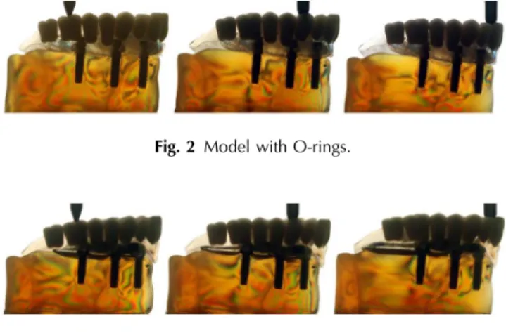
Regarding stress distribution in the model without implants, the fringes were located on the region of alveolar ridge crest (Fig. 1). In the models with implants, regardless attachment
system, the photoelastic fringes were observed at the apical region of the implants (Figs.2and3).
4 Discussion
The hypothesis that the system with three individualized O-rings provides the lowest stress on the implants and support tissues was accepted, since this system exhibited lowest stress values. The palatal obturator prostheses aim to seal the communi-cation among the oral, nasal, and orbital cavities, allowing the restoration of the speech, mastication, swallowing, and aes-thetics, to provide a better quality of life to the patients.4,5And according to the results, the conventional obturator prosthesis (without implants) exhibit low stress values (Table1and Fig.1). However, the stability and retention of these prostheses have been a problem for the prosthodontists and the patients3,4,12
because specific anatomic conditions in each case of maxillary surgical resection demand distinctive planning since the exten-sion and location of surgical resection, as well as the mucosa and bone condition, are determinant aspects for the planning, in order to reach a medium retention in a palatal obturator prosthesis.
So, the use of implant-retained palatal obturator prostheses to rehabilitate partial or total maxillectomized patients has been growing since it provides higher retention and stability of the prostheses, reducing their movingwhich can lead to lower stress on bone support.2–5,7,13 In our study, the O-ring attachment
system for the implant-retained obturator prostheses exhibited lower stress values when comparing it with the bar-clip system (Table1and Fig.2).
These results corroborate with some studies14–16which con-sider that the O-ring attachment system (Fig.2) transfers less stress to the implants in comparison to the bar-clip system (Fig.3), because the O-ring system reduced stress and provided great stability. According to the authors, it can result from stress absorption by the female component of the system, which generally presents a rubber ring into a metallic capsule that may absorb or homogenously distribute the stress they are submitted to.
Most of the failures related to total implant-retained pros-theses happen due to the excess of stress transmitted to the implants and attachment systems. The systems fatigue may cause fracture on the implants components, overload them and the bone tissue, which would also result in a possible loss of osseointegration, generating prostheses instability and loss retention.2,5,7 The photoelasticity method analyzed through
Table 1 Number of photoelastic fringes according to the crowns in
which the load was applied.
Attachment system
Axial load
Crown
16 13 11
Conventional 0 1 1
O-ring 3 4 4
Bar clip 5 6 6
Fig. 1 Model without implants.
Fig. 2 Model with O-rings.
Fig. 3 Model with bar-clips.
Journal of Biomedical Optics 061203-2 June 2013 • Vol. 18(6)
Pesqueira et al.: Stress analysis in oral obturator prostheses: imaging photoelastic
images aims to support the selection of a better retention method with dental implants to reach the correct rehabilitation planning for these patients with oronasal communication.
5 Conclusion
The photoelastic method is efficient for better oral rehabilitation planning.
The system with three individualized O-rings provided the lower values of stress in the implants and support tissues, reach-ing, in this way, the biomechanical success of the implant-retained palatal obturator prostheses.
Acknowledgments
This investigation was supported by a research grant from Fun-dação de Amparo à Pesquisa do Estado de São Paulo.
References
1. S. M. Ortegon, J. W. Martin, and J. S. Lewin,“A hollow delayed sur-gical obturator for a bilateral subtotal maxillectomy patient clinical report,”J. Prosthet. Dent.99(1), 14–18 (2008).
2. O. C. Dilek and E. Tezulas,“A mini implant-supported obturator appli-cation in a patient with partial maxillectomy due to tumor: case report,” Oral Surg. Oral Pathol. Oral Radiol. Endod.103(3), e6–e10 (2007). 3. B. Bagis, E. Aydogan, and U. Hasanreisoglu,“Rehabilitation of a con-genital palatal defect with a modified technique: a case report,”Cases J. 1(1), 39 (2008).
4. M. C. Goiato et al.,“Positioning magnets on a multiple/sectional max-illofacial prosthesis,”J. Contemp. Dent. Pract.8(7), 101–107 (2007).
5. P. do Prado Ribeiro et al., “Photoelastic stress analysis of different attachment systems on implant-retained and conventional palatal obturator prostheses,”J. Craniofac. Surg.22(3), 523–526 (2011). 6. G. Celik and B. Uludag,“Photoelastic stress analysis of various
reten-tion mechanisms on 3-implant-retained mandibular overdentures,” J. Prosthet. Dent.97(4), 229–235 (2007).
7. M. C. Goiato et al.,“Retention systems to implant-supported cranio-facial prostheses,”J. Craniofac. Surg.20(3), 889–891 (2009). 8. R. A. Markarian et al.,“Stress distribution after installation of fixed
frameworks with marginal gaps over angled and parallel implants: a photoelastic analysis,”J. Prosthodont.16(2), 117–122 (2007). 9. K. Turcio et al.,“Photoelastic analysis of stress distribuition in oral
reha-bilitation,”J. Craniofacial Surg.20(2), 471–474 (2009).
10. A. A. Caputo,“Stress analysis,”Seminário de Biomateriais, Science Section. Abstracts, UCLA School of Dentistry, Los Angeles (1993). 11. A. A. French et al.,“Comparison of periimplant stresses transmitted by
four commercially available osseointegrated implants,”Int. J. Period-ontics Restorative Dent.9(3), 221–230 (1989).
12. K. M. Lyons, J. Beumer, and A. A. Caputo,“Abutment load transfer by removable partial denture obturator frameworks in different acquired maxillary defects,”J. Prosthet. Dent.94(3), 281–288 (2005). 13. W. Oh and E. Roumanas,“Dental implant-assisted prosthetic
rehabili-tation of a patient with a bilateral maxillectomy defect secondary to mucormycosis,”J. Prosthet. Dent.96(2), 88–95 (2006).
14. R. Kenney and M. W. Richards,“Photoelastic stress patterns produced by implant-retained overdentures,”J. Prosthet. Dent.80(5), 559–564 (1998).
15. M. Tokuhisa, Y. Matsushita, and K. Koyano,“In vitrostudy of mandib-ular implant overdenture retained with ball,magnet, or bar attachements: comparison of load transfer and denture stability,”Int. J. Prosthodont.
16(2), 128–134 (2003).
16. M. C. Goiato et al.,“Photoelastic analysis to compare implant-retained and conventional obturator dentures,”J. Biomed. Opt.17(6), 061203 (2012).
Journal of Biomedical Optics 061203-3 June 2013 • Vol. 18(6)
Pesqueira et al.: Stress analysis in oral obturator prostheses: imaging photoelastic