199
Iranian Journal of Otorhinolaryngology, Vol.27 (3), Serial No.80, May 2015
Original Article
Association between Serum Levels of Interleukin-6
and Stage of Laryngeal Cancer
Soheila Nikakhlagh1, Nastran Ranjbari1, Elaheh Khorami1,*Nader Saki1
Abstract
Introduction:
The purpose of this study was to investigate the relationship between serum levels of interleukin-6 (IL-6) and the severity and extent of squamous cell carcinoma (SCC) of the larynx based on stage of tumor progression and histological grade.
Materials and Methods:
All patients with a diagnosis of laryngeal cancer who underwent laryngoscopy and biopsy while hospitalized in Imam Khomeini Hospital in Ahvaz were enrolled. Tumor stage was calculated based on the TNM system, and divided into early (stage 1,2) or advanced stage (stage 3,4). In addition, patients were divided into low-grade (well differentiated) or high-grade (moderate and poorly differentiated) groups based on pathology reports from biopsy specimens. Several healthy volunteers were also enrolled as the control group. After collecting the blood samples, quantitative serum levels of IL-6 were measured (pmol/L) using IL-6 kits (Bender MedSystem, Germany). Results for quantitative variables are presented as mean and standard deviation and qualitative variables as percentages. Mann-Whitney, Kruskal-Wallis
and Pearson’s chi square tests were used for statistical analyses. P<0.05 was considered statistically significant. Statistical analysis of data was performed using SPSS version 13.
Results:
Thirty-eight patients (82.6%) were male and eight patients (17.4%) were female. IL-6 serum level was 28.8±4.7 pmol/L in the patient group and 2.64±2.88 pmol/L in the control group (P=0.0001). The serum level of IL-6 was 7.27 ± 5.31 pmol/L in early-stage patients and 54.43 ± 6.06 pmol/L in advanced-stage patients (P<0.0001). IL-6 levels increased significantly with increasing N (according to TNM) (P=0.002). Levels of IL-6 in patients with metastasis were significantly higher than in the group without metastasis (P=0.024). Moreover, IL-6 levels increased significantly with increasing local tumor spread (T) (P<0.0001).
Conclusion:
This study shows that IL-6 is a gender-independent factor, serum levels of which are higher in patients with laryngeal SCC than in normal subjects. The results of this study also show that serum levels of this cytokine increase significantly with progression of this malignancy.
Keywords:
Biomarker, Laryngeal Cancer, Squamous Cell Carcinoma.
Received date: 25 Jun 2014 Accepted date: 12 Aug 2014
1Hearing & Speech Research Center, Ahvaz Jundishapur University of Medical Sciences, Ahvaz, Iran.
*
Corresponding Author:
Introduction
Laryngeal cancer is the second most prevalent cancer in the upper respiratory tract, affecting 11,000 individuals in the United States each year. Approximately 85– 95% of laryngeal cancers are squamous cell carcinomas (SCC). Successful treatment requires accurate and timely diagnosis, staging and appropriate assessment of the patient. Laryngeal tumor staging is performed based on the sixth edition of the TNM system, published by the American Joint Committee on Cancer/Union for Internationals Cancer Control (AJCC/ UICC) (1).
Tumor grading is also expressed based on the pathologist’s report of biopsy specimens and according to cell differentiation (well, moderate, or poorly differentiated) (1).
Several factors affect the prognosis of patients with laryngeal cancer, including clinical stage and histological forms. Recent studies have examined genetic factors and biomarkers, in which the role of different interleukins has been described (2).
Interleukin-6 (IL-6) levels increase in many inflammatory diseases and malignancies. This cytokine which has multiple functions, and is involved in the differentiation of B lymphocytes into plasma cells. Studies have shown that IL-6 level is significantly associated with tumor invasion level, disease severity, and metastasis (3). IL-6 in conjunction with IL-1 and tumor necrosis factor-alpha (TNF-α) is one of the most important cytokines inducing acute phase inflammatory responses. This cytokine is secreted by B and T lymphocytes, macrophages, fibroblasts, keratinocytes, and other cells. In addition to the acute phase response, IL-6 also reinforces chronic phase inflammation, providing an ideal environment for the growth of tumors. On the other hand, IL-6 is also secreted by cancerous cells and plays a role in regulating proliferation and apoptosis (4). IL-6 activates Janus kinase (JAK) after binding to its receptor, where subsequent
phosphorylation of the signal transducer and activator of transcription (STAT) occurs. The phosphorylated STAT gene is transferred into the nucleus and activates target genes such as vascular endothelial growth factor (VEGF) and rho which increase the rate of tumor invasion (5,6).
The purpose of this study was to investigate the relationship between serum levels of IL-6 and the severity and extent of the disease based on tumor stage and histological grade in order to explore use of this biomarker as a prognostic factor and tumor marker. In the case of a relationship being identified, immunologic approaches could be used for the treatment of patients in the future.
Materials and Methods
Iranian Journal of Otorhinolaryngology,Vol.27(3), Serial No.80, May 2015 201
immediately transferred to the laboratory for serum separation. In order to estimate the serum levels of IL-6 in normal subjects, samples were also taken from a number of non-patient volunteers. Then the sera were stored in a freezer at −20 °C. Melting and refreezing of the samples was prevented during storage. After collecting the samples, quantitative serum levels of IL-6 were measured (pmol/L) according to the enzyme-linked immunosorbent assay (ELISA) method using IL-6 kits (Bender MedSystem, Germany). After preparing the diluted solutions and adding substrate and stop solution, the levels of IL-6 were obtained using a spectrophotometer based on the absorption rate and a comparison with standard solutions. Results for quantitative variables are expressed as mean and standard deviation (mean ± SD) and as percentages for qualitative variables. Mann-Whitney, Kruskal-Wallis
and Pearson’s chi square tests were used for statistical analysis. P<0.05 was considered to be statistically significant. Statistical analysis of data was performed using SPSS version 13. The Ethics Committee number is AJUMS. REC. 1393.68.
Results
In this study, 63 individuals were examined; 46 of whom were patients with laryngeal cancer and 17 of whom were in the control group. Thirty-eight patients (82.6%) were male and eight (17.4%) were female. The age of patients was 63.78 ± 11.19 years. Serum IL-6 level was 28.8 ± 4.7 pmol/L in the patient group (range, 96– 182.81 pmol/L) and 2.64 ± 2.88 pmol/L (range, 0.16–9.9 pmol/L) in the control group. Given the non-normal distribution of IL-6 levels, the Mann-Whitney statistical test was used to determine that mean IL-6 levels in patients with laryngeal carcinoma were significantly higher than those in the control group (P= 0.0001).
Of the 46 patients with laryngeal cancer, 25 (54.3%) were in the early-stage group and 21 (45.7%) were in the advanced-stage group. IL-6 levels were 7.27±5.31 pmol/L in the early-stage group and 54.43 ± 6.06 pmol/L in the advanced-stage group (P<0.0001) (Table. 1).
Table 1: IL-6 level according to stage of disease.
Lymph node involvement
Mean SD P-value
Early 7.27 5.31
<0.0001
Advanced 49 56.5
Metastatic 106 101.3
IL-6 levels were 28.18±4.8 pmol/L in male patients and 31.72±4.51 pmol/L in female patients. No significant difference was observed between the two sexes in terms of IL-6 levels (P=0.653).
Tumors were low grade in 24 (52.2%) patients and high grade in 22 (47.8%) patients. Statistically, the difference between the two tumor grades was at a borderline level (P = 0.057) (Table. 2).
Table 2: Levels of IL-6 according to tumor grade.
Tumor grade Mean SD P-value
Low grade 15.1 1.9
0.057
High grade 43.7 6.2
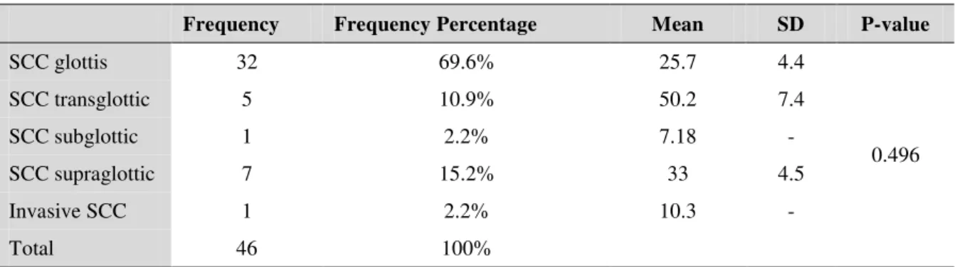
Table 3: Frequency distribution of various types of laryngeal tumors and IL-6
Frequency Frequency Percentage Mean SD P-value
SCC glottis 32 69.6% 25.7 4.4
0.496
SCC transglottic 5 10.9% 50.2 7.4
SCC subglottic 1 2.2% 7.18
-SCC supraglottic 7 15.2% 33 4.5
Invasive SCC 1 2.2% 10.3
-Total 46 100%
Distant metastases were observed in four (8.7%) patients. Mean ± SD IL-6 level was 72.1±7.2 pmol/L among this group, compared with 24.6±4.2 pmol/L in the group without distant metastases (P= 0.024).
In terms of local tumor spread, most patients (70%) were T2 or T3. IL-6 levels increased significantly with increasing local tumor spread only (Table. 4).
Table4: IL-6 and tumor extension
Tumor extension Frequency Frequency percentage Mean SD P-value
T1 8 17.4% 6.6 6.7
P<0.0001
T2 17 37% 7.5 4.7
T3 15 32.6% 4.7 5
T4 6 13% 88.7 7.4
In terms of lymph node involvement, 26 (69.6%) cases were N0, 12 (26.1%) cases were N1, and two (4.3%) cases were N2. No patient was observed with N3. There was a
significant difference between different levels of lymph node involvement and levels of IL-6 (P=0.002) (Table. 5).
Table 5: IL-6 level and lymph node involvement
Lymph node involvement Frequency Frequency percentage Mean SD P-value
N0 32 69.6 16.3 3.2
0.002
N1 12 26.1 49.1 5.4
N2 2 4.3 105.8 108.8
Discussion
Head and neck squamous cell carcinoma (HNSCC) is the eighth most common cancer worldwide and accounts for nearly six percent of all cancers (7). The larynx is the most common site of HNSCC. In Iran, as in other places of the world, the most common malignant tumor of the larynx is
the SCC (8,9). The most common age of onset of this tumor is 60 years in Iran, and the male-to-female ratio is 9.5 to 1. In Iran, tumors located in the supraglottic region seem to be more common than those in the glottic area (10).
Iranian Journal of Otorhinolaryngology,Vol.27(3), Serial No.80, May 2015 203
one of the most important cytokines involved in inducing acute phase inflammatory responses. This cytokine also enhances the chronic phase of inflammation, providing an ideal environment for tumor growth (5,6).
Few studies have been conducted on the role of interleukins in whole head and neck carcinomas. Although the relationship between IL-6 and cancer has been extensively studied, the role of the production of this interleukin in the microenvironment of the tumor which progresses via malignant cells and tumoral infiltrated lymphocytes and macrophages is still controversial (11). Generally, it appears that early diagnosis or prevention of this cancer can be the most effective factor in improving patient outcomes. The absence of warning clinical signs for most HNSCCs increases the importance of identifying a biomarker with good sensitivity and specificity for screening patients at risk. So far, a number of molecular markers have been identified for the diagnosis of tumors, with varying degrees of sensitivity and specificity. IL-6 is a multifunctional cytokine which plays a pivotal role as a growth and differentiation factor in different cells such as hematopoietic progenitor cells, B-cells, T-cells, keratinocytes, neuronal cells, osteoclasts, and endothelial cells (12).
In different studies, IL-6 production has been reported in several tumors in humans such as esophageal SCC, multiple myeloma, renal cell carcinoma, glioblastoma, and lung carcinomas (13-17). Moreover, cycle of IL-6 - IL-6 receptoras been reported in a variety of tumors in several studies (14). In addition, the release of IL-6 has been reported by apoptotic T-cells in mice (18). These findings suggest that IL-6 produced by tumor cells enhances the survival and proliferation of cancer cells as a growth factor interacting with specific receptors on the surface membrane of cancer cells (13). Although recent studies have shown that an
increased serum level of IL-6 is aligned with patient prognosis in various malignancies (19–22), the mechanism that causes this reaction in these diseases is still being discussed. The strongest hypothesis relating to the mechanism is that the increased level of IL-6 in cancer patients is due to excessive production from the tumor, and reflects the increase in serum levels of IL-6 as a biologic characteristic of the tumor However, the role of IL-6 in the growth of head and neck cancers is not yet well known.
The present study aimed to investigate the relationship between serum levels of IL-6 and severity of laryngeal cancer. This study is among the few studies that examined the association of this cytokine with the severity of laryngeal malignancies. In this study, 46 patients with laryngeal cancer at different stages were studied. The mean age of patients was 63.8 years, and there were approximately five times as many men as women with this cancer. The mean age and gender prevalence in this study is consistent with other studies in this field (23).
The results of the present study primarily showed that the serum levels of IL-6 in patients with laryngeal cancer are significantly higher than in normal subjects in the control group. In a 2013 study conducted by Hao et al., patients with laryngeal squamous cell cancer (LSCC) had significantly higher levels of IL-6 compared with normal subjects, similar to the present study (24). However, in a 2009 study by Dae-Young Hong et al. investigating a number of biomarkers in patients with HNSCC, no significant difference was observed between the serum levels of IL-6 in patients with malignancy compared with the control group, indicating higher specificity of this cytokine in LSCC.
pmol/L in advanced-stage patients. This mean IL-6 level was significantly higher in advanced stages compared with early stages. In studies conducted in association with HNSCC, similar results to the present study were observed. For instance, a study by Riedel et al. in 2005 showed that IL-6 levels increased with increasing severity of disease (25). In a study conducted by Hao et al. regarding the diagnostic value of IL-6 and IL-8 in LSCC, it was determined that serum levels of IL-6 are directly correlated with metastasis to lymph nodes, local tumor spread, and clinical stage of tumor (P<0.05). Moreover, multivariate analysis showed that the serum level of IL-6 is an independent predictor for specific survival of LSCC (24). The Hao study was the closest to the present study, the results of which were also largely similar to our results. In another study by Duffy in 2007 in the United States in HNSCC patients, it was shown that the recurrence rate is higher in patients with higher levels of interleukin (P=0.002) and the survival rate is lower (3). Regarding the evaluation of serum levels of cytokines in patients with laryngeal cancer, a study was conducted in Southwest of Iran in levels of IL-10 in patients with laryngeal cancer and the associated metastases. No significant differences were found between patients with local invasion of laryngeal cancer and patients without local invasion. Furthermore, no statistically significant correlation was observed between IL-10 gene expression and different stages of tumor (26). The results of this study compared with the present study suggest that IL-6 is a more specific cytokine in laryngeal malignancies, levels of which increase considerably both compared with normal subjects and in advanced stages of tumor. The results of the present study showed that IL-6 is a gender-independent factor whose serum level in patients with laryngeal SCC is higher than in normal
subjects. Moreover, this study indicated that the serum level of this cytokine dramatically increases with the progression of malignancy, so that serum levels of IL-6 were significantly higher in advanced-stage patients compared with early-stage patients. Furthermore, patients with high-grade tumors, more lymph node involvement, distant metastases and higher levels of T classification had higher serum levels of IL-6.
Conclusion
Laryngeal cancer is the second most common cancer in the upper respiratory tract. Finding new cellular and molecular methods for early detection of aggressive cases and timely treatment can reduce the associated mortality. According to the results of the present study, serum levels of IL-6 in patients were significantly associated with the severity of laryngeal cancer. Therefore, this biomarker can be used in the diagnosis and treatment of invasive cases. Further studies are needed to accomplish the goals described.
Acknowledgements
The authors would like to thank the research deputy Chairman in his valuable cooperation. This research work was the result of an ear, nose, and throat (ENT) Specialist thesis (Ref No: HRC9203).
References
1. Armstrong WB, Vokes D, Maisel RH. Malignant tumor of the larynx. In: Cummings CW, Flint PW, Harker LA, Haughey BH, Richardson MA, Robbins KT, et al.(editors).Cummings Otolaryngology Head & Neck Surgery. 5th ed.Philadelphia: Elsevier Mosby; 2010:1482–1510.
2. Lopez RVM, Zago MA, Eluf-Neto J. Education, tobacco smoking, alcohol consumption, and IL-2 and IL-6gene polymorphisms in the survival of head and neck cancer. Medical and Biological Res 2011; 44(10):1006–12.
Iranian Journal of Otorhinolaryngology,Vol.27(3), Serial No.80, May 2015 205 4. Mojtahedi Z, Khademi B, Hashemi SB. Serum
interleukin-6 concentration but not Interleukin-18, is associated with head and neck squamous cell carcinoma progression. Pathol Oncol Res 2011; 17(1): 7–10.
5. Blay JY, Negrier S, Combaret V. Serum level of interleukin 6 as a prognosis factor in metastatic renal cell carcinoma. Cancer Res1992; 52:3317–22. 6. Scambia G, Testa U, Benedetti Panici P. Prognostic significance of interleukin 6 serum level in patient with ovarian cancer. Br J Cancer 1995; 71(2):354–6.
7. O’Regan EM, Timon C, Sheils O, Codd M,
Oleary JJ, Toner M. Squamous cell carcinoma of the head and neck in young Irish adults. Br J Oral Maxillofac Surg 2006; 44(3): 203–6.
8. Mafi N, Kadivar M, Hosseini N, Ahmadi S, Zare Mirzaie A, et al. Head and neck squamous cell carcinoma in Iranian patients and risk factors in young adults: a fifteen-year study. Asian Pac J Cancer Prev 2012; 13: 3373–8.
9. Saki N, Saki G, Maasomi SA, Rahim F, Emad Mostofi N, Nikakhlagh S. Effect of smoking in the etiology of cancer of larynx in Khuzestan. Jentashpir Journal 2011; 2(3):99–103.
10. Saedi B, Razmpa E, Sadeghi M, Mojtahed M, Mojtahed A, et al. The epidemiology of laryngeal cancer in a country on the esophageal cancer belt. Indian J Otolaryngol Head Neck Surg 2009; 61:213-17.
11. Zou W. Immunosuppressive networks in the tumour environment and their therapeutic relevance. Nat Rev Cancer 2005; 5: 263–74. 12. Kinoshita T, Ito H and Miki C. Serum interleukin-6 level reflects the tumor proliferative activity in patients with colorectal carcinoma. Cancer 1999; 85(12):2526–31.
13. Oka M, Iizuka N, Yamamoto K, Gondo T, Abe T, Hazama S, Akitomi Y, Koishihara Y, Ohsugi Y, Ooba Y, Ishihara T, et al. The influence of interleukin-6 on the growth of human esophageal cancer cell lines. J Interferon Cytokine Res 1996; 16(12): 1001–6.
14. Kawano M, Hirano T, Matsuda T, Taga T, Horii Y, Iwato K, et al. Autocrine generation and requirement of BSF-2/IL-6 for human multiple myelomas. Nature 1988; 332: 83–5.
15. Miki S, Iwano M, Miki Y, Yamamoto M, Tang B, Yokokawa K, et al. Interleukin-6 (IL-6) functions as
an in vitro autocrine growth factor in renal cell carcinomas. FEBS Lett 1989(2); 250: 607–10. 16. Meir EV, Sawamura Y, Diserens A, Hamou M, Tribolet N. Human glioblastoma cells release interleukin-6 in vivo and in vitro. Cancer Res 1990; 50: 6683–8.
17. Takizawa H, Ohtoshi T, Ohta K, Yamashita N, Hirohata S, Hirai K, et al. Growth inhibition of human lung cancer cell lines by interleukin-6 in vitro: a possible role in tumor growth via an autocrine mechanism. Cancer Res 1993; 53: 4175–81.
18. Teague TK, Marrack P, John WK, Anthony TV. IL-6 rescues resting mouse T cell from apoptosis. J Immunol 1997; 158(12): 5791–96. 19. Bataille R, Jourdan M, Zhang XG, Klein B. Serum levels of interleukin-6, a potent myeloma growth factor, as a reflection of disease severity in plasma cell dyscrasias. J Clin Invest 1989; 84(6): 2008–11.
20. Tsukamoto T, Kumanoto Y, Miyao N, Masumori N, Takahashi A, Yanase M. Interleukin-6 in renal cell carcinoma. J Urol 1982; 148(6); 1778–82.
21. Oka M, Yamamoto K, Takahashi M, Hakozaki M, Abe T, Iizuka N, et al. Relationship between serum levels of interleukin 6, various disease parameters and malnutrition in patients with esophageal squamous cell carcinoma. Cancer Res 1996; 56: 27–2780.
22. Wu CW, Wang SR, Chao MF, Wu TC, Lui WY, Peng FK, et al. Serum interleukin-6 levels reflect disease status of gastric cancer. Am J Gastroentrol 1996; 91(7):1417–22.
23. Jacobs C. The internist in the management of head and neck cancer. Ann Intern Med 1990; 113(10):771–8.