EXTRA-ARTICULAR MANIFESTATIONS OF
RHEUMATOID ARTHRITIS, NOW
*Paloma Vela
Section of Rheumatology, Hospital General University of Alicante; Department of Medicine, Miguel Hernández de Elche University, Alicante, Spain
*Correspondence to palomavela62@gmail.com
Disclosure: The author has received honoraria for lectures from Abbvie, Pizer, Roche, Bristol-Myers Squibb, MSD, Lilly and UCB, and honoraria for participation in advisory boards from Roche, UCB, and Pizer.
Received: 23.03.14 Accepted: 15.05.14 Citation: EMJ Rheumatol. 2014;1:103-112.
ABSTRACT
Rheumatoid arthritis (RA) is a chronic systemic inlammatory disease, characterised by polyarthritis and extra-articular organ disease, including rheumatoid nodules, ophthalmologic manifestations, cardiopulmonary disease, vasculitis, neuropathy, glomerulonephritis, Felty’s syndrome, and amyloidosis. Extra-articular manifestations of RA (ExRA) occur in 17.8–40.9% of RA patients, 1.5–21.5% of them presenting as severe forms and usually associated with increased morbidity and mortality. They can develop at any time during the course of the disease, even in the early stages, and are associated with certain predisposing factors, such as the presence of rheumatoid factor, smoking, and long-standing severe disease. Rheumatoid nodules, the most common ExRA, have been found to be associated with the development of severe features, such as vasculitis, rheumatoid lung disease, pericarditis, and pleuritis, especially in those patients who develop them within 2 years from RA diagnosis. There is no uniformity in the deinition of the term ExRA, which limits comparability between diferent studies. Several recent surveys suggest a lower frequency, probably due to a better control of disease activity. Diagnosis of ExRA is a challenge for clinicians, given its variable and complex presentation, and the lack of speciic diagnostic tests; it must be based on clinical recognition and exclusion of other causes of the signs and symptoms. Furthermore, management continues to be diicult with a bad prognosis in many conditions. This article reviews the clinical aspects of major ExRA, focusing on incidence, clinical features, and therapeutic approaches, and how modern immunosuppressive therapy can change the outcome.
Keywords: Rheumatoid arthritis, extra-articular manifestation, management, biologic treatment.
INTRODUCTION
Rheumatoid arthritis (RA) is an immune-mediated chronic inlammatory disease. Despite being characterised by inlammation of the synovial membrane and progressive destruction of the articular cartilage and bone, RA is a systemic disease often associated with other extra-articular manifestations, with a signiicant impact on mortality and morbidity.1 A better control of disease activity
in the last decades due to the availability of more eicacious drugs has resulted in a lower frequency of extra-articular manifestations of RA (ExRA), as well as better outcomes in many patients.
Epidemiology
ExRA can develop at any time during the course of the disease, even in the early stages.2 They occur
in 17.8–40.9% of RA patients, 1.5–21.5% of them presenting as severe forms and usually associated with greater comorbidity and premature death.3
Higher frequencies are seen in Northern European countries, rather than Southern Europe, suggesting a role of environmental and genetic factors in their pathogenesis. Hospital-based studies also show more prevalence, probably due to inclusion of patients with more severe disease.4 Until now
studied in numerous RA cohorts. This partly justiies the diferences seen in descriptive reports and incidence studies.
Predisposing Factors
Prete et al.4 recently reviewed all relevant articles
related to ExRA published up to June 2011, and found smoking, early disability, antinuclear antibodies, and rheumatoid factor (RF) positivity as the principal predictor factors of severe ExRA. Gender was not found to have any efect, with the exception of one Italian study, which reported higher risk of developing any ExRA in men than in women (odds ratio, 1.68). Moreover, Turesson et al.5 described the presence of rheumatoid nodules
(RNs) as a predisposing factor for development of severe ExRA, such as vasculitis, rheumatoid lung disease, pericarditis, and pleuritis, especially in those patients who develop them within 2 years from RA diagnosis, and found a particularly high risk in those patients with long-standing, severe disease.
CLINICAL MANIFESTATIONS
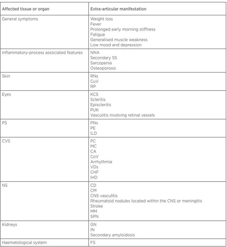
Many diferent tissues and organs can be involved in RA patients in addition to the characteristic peripheral polyarthritis (Table 1). Several general symptoms can represent a major problem during the course of RA, many of them also being present before its diagnosis. Weight loss, fever, prolonged early morning stifness, fatigue, generalised muscle weakness, low mood, and depression are often responsible for a signiicant loss in the quality of life of patients. Fatigue is reported in 40–80% of RA patients as their most disabling symptom.6 The
OMERACT (Outcome Measures in Rheumatology) network of international researchers highlighted fatigue as a main outcome, recommending its measure whenever possible.7
As a result of the inlammatory process, RA patients frequently developed normochromic-normocytic anaemia.8 Other manifestations
associated with chronic inlammation include injury of exocrine glands with the development of a secondary Sjögren’s syndrome (SS), sarcopaenia, and osteoporosis.
RNs are the most frequent skin manifestation. They occur in about 30% of RA patients, mostly in RF-positive subjects, and are usually located subcutaneously on pressure areas, including the olecranon process and proximal ulna, inger joints,
Usually painless, they have a variable consistency from a soft, mobile to a hard, rubbery mass attached irmly to the periosteum. Histologically they are characterised by a central necrotic area rimmed by a corona of palisading ibroblasts that is surrounded by a zone of tissue afected by perivascular cellular iniltration enriched with lymphocytes, plasma cells, and histiocytes.9
Regression of nodules may occur during treatment with disease modifying anti-rheumatic drugs (DMARD). Paradoxically, in 8–11% of methotrexate-treated RA patients an accelerated rheumatoid nodulosis can occur, with nodules usually located in the ingers or in the metacarpophalangeal and proximal interphalangeal joints. The condition regresses when methotrexate is reduced or withdrawn and if hydroxychloroquine or sulphasalazine treatment is started. Etanercept has also been related with the development of this type of nodulosis.10 No efective treatment
is available.
Ocular involvement occurs in 27% of RA patients.11
Table 1: Extra-articular manifestations in rheumatoid arthritis.
NNA: normochromic-normocytic anaemia; SS: Sjögren’s syndrome; RNs: rheumatoid nodules; CuV: cutaneous vasculitis; RP: Raynaud’s phenomenon; KCS: keratoconjunctivitis sicca; PUK: peripheral ulcerative keratitis; PS: pulmonary system; PNs: pulmonary nodules; PE: pleural efusion; ILD: interstitial lung disease; CVS: cardiovascular system; PC: pericarditis; MC: myocarditis; CA: cardiac amyloidosis; CoV: coronary vasculitis; VDs: valve diseases; CHF: congestive heart failure; IDH: ischaemic heart disease; NS: nervous system; CD: cognitive dysfunction; CNS: central nervous system; CM: cervical myelopathy; MM: mononeuritis multiplex; SPN: sensory peripheral neuropathy; GN: glomerulonephritis; IN: interstitial nephritis; FS: Felty’s syndrome.
Afected tissue or organ Extra-articular manifestation
General symptoms Weight loss Fever
Prolonged early morning stifness Fatigue
Generalised muscle weakness Low mood and depression
Inlammatory-process associated features NNA
Secondary SS Sarcopenia Osteoporosis
Skin RNs
CuV RP
Eyes KCS
Scleritis Episcleritis PUK
Vasculitis involving retinal vessels
PS PNs
PE ILD
CVS PC
MC CA CoV
Arrhythmia VDs
CHF IHD
NS CD
CM
CNS vasculitis
Rheumatoid nodules located within the CNS or meningitis Stroke
MM SPN
Kidneys GN
IN
Secondary amyloidosis
This condition is often painless and can evolve to scleral perforation when it goes untreated.12
Pulmonary involvement in RA is frequent, although not always clinically recognised, and includes RNs, pleural efusion (PE), interstitial lung disease (ILD), small airway disease, and pulmonary vasculitis. It is responsible for 10-20% of overall mortality,13,14
and can occur before the development of joint symptoms.15,16 Parenchymal pulmonary nodules
(PNs) are usually asymptomatic, but may cavitate and cause PEs (Figure 1); they also increase the risk of infections and pneumothorax. They are usually found in RF-positive patients with nodules elsewhere. Sometimes diferentiation with neoplasms and infections can be diicult. PE, usually an exudate with mixed cell counts and high protein concentration, is common but frequently asymptomatic; autopsy studies reported pleural involvement in 50% of cases, with only 10% clinically detected.17
ILD is the most important pulmonary manifestation of RA, being the commonest pulmonary cause of death and a signiicant contributor to morbidity.13,14,18,19 The most frequent
histopathological patterns of ILD in RA are usual interstitial pneumonia (UIP) and nonspeciic interstitial pneumonia (NSIP) (44–56% and 33–44%, respectively),20 followed by mixed disease (0–12%).
Other forms, such as obliterative bronchiolitis, are rare but associated with a high mortality.21 Although
it tends to occur more often in RF positive male patients with long-standing nodular disease,22
studies in new onset RA have found lung abnormalities in a high percentage of patients.23,24
Clinical presentation and course are similar to that of idiopathic pulmonary ibrosis, but the response to immunosuppressants is usually better. Diagnosis is based on clinical presentation, blood gases, pulmonary function tests, and high resolution computed tomography (HRCT).25 As abnormalities
can be detected by HRCT in about 50% of RA patients, but only 10% have clinically signiicant symptoms,26 diagnosis should be supported not
only on clinical signs and symptoms, but also in abnormal pulmonary function tests and either a compatible HRCT or lung biopsy. Physiological abnormalities include a reduction in lung volume, a low difusing capacity for carbon monoxide (which is the measure best associated with the extent of disease in ILDs and a poorer prognosis in RA-ILD27), and oxygen desaturation during a
Cardiovascular (CV) features in RA are common,28
including pericarditis, myocarditis, cardiac amyloidosis, coronary vasculitis (CoV), arrhythmia, valve diseases, and, most importantly, congestive heart failure29 and ischaemic heart disease (IHD).
The last two have been associated with an increased morbidity and mortality in RA patients compared with the general population due to an accelerated atherogenesis process that cannot be fully explained by the classic atherosclerosis risk factors;30,31 the presence of chronic inlammation
and a possible genetic component are important contributing agents.32,33
Within the classical cardiac manifestations, pericarditis is the most common; both echocardiography and autopsy studies reveal evidence of pericardial inlammation in 50% of patients, although symptoms are relatively uncommon, occurring in about 1–4% of patients. It usually occurs in RF-positive nodular RA, and pericardial luid analysis reveals features similar to those found in rheumatoid PEs. Conversely, symptomatic myocarditis, endocarditis, and CoV rarely occur, and are almost exclusively demonstrated by autopsy.34 Cognitive dysfunction is frequently
found in RA patients, with prevalence rates ranging from 38-71%.35,36 Education, income, glucocorticoid
use, and cardiovascular disease (CVD) risk factors are independent predictors of its development.37
or meningitis. Stroke also occurs with increased frequency.38 CNS vasculitis is extremely rare.
The diagnosis is supported by magnetic resonance imaging (MRI), alone or with magnetic resonance angiography (MRA), showing the segmental vascular stenosis characteristic of vasculitis.39
Peripheral neuropathy is usually manifested as sensorimotor neuropathy or mononeuritis multiplex. The underlying mechanism is small vessel vasculitis of the vasa vasorum of the nerves with ischaemic neuropathy and demyelinisation as part of the rheumatoid vasculitis (RV) syndrome.
RA and kidney disease (KD) often coincide. There are several potential causes of nephropathy such as drug-related renal disease, secondary renal amyloidosis, and various types of glomerulonephritis (GN). Mesangial proliferative GN is the most frequent histological lesion followed by membranous GN,40,41 the latter usually being
related to gold or D-penicillamine, with both therapies not currently in use. Other infrequent causes of KD can be interstitial nephritis, minimal change glomerulopathy, IgA nephritis, focal proliferative GN, or rapidly progressive GN due to microscopic polyangiitis.42 Prevalence has been
recently established; the MATRIX study43 found
KD in 46.3% of RA patients according to the National Kidney Foundation (NKF) classiication44
with a stage distribution of 11.3% in Stage 1 (normal kidney function with kidney damage), 20.0% in Stage 2 (mild renal insuiciency with kidney damage), 15.0% in Stage 3 (moderate renal insuiciency), and no patients in Stages 4 or 5. The study was not designed to identify the potential causes of KD. A recent retrospective review45 has shown that RA patients are more
likely to develop reduced kidney function over time, with CVD at baseline and elevated erythrocyte sedimentation rate as predisposing factors, and found a relationship between renal impairment and increased morbidity from CVD development.
Secondary (reactive AA) amyloidosis can be seen in long-standing disease and poor response to therapy, and markedly inluences these patients’ outcomes.46 Prevalence is around 7%,47 with clinically
symptomatic amyloidosis much lower.48 Common
clinical signs of reactive AA amyloidosis in patients with RA can be found by careful observation for the onset of proteinuria, kidney insuiciency, or gastrointestinal tract symptoms. Biopsy is often necessary to make an accurate diagnosis.
RV is a rare but potentially serious necrotising vasculitis, which can develop in patients with RA sometimes in the absence of active joint disease. RV typically occurs in male patients with long-standing RF-positive erosive nodular RA,49 in
association with a severe disease course and other ExRA features including episcleritis, pleural, and pericardial efusions or pulmonary ibrosis.50
Smoking is associated with an increased risk of vasculitis among patients with RA51 and there
also appears to be a genetic predisposition, with major histocompatibility complex, Class 2, DR beta 1 (HLA-DRB1)-shared epitope genotypes strongly associated.52 Any size of blood vessel may be
involved, but capillaries, small venules, veins, arterioles, and medium-sized arteries are the most frequently afected. Histopathologically, it is characterised by a necrotising panarteritis showing ibrinoid necrosis of the vessel wall, with an inlammatory cell iniltrate in early lesions. Later on, arterial wall ibrosis with occlusion can appear. It may present with palpable purpura, distal vasculitis (ranging from splinter haemorrhages and ingertips infarction to gangrene), cutaneous ulceration, mononeuritis multiplex, or arteritis of viscera, including heart, lungs, bowel, kidney, liver, spleen, pancreas, lymph nodes, and testis. Sometimes vasculitis is limited to the nail folds, and this has a better prognosis and does not usually herald the onset of systemic disease.53
FS is an uncommon ExRA, occurring in <1% of RA patients. It is deined as a combination of RA with neutropaenia and splenomegaly, and occurs mostly among women around the age of 60 with a long history of severe articular disease, RF-positive in association with antibodies to cyclic citrullinated peptides, and who have the HLA-DR4*0401 antigen.54 Almost 75% of patients with FS will
present cutaneous nodules. Other features that are usually present include lymphadenopathy, hepatopathy, vasculitis, leg ulcers, and skin pigmentation. Its poor prognosis is due to a higher incidence of severe infection related to the neutropaenia that normally accompanies it, whose cause lies in both decreased granulopoiesis and increased peripheral destruction of granulocytes.55
associated with an increased risk of malignant lymphoproliferative disease compared to other patients with RA. This highlights the importance of careful evaluation of these cases.
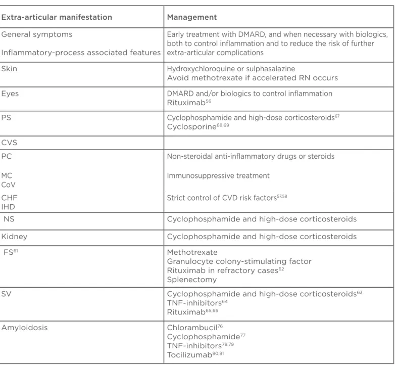
MANAGEMENT
The irst step in management of RA with or without extra-articular manifestations must be
early treatment with DMARD, both to control inlammation with subsequent articular progression and to reduce the risk of further extra-articular complications (Table 2). Progression of scleromalacia perforans can be prevented with this approach, although refractory cases may beneit from the use of biological agents; several papers have shown good results in controlling ocular complications, but clinical trials have not yet been reported.56
Table 2: Management of extra-articular manifestations in rheumatoid arthritis.
DMARD: disease modifying anti-rheumatic drugs; RN: rheumatoid nodulosis; PS: pulmonary system; CVS: cardiovascular system; PC: pericarditis; MC: myocarditis; CoV: coronary vasculitis; CHF: congestive heart failure; IHD: ischaemic heart disease; CVD: cardiovascular disease; NS: nervous system; FS: Felty’s syndrome; SV: systemic vasculitis; TNF: tumour necrosis factor.
Extra-articular manifestation Management
General symptoms
Inlammatory-process associated features
Early treatment with DMARD, and when necessary with biologics, both to control inlammation and to reduce the risk of further extra-articular complications
Skin Hydroxychloroquine or sulphasalazine
Avoid methotrexate if accelerated RN occurs
Eyes DMARD and/or biologics to control inlammation Rituximab56
PS Cyclophosphamide and high-dose corticosteroids67
Cyclosporine68,69
CVS
PC Non-steroidal anti-inlammatory drugs or steroids
MC CoV
Immunosuppressive treatment
CHF IHD
Strict control of CVD risk factors57,58
NS Cyclophosphamide and high-dose corticosteroids
Kidney Cyclophosphamide and high-dose corticosteroids
FS61 Methotrexate
Granulocyte colony-stimulating factor Rituximab in refractory cases62
Splenectomy
SV Cyclophosphamide and high-dose corticosteroids63
TNF-inhibitors64
Rituximab65,66
Amyloidosis Chlorambucil76
Cyclophosphamide77
TNF-inhibitors78,79
Although there is some evidence that CV risk in RA is reduced by successful suppression of inlammation, it remains important to identify and target traditional CVD risk factors as well. Guidelines emphasising the need for regular screening of patients with RA for CV risks have been recently published.57,58 Congestive heart failure
requires special consideration, since it does not appear to be fully related to traditional CVD risk factors or clinical IHD. Findings from a number of studies have shown that inlammatory cytokines are related to the echocardiographic indices of both systolic and diastolic left ventricular function.59
In addition, the inhibition of interleukin-1 (IL-1) showed an ability to improve myocardial deformation in these patients.60
The majority of traditional cardiac complications in RA are silent and do not require treatment. Symptomatic pericardial disease without haemodynamic compromise can be resolved with non-steroidal anti-inlammatory drugs or steroids, but recurrent forms may need immunosuppressive treatment. Constrictive pericarditis, and rapidly progressive pericarditis, require emergency intervention and can worsen the outcome of patients.
In patients with FS, neutropaenia can be efectively managed with DMARDs, the widest experience being with methotrexate. Splenectomy results in immediate improvement of neutropaenia in 80% of patients, but the rate of infection decreases to a lesser degree. Granulocyte colony-stimulating factor can be useful too. It seems to be logical to suppose that early aggressive treatment of RA may prevent the development of FS, but there are no epidemiological data to support this hypothesis.61 Some investigators reported a
response of FS to rituximab, while others found this treatment questionable. A systematic review evaluating biological treatment in FS concludes that the use of RTX can only be recommended as a second-line therapy in patients with refractory FS; experience with anti-tumour necrosis factor (TNF) agents is very limited with no improvement in neutrophil count.62 Spontaneous remission of the
syndrome can also occur.
For severe ExRA, such as systemic vasculitis, treatment with cyclophosphamide and high-dose corticosteroids has been considered as the recommended approach.63 According to other
reports,64 we have observed a dramatic response
of severe cutaneous vasculitic ulcers after anti-
TNF treatment in a RF-positive RA patient with long-standing severe disease (data unpublished,
Figure 2). On the other hand, rituximab has proved beneicial in RV, being an alternative to cyclophosphamide in selected patients.65,66
RA-associated lung disease treatment is controversial. It is mainly based on systemic steroids and cyclophosphamide,67 existing also positive data
with cyclosporine.68,69 Although a beneicial efect
of anti-CD20 therapy has been described in several case reports, physicians should be aware that this drug could trigger or worsen RA-related pulmonary ibrosis.70 Conlicting results have been published
with TNF-inhibitors, with reports on excellent response in refractory patients,71 as well as worsening
of ILD in others.72-74 Regression of parenchymal
PNs after tocilizumab treatment has been observed.75
Figure 2: Complete healing of severe cutaneous vasculitic ulcers after anti-tumour necrosis factor treatment (A: pre-treatment; B: post-treatment).
A
Standard treatment for AA amyloidosis has been based for a long time on colchicine, chlorambucil,76
or cyclophosphamide,77 but TNF inhibitors have
proved efective in controlling the progression of renal amyloidosis in patients with RA.78,79 Since
the activation of the serum amyloid A gene depends more on the presence of IL-6 than on the presence of TNF alpha, tocilizumab will probably be a irst-line treatment in the future.80,81
EXTRA-ARTICULAR MANIFESTATIONS
OF RA, NOW
A better control of disease activity during the last decades has improved the outcome of RA.82
ExRA incidence seems to be reduced too, although it is not equal for all manifestations: a decline in RV incidence has been found in several studies,83,84
even though clinical manifestations remain similar and its prognosis remains poor despite modern immunosuppressive therapy.85 Secondary
amyloidosis with clinically apparent organ manifestations is not present in the most recent series of RA patients. On the contrary, it has not been a signiicant change in the incidence of other severe ExRA manifestations, like RA-associated lung disease. Moreover, milder ExRA manifestations, such as KCS, has been diagnosed more frequently among patients with a more recent onset of RA, possibly because of improved clinical surveillance.86
The clinician must be familiar with ExRA diagnosis, which is often complicated and diicult, and its management. New immunosuppressive drugs may ofer interesting possibilities, although until now quality studies are lacking and there is a need to act with care. Undoubtedly, the future for RA patients is encouraging.
REFERENCES
1. Gabriel SE et al. Survival in rheumatoid arthritis: a population-based analysis of trends over 40 years. Arthritis Rheum. 2003;48(1):54–8.
2. Young A, Koduri G. Extra-articular manifestations and complications of rheumatoid arthritis. Best Pract Res Clin Rheumatol. 2007;21(5):907–27.
3. Turesson C et al. Multiple extra-articular manifestations are associated with poor survival in patients with rheumatoid arthritis. Ann Rheum Dis. 2006;65(11):1533–4.
4. Prete M et al. Extra-articular manifestations of rheumatoid arthritis: an update. Autoimmun Rev. 2011;11(2):123–31. 5. Turesson C et al. Predictors of extra-articular manifestations in rheumatoid arthritis. Scand J Rheumatol. 2000;29(6):358-64.
6. Balsamo S et al. Exercise and fatigue in rheumatoid arthritis. Isr Med Assoc J. 2014;16(1):57-60.
7. Kirwan JR et al. OMERACT 10 Patient Perspective Virtual Campus: valuing health; measuring outcomes in rheumatoid arthritis fatigue, RA sleep, arthroplasty, and systemic sclerosis; and clinical signiicance of changes in health. J Rheumatol. 2011;38(8):1728-34.
8. Wilson A et al. Prevalence and outcomes of anaemia in rheumatoid arthritis: a systematic review of the literature. Am J Med. 2004;116:50S–7S.
9. Zif M. The rheumatoid nodule. Arthritis Rheum. 1990;33(6):761–7.
10. Cunnane G et al. Accelerated nodulosis
Rheum. 2002;47(4):445–9.
11. Zlatanovic G et al. Ocular manifestation of rheumatoid arthritis-diferent forms and frequency. Bosn J Basic Med Sci. 2010;10(4):323–7.
12. Smith JR et al. Therapy Insight: scleritis and its relationship to systemic autoimmune disease. Nat Clin Prac Rheumatol. 2007;3(4):219-26.
13. Bongartz T et al. Incidence and mortality of interstitial lung disease in rheumatoid arthritis: a population-based study. Arthritis Rheum. 2010;62(6): 1583–91.
14. Young A et al.; Early Rheumatoid Arthritis Study (ERAS) group. Mortality in rheumatoid arthritis. Increased in the early course of disease, in ischaemic heart disease and in pulmonary ibrosis. Rheumatology (Oxford). 2007;46(2): 350–7.
15. Gizinski AM et al. Rheumatoid arthritis (RA)-speciic autoantibodies in patients with interstitial lung disease and absence of clinically apparent articular RA. Clin Rheumatol. 2009;28(5):611–3.
16. Chen J et al. Asymptomatic preclinical
rheumatoid arthritis-associated interstitial lung disease. Clin Dev Immunol.
2013;2013:406927.
17. Mielants H, Van den Bosch F. Extra-articular manifestations. Clin Exp Rheumatol. 2009;27(4 Suppl 55):S56-61. 18. Kelly C, Hamilton J. What kills patients with rheumatoid arthritis? Rheumatology (Oxford). 2007;46(2):183–4.
19. Sihvonen S et al. Death rates and causes
J Rheumatol. 2004;33(4):221–7.
20. Lee HK et al. Histopathologic pattern and clinical features of rheumatoid arthritis-associated interstitial lung disease. Chest. 2005;127(6):2019–27.
21. Devouassoux G et al. Characterisation of severe obliterative bronchiolitis in rheumatoid arthritis. Eur Respir J. 2009;33:1053–61.
22. Anaya JM et al. Pulmonary involvement in rheumatoid arthritis. Semin Arthritis Rheum. 1995;24(4):242-54.
23. Youssef AA et al. Respiratory symptoms in rheumatoid arthritis: relation to pulmonary abnormalities detected by high-resolution CT and pulmonary functional testing. Rheumatol Int. 2012;32(7):1985–95.
24. Wilsher M et al. Prevalence of airway and parenchymal abnormalities in newly diagnosed rheumatoid arthritis. Respir Med. 2012;106(10):1441–6.
25. Lake F, Proudman S. Rheumatoid arthritis and lung disease: from mechanisms to a practical approach. Semin Respir Crit Care Med. 2014;35: 222–38.
26. Lioté H. [Pulmonary manifestation of rheumatoid arthritis]. Rev Mal Respir. 2008;25(8):973-88.
27. Dawson JK et al. Fibrosing alveolitis in patients with rheumatoid arthritis as assessed by high resolution computed tomography, chest radiography, and pulmonary function tests. Thorax. 2001;56(8):622–7.
2010;9:849–52.
29. Nicola PJ et al. The risk of congestive heart failure in rheumatoid arthritis: a population-based study over 46 years. Arthritis Rheum. 2005;52(2):412–20. 30. del Rincon ID et al. High incidence of cardiovascular events in a rheumatoid arthritis cohort not explained by traditional cardiac risk factors. Arthritis Rheum. 2001;44:2737–45.
31. Dessein PH et al. Traditional and non-traditional cardiovascular risk factors are associated with atherosclerosis in rheumatoid arthritis. J Rheumatol. 2005;32:435–42.
32. Gonzalez-Gay MA et al. HLA–DRB1 and persistent chronic inlammation contribute to cardiovascular events and cardiovascular mortality in patients with rheumatoid arthritis. Arthritis Rheum. 2007;57:125–32.
33. Rodriguez-Rodriguez L et al. TNFA-308 (rs1800629) polymorphism is associated with a higher risk of cardiovascular disease in patients with rheumatoid arthritis. Atherosclerosis. 2011;216:125–30.
34. Voskuyl AE. The heart and cardiovascular manifestations in rheumatoid arthritis. Rheumatology. 2006;45 Suppl 4:iv4-7.
35. Bartolini M et al. Are behaviour and motor performances of rheumatoid arthritis patients inluenced by subclinical cognitive impairments? A clinical and neuroimaging study. Clin Exp Rheumatol. 2002;20:491–7.
36. Appenzeller S et al. Cognitive impairment in rheumatoid arthritis. Methods Find Exp Clin Pharmacol. 2004;26:339–43.
37. Shin SY et al. Cognitive impairment in persons with rheumatoid arthritis. Arthritis Care Res (Hoboken). 2012;64(8):1144–50. 38. Aviña-Zubieta JA et al. Risk of cardiovascular mortality in patients with rheumatoid arthritis: a meta-analysis of observational studies. Arthritis Rheum. 2008;59:1690.
39. Caballol Pons N et al. Isolated cerebral vasculitis associated with rheumatoid arthritis. Joint Bone Spine. 2010;77(4):361–3.
40. Nakano M et al. Analysis of renal pathology and drug history in 158 Japanese patients with rheumatoid arthritis. Clin Nephrol. 1998;50:154–60. 41. Helin HJ et al. Renal biopsy indings and clinicopathologic correlations in rheumatoid arthritis. Arthritis Rheum. 1995;38:242–7.
42. Palomar R et al. [Microscopic polyangiitis in a patient with rheumatoid arthritis.] Nefrologia. 2005;25:438–41. 43. Karie S et al. Kidney disease in RA patients: prevalence and implication
on RA-related drugs management: the MATRIX Study. Rheumatology (Oxford). 2008;47(3):350-4.
44. National Kidney Foundation. K/DOQI Clinical Practice Guidelines for Chronic Kidney Disease: evaluation, classiication, and stratiication. Am J Kidney Dis. 2002:39(suppl 1):S1-S266.
45. Hickson LJ et al. Development of reduced kidney function in rheumatoid arthritis. Am J Kidney Dis. 2014;63(2): 206-13.
46. Lachmann HJ et al. Natural history and outcome in systemic AA amyloidosis. N Engl J Med. 2007;356(23):2361-71. 47. Ishii W et al. Abdominal fat aspiration biopsy and genotyping of serum amyloid A contribute to early diagnosis of reactive AA amyloidosis secondary to rheumatoid arthritis. Internal Medicine. 2003;42(9):800-5.
48. Gomez-Casanovas E et al. The clinical signiicance of amyloid fat deposits in rheumatoid arthritis: a systematic long-term follow-up study using abdominal fat aspiration. Arthritis Rheum. 2001;44: 66-72.
49. Scott DG et al. Systemic rheumatoid vasculitis: a clinical and laboratory study of 50 cases. Medicine (Baltimore). 1981;60:288.
50. Turesson C et al. Clustering of extraarticular manifestations in patients with rheumatoid arthritis. J Rheumatol. 2008;35:179–80.
51. Turesson C et al. Association of HLAC3 and smoking with vasculitis in patients with rheumatoid arthritis. Arthritis Rheum. 2006;54:2276–83.
52. Turesson C et al. The impact of HLA-DRB1 genes on extra-articular disease manifestations in rheumatoid arthritis. Arthritis Res Ther. 2005;7:1386–93.
53. Watts RA et al. Isolated nail fold vasculitis in rheumatoid arthritis. Ann Rheum Dis. 1995;54:927.
54. Campion G et al. The Felty syndrome: a case-matched study of clinical manifestations and outcome, serologic features, and immunogenetic associations. Medicine. 1990;69:69–80. 55. Breedveld FC et al. Factors inluencing the incidence of infections in Felty’s syndrome. Arch Intern Med. 1987;147: 915–20.
56. Iaccheri B et al. Rituximab treatment for persistent scleritis associated with rheumatoid arthritis. Ocul Immunol Inlamm. 2010;18(3):223–5.
57. Martín-Martínez MA et al. Recommendations for the management of cardiovascular risk in patients with rheumatoid arthritis: scientiic evidence and expert opinion. Semin Arthritis Rheum. 2014;doi:10.1016/j.semarthrit.2014.01.002. [Epub ahead of print].
58. Peters MLJ et al. EULAR evidence-based recommendations for cardiovascular risk management in patients with rheumatoid arthritis and other forms of inlammatory arthritis. Ann Rheum Dis. 2010;69:325-31.
59. Chrysohoou C et al. Chronic systemic inlammation accompanies impaired ventricular diastolic function, detected by Doppler imaging, in patients with newly diagnosed systolic heart failure (Hellenic Heart Failure Study). Heart Vessels. 2009;24:22-6.
60. Ikonomidis I et al. Lowering interleukin-1 activity with anakinra improves myocardial deformation in rheumatoid arthritis. Heart. 2009;95(18):1502-7. 61. Balint GP, Balint PV. Felty’s syndrome. Best Pract Res Clin Rheumatol. 2004;18(5):631-45.
62. Narváez J et al. Biological agents in the management of Felty’s syndrome: a systematic review. Semin Arthritis Rheum. 2012;41(5):658-68.
63. Scott DG, Bacon PA. Intravenous cyclophosphamide plus methylprednisolone in treatment of systemic rheumatoid vasculitis. Am J Med. 1984;76:377–84.
64. Puechal X et al. Anti-tumour necrosis factor treatment in patients with refractory systemic vasculitis associated with rheumatoid arthritis. Ann Rheum Dis. 2008;67:880–4.
65. Hellmann M et al. Successful treatment of rheumatoid vasculitis-associated cutaneous ulcers using rituximab in two patients with rheumatoid arthritis. Rheumatology (Oxford). 2008;247: 929-30.
66. Assmann G et al. Rituximab in patients with rheumatoid arthritis and vasculitis associated cutaneous ulcers. Clin Exp Rheumatol. 2010;28:81.
67. Turesson C. Extra-articular rheumatoid arthritis. Curr Opin Rheumatol. 2013;25(3):360-6.
68. Ogawa D et al. Successful use of cyclosporin A for the treatment of acute interstitial pneumonitis associated with rheumatoid arthritis. Rheumatology (Oxford). 2000;39:1422–4.
69. Song J-W et al. Clinical course and outcome of rheumatoid arthritis-related usual interstitial pneumonia. Sarcoidosis Vasc Difuse Lung Dis. 2013;30(2):103–12. 70. Lioté H et al. Rituximab-induced lung disease: a systematic literature review. Eur Respir J. 2010;35(3):681-7.
71. Vassallo R et al. Clinical response of rheumatoid arthritis-associated pulmonary ibrosis to tumor necrosis factor-alpha inhibition. Chest. 2002;122:1093–6.
by DMARDS and biologic agents in rheumatoid arthritis: a systematic literature review. Semin Arthritis Rheum. 2014;43:613–26.
73. Wolfe F et al. Rheumatoid arthritis treatment and the risk of severe interstitial lung disease. Scand J Rheumatol. 2007;36(3):172-8.
74. Perez-Alvarez R et al. Interstitial lung disease induced or exacerbated by TNF-targeted therapies: analysis of 122 cases. Semin Arthritis Rheum. 2011;41(2):256–64. 75. Andres M et al. Marked improvement of lung rheumatoid nodules after treatment with tocilizumab. Rheumatology. 2012;51(6):1132-4.
76. Ortiz-Santamaria V et al. Treatment of AA amyloid with chlorambucil. Rheumatology (Oxford). 2002;41:833. 77. Nakamura T et al. Eicacy of cyclophosphamide combined with prednisolone in patients with AA
amyloidosis secondary to rheumatoid arthritis. Clin Rheumatol. 2003;22:371–5. 78. Fernández-Nebro A et al. Treatment of rheumatic inlammatory disease in 25 patients with secondary amyloidosis using tumor necrosis factor alpha antagonists. Am J Med. 2005;118(5):552-6.
79. Nakamura T et al. Eicacy of etanercept in patients with AA amyloidosis secondary to rheumatoid arthritis. Clin Exp Rheumatol. 2007;25:518–22.
80. Nishida S et al. Rapid improvement of AA amyloidosis with humanised anti-interleukin 6 receptor antibody treatment. Ann Rheum Dis. 2009;68:1235–6.
81. Inoue D et al. Excellent therapeutic efect of tocilizumab on intestinal amyloid a deposition secondary to active rheumatoid arthritis. Clin Rheumatol. 2010;29:1195–7.
82. Van Nies JA et al. What is the evidence for the presence of a therapeutic window
of opportunity in rheumatoid arthritis? A systematic literature review. Ann Rheum Dis. 2014;73(5):861-70.
83. Turesson C, Matteson EL. Vasculitis in rheumatoid arthritis. Curr Opin Rheumatol. 2009;21(1):35- 40.
84. Bartels CM et al. Changing trends in serious extra-articular manifestations of rheumatoid arthritis among United States veterans over 20 years. Rheumatology. 2010;49(9):1670-5.
85. Ntatsaki E et al. Systemic rheumatoid vasculitis in the era of modern immunosuppressive therapy. Rheumatology. 2014;53(1):145-52.