© 2012 A. Pandey et al., This open access article is distributed under a Creative Commons Attribution (CC-BY) 3.0 license
doi:10.3844/ojbssp.2012.123.133 Published Online 12 (4) 2012 (http://www.thescipub.com/ojbs.toc)
Corresponding Author: Ashutosh Pandey, Molecular Plant Pathology Laboratory, Central Institute for Subtropical Horticulture, Lucknow- 227 107, Uttar Pradesh, India Tel: +91 933 616 2218, Fax: +91 5278 265201
EFFECTIVENESS OF CULTURAL PARAMETERS ON THE
GROWTH AND SPORULATION OF COLLETOTRICHUM
GLOEOSPORIOIDES CAUSING ANTHRACNOSE DISEASE OF
MANGO (MANGIFERA INDICA L.)
1
Ashutosh Pandey,
1Lallan Prasad Yadava,
1Muthukumar Manoharan,
2
Ugam Kumari Chauhan and
1Brajesh Kumar Pandey
1Central Institute for Subtropical Horticulture,
Molecular Plant Pathology Laboratory, Lucknow-227 107, Uttar Pradesh, India
2
Center for Biotechnology, School of Environmental Biology, A.P.S. University, Rewa- 486 003, Madhya Pradesh, India
ABSTRACT
Colletotrichum gloeosporioides causing anthracnose which is a serious post harvest disease in mango accounting for 15-20% loss. The variation in nutritional and physiological characteristics among five isolates of C. gloeosporioides collected from different agro-climatic regions of India was investigated. All the isolates showed differential response in requirements of media, temperature and media pH for growth and sporulation. Malt Extract Agar (MEA) medium was best suited for growth in terms of radial mycelial diameter for all the isolates. Among the studied isolates, Cg 72 (from Maharashtra) showed more virulence and maximum sporulation (137.5×10³ mL−1) at 28°C and media pH 6. Maximum growth and virulence at 28°C was observed with Cg 62 isolate. Media of pH 6 was found to be most suitable for the growth of respective isolates (s), but Cg 62 which was collected from Bihar found most virulent in this experiment.
Keywords: Colletotrichum gloeosporioides, Mangifera Indica L., Radial Growth, Sporulation, Culture Media, Temperature, Media pH
1. INTRODUCTION
Mango (Mangifera indica L.) is one of the most popular seasonal fruit found mainly in the tropical and subtropical countries (Shad et al., 2002). It is widely grown in different countries of the world and is attacked by a number of diseases of which anthracnose is one of the most common especially in India and loss due to this disease is substantial (Slade et al., 1987). Anthracnose caused by C. gloeosporioides is reported on a wide variety of crop, including almond, avocado, apple, arabica coffee, guava, mango, dragon fruit, cassava, sorghum and strawberry (Amusa et al., 2005; Masyahit et al., 2009; Owolade et al., 2009; Erpelding, 2010).
The optimum pH was between 5.8 and 6.5 and temperature was optimum at 25°C for growth but it ceased beyond 35°C. The fungus grew vigorously on starch and peptone. Glutamic acid and alanine supported best growth and sporulation (Prakash and Srivastava, 1987). Keeping in view of the above facts the present investigation was performed to assess the effect of media, temperature and media pH on the growth and sporulation of isolates of C. gloeosporioides collected from Andhra Pradesh, Madhya Pradesh, Bihar, Maharashtra and Uttar Pradesh.
2. MATERIALS AND METHODS
2.1. Collection and Isolation of Pathogens
The isolates of C. gloeosporioides were isolated from the samples collected from Chittoor (Andhra Pradesh), Rewa (Madhya Pradesh), Lucknow (Uttar Pradesh), Muzaffarpur (Bihar) and Dapoli (Maharashtra). A small section of anthracnose infected leaf was surface sterilized with 0.1% HgCl2 and washed thoroughly with
sterile distilled water. It was then inoculated on Potato Dextrose Agar (PDA) medium (Potato 200 g, Glucose 20 g, Agar 15 g, distilled water 1000 mL) and incubated at 28±2°C for 6 days.
A total of 5 C. gloeosporioides isolates (Cg 1, Cg 19, Cg 30, Cg 62 and Cg 72) were used in this study. These isolates were isolated, purified and maintained on Potato Dextrose Agar (PDA) slants under controlled temperature. Pathogenicity of these isolates was also confirmed suggested by Bhuvanaeswari and Rao (2001). All the collected isolates of C. gloeosporioides were submitted to National Agriculture Important Microbial Culture Collection (NAIMCC), Mau, Uttar Pradesh, India and accessions number were allotted (Table 1).
2.2. Preparation
of
Different
Media
and
Inoculation
The fungal pathogen was inoculated on various types of media to identify the best suited media for its growth and sporulation. In this experiment, fourteen media (Himedia, Mumbai), viz., Potato Dextrose Agar (PDA),
Corn Meal Agar (CMA), Cooke rose Bengal agar base (CRBA), Czapek Dox Agar (CDA), Pseudomonas Agar (PA), Limabean Agar (LA), Conn’s Agar (CA), Yeast Dextrose Agar (YDA), Corn Meal Peptone Yeast Agar (CMPYA), Modified Czapex Dox Agar (MCDA), V-8 Juice Agar (VJA), Potato carrot agar (PCA), Malt extract agar base Mycological peptone (MEA) and Oat Meal Agar (OMA) were used 39, 17, 36.54, 49, 37.3, 23, 38, 35, 64, 45.36, 44.3, 24, 50 and 72.5 gram per liter respectively. All these were autoclaved at 121°C under 15 psi for 20 min. Five mm diameter identical fresh culture discs of different isolates (Cg 1, Cg 19, Cg 30, Cg 62 and Cg 72) were grown for 7 days old culture at 27±2°C dissolve in 1 ml distilled water and count.
2.3. Preparation of Media at Different level of
pH and Inoculation
To assess the optimum media pH for the growth of C. gloeosporioides the fresh culture on Malt Extract Agar (MEA) medium of different pH level (5.5, 6.0, 6.5, 7.0, 7.5 and 8.0) was used in experiment. The media pH of the medium was adjusted with 0.1N NaOH or 0.1N HCl (Naik et al., 1988). The medium was buffered with disodium hydrogen phosphate citric acid buffer according to the protocol (Vogel, 1951). For inoculation same method was adopted as mentioned earlier. The diameter of colony was observed at 3rd, 5th and 7th days after inoculation while the level of sporulation was recorded at 7th Day After Inoculation (DAI) by modifying the methods (Tastwal and Enagi, 2009).
2.4. Incubation
at
Different
Temperature
Regime
To study the growth and sporulation of C. gloeosporioides at different temperature regime fresh culture was prepared in the solid media viz. Malt Extract Agar (MEA). For inoculation same method was adopted as mentioned earlier and culture of different isolates were placed at different temperature regimes to study the best suited temperature level viz. (12, 16, 20, 24, 28 and 32°C). Data were recorded with the method(Kumara and Rawal, 2008).
Table 1. Morphological characteristic of different isolates from different geographical location of Colletotrichum gloeosporioides penz
Culture Culture Geographical Symptom of Mango Percent Sporu Mean spore size Colour of
Code accession no. location anthracnose substrate infection lation (Length × breadth) ( m) mycelial mat
Cg 1 NAIMCC-F-02694 Chittoor (Andhra Pradesh) Partial Leaf 50 ** 13.49×4.75 White black
Cg 19 NAIMCC-F-02707 Rewa (Madhya Pradesh) Typical Leaf 100 *** 14.20×5.25 Yellow white
Cg 30 NAIMCC-F-02719 Lucknow (Utter Pradesh) Typical Fruit 70 Nil 16.15×7.28 Grayish white
Cg 62 NAIMCC-F-02730 Muzaffarpur (Bihar) Typical Leaf 100 *** 15.78×4.38 White
Cg 72 NAIMCC-F-02735 Dapoli (Maharashtra) Partial Leaf 75 *** 16.87×4.57 Cottony white
2.5. Statistical Analysis
All treatments were designed in three factors factorial Completely Randomized Design (CRD) with five replications. Experimental data was statistically analyzed using O. P. Sheoran software version 1.0 (CCS HAU, Hisar). The Dendrogram was generated by Un-weighted Pair-Group Arithmetic Mean (UPGMA) using FREE TREE software version 0.9.1.50.
3. RESULTS
The effect of various factors such as media, media pH, temperature regimes and their combinations with days after inoculation on the growth (Table 2) and sporulation of C. gloeosporioides were studied for 5 isolates representing different agro climatic zones.
3.1. Effect of Media on Growth and Sporulation
of C. Gloeosporioides Isolates
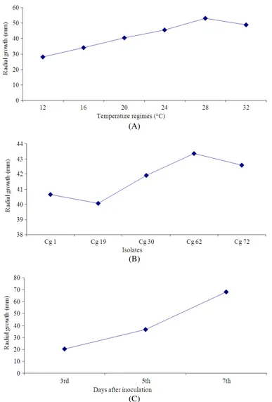
Malt Extract Agar (MEA) showed higher mean value (54.98 mm) of mycelia growth as compared to other evaluated media followed by Modified Czapex Dox agar medium MCDA (53.01 mm). Cooke rose Bengal agar base (CRBA) media exhibited the least mycelial diameter (18.29 mm). Pseudomonas Agar (PA) and Limabean Agar (LA) medium also recorded lower growth next to CRBA medium but both of them were found to be similar in response (Fig. 1A).
Maximum (46.12 mm) growth was noticed with Cg 72 isolate in over all media while the least growth was recorded in Cg 1 (36.34 mm) (Fig. 1B). Cg 1 showed comparatively slow growth in all the media evaluated for the radial growth. As far as the effects of interaction between media and isolates are concerned, maximum (60.88 mm) growth was recorded with Cg 72 isolate in the MEA media followed by MCDA (60.22 mm).
A significant interaction effect of media and incubation period revealed highest growth in MEA medium followed by MCDA both at 5 and 7th day after inoculation CDA and OMA medium were also at par with MEA medium at 7th days after inoculation. The period of incubation clearly indicated that radial growth of the mycelia increased with increase in incubation period (days after inoculation) (Fig. 1C). The data of interaction between isolates and incubation period clearly showed that mycelial growth in all the isolates increased with advancement of incubation period. CRBA medium did not follow significantly for growth enhancement throughout the incubation period. With respect to the isolate effect, maximum radial growth was
recorded at 7th day in Cg 72 (71.27 mm) while minimum was found in Cg 1 isolate at 3rd day after inoculation. It is also evident that mycelial growth of C. gloeosporioides was significantly influenced by interaction of media × isolates × incubation period. Higher radial growth (87.67 mm) was observed in Cg 19 isolate in Oat Meal Agar (OMA) medium at 7th day of incubation followed by the same isolates in Malt extract agar (86.33 mm), Cg 72 (85.33 mm) and Cg 19 (85.17 mm) in CDA, Cg 72 in MEA (84.0 mm) and MCDA (83.67 mm) at 7th day of incubation.
In the experiment of sporulation in different isolates was also significantly varied by media (Table 3). Highest spore count (137.5×10³ mL−1 of culture suspension) was recorded in Cg 72 isolate on MEA medium followed by Cg 19 on CDA medium (102.5×10³ mL−1 of culture suspension).
3.2. Effect of Media pH on Growth and
Sporulation of C. Gloeosporioides Isolates
The media pH is another important factor like temperature and media that influences growth and sporulation significantly. The effect of different media pH on the growth was observed considering test isolates at different incubation period.
It is evident from the data, that mycelial growth in different isolates significantly differed by pH range of the media. The media pH of 6.0 exhibited higher mycelial growth (54.43 mm) over other level of pH studied (Fig. 2A). Among the isolates, Cg 62 resulted in highest growth (43.86 mm) at this pH followed by Cg 1 (Fig. 2B).
The effect of media pH × incubation period interaction with respect to radial mycelial growth was significant. The radial growth pattern was single sigmoid with respect to media pH with peak inferring highest radial growth at 7th day of incubation in media pH 6. Cg 62 and Cg 1 isolate showed the highest radial growth (67.60 mm) at the 7th day of incubation, while minimum was found with Cg 19 isolate at 3rd day of incubation. It is interesting to note that radial growth of all the isolates was at par with each other on 5th day of incubation. However, radial growth of all the isolates increased with the advancement of incubation period (Fig. 2C).
Three factors interactions among media pH, isolates and incubation period revealed that mycelial growth of all the test isolates increased with incubation period in corresponding order. Higher radial growth (83.20 mm) was recorded with Cg 1 isolate followed by Cg 62 (82.40 mm) and Cg 30 (81.40 mm) grown in the media 6.0 pH
Table 2. ANOVA for different isolates of C. gloeosporioides under different media, media pH and temperature regimes
Source Degree Sum of Mean of sum F- Critical
of variance of freedom squires of squires calculated P value Deference (C.D.)
Media 13 53381.45 4106.27 393.35 0.00001 1.339
Isolates 4 8248.73 2062.18 197.54 0.00001 0.800
Day after inoculation 2 231091.70 115545.80 11068.40 0.00001 0.620
Media × isolates 52 6161.30 118.49 11.35 0.00001 2.994
Media × Day after inoculation 26 30117.31 1158.36 110.96 0.00001 2.319
Isolates × Day after inoculation 8 2282.16 285.27 27.33 0.00001 1.386
Media × isolates × Day after inoculation 104 5671.09 54.53 5.22 0.00001 5.186
Media pH 5 26177.96 5235.59 2008.54 0.00001 0.519
Isolates 4 1685.27 421.32 161.63 0.00001 0.473
Days after inoculation 2 178894.50 89447.26 34314.81 0.00001 0.367
Media pH × isolates 20 740.12 37.01 14.20 0.00001 1.159
Media pH × Days after inoculation 10 5125.64 512.56 196.64 0.00001 0.898
Isolates × Days after inoculation 8 338.65 42.33 16.24 0.00001 0.820
Media pH × isolates × Days after inoculation 40 1641.72 41.04 15.75 0.00001 2.008
Temperature regime 5 33325.10 6665.02 4697.35 0.00001 0.383
Isolates 4 659.96 164.99 116.28 0.00001 0.349
Days after inoculation 2 176546.00 88272.98 62212.75 0.00001 0.271
Temperature regime × isolates 20 102.86 5.14 3.62 0.00001 0.855
Temperature regime × Days after inoculation 10 6349.33 634.93 447.49 0.00001 0.663
Isolates × Days after inoculation 8 32.38 4.05 2.85 0.00437 0.605
Temperature regime × 40 118.47 2.96 2.09 0.00023 1.482
isolates × days after inoculation
Table 3. Sporulation of different Colletotrichum gloeosporioides isolates under different media pH
pH ranges Cg 1 Cg 19 Cg 30 Cg 62 Cg 72 Mean
5.5 74.0000 151.00 200.3300 80.00 135.0000 128.06
6 51.0000 65.00 101.0000 63.00 94.0000 74.80
6.5 12.0000 26.00 40.0000 34.00 21.0000 26.60
7 11.0000 11.00 9.0000 15.00 21.0000 13.40
7.5 7.0000 7.00 10.0000 11.00 15.0000 10.00
8 7.0000 4.33 9.0000 3.66 1.6600 5.13
Mean 27.0000 44.05 61.5500 34.44 47.9400
Factors C.D. SE(d) SE(m)
pH ranges 0.6973 0.3485 0.2465
Isolates 0.6366 0.3182 0.2250
pH ranges x isolates 1.5590 0.7794 0.5511
Table 4. Sporulation of different Colletotrichum gloeosporioides isolates under different temperature regimes
Temperature regimes (°C) Cg 1 Cg 19 Cg 30 Cg 62 Cg 72 Mean
12 2.00 4.0000 3.33 2.0000 7.3300 3.73
16 6.33 8.0000 9.00 3.0000 12.0000 7.66
20 20.00 16.0000 15.00 15.0000 12.0000 15.60
24 51.00 43.0000 34.66 30.0000 42.0000 40.13
28 103.00 99.0000 111.00 126.0000 114.0000 110.60
32 76.33 59.0000 66.66 60.0000 62.6600 64.93
Mean 43.11 38.1600 39.94 39.3300 41.6600
Factors C.D. SE(d) SE(m)
Temperature regimes 0.7466 0.3732 0.2639
Isolates 0.6816 0.3407 0.2409
(A)
(B)
(C)
Fig. 1. (A) Radial growth of Colletotrichum gloeosporioides on the different media (B) Radial growth of different Colletotrichum gloeosporioides isolates under different media (c)Radial growth of Colletotrichum gloeosporioides at different incubation period under different media
(B)
(C)
Fig. 2. (A) Radial growth of Colletotrichum gloeosporioides on the different media pH (B) Radial growth of different Colletotrichum gloeosporioides isolates under different media pH (C) Radial growth of Colletotrichum gloeosporioides at different incubation period under different media pH
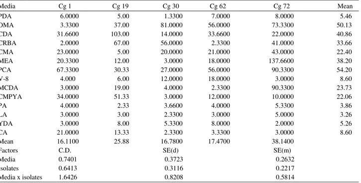
Table 5. Sporulation of different Colletotrichum gloeosporioides isolates under different media
Media Cg 1 Cg 19 Cg 30 Cg 62 Cg 72 Mean
PDA 6.0000 5.00 1.3300 7.0000 8.0000 5.46
OMA 3.3300 37.00 81.0000 56.0000 73.3300 50.13
CDA 31.6600 103.00 14.0000 33.6600 22.0000 40.86
CRBA 2.0000 67.00 56.0000 2.3300 41.0000 33.66
CMA 23.0000 5.00 20.0000 21.0000 43.0000 22.40
MEA 20.3300 12.00 3.0000 18.0000 137.6600 38.20
PCA 67.3300 30.33 27.0000 56.0000 90.3300 54.20
V-8 4.000 6.00 12.0000 18.0000 3.0000 8.60
MCDA 3.0000 19.00 4.0000 2.3300 90.3300 23.73
CMPYA 34.0000 51.33 3.0000 12.0000 10.0000 22.06
PA 4.0000 2.33 3.6600 4.0000 5.3300 3.86
LA 3.0000 3.00 2.3300 3.0000 5.0000 3.26
YDA 3.0000 8.00 5.3300 8.0000 2.0000 5.26
CA 21.0000 13.33 2.3300 3.3300 3.0000 8.60
Mean 16.1100 25.88 16.7800 17.4700 38.1400
Factors C.D. SE(d) SE(m)
Media 0.7401 0.3723 0.2632
Isolates 0.6413 0.3116 0.2217
(A)
(B)
(C)
Fig. 3. (A) Radial growth of Colletotrichum gloeosporioides on different temperature regimes (B) Radial growth of different Colletotrichum gloeosporioides isolates under different temperature regimes (C) Radial growth of Colletotrichum gloeosporioides at different incubation period under different temperature regimes
Sporulation was highest at media pH 5.5 in all the isolates followed by media pH 6. However, as media pH increased the sporulation was increased consistently in all the test isolates (Table 4).
3.3. Effect of Temperature Regimes on Growth
and Sporulation of C. Gloeosporioides Isolates
It was observed that growth of mycelia in different isolates significantly varied with temperature regimes and incubation period. Higher (53.17 mm) radial growth of mycelia was observed at 28°C while lesser growth (28.03 mm) was at 12°C (Fig. 3A). Among the isolate Cg 62 showed highest growth (43.36 mm) than other isolates, whereas least growth (40.07 mm) was observed in Cg 19 (Fig. 3B). A significant effect of temperature x isolates interaction on the radial growth of mycelia was noticed where in maximum growth was recorded with Cg 62 isolate followed by isolate Cg 72 at 28°C.
The radial growth of mycelia increased with advancement of incubation period of the culture (Fig. 3C). The interaction between temperature and incubation period of culture also showed significant effect on mycelial growth. Statistically significant average growth (82.24 mm) was observed at 28°C after 7th days of incubation.
It is evident from the data that interaction between isolates and incubation period exhibited significant difference in mycelial growth of all the isolates. Maximum radial growth was recorded at 7th days of incubation with Cg 62 isolate (69.80 mm) while minimum was with Cg 19 isolate (18.57 mm) at 3rd day of incubation. Radial growth of mycelia was significantly influenced by interaction of temperature x isolates x incubation period. Higher radial growth was observed with Cg 62 isolate (84.60 mm) followed by Cg 72 isolate (84.20 mm) at 7th day of incubation in 28°C.
Temperature showed marked effect on the sporulation in the culture of test isolates. The best sporulation was recorded (125×103 mL−1 of culture suspension) in the culture of Cg 62 isolate at 28°C. Although, it was observed that sporulation in all the culture of test isolates increased in temperature regime upto 28°C, but rate of sporulation decreased when temperature regime of the culture increased from 28° to 32°C during experimentation (Table 5).
The dendrogram profile showed that the isolates were grouped into two major cluster, sharing 72% maximum similarity. The entire two cluster consisted test isolates collected from different state showed higher degree of cultural variability. Avery close association was found between isolates Cg 1 and Cg 30 collected from Chittoor (Andhra Pradesh) and Lucknow (Uttar Pradesh)
respectively in the first cluster. Second cluster contains Cg 19, Cg 62 and Cg 72 collecting from Rewa (Madhya Pradesh), Muzaffarpur (Bihar) and Dapoli (Maharashtra) respectively showing more than 56% similarity with each other (Fig. 4).
4. DISCUSSION
Colletotrichum gloeosporioides, a filamentous fungus, causing anthracnose disease in fruit crops, is reported to exhibit different requirements of nutrients and optimum conditions either for growth or sporulation (Shin et al., 2000). There is, therefore, a need to study these parameters for mango anthracnose pathogen in order to establish the survivability of C. gloeosporioides in soil(Green, 1994). The present study has focused on resolving these issues pertaining to optimum conditions for growth and sporulation of C. gloeosporioides.
Growth of mycelium and sporulation are influenced by the medium, pH and temperature (Kumara and Rawal, 2008). These factors independently and or in combination have positive and negative effects in most of the fungi have been reported by several workers. Media containing carbohydrates, lipids, proteins and elements are basic requirements and needed by the microorganisms as these nutrients provide energy for biosynthesis and cell maintenance (Hilton, 1999). Production of biomass in fungi and growth-associated products requires nutrient-balanced media (Hilton, 1999). Some dimorphic fungi require optimal nutrition to produce high biomass, but for sporulation require nutritionally poor media which trigger differentiation of conidia from vegetative growth (Vega et al., 2003).
In a study on growth of C. gloeosporioides under in vitro conditions, maximum growth was obtained after 10th day of incubation on potato dextrose broth, with optimum temperature in the range of 25-35°C (Hegde et al., 1990). Temperature affects almost every function of the fungi including sporulation(Lilly and Barnett, 1951). A temperature range of 15-35°C was also suggested to be most suitable for maximum sporulation (Sattar and Malik, 1939). Meanwhile, 15-20°C favoured the conidia formation by C. lindemuthianum in culture (Mathur et al., 1950). Further they reported that the sporulation was less at 25°C and ceased at 30°C. Ideally, 20 and 25°C was reported as the most favorable temperature for colony growth and sporulation in many fungi. Recently, maximum growth of C. gloeosporioides isolates (from Dapoli, Hessarghatta and Tumkur) at 28°C was identified while, 30°C supported growth of Hassan and Raichur isolates (Sangeetha and Rawal, 2009). Earlier, recorded maximum growth of different isolates of C. gloeosporioides at a temperature range of 25-30°C in the mangobut sporulation was at an optimum range of 25-28°C (Sangeetha, 2003). In a study C. gloeosporioides produced maximum radial mycelial growth at 25°C after 6 days (Prabakar et al., 2003). But in the present study, maximum growth was achieved on 7th day after inoculation.
In the present study, maximum sporulation was observed at 28°C in all five C. gloeosporioides isolates which was in conformity with the reports (Banik et al., 1998). The sporulation of fungi of the genus Colletotrichum is favored by temperatures in the range of 20-24°C, while temperatures above 30°C may have an inhibitory effect, in total agreement with the results obtained in the present study (Slade et al., 1987). The isolates of Colletotrichum respond differently in their growth and sporulation when exposed to different temperature conditions (Jayasinghe and Fernando, 1998). The pH of the medium determines the rate and amount of growth including many other life processes of fungi (Lilly and Barnett, 1951). A medium with a specific pH which favours the growth but be unfavorable for sporulation or other processes. A medium having pH values between 5 and 6 at the time of inoculation was suitable for sporulation in most fungi which are also in accordance with the present study (Lilly and Barnett, 1951). According to them, fungi generally tolerate more acid than alkali. Similar observations were also recorded by some other workers with various species of Colletotrichum (Naik et al., 1988). Similarly, the optimal growth pH of 6, are in agreement with temperature and pH optima reported for this species (Wastie, 1972). In papaya fruit crop C. gloeosporioides grew well in a medium of pH 5 (Kumara and Rawal, 2008).
5. CONCLUSION
Isolates of C. gloeosporioides have shown differential response for the parameters viz. media, media pH and temperature regimes in respect of growth and sporulation. The optimum temperatures for maximum growth of C. gloeosporioides were 28°C followed by 32°C with 6.0 media pH. Thus, the C. gloeosporioides pathogen can grow maximum under the temperature ranging 28 to 32°C with media pH of 5.5 to 6.0. Thus it may be concluded that the temperature and media pH are the critical factors for the growth of pathogen, which might be the main reason for the expression of mango anthracnose symptoms under field conditions in the Northern parts of India.
6. ACKNOWLEDGEMENT
Researchers are thankful to Director of Central Institute for Subtropical Horticulture (CISH), Lucknow for providing necessary research facilities and research grant in the form of ICAR-SRF.
7. REFERENCES
Amusa, N.A., O.A. Ashaye, M.O. Oladapo and M.O. Oni, 2005. Guava fruit anthracnose and the effects on its nutritional and market values in Ibadan, Nigeria. World J. Agric. Sci., 1: 169-172.
Banik, A.K., S.A.K.M. Kaiser and R.S. Dhua, 1998. Influence of temperature and humidity on the growth and development of C. gloeosporioides and Diplodia natalensis causing postharvest fruit rot of mango. Adv. Plant Sci., 11: 51-57.
Bhuvanaeswari, V. and M.S. Rao, 2001. Evaluation of Trichoderma viride antagonistic to post harvest pathogens on mango. Indian Phytopathol.,54: 493-494. Erpelding, J.E., 2010. Field assessment of anthracnose disease response for the sorghum germplasm collection from the mopti region. Am. J. Agric. Biol.
Sci., 5: 363-369. DOI:
10.3844/ajabssp.2010.363.369
Green, K.R., 1994. Studies on the epidemiology and control of yam anthracnose. Ph.D. Thesis, University of Reading, Reading, UK.
Hegde, Y.R., R.K. Hegde and S. Kulkarni, 1990. Studies on nutritional requirements of C. gloeosporioides (Penz.) Penz. and Sac. a causal agent of anthracnose of arecanut. Mysore J. Agric. Sci., 24: 358-359. Hilton, M.D., 1999. Small Scale Liquid Fermentations.
Jayasinghe, C.K. and T.H.P.S. Fernando, 1998. Growth at different temperatures and on fungicide amended media: two characteristics to distinguish Colletotrichum species pathogenic to rubber. Mycopathologia, 143: 93-95. PMID: 16284845 Kumara, K.L.W. and R.D. Rawal, 2008. Influence of
carbon, nitrogen, temperature and pH on the growth and sporulation of some Indian isolates of C. gloeosporioides causing anthracnose disease of papaya (Carrica papaya L.). Tropical Agric. Res. Extension, 11: 7-12.
Lilly, V.G. and H.L. Barnett, 1951. Physiology of Fungi. 1st Edn., McGraw Hill Book Company Inc., New York, pp: 464.
Marikar, F.M.M.T., 2009. Cocos nucifera’s watery endosperm as a potential culture medium for fungal growth. Micologia Aplicada Int., 21: 63-66.
Masyahit, M., S. Kamaruzaman, A. Yahya and M.G.M. Satar, 2009. The First Report of the Occurrence of Anthracnose Disease Caused by Colletotrichum gloeosporioides (Penz.) Penz. Sacc. on Dragon Fruit (Hylocereus spp.) in Peninsular Malaysia. Am. J. Applied Sci., 6: 902-912. DOI: 10.3844/ajassp.2009.902.912
Mathur, R.S., H.L. Barnett and V.G. Lilly, 1950. Factors influencing growth and sporulation of C. lindemu thianum in culture. Phytopathology,40: 104-114. Mello, A.F.S., A.C.Z. Machado and I.P. Bedendo, 2004.
Development of C. gloeosporioides isolates from green papper in different culture media, temperature and light regimes. Scientia Agricola, (Piracicaba, Brazil), 61: 542-544.
Naik, M.K., P.C. Hiremath and R.K. Hegde, 1988. Physiological and nutritional studies on C. gloeosporioides, a causal agent of anthracnose of beetlevine. Mysore J. Agric. Sci.,22: 471-474. Owolade, O.F., A.G.O. Dixon, S.R. Akande and S.A.
Olakojo, 2009. A combining ability analysis of cassava manihot esculenta crantz genotypes to anthracnose disease. Am. J. Applied Sci., 6: 172-178. DOI: 10.3844/ajassp.2009.172.178
Prabakar, K., P. Muthulakshmi, T. Raguchander and V.K. Parthiban, 2003. Influence of temperature and relative humidity on anthracnose pathogen growth and diseases development in mango under in vitro. Madras Agric. J., 90: 495-501.
Prakash, O. and K.C. Srivastava, 1987. Book on Mango Diseases and their Management: A World Review. 1st Edn., Today and Tomorrow’s Printer and Publishers, New Delhi, ISBN-10: 155528101X, pp: 175.
Rani, S.G. and K.V.M.K. Murthy, 2004. Cultural and nutritional characteristic of C. gloeosporioides, the causal organism in cashewnut anthracnose. J. Mycol. Plant Pathol., 34: 317-318.
Sangeetha, C.G. and R.D. Rawal, 2009. Temperature requirement of different isolates of C. gloeosporioides isolated from mango. Am. Eurasian J. Sci. Res., 4: 20-25.
Sangeetha, C.G., 2003. Studies on anthracnose of mango caused by C. gloeosporioides (Penz.) Penz. and Sacc. PhD Thesis, University Agriculture Science, Bangalore, pp: 77-156.
Santoso, U., K. Kubo, T. Ota, T. Tadokoro and A. Maekawa, 1996. Nutrient composition of kopyor coconuts (Cocos nucifera L.). Food Chem., 57: 299-304. DOI: 10.1016/0308-8146(95)00237-5
Sattar, A. and S.A. Malik, 1939. Some studies on anthracnose of mango caused by Glomerella cingulata (Stonem.) Spauld. Sch. (C. gloeosporioides Penz.). India J. Agric. Sci., 1: 511-521.
Shad, M.A., T.M. Ansari, H. Pervez, M. Rubab and T. Mahmood, 2002. Changes in sugar, amino acid and mineral content of leaves of two mango varieties affected by quick decline disease. OnLine J. Biol. Sci., 2: 694-696. DOI: 10.3844/ojbssp.2002.694.696 Shih, J., Y. Wei and P.H. Goodwin, 2000. A comparison
of the pectate lyase genes, pel-1 and pel-2, of Colletotrichum gloeosporioides f.sp. malvae and the relationship between their expression in culture and during necrotrophic infection. Gene,243: 139-150. DOI: 10.1016/S0378-1119(99)00546-6
Shin, H.J., T. Xu, C.L. Zhang and Z.J. Chen, 2000. The comparative study of capsicum anthracnose pathogens from Korea with that of China. J. Zhejiang Univ., 26: 629-634.
Slade, S.J., R.F. Harris, C.S. Smith and J.H. Andrews, 1987. Microcycle conidiation and spore-carrying capacity of colletotrichum gloeosporioides on solid media. Applied Environ. Microbiol., 53: 2106-2110. PMID: 16347433
Sudhakar, 2000. Biology and management of stylosanthes anthracnose caused by C. gloeosporioides (Penz.) Penz. and Sacc. M.Sc. (Agri.) Thesis, University Agriculture Science, Dharwad, India.
Vega, F.E., M.A. Jackson, G. Mercadier and T.J. Poprawski, 2003. The impact of nutrition on spore yields for various fungal entomopathogens in liquid culture. World J. Microbiol. Biotechnol., 19: 363-368. DOI: 10.1023/A:1023924304456
Vogel, A.I., 1951. Inorganic Quantitative Analysis. 1st Edn., Long-mans Green and Company, Toranto, pp: 872.
Wastie, R.L., 1972. Secondary leaf fall of Hevea brasiliensis: meteorological and other factors affecting infection by colletotrichum gloeosporioides. Annals Applied Biol., 72: 283-293. DOI: 10.1111/j.1744-7348.1972.tb01295.x