LUCAS FERNANDO DOS SANTOS
INVESTIGATION OF GENETIC DIVERSITY IN MYCOPLASMA HYOPNEUMONIAE
AND MYCOPLASMA HYORHINIS
VIÇOSA
MINAS GERAIS - BRASIL
2015
i
Ficha catalográfica preparada pela Biblioteca Central da Universidade
Federal de Viçosa - Câmpus Viçosa
T
Santos, Lucas Fernando dos, 1983-
S237i
Investigation of genetic diversity in
Mycoplasma
2015
hyopneumoniae
and
Mycoplasma hyorhinis
/ Lucas Fernando
dos Santos.
–
Viçosa, MG, 2015.
xiii, 98f. : il. ; 29 cm.
Orientador: Maria Aparecida Scatamburlo Moreira.
Tese (doutorado) - Universidade Federal de Viçosa.
Referências bibliográficas: f.77-98.
1. Mycoplasma hyopneumoniae. 2. Mycoplasma hyorhinis.
3. Variação (Genética). 4. Suíno - Doenças. 5. Epidemiologia
veterinária. I. Universidade Federal de Viçosa. Departamento de
Medicina Veterinária. Programa de Pós-graduação em Medicina
Veterinária. II. Título.
iii
ACKNOWLEDGMENTS
I am very thankful for all of the people who have provided invaluable support, time and guidance
during this amazing journey. The work presented here was only possible because of such people
and I have no words to express my gratitude.
I will always be grateful for my advisor, Dr. Maria Aparecida Scatamburlo Moreira (Cidinha).
Thank you for always teaching me to be critical. I really appreciate you giving me the flexibility
to pursue my scientific interests and for always respecting my opinions.
I thank my co-advisor, Dr. Maria Pieters. Thank you for accepting me as your student. For
always asking the most simple, yet most challenging and thought provoking questions. You have
taught me to think out of the box and to approach situations in a critical manner. I have learned
the most from you and I look forward to future scientific collaborations.
I thank Dr. Srinand Sreevatsan, co-adviser. I thank you as well for accepting to be part of my
committee. You are continuously encouraging students to be the best they can. Thank you for
always being available for me, for sharing your ideas and for giving me valuable feedback and
advice.
I would like to thank Dr. Roberto Guedes and Prof. Walter for being part of my committee.
iv
I would also like to acknowledge all of the funding agencies for their support and for making
these studies possible: National Pork Board, the UMN Swine disease eradication Center,
FAPEMIG and CAPES Foundation.
My time In Minnesota would never have been the same without the support and fun moments
spent with my swine group friends. I will never forget you guys. I really enjoyed getting to know
each one of you. Very special thanks to: Andres, Carmen, Catalina, Jonathan, Dane, Doug, Ema,
Fernanda, Jisun, Nitipong, Nubia, Raffaella, Steve, Sunil and Victor. Also I would like to thank
my special lab mates: Alyssa, Cesar, Jason, Katie, Ang Su.
I thank the most important people in my life, my family. It was their endless support, love and
encouragement that gave me the strength to go through this journey. Thanks Mom, Dad, Victor,
Daniel, and Vó Zita.
My sincerely
tribute to one of my eternal buddy who left me in 2013. “ Indu”
, Friends, come
and go, but I would like that you know that the good ones, like you. Always going to be in my
heart as eternal mark. One day we will all meet, and hopefully until that happens, you always
v
TABLE OF CONTENTS
LIST OF TABLES
………
.
……
vii
LIST OF FIGURES
………
.
………….
.viii
RESUMO
………
...
……….
x
ABSTRACT
………
....xii
,
General introduction ... 1
Literature Review ... 4
1.1 Mycoplasmas ... 4
1.2 Importance of Mycoplasma in porcine respiratory diseases complex ... 6
1.3 Porcine Mycoplasmas highlighting Mycoplasma hyopneumoniae and Mycoplasma hyorhinis ... 8
Mycoplasma hyopneumoniae ... 9
Mycoplasma hyorhinis ... 14
1.4 Mycoplasmas & Variable number of tandem repeat (VNTR) ... 18
1.5 Molecular technique to achieve the VNTRs ... 19
1.5 Aims of this study ... 22
CHAPTER I - Genotype distribution of Mycoplasma hyopneumoniae in swine herds from
different geographical regions ... 24
Title Page ... 25
Abstract ... 26
1 Introduction ... 27
2 Material and methods ... 28
2.1 M. hyopneumoniae strains... 28
2.2 Previous experimental infection study ... 28
2.3 Clinical specimens ... 28
vi
2.5 Multilple-Locus Variable number tandem repeat Analysis (MLVA) ... 30
2.6 Locus stability and reproducibility tests ... 30
2.7 Data analysis ... 31
3 Results ... 31
3.1 Locus stability and reproducibility tests ... 32
3.2 Discriminatory power ... 32
3.3 MLVA typing ... 33
4 Discussion ... 34
5 Conclusion ... 36
Conflict of Interests ... 37
Acknowledgments ... 37
References ... 37
Figures ... 41
Table ... 45
CHAPTER II - Genotyping of Mycoplasma hyorhinis using multiple-locus variable number
tandem repeat analysis ... 46
Title Page ... 47
Abstract ... 48
1 Introduction ... 49
2 Materials and methods ... 50
2.1 Bacterial strains and culturing conditions ... 50
2.2 Clinical specimens ... 50
2.3 In vitro stability and reproducibility tests ... 51
2.4 DNA extraction ... 51
2.5 Identification of VNTRs ... 51
2.5 PCR amplification for MLVA ... 52
2.6 Data analysis ... 53
3 Results ... 53
3.1 Identification of VNTRs ... 53
3.2 In vitro stability and reproducibility tests ... 54
3.3 MLVA typing ... 54
vii
5 Conclusions ... 57
Conflict of Interests ... 58
Acknowledgments ... 58
References ... 58
Figure ... 62
Tables ... 63
CHAPTER III - Association between the number of tandem repeats in two important
Mycoplasma hyopneumoniae adhesins ... 67
Communication ... 68
RESUMO ... 70
ACKNOWLEDGMENTS ... 71
REFERENCES ... 71
General conclusions ... 74
viii
LIST OF TABLES
Literature Review
Table 1
–
Major disease associated with mycoplasma species of veterinary significance, the host
and the
disease conditions which they cause………05
Table 2
–
Summary of molecular techniques used to genotype M. hyopneumoniae and M.
hyorhinis
…
……….20
CHAPTER I
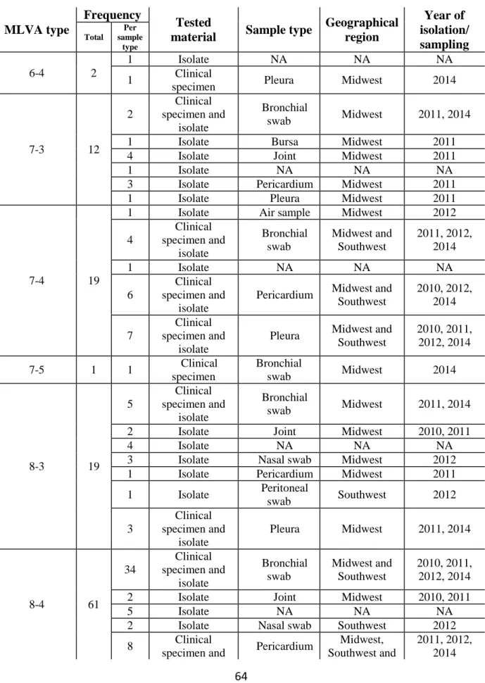
Table 1
–
Clinical specimens processed for M. hyopneumoniae
MLVA Typing ………45
CHAPTER II
Table 1
–
Primers used for multiplex PCR amplification for MLVA………...63
Table 2
–
Relevant information on the M. hyorhinis isolates and clinical samples used in this
study (n=165)……….64
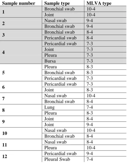
Table 3 - M. hyohinis MLVA types identified in pigs from which more than one sample type was
evaluated………66
CHAPTER III
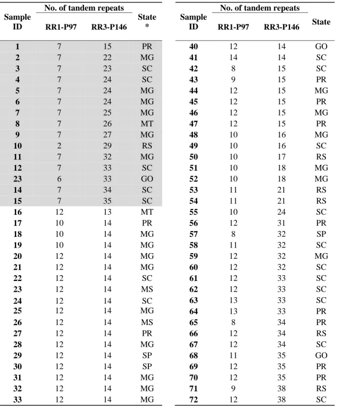
Table 1
–
Mycoplasma hyopneumoniae MLVA types identified in Brazil during Jan 2013
–
ix
LIST OF FIGURES
CHAPTER I
Figure 1
–
Dendrogram showing the distribution of M. hyopneumoniae MLVA types in clinical
specimens in the USA. The MLVA type is indicated by the number of repeats in each locus (e.g.
14-21), followed by the production system where the type was identified (A to R, UNK =
unknown) and the state where the type came from (USA state code). The size of the gray triangle
represents the frequency of the type.
The MLVA type with asterisk represents the reference
strain (ATCC-25095). The dendrogram was generated using the unweighted pair group method
with arithmetic means (UPGMA) a
nd cut to fit in the page………
..41
Figure 2
–
Dendrogram showing the distribution of M. hyopneumoniae MLVA types in clinical
specimens in (A) Mexico and (B) Spain. (A) The MLVA type is indicated by the number of
repeats in each locus (e.g. 14-21), followed by the production system in which the sample was
identified (A and B). The black triangle
represents the production system A and the gray
triangles the productions system B. (B) The size of the gray triangle represents the frequency of
the referred type. Dendrograms were generated using UPGMA
………
42
Figure 3
–
Dendrogram of the distribution of M. hyopneumoniae MLVA types in clinical
specimens in Brazil. The MLVA type is indicated by the number of repeats in each locus (e.g.
14-21), followed by state in which the samples were obtained (state code). The size of the gray
x
the reference strain used in the analysis (ATCC-25095).
The dendrogram was generated using
UPGMA
and cut to fit in the page………
.43
CHAPTER II
Figure 1
–
Dendrogram showing the distribution of M. hyorhinis MLVA types. The MLVA type
is indicated by the number of repeats in each locus for genesMHR_0152
–
MHR_0298 (e.g. 8-4).
The size of the gray triangle represents the frequency of the type. The dendrogram was generated
using the unweighted pair group method with arithmetic means (UPGMA). The algorithm
considers that pairwise distances contribute equally to clustering the data, clustering variations in
one or the other repeats automatically. The dendrogram was cut to fit the page
………
..62
CHAPTER III
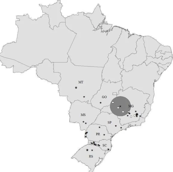
Figure 1
–
Distribution of MLVA types observed in 63 farms in Brazil. Repeat region 1 (RR1)
P97 was categorized as high (≥ 8 tandem repeats, black stars) and low (< 8 tandem repeats, white
circles). Representation of the MLVA types on the map is based on the actual herd location. The
gray circle represents a significant (P=0.05) cluster of MLVA types with low number of tandem
repeats in RR1 P97 (observed to expected ratio of 4.22). States identified with the two letter code
are the most important areas of swine production in Brazil: Goiás (GO), Minas Gerais (MG),
Mato Grosso (MT), Mato Grosso do Sul (MS), Paraná (PR), Rio Grande do Sul (RS), São Paulo
xi
RESUMO
SANTOS, Lucas Fernando dos, D.Sc., Universidade Federal de Viçosa, agosto de 2015.
Investigation of genetic diversity in Mycoplasma hyopneumoniae and Mycoplasma
hyorhinis. Orientador: Maria Aparecida Scatamburlo Moreira. Coorientadores: Maria G. Pieters
e Srinand Sreevatsan
xii
xiii
ABSTRACT
SANTOS, Lucas Fernando dos, D.Sc., Universidade Federal de Viçosa, August 2015.
Investigation of genetic diversity in Mycoplasma hyopneumoniae and Mycoplasma
hyorhinis. Adviser: Maria Aparecida Scatamburlo Moreira. Co-advisers: Maria G. Pieters and
Srinand Sreevatsan
The two most prevalent Mycoplasmas species present in the swine industry are
Mycoplasma hyopneumoniae and Mycoplasma hyorhinis. These pathogens can cause significant
economic losses for producers and for the industry. Genetic variability has been observed in M.
hyopneumoniae using different techniques and heterogeneity of M. hyorhinis has been the focus
of a limited number of studies in the past years. The fact that Mycoplasma genomes contain
numerous repetitive regions (VNTR) within their DNA and that they are known to be active sites
for genetic recombination has prompted the hypothesis that Multiple locus variable number
tandem repeat analysis (MLVA) would be an appropriate technique to investigate genetic
variation in Mycoplasma. Therefore, the main goal of this dissertation was to investigate the non
random distribution of genotypes from different geographical location using multiple locus
variable tandem repeat analysis (MLVA) to type M. hyopneumoniae and M. hyorhinis and
advance the knowledge on the genetic diversity and the epidemiology of these pathogens.When
the M. hyopneumoniae study was taken in account, a high number of M. hyopneumoniae MLVA
types appear to circulate among swine herds with a non-random distribution of the MLVA types
among regions, also a common type was not identified among samples obtained from all regions
in this study. Likewise, heterogeneity of M. hyorhinis was observed, with limited variation in one
of the target VNTR in the M. hyorhinis study. A relevant point observed in this study was that
different MLVA types were identified in the same animal in different sample types, which
xiv
one of the most important in the world, a specific study observed a high heterogeneity of M.
hyopneumoniae strains circulating in the country, and the comparison between the number of
tandem repeats (TR) in RR1 P97 and RR3 P146 showed a significant negative correlation that
may suggest a possible compensatory mechanism that would allow the bacterium to keep its full
adhesion capacity even after reduction of TR in RR1-P97. In conclusion, the identification of the
genetic heterogeneity of M. hyopneumoniae and M. hyorhinis will assist local epidemiological or
outbreak investigations, design future control strategies as well as serve as a potential tool to
study the evolutionary biology of this species.
1
General introduction
The two most prevalent Mycoplasmas species present in the swine industry are
Mycoplasma hyopneumoniae and Mycoplasma hyorhinis. These pathogens can cause significant
economic losses for producers and for the industry. The first one is related to the Enzootic
Pneumonia (EP), a chronic respiratory disease. M. hyopneumoniae causes reduction in the
growth rate, in the feed conversion rate, increase in medication costs, increased rates of mortality
and depreciation of carcasses in the slaughterhouse due to the adherence of pleura and lung
abscesses in interaction with other pathogens.
Despite the efforts and control strategies adopted, M. hyopneumoniae remains a
widespread organism in swine populations, which makes the eradication of the disease a very
difficult and frustrating task. Failure in the control strategies of EP could be associated to the
strain variability considering that infection with one type of strain does not confer cross
protection against an infection with different strain.
The second agent, M. hyorhinis, is considered an emerging pathogen, which are
associated with polyserositis and arthritis in animals from 3 to 10 weeks of age. M. hyorhinis can
be present in normal lungs, but it is more frequently found in pneumonic lungs, where it is
considered a secondary agent.
Heterogeneity of M. hyorhinis strains in the swine populations could result in a difference
in the dynamics of infection and spreading. The genetic diversity of M. hyorhinis has been the
focus of a limited number of studies in the past years. Understanding the genetic diversity of M.
2
Molecular typing methods have been described to investigate the genetic variability of M.
hyopneumoniae and M. hyorhinis. However, these tools cannot be used effectively until they
have been validated for each pathogen. A major purpose of the research described within this
thesis was to investigate the non random distribution of genotypes from different geographical
location using multiple locus variable tandem repeat analysis (MLVA) to type M.
hyopneumoniae and M. hyorhinis. The MLVA method has been used to genotype several species
of bacteria, including different Mycoplasma species. The use of MLVA has improved the
understanding of bacterial epidemiology as it presents a high discriminatory power and can be an
effective tool useful in outbreaks and epidemiological investigations. Other advantages for the
MLVA include the ability to use genetic material without the need to grow the bacterium, to be
performed directly from clinical specimens, and to be used as an initial high throughput
screening and typing method for diagnostic laboratories. The fact that Mycoplasma genomes
contain numerous repetitive regions within their DNA and that they are known to be active sites
for genetic recombination has prompted the hypothesis that MLVA is an appropriate technique
to investigate genetic variation in M. hyopneumoniae and M. hyorhinis.
Therefore, the main goal of this dissertation was to advance the knowledge on the genetic
diversity and the epidemiology of M. hyopneumoniae and M. hyorhinis in swine populations.
The results of this study will contribute to the better implementation of control and prevention
strategies for both pathogens.
In Chapter I, a modified and standardized high-resolution multiple locus variable number
tandem repeat analysis (MLVA) was used to investigate the genetic variability of M.
hyopneumoniae circulating in the United States of America (USA), Brazil, Mexico and Spain. In
3
molecular tool based on the variable number of tandem repeats present in the genome of this
bacterium. An MLVA assay was developed and applied to a strain collection for analysis of the
diversity. And in the third chapter, the MLVA assay developed in the first chapter was applied to
a set of samples from Brazil with the objective of describe the spatial distribution and genetic
heterogeneity of M. hyopneumoniae in this country, and to investigate the association between
number of tandem repeats in the two adhesion genes that were target in the MLVA assay
4
Literature Review
1.1 Mycoplasmas
Members of the Class Mollicutes, Mycoplasma belongs to a group of cell
wall-less bacteria that infect a wide variety of plants and animals (including humans), Family
Mycoplasmataceae and Genus Mycoplasma. They are small pleomorphic organisms whose
diameter can vary from 0.2 to 0.3 µm (Razin et al., 1998), this allows the organism to pass
through filters resulting in the contamination of cell culture (Kobish et al., 1996). Their
morphology seems as
“fried egg” colony shape
, this can be explained by their total lack of cell
wall (Razin et al., 1998). However some species have a cytoesqueleton, which is used to
modulate and control their shape in cell division and the motility (Balish and Krause 2006; Razin
et al., 2010). Mycoplasmas can replicate by binary fission like other prokaryotes (Razin et al.,
2010). Usually, this organism is a surface parasite, although some species as Mycoplasma
fermentans, Mycoplasma penetrans and Mycoplasma pneumoniae show intracellular location
(Lo et al., 1993; Yavlovich et al., 2004 a; b) that may be considered a form of escape from the
host immune system and from antibiotic action, resulting in the establishment of a latent or
chronic infection state (Razin et al., 2010).
Mycoplasma presents a reduced genomic size that ranges from 580 kb to 1380 kb (Razin
et al., 1998). The lack of genes involved in amino acid synthesis, making some species
dependent of the host amino acid supplies (Himmelreich et al., 1996), which implies a limited
biosynthetic capacity. For this reason, they become parasitic and obtain most of their nutrients
5
and Montague, 2002). In turn this can make mycoplasmas particularly difficult to culture as they
have very specific growth requirements (Razin et al., 1998).
Mycoplasmas are facultative anaerobes that grow at 37
oC with 5-10 % of CO
2and need
special media with growth requirements when cultured in vitro. They can be found on mucosal
surfaces of the conjunctive, nasal cavity, genital and intestinal tracts of animals and humans.
Mycoplasmas can be differentiated by colonial morphology, requirements for cholesterol,
biochemical reactivity and host specificity. The hosts and disease conditions are shown in Table
1.
Table 1 - Major diseases associated with mycoplasma species of veterinary significance, the
host and the disease conditions which they cause.
Mycoplasma Species
Hosts Disease conditions
M. mycoides subsp.
mycoides Cattle Contagious bovine
pleuropneumonia
M. bovis
Cattle Mastitis, pneumonia, arthritis
M. agalactiae
Sheep, Goats Contagious agalactia
M. capricolum subsp.
capripneumoniae Goats Contagious caprine
pleuropneumoniae
M. capricolum subsp.
capricolum Sheep, Goats
Septicaemia, mastitis , polyarthritis, pneumonia
M. mycoides subsp. capri Goats, Sheep Septicaemia, pleuropneumonia,
arthritis, mastitis
M. hyopneumoniae
Pigs Enzootic pneumonia
M. hyorhinis
Pigs (3-10 weeks of age) Polyserositis and arthiritis
6
M. gallisepticum Chickens
Turkeys
Chronic respiratory disease Infectious sinusitis
M. synoviae Chickens , Turkeys Infectious synovitis
M. meleagridis Turkeys
Airsacculitis, bone deformities, reduced hatchability and growth
rate
M. haemofelis Cats Feline infectious anemia
Adapted by Quinn et al., 2011
1.2
Importance of Mycoplasma in porcine respiratory diseases complex
During the last decades pork production has enhanced substantially. As consequence of the
increase in herd size and the improvements of infectious disease diagnostic, the identification of
infectious diseases has become consistently. Respiratory disease in swine is the most important
health concern for swine producers and has been identified as the greatest causes of economic
losses in pig production (Opriessnig et al., 2011). These losses are represented by increased
spending on medicines, losses in animal performance and carcasses at slaughterhouses (Sorense
et al., 2006; Martínez et al., 2007).
With the improvement in the pathogen detection methods was possible to identify a multiple
microbial infections causing respiratory disease in pigs. This polymicrobial infection is often the
result of a combination of primary and opportunistic infectious agents (Opriessnig et al., 2011).
To the multiple etiologies pneumoniae was created the term porcine respiratory disease complex
- PRDC (Halbur, 1997). Slow growth, decreased feed efficiency, lethargy, anorexia, fever,
cough, and dyspnea are symptoms of PRDC (Holko et al., 2004). Pneumonia and pleuritis are
7
prevalence of pneumonia ranging from 19% to 79% and for pleuritis from 3.8% to 62% (Wilson
et al., 1986; Hartley et al., 1988; Enoe et al., 2002; Leneveu et al., 2005; Fraile et al., 2010;
Meyns et al., 2011). The severity of PRDC manifestation in pigs are direct related to the
interactions and synergy of infectious factors (viral and bacterial pathogens), environmental
factors, type of production system and management, and genetics, age and immunological status
of the animals. Different bacteria and viruses are involved in the development of PRDC.
Mycoplasma hyopneumoniae (M. hyopneumoniae), Pasteurella multocida (P. multocida),
Actinobacillus pleuropneumoniae (A. pleuropneumoniae) which can act as primary bacterial
respiratory pathogens;
viral respiratory pathogens as swine influenza viruses (SIV), porcine
reproductive and respiratory syndrome virus (PRRSV) and porcine circovirus type 2 (PCV2)
and opportunistic bacterial respiratory pathogens as Streptococcus suis (S. suis) and
Haemophilus parasuis (Hps) (Holko et al., 2004; Sorensen et al., 2006; Opriessnig et al., 2011;
Savic et al.,2015).
The importance of the Mycoplasmas in the respiratory diseases is focused in the M.
hyopneumoniae. M. hyopneumoniae is especially important for PRDC because infects the cilia of
the epithelial cells of the respiratory tract resulting in destruction of the cilia (DeBey and Ross,
1994; Young et al., 2000). Mucociliary apparatus are an important mechanism used as host
defense mechanism to move foreign materials out of the airways. The damage and loss of the
cilia results the increased incidence of secondary bacterial infections associated with M.
hyopneumoniae infection (Thacker and Thanawongnuwech, 2002; Opriessnig et al., 2011).
Studies indicate that other Mycoplasma can be important in PRDC, M. hyorhinis. M. hyorhinis
appears to be frequently associated with M. hyopneumoniae in PRDC cases (Thacker, 2006; Lin
8
1.3
Porcine Mycoplasmas highlighting Mycoplasma hyopneumoniae and Mycoplasma
hyorhinis
Infections by Mycoplasmas represent one of major economic losses in animals.
Mycoplasma can infect the respiratory tract especially in pigs, ruminants and poultry (Table 1).
Some mycoplasmas have been isolated from pigs and they have been implicated in respiratory
and systemic diseases. Mycoplasma hyopneumoniae is the primary agent of enzootic pneumonia
(EP) in pigs, a chronic disease that affects the growth performance, characterized by high
morbidity and low mortality and increased susceptibility to co-infections for other pathogens
(Ciprian et al., 1988).
Also M. hyopneumoniae is considered one of the primary agents involved
in respiratory disease complex in pigs (Thacker et al., 2006).
This infection is highly prevalent
where causing significant economic losses in pig production systems (Ross et al., 1999). M.
hyorhinis is a common inhabitant of the respiratory tract of pigs and can cause polyserositis in
animals from 3- 10 weeks of age when it becomes systemic. The mechanism that M. hyorhinis
uses to become systemic is unclear, however the first step of the colonization is attachment to the
cilia in the upper respiratory tract (Gois et al., 1974). Also, M. hyorhinis can be present in cell
culture lines as contaminant (Kobish et al., 1996).
Another mycoplasma, M. hyosynoviae, affects pigs from 10 to 30 weeks of age and has
an affinity for synovial tissue (Kobish et al., 1996). The sow is the main source of infection of
the piglets, which appear resistant in young ages (Ross et al., 1971). M. hyosynoviae spreads
hematogenously and presents a special affinity to joint synovial tissues (Ross et al., 1971). High
morbidity rates range between 10
–
50% among grower and finisher animals and gilts (Kobish et
al., 1996). Polyarthritis is a common problem in young breeding animals and grower finisher
9
et al., 2001). In general, the clinical signs observed are difficulty in moving, stiffness, lameness,
arched backs and inability to get up (Kobish and Friis , 1996). Mycoplasma suis, a hemotropic
mycoplasma that parasitizes the surface of erythrocytes and causes porcine eperythrozoonosis, an
immune mediated hemolytic anemia (Messick, 2004). This can affect young and growing pigs,
causing disease characterized by listlessness, fever, anorexia, hemolytic anemia and icterus
(Hoelzle, 2008). Infected animals can be subclinical or a chronically infected carrier and spread
the disease in the herd or apparently normal for months before developing the symptoms
(Hoelzle, 2008). M. suis suppresses the host immune system, increasing the susceptibility to
other pathogens (Zachary and Smith, 1985). Mycoplasma flocculare is present in the nasal
cavities, and is similar to M. hyopneumoniae but there is no diseases related with this pathogen
(Kobish and Friis, 1996). M. flocculare can attached to the cilia but is different from M.
hyopneumoniae, as it does not cause destructions of the epithelium (Young et al.,2000). Siqueira
et al., (2013) found that despite M. flocculare and M. hyopneumoniae displaying a high degree
of similarity, the structure and organization of genes that encode adhesins exhibit differences in
the sequence for important domains for adhesion to host cells.
Mycoplasma hyopneumoniae
M. hyopneumoniae belongs to the class Mollicutes. Like all member of this class, M.
hyopneumoniae has no cell wall, presents a reduced genome and a low G+C content (Razin et
al., 1998). So far, the whole genome of six strains has been sequenced (Minion et al., 2004;
Vasconcelos et al., 2005; Liu et al., 2011; Siqueira et al., 2013). The genome size ranges from
892 to 926 kb with a G+C content of 28 %. Like most Mycoplasma, M. hyopneumoniae codon
usage differs from the universal genetic code. The UGA codon encodes for tryptophan instead of
10
The bacterium was isolated for the first time in 1965 by Mare et al (1965) and was
identified as M. hyopneumoniae. Other research group led by Goodwin isolated the
microorganism and named it Mycoplasma suipneumoniae (Goodwin et al., 1967) EP was
reproduced experimentally showing the pathogenicity of the microorganism. Later, it was shown
that both isolates were the same and the bacterium was named M. hyopneumoniae.
The M. hyopneumoniae is spread worldwide especially in the countries where the swine
industry is important (Sibila et al., 2004; Maes et al., 2008).
Studies in different countries
indicate that lesions suggestive of EP occur in 30 to 80 % of slaughtered pigs. In southern Brazil,
which accounts for about 80 % of the national slaughter, it was found that 55 % of pigs had
lesion suggestive of EP and 100 % of herds examined were affected by M. hyopneumoniae
(Sobestiansky, 1999).
Maes et al, (1999) reported that over 90 % of Belgian herd were infected
with M. hyopneumoniae
.Introduction of M. hyopneumoniae in a naive herd is caused mainly by
airborne transmission or by introduction of subclinical infected animals (Sibila et al., 2009).
Several risk factors are associated to (re) infection of a swine herd with M. hyopneumoniae. In
endemic regions, replacement of boars and gilts as part of the restocking policy, the distance to
neighboring pig farms and the distance to the next countryside highway can increase the risk of a
herd become infected (Maes et al., 2000).
The persistence and maintenance of the M. hyopneumoniae in infected herds may be
related to three factors. 1 - When transmission occurs from infected sows to their offsprings,
which in general may occurs in the first week of age (Sibila et al., 2007; Holst et al., 2013).
Pieters et al. (2014) demonstrate that the prevalence of M. hyopneumoniae at weaning increased
with piglet's age if at least one dam was M. hyopneumoniae positive. 2 - by transmission between
11
transmission between older pigs (growing or finisher animals) to younger piglets that enter in the
facilities without all in all out protocol (Ross, 1999; Casamiglia and Pijoan 2000, Rautianen and
Wallgreen, 2001). Gilts and sows with low parity have more chance to transmit the pathogen to
their offspring (Fano et al., 2007), but sow with higher parity, up to the 7th parity, also may
shedding the organisms (Calsamiglia and Pijoan, 2000). M. hyopneumoniae infected pigs can
remain infectious and be a reservatory of the organism for at least 214 days post infection
(Pieters et al., 2009). The transmission rate of M. hyopneumoniae between pigs is slower
compared to viral infections such as swine influenza. One M. hyopneumoniae positive pig at
weaning will transmit to one pen mate during a nursery period of 6 weeks (Meyns et al., 2004;
Villarreal et al., 2011). The dynamics of infections and clinical implication may vary according
to the type of production system (Sibila et al, 2004).
Infection occurs via the respiratory route and the incubation period may vary from one
day to 10 months, with an average of five weeks (Sobestiansky, 1999). The pathogenesis of the
disease caused by M. hyopneumoniae depends on adherence of these microorganisms to the host
lung ciliated epithelium (Wannemuehler & Galvin, 1994).
Although virulence factors of M. hyopneumoniae have not been well characterized, the
adherence to the respiratory epithelium is recognized as an important step for colonization and
infection (Ross, 1999). M. hyopneumoniae infects epithelial cell of the trachea, bronchi and
bronchioles, attaching to the cilia and promoting a reduction in ciliary activity, with damage and
loss of cilia (Debey and Ross, 1994).
Thus, the loss in efficiency of the mucociliary system, an
important non-specific defense mechanism and an increase in mucus production favors
secondary infections (Ciprian et al., 1988; Debey and Ross, 1994; Sorensen et al., 1997). The
12
host immune system and interfere to the effectiveness of antimicrobial treatment for hindering
their release into the site of infection (Schwartz, 2001). M. hyopneumoniae interacts with
alveolar macrophages and lymphocytes, stimulating them to produce proinflammatory cytokines
(TNF-
α, IL
-1 and IL-6) being responsible for lung injury by perivascular and peribronchial
lymphoid hyperplasia (Rodriguez et al., 2004).
The advancement results in the peribronchial
lymphoid hyperplasia are the obstruction of airway, leading to the formation of atelectatic lung
lesions, with coloration varies from dark red to purple in acute cases to grayish brown in chronic
cases (Thacker and Minion, 2012), located mainly in the cranioventral region of apical and
cardiac lobes of the lung (Sobestiansky et al., 1999).
Although the isolation of M. hyopneumoniae is the "gold standard" diagnostic, this
microorganism is very difficult to be isolated and identified as they require special growth
medium and may take 4 to 8 weeks to multiply. Furthermore, successful isolation of M.
hyopneumoniae may be difficult due to the presence of other mycoplasmas with rapid growth,
such as M. hyorhinis, making diagnosis via cultivation sometimes not feasible (Thacker, 2006).
As a result, bacterial isolation is not used as a routine diagnostic (Sibila et al., 2009).
Serological tests are the most commonly used tool to diagnose EP. Detection of
antibodies against M. hyopneumoniae by ELISA may be carried out, and less frequently by the
complement fixation test. ELISA is a quick and inexpensive automated method that provides
useful information about the presence of maternally derived or acquired antibodies (Sibila et al.,
2009). Currently, the tests used are blocking ELISA and indirect ELISA. However, comparative
studies have reported differences in terms of sensitivity and specificity of these kits (Sorensen et
13
The fluorescent antibody technique (FA) and immunohistochemistry (IHC) are used for
identification of M. hyopneumoniae in tissues or swab blades. However, these techniques have
the disadvantage of only be done post mortem (Sibila et al., 2009).
The accurate detection of M. hyopneumoniae increased significantly with advances in
molecular biology methods based on the application of recombinant DNA technology, such as
polymerase chain reaction (PCR). Since then, several tests have been developed (Stark et al,
1998; Calsamiglia et al., 1999; Caron et al., 2000; Verdin et al., 2000; Kurth et al, 2002;
Thacker, 2004; Strait et al., 2008).
The strain variability of M. hyopneumoniae has been suspected for a long time and has
been the main focus of several studies (Ro and Ross, 1983; Zielinski and Ross, 1990; Frey et al.,
1992; Artiushin and Minion, 1996). Frey et al. (1992) observed the existence of different strains
with differences in virulence by chromosomal restriction patterns.
High variation in virulence
was observed between M. hyopneumoniae strains isolated from different swine herds.
A
fragment of 5kb in RAPD analysis was associated with highly and moderately virulent strains,
and interestingly this fragment was not observed in low virulent strains (Vicca et al., 2003).
D
ifferences in pathogenicity between the highly and low virulent isolates was associated with a
faster in vitro growth, a higher capacity to multiply in the lungs and the induction of a more
severe inflammation process by the highly virulent isolate (Meyns et al., 2007).
Recent studies using different typing methods to analyze genetic diversity of M.
hyopneumoniae, including amplified fragment length polymorphism (AFLP), pulse-field gel
electrophoresis (PFGE), multi-locus sequence typing (MLST), random amplified polymorphic
DNA (RAPD) analysis, restriction fragment length polymorphism (RFLP) and Mulitple locus of
14
hyopneumoniae strains from different sources (Stakenborg et al., 2005; Stakenborg et al., 2006;
Mayor et al., 2007; Mayor et al., 2008; Vranckx, et al., 2011; Charlebois et al., 2014).
It is clear
that even with several studies using different typing methods being performed on the
investigation of genetic diversity of M. hyopneumoniae, many aspects remain unknown. The
high diversity between strains from different herds and the difficulties in showing the association
between the genotype and the virulence of the strain still unclear.
Mycoplasma hyorhinis
M. hyorhinis is a swine pathogen (Ross et al., 1973) and it is usually found as a
contaminant of mammalian tissue cultures. M. hyorhinis was first isolate by Switzer (1953), and
was considered to be the cause of enzootic pneumonia, which was later correctly then associated
to M. hyopneumoniae (Mare and Switzer, 1965; Goodwin et al., 1967). M. hyorhinis play a role
as an obligate parasite of swine, and can cause disease as a secondary pathogen. Possible features
that distinguish field isolates from tissue culture strains have only recently begun to be explored
at the genome level. To date, five strains had genomes completed sequence (Liu et al., 2010;
Kornspan et al., 2011;
Calcutt et al., 2012; Goodison et al., 2013; Dabrazhynetskaya et al.,
2014). The completed genome range from 830 to 840 kb with an average G+C content of about
26 %. Even with their small genome, these organisms are able to survive on the host, producing
the metabolics needed for their growth and escape from the host defense system. Chromosomal
gene rearrangement is one strategy used by mycoplasmas to achieve these tasks (Bork et al.,
1995).
M. hyorhinis is commonly present in the respiratory tract of pigs (Switzer, 1955). M.
hyorhinis can be present in normal lungs, but it is more frequently found in pneumonic lungs,
15
Lin et al., 2006). M. hyorhinis can cause polyserositis and arthritis in animals from 3 to 10 weeks
of age when it becomes systemic (Potgieter and Ross, 1972; Friis and Feestra, 1994). This
bacterium has affinity for serosal surfaces and may lead to acute inflammation of the serosa of
body cavities and joint.
An increase in the detection of M. hyorhinis as an etiological agent of polyserositis and
arthritis in piglets in recent years demonstrates the relevance of this pathogen in the swine
industry as an emerging challenge (Rovira et al., 2010).
Most of swine producing countries worldwide have reported the presence of M. hyorhinis
associated with clinical disease. However, most studies that investigate the epidemiology of this
pathogen were done in slaughter pigs or in pigs submitted to diagnostic laboratories, which could
bias the analyses of the epidemiology of M. hyorhinis in swine farms (Rovira et al 2010).
A small proportion of piglets become colonized with this microorganism by contact with
the dams and later nose-to-nose contact among pigs in the nursery increases transmission among
pen mates (Kobish and Friis, 1996; Clavijo et al., 2012). In a longitudinal study, Clavijo et al
(2012) evaluated by PCR of nasal swab the dynamics of colonization by M. hyorhinis in farms
with polyserositis history. Neonatal piglets had 1.7 % of M. hyorhinis positivity and an
increasing in the percentage of positivity of 85 % was observed after weaning. Palzer et al
(2008) found that 80 % of pneumonic lungs and 37 % of normal lungs from slaughter pigs were
positive for M. hyorhinis by PCR in Germany.
In addition to infect pigs, M. hyorhinis has the ability to infect tumor cells or cell culture.
The infection of M. hyorhinis in benign tumors can cause phenotypic changes and differential
16
ability of this agent to infect gastric tumor cells and prostate (Urbanek et al, 2011;. Xu et al,
2013).
To date no virulence factors of M. hyorhinis have been described. However, diverse
studies have been showed the differences in virulence between isolates (Gois and Kuksa, 1974;
Lin et al., 2006; Ross and Switzer, 1963).
Experimental studies have shown that immune suppressive factors make the colonization
by M. hyorhinis more intense in the respiratory tract and serosa. It is noted that in elderly and
immuno competent animals frequently occur the development of arthritis (Magnusson et al.,
1998). Observations under field conditions showed increased diagnosis of M. hyorhinis in cases
of coinfection (PCV2 and PRRSV), suggesting that viral agents are immunosuppressive and
facilitate colonization and spread of M. hyorhinis in the host (Kawashima et al., 1996;
Kobayashi
et al., 1996; Kawashima et al., 2007; Kixmöller et al., 2008).
In most infections, animals show mainly fever, dyspnea, and reluctance to move
(Kobisch
and Friis, 1996). Infection in pigs is usually characterized by arthritis and polyserositis but most
of the infections are subclinical (Potgieter and Ross, 1972). Other clinical presentations
associated with M. hyorhinis include rhinitis, pneumonia, otitis, conjunctivitis and abortions
(Friis, 1971; Poland et al., 1971; Morita et al., 1993; Morita et al., 1999; Shin et al., 2003;
Thacker and Minion, 2012).
The isolation of M. hyorhinis is less laborious as compared to M. hyopneumoniae,
however, it is not frequently used in routine. The analysis of clinical signs and gross lesions
represents a good strategy to diagnostic M. hyorhinis. However, different etiologies can causes
polyserositis, like Haemophillus parasuis and Streptococcus suis. This facts is should take into
17
2012). Techniques like immunohistochemistry and immunofluorescence had been used to detect
the presence of M. hyorhinis in the tissues
(L’Ecuyer and Boulanger, 1970; Gois
et al., 1971;
Potgieter and Ross, 1972). Several PCR based diagnostic tests have been used to identify
M .hyorhinis (Stemke et al., 1994; Kobayashi et al., 1996; Caron et al., 2000; Lin et al., 2006;
Timenetsky et al., 2006; Kang et al., 2012; Tocqueville et al., 2014, Clavijo et al., 2014)
however, little is known about the sensitivity and specificity.
Real-time PCR is the most sensitive
assay available to detect and quantify M. hyorhinis and can be conducted directly on clinical
specimens (such as joint fluid), joint swabs, nasal swabs, or tonsil scrapings. Currently, there are
no immunological tests commercially available for M. hyorhinis.
An important feature of mycoplasmas is the ability to change their surface antigens.
Through this mechanism, these microorganisms are able to evade the immune response mounted
by the host and establish a chronic infection (Muneta et al, 2008). When compared to other
mycoplasmas, M. hyorhinis exhibits an aggressive growth due to differences found in
components involved in metabolism and suggestive of evasion of the host immune system
(Siqueira et al., 2013). The occurrence of variation in the expression of surface lipoproteins in M.
hyorhinis resulted in the generation of population diversity (Rosengarten and Wise, 1991). It has
been suggested that the source of genetic variation in the variable lipoprotein (Vlp) is the
insertion and deletion of tandem repeats (VNTR) within the 3’region in the
Vlp genes
(Rosengarten and Wise, 1991; Yogev et al., 1991; Citti et al., 2000).
The heterogeneity of M. hyorhinis has been the focus of a limited number of studies in
the past years. Restriction endonuclease analysis was applied by Darai and collegues (1982) in a
strain collection of M. hyorhinis to verify the characteristic of fragment pattern of DNA bands.
18
namely sequencing of the p37 gene and a multilocus sequencing typing (MLST; Tocqueville et
al., 2014).
1.4
Mycoplasmas & Variable number of tandem repeat (VNTR)
The surface of the Mycoplasma membrane establish a critical role in the infection
process, due the importance of this component in mediating the basic functions, such as host cell
interactions, transport of nutrients and host immune defenses (Citti et al.,2010). A sets of related
genes are responsible to encoding surface lipoproteins and these genes are susceptible to
high-frequency, reversible mutations that could lead variable expression patterns of corresponding
gene products (Razin et al., 1998). Variation of lipoproteins would results in a modification of
structures at the bacterial cell surface which could be involved in antigenicity (Rosengarten et
al., 2000). Lipoprotein variations has been described in many Mycoplasmas species and are
responsible to generate surface antigen diversity which could be result of phase and/or size
variation (Rocha and Blanchard, 2002). Genomic repeats, such as tandem repeats constitute
source of these variation which occurs in recombination events.
Mycoplasmas contain various repeated sequences, which could be structured in tandem,
in their genome. The variation of the number of the repeat sequence in one gene could lead a size
variation of expressed lipoproteins genes (Citti et al.,2010). Functions of these VNTRs are
partially understood, however, appear that can shield the mycoplasma cells from complement
and modulate biofilm formation (Simmons et al., 2007).
Size variation of surface proteins has been reported in different Mycoplasmas species,
19
al.,2000; Rocha and Blanchard, 2002). M. hyorhinis the size variation of the Vlp gene occurs in
high frequency (Rosengarten and Wise, 1991). The expression of the Vlp genes is controlled by
variation in the size of the polyadenosine tract, the length of 17 polyadenosine tract is necessary
for the gene transcription (Yogev et al.,1991; Citti and Wise, 1995). Longer variants of the Vlp
genes increase resistance to growth inhibitory antibody (Citti et al., 1997). In M. pulmonis, the
expression of Vsa proteins with high number of VNTRs concedes protection against killing by
complement and instigates formation of microcolonies. However, stimulation of the
cytoadherence are achieved with the expression of the VSA proteins with few VNTRs (Simmons
and Dybvig, 2003; Simmons et al., 2004). Also, the lenght of the Vsa proteins is correlated with
the thickness of the surface of the M. pulmonis. More repeats the Vsa protein presents, more
thick it will be (Simmons and Dybvig, 2003).
Expressions of lipoproteins encoded by single copy gene are subject to high frequency
phase variation and the control of the expression is achieved by variation in the length of
repeated sequence. An example is in M. hominis, which the variation in the VNTRs results in
difference in the adherence (Zhang and Wise, 1997).
1.5 Molecular technique to achieve the VNTRs
The molecular detection of a pathogen plays a key role in the identification, typing and
association or such pathogens with disease. Different molecular typing methods can be used to
investigate outbreaks and the epidemiology of bacterial pathogens (Sabat et al., 2013). Most of
mycoplasmal diseases need the specific laboratory diagnostic strategies to identify and type the
pathogen and the strategies are quite different than those for fast-growing bacteria due the
20
Several different typing techniques of mycoplasma have been developed, each one of
which has its advantages and limitations with respect to cost, time, reliability, specificity, and
sensitivity. A selection of typing methods that were used to date and may be usefully applied to
M. hyopneumoniae and M. hyorhinis are shown in table 2.
The fact that mycoplasma genomes contain numerous repetitive regions within their
DNA and that they are known to be active sites for genetic recombination has prompted the
hypothesis that MLVA is an appropriate technique to investigate genetic variation in M.
hyopneumoniae and M. hyorhinis. MLVA analysis is a feasible and suitable method to
characterize bacteria without the necessity of any culture steps. It was reported as being a useful
tool for typing different mycoplasmas. It was demonstrated that specific VNTRs are numerous in
the genome of mycoplasmas and could be associated to functional properties (Hsu and Minion,
1998). These facts might approve MLVA as the method of choice when it is necessary to
characterize a large numbers of mycoplasmas strains. Therefore, the objective of this thesis was
to investigate the nonrandom distribution of genotypes of M. hyopneumoniae and M. hyorhinis
using the MLVA approaches.
Table 2
–
Summary of molecular techniques used to genotype M. hyopneumoniae and M.
hyorhinis.
Organism
Typing method
Target/gene
Reference
Mycoplasma
hyopneumoniae
PFGE
ApaI, Sal I,
ApaL, Asp718
Stakenborg et al., 2005
21
Stakenborg et al., 2006a
RAPD - PCR
OPA-3 primer
Artiushin and Minion 1996
Vicca et al., 2003
Stakenborg et al., 2006a
Nathues et al., 2011
PCR - RFLP
P146
Stakenborg et al., 2006a
PCR -
Sequencing
P97
P146
Wilton et al., 1998
Mayor et al., 2007a
MLST
adk, rpoB, tpiA
Mayor et al., 2007b
VNTR
P97
VNTR genes
P97, P146, H4
Stakenborg et al., 2006a
De Castro et al., 2006
Nathues et al., 2011
MLVA
Variable number
of tandem
repeats genes
Vranckx et al., 2011;
Charlebois et al., 2014.
Mycoplasma
hyorhinis
REA
Darai et al., 1981
PCR-
Sequencing
P37
Tocqueville et al., 2014
MLST
dnaA, rpoB,
gyrB, gltX, adk,
and gmk.
22
PCR, polymerase chain reaction; RAPD, randomly amplified polymorphic DNA; VNTR,
variable number tandem repeats; AFLP, amplified fragment length polymorphism; RLFP,
restriction fragment length polymorphism; PFGE, pulsed-field gel electrophoresis; REA,
restriction endonuclease analysis; MLST, multi locus sequence typing; MLVA, multiple locus of
variable number of tandem repeat analysis.
1.5 Aims of this study
A high strain diversity of M. hyopneumoniae and M .hyorhinis has been observed.
Differences in strains within and among swine populations could impact the observed dynamics
of infection and spread. The genetic diversity within a herd, the clinical importance of this
diversity as well as the persistence of different strains in pigs under field conditions are not
known.
Our hypothesis is that a nonrandom distribution of genotypes of M. hyopneumoniae and
M. hyorhinis strain vary according to distinct geographical regions and that those strains
differences account for the different clinical presentation and diverse response to control
measures observed in the field. The specific objectives are:
Standardize a highly sensitive and discriminatory technique (Multiple-Locus
Variable-Number Tandem-Repeat Analysis (MLVA)) for differentiation of M.
hyopneumoniae strains.
Investigate the genetic variability of M. hyopneumoniae strains in clinically
23
Standardize a highly sensitive, highly discriminatory technique (Multiple-Locus
Variable-Number Tandem-Repeat Analysis (MLVA)) for differentiation of M.
hyorhinis strains.
Investigate the genetic variability of M. hyorhinis strains in clinically relevant
specimens from geographically distinct US farms regions
Such information will contribute to the better understanding of the epidemiology of both
pathogens, which can help the implementation of control and prevention strategies.
CHAPTER I - Genotype distribution of Mycoplasma hyopneumoniae in swine herds from
different geographical regions
Dos Santos, L.F., et al., Genotype distribution of Mycoplasma hyopneumoniae in swine
herds from different geographical regions. Veterinary Microbiology 2015,
Feb 25; 175 (2-4):374-81. doi: 10.1016/j.vetmic.2014.11.018.
This chapter has been published in Veterinary Microbiology and presented as the final
25
Title Page
Short Communication
Genotype distribution of Mycoplasma hyopneumoniae in swine herds from different
geographical regions
Lucas F. Dos Santos
a,b, Srinand Sreevatsan
a, Montserrat Torremorell
a, Maria A.S. Moreira
b,
Marina Sibila
c, Maria Pieters
a,*
a
Department of Veterinary Population Medicine, College of Veterinary Medicine, University of
Minnesota, 1365 Gortner Avenue, St. Paul, MN 55108, USA
b
Departamento de Veterinária, Universidade Federal de Vicosa, Av. PH Rolfs s/n
–
Campus
Universitário, Viçosa, MG 36570 000, Brazil
c
Centre de Recerca en Sanitat Animal (CReSA), UAB-IRTA, Campus de la Universitat
Autonoma de Barcelona, 08193 Bellatera (Cerdanyola del Valle`s), Spain
* Corresponding author at: 1365 Gortner Avenue, 225 VMC, St. Paul, MN 55108, USA. Tel.: +1
612 624 7947; fax: +1 612 625 6241.
26
Abstract
Genetic heterogeneity of Mycoplasma hyopneumoniae in pigs has been reported, however there
has been limited reproducibility on the molecular methods employed so far. The aim of this
study was to modify and standardize a high-resolution multiple locus variable number tandem
repeat analysis (MLVA), to investigate the genetic variability of M. hyopneumoniae circulating
in the United States of America (USA), Brazil, Mexico and Spain. The MLVA was standardized
on the basis of the number of tandem repeats in two Mycoplasma adhesins, P97 and P146, which
are proteins involved in the adherence of the pathogen to cilia. A total of 355 samples obtained
from the four countries were analyzed. The Simpson’s diversity index for the assay was D =
0.976 when samples from all countries were combined. A large number of MLVA types (n =
139) were identified, suggesting that multiple M. hyopneumoniae variants are circulating in
swine. The locus P97 had 17 different types with 2
–
18 repeats. The P146 locus showed higher
heterogeneity, with 34 different types, ranging from 7 to 48 repeats. MLVA types that presented
more than 30 repeats in P146 were found in Spain and Brazil, while shorter repeats were
observed in the USA and Mexico. This simplified MLVA method proved to be an efficient tool
for typing M. hyopneumoniae with a high degree of stability, repeatability, and discriminatory
power. In conclusion, M. hyopneumoniae showed a high variable number tandem repeat
heterogeneity and this assay can be applied in molecular epidemiology investigations within
farms and productions systems.
27
1 Introduction
Mycoplasma hyopneumoniae (M. hyopneumoniae) is the etiological agent of enzootic
pneumonia, an important respiratory disease affecting pigs (Mare and Switzer, 1965; Goodwin et
al., 1965). M. hyopneumoniae infections have been reported worldwide, and have an important
economic impact in swine production, reducing growth rate and feed efficiency (Maes et al.,
2008. Various reports have suggested the circulation of genetically diverse strains of M.
hyopneumoniae in swine, as detected by different molecular techniques (Frey et al., 1992;
Kokotovic et al., 1999; Vicca et al., 2003; Dubosson et al., 2004; Stakenborg et al., 2005;
Stakenborg et al., 2006; Mayor et al., 2008; Nathues et al., 2011; Vranckx et al., 2011;
Charlebois et al., 2014). Multiple locus variable number tandem repeat analysis (MLVA) has
been used to genotype several species of bacteria, including Mycoplasma species. The use of
MLVA has improved the understanding of bacterial epidemiology as it presents a high
discriminatory power and can be a useful tool in outbreaks and epidemiological investigations.
Other advantages include the ability to use genetic material without the need to grow the
bacterium, to be performed directly from clinical specimens, and to be used as an initial high
throughput screening and typing method. The fact that Mycoplasma genomes contain numerous
repetitive regions within their DNA and that they are known to be active sites for genetic
recombination (Rocha et al., 2002) has prompted the hypothesis that MLVA is an appropriate
technique to investigate genetic variation in M. hyopneumoniae. Previously, two different
MLVA assays have been developed for typing M. hyopneumoniae (Vranckx et al., 2011;
Charlebois et al., 2014). However, little is known about M. hyopneumoniae heterogeneity in
United States of America (USA), Brazil, Mexico, Spain, and other countries. In this study, we
28