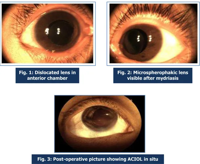
A RARE CASE OF BILATERAL MICROSPHEREPHAKIA
Texto
Imagem


Documentos relacionados
Distribution of 64 eyes of 32 patients according to the horizontal corneal diameter of the right eye and the left eye in the first and the last consutations in number (mm), and the
Figure 1: Left eye with interstitial corneal infiltrates associated to reactive conjunctival hyperemia and deep corneal vascularization, similar to that of the right eye
There were statistically significant differences among the phases of the menstrual cycle in the following tests: CAL latency in the right eye, SM precision in the left eye,
Regarding the abduction of the left eye, a slight widening of the palpebral issure was observed in the left eye, characterizing bilateral type III Duane retraction syndrome, with
The right eye had an uneventful evolution on the postoperative visits, by contrast, in the left eye, the patient reported on the first postoperative month visit a dark shadow in
Case 2: Figure 2 - A) Preoperative photograph demonstrating face turned to the left; B) preoperative photograph in left gaze demonstrating limitation of adduction (-4) in the right
To proof the retinal origin of the AxEvg the electrodes in left eye were placed as mentioned by Cella at al., whereas, the electrodes in right eye were placed at the conjunctiva
Left eye showing bulbar and palpebral hyperemia, secretion in the meibomian glands, sparse inferior eyelashes, superior (right) and inferior (left) fornix foreshortening,