Study on The Reproductive Organs and Fertility of The Male
Mice following Administration of Metronidazole
Mrinalini Kumari, M.Sc., Poonam Singh, Ph.D.*
Department of Zoology, MMV, Banaras Hindu University, Varanasi, India
Abstract
Background:Metronidazole (MTZ) is commonly used as an antibacterial and antipro-tozoal drug. Various doses of MTZ have been reported to inhibit spermatogenic activity and sperm indices.
Materials and Methods: In this experimental study, dose-dependent effects of MTZ on the structural and functional integrity of the testis and accessory reproductive organs have been investigated. Adult male mice of Swiss strain were administered orally with MTZ at the doses of 250 mg/kgBW/day and 500 mg/kgBW/day for 28 consecutive days to study the changes in the testis, epididymis, seminal vesicle, sperm indices and fertility. Reversal effects of the drug were also studied on the same mice, 42 days after cessation of the treatment.
Results:Therapeutic dose of MTZ (250 mg/kgBW/day) neither altered the weights of the testis, epididymis and seminal vesicle nor their histoarchitecture and sperm indi-ces. The drug at the high dose (500 mg/kg BW/day) caused signiicant reductions in the weights of the testis and epididymis. Histoarchitecture of the testis and epididymis at the high dose revealed marked regressive changes while that of seminal vesicle remained unaffected. Signiicant reductions were noticed in the motility, viability and count of epididymal spermatozoa while the concentrations of epididymal sialic acid and seminal vesicular fructose remained unaltered after the treatment. No signiicant changes were noticed in the mating ability as well as in the level of serum testosterone in the treated mice. Fertility of the male mice treated with high dose of MTZ declined markedly lead-ing to an increase in pre- and postimplantation loss while a signiicant decrease was noticed in the number of live blastocysts in females impregnated with such males. MTZ-induced changes in the male reproductive organs and fertility were reinstated 42 days after cessation of the treatment.
Conclusion:High dose of MTZ induced reversible deleterious effects on the male repro-duction and fertility.
Keywords: Epididymis, Metronidazole, Seminal Vesicle, Sperm, Testis
Citation: Kumari M, Singh P. Study on the reproductive organs and fertility of the male mice following administra-tion of metronidazole. Int J Fertil Steril. 2013; 7(3): 225-238.
Received: 21 Aug 2012, Accepted: 20 Nov 2013
* Corresponding Address: P.O. Box: Department of Zoology, MMV, Banaras Hindu University, Varanasi-221005, India
Email: poonom@gmail.com
Royan Institute
International Journal of Fertility and Sterility
Introduction
The increase in incidences of infertility in men due to frequent use of a number of therapeutic drugs has made efforts to study their untoward side effects on the male reproduction. Various drugs used for treating diseases are reported to cause male infertility (1, 2). Among them certain
deriva-tives of nitroimidazole such as ornidazole, metroni-dazole, tinidazole and nimorazole are reported to im-pair the fertility potential by exerting adverse effects on the spermatogenesis and sperm parameters (3-6).
1-[2-hydroxyethyl]-2-methyl-5-nitroimidazole), a drug of irst choice,
recommended by the clinicians to be consumed at maximum for seven to ten days for the treat-ment of Helicobacter pylori infection, amoebia-sis, giardiaamoebia-sis, trichomoniaamoebia-sis, bacterial vaginosis and several other anaerobic bacterial and parasitic infections. However, for the treatment of several complications like Chagas disease, Crohn’s dis-ease, osteomyelitis, endocarditis, deep neck infec-tion, joint infection and liver abscess, this drug is advised to be consumed for 4-8 weeks. Inspite of its long-term clinical use, untoward side effects of MTZ on the male fertility has been studied in labo-ratory rodents (3, 7, 8). Administration of various doses of MTZ (200 mg/kgBW/day and 400 mg/ kgBW/day) for 6 and 8 weeks causes suppressive effects on the spermatogenesis and fertility in the rats (3, 9, 10).
Quantitative studies have indicated marked al-terations in the number of germ cells at stage I, V and XII following intraperitonial administration of 130 mg/kgBW/day of MTZ for seven days in mice (9) while the drug at the doses of 200 mg/kgBW/ day and 400 mg/kgBW/day for 60 days causes sup-pressive effect on spermatogenesis by altering the number of germ cells at stage VII of seminiferous tubule cycle in rats (10). Various doses of MTZ cause marked alterations in the count (3, 11, 12), motility (11, 12) and morphology of epididymal spermatozoa (3, 8) in laboratory rodents. Oda (13) reported dose-dependent decrease in the luminal content of epididymal spermatozoa in the MTZ-treated rat. Decreased levels of gonadotropins and testosterone result in MTZ-induced suppressive effects on spermatogenesis (9-13).
From the foregoing it is clearly seen that MTZ at various doses impairs fertility in the males by in-hibiting spermatogenic activity and sperm indices. However, a detailed study regarding the effects of therapeutic dose of MTZ for long duration, such as for 4-8 weeks on the male reproductive organs and fertility is still required. Therefore, the aim of the present study is to investigate the effects of the therapeutic and high doses of MTZ on the testis, epididymis, seminal vesicle and fertility as well as on the secretory activities of the latter two organs. For the safety evaluation of the potential effect of the drug on the male reproductive organs, a study with a dose higher than the therapeutic one may be considered in a non clinical trial. The study also
deals with the withdrawal effects of high dose of MTZ, 42 days after cessation of the treatment.
Materials and Methods
Animal selection
In this experimental study, ifty Swiss strain adult (12 weeks old) male mice weighing about 25-30 g were used for the present investigation. The animals were housed under standard labora-tory conditions and maintained on pelleted diet and water ad libitum. Approval from the Animal Ethical Committee, Banaras Hindu University, Varanasi, India was obtained for the animal study plan (No. Dean/11-12/CAEC/263).
Experimental design, drug and dosage
After recording the initial body weights, all the
animals were divided into ive groups of ten each
and treated as follows: Group I Untreated controls
Group II Vehicle-treated controls (distilled water) Group III Administration of MTZ (250 mg/kgBW/ day) for 28 days
Group IV Administration of MTZ (500 mg/kgBW/ day) for 28 days
Group V Administration of MTZ (500 mg/kgBW/
day) for 28 days followed by sacriicing the ani
-mals 42 days after cessation of the treatment.
MTZ (CDH, India) was dissolved in double distilled water and administered orally. The human therapeutic dose of MTZ was selected and translated to mice (14). The doses 250 mg/ kgBW/day and 500 mg/kgBW/day of MTZ were administered to mice, equivalent to hu-man therapeutic dose (20 mg/kgBW/day) and its higher dose (40 mg/kgBW/day), respective-ly. The procedure for the oral administration of the drug through gavage was based on the prior studies (8, 15).
Animal sacriice and collection of reproductive organs
After recording the inal body weights the ani
-mals were sacriiced by cervical dislocation. Among ten animals from each group, ive animals
were used for the histological studies and sperm
assessment while the other ive were used for bio
testoster-one level. Blood was collected by cardiac puncture to measure the level of serum testosterone. The re-productive organs were dissected out, blotted free of blood and processed for the following studies:
Organs weight
Wet weights of the testis, epididymis and semi-nal vesicle were recorded to calculate the gonad-osomatic index by using the following formula: Gonadosomatic Index (GSI)=(Gonad weight/total body weight) ×100.
Histological studies
Bouin’s ixed testis, epididymis and seminal
vesicle were dehydrated and embedded in
paraf-in. Sections of 5 μm thickness were taken from
the mid portion of each testis, all the three regions of epididymis and seminal vesicle, dehydrated in graded series of alcohol and stained with Periodic Acid Schiff reagent followed by counterstaining with Ehrlich’s Hematoxylin.
Quantitative study of the testis
Frequency of the stages was determined from
one cross section of the testis of the ive animals
in each group. All the seminiferous tubules with-in a cross section of the testis were examwith-ined at
×40 and classiied according to the stages of the
cycle. The stages of the seminiferous tubules were
classiied according to the method of Hess and
Franca (16). Due to severe degenerative changes
in the seminiferous tubules, accurate identiication
of each stage was not possible; therefore, the tu-bules were grouped as stages I-IV, V-VI, VII-VIII, IX-X and XI- XII. The percentage frequency of all the grouped stages in one cross section of the
testis in each of the ive animals was calculated
and analysed statistically. The relative number of each variety of germ cells at stage VII of the spermatogenic cycle (i.e. type-A spermatogonia (Asg), preleptotene spermatocytes (PLSc), pachy-tene spermatocytes (PSc) and stage 7 spermatids (7Sd)) was counted according to the method of Russell et al. (17).
Morphometric study of the seminiferous tubules
The diameter of the seminiferous tubules was measured using ocular micrometer at ×40 objec-tive piece.
Biochemical studies
Concentrations of epididymal sialic acid and seminal vesicular fructose were estimated using the methods of Aminoff (18) and Linder and Mann (19) respectively.
Assessment of sperm parameters
Cauda epididymidis of ive mice in each group
was minced thoroughly in the physiological nor-mal saline at 37˚C and used for the assessment of motility, viability and count according to the WHO Laboratory Manual (20). The sperm morphology was assessed by observing the smear prepared on a clean glass slide under microscope at ×40.
Evaluation of sperm abnormality was based on the criteria of Wyrobek and Bruce (21) and Zaneveld and Polakoski (22).
Serum testosterone assay
Serum testosterone was measured by ELISA, as described in the instructions provided in the kit (LDN, Germany).
Mating ability and fertility
Each male was caged with two proestrus fe-males overnight and according to presence of vaginal plug and implantation sites in females, the mating ability and fertility of the males were assessed respectively. The females were sacri-ficed by cervical dislocation on the fifteenth day of cohabitation with males. The ovaries were removed to count the number of corpus luteum. To determine the total number of implantation sites, the dissected out uteri were placed in 10% ammonium sulfide solution, which stained the hemosiderin pigment of resorbed implanted sites blue-black (23). The number of live im-plants, as well as pre- and post-implantation loss was recorded. Preimplantation loss was calculated using the following formula:
Corpus luteum – [number of resorbed implants + number of live implants + number of dead implants] Postimplantation loss was equal to the total number of resorbed and dead implants.
Statistical analysis
Body weight and number of live implants as well as pre- and postimplantation loss were analyzed using Student’s t test. Values were considered
sig-niicant at p<0.05.
Results
Body weight
No signiicant differences were found between the initial and the inal body weights of the
MTZ-treated mice and the controls at therapeutic and high dose (Table 1).
Organs weight
Administration of MTZ at the therapeutic dose did not induce significant changes in the weights of the testis and epididymis while the drug at the high dose resulted in significant re-ductions in the weight of these organs as com-pared with the controls. Forty two days after cessation of the treatment, weight of the organs recovered to the control values. Administration of MTZ at any dose did not induce significant reduction in the weight of the seminal vesicle compared with that of controls (Table 1).
Table 1: Effect of the oral administration of MTZ on body weight and weight of testis, epididymis and seminal vesicle (values are mean ± SE of ive animals)
Weight of the reproductive organs (mg/100 gBW) Body weight (g)
Groups
Seminal vesicle Epididymis
Testis Final BW
Initial BW
230.79 ± 22.41 131.36 ± 9.52
316.87 ± 20.6 28.0 ± 1.52
23.2 ± 1.35
I. Untreated control
230.84 ± 27.1 134.21 ± 6.15
310.40 ± 21.17 26.8 ± 0.58
23.8 ± 0.19
II. Vehicle-treated control
200.05 ± 20.11 122.54 ± 9.28
297.97 ± 9.09 28.4 ± 0.51
25.8 ± 0.48
III. MTZ (250 mg/kgBW/day)
169.45 ± 14.53 101.23 ± 5.3a
189.96 ± 4.95 a
27.8 ± 0.79 23.4 ± 1.32
IV. MTZ (500 mg/kgBW/day)
245.97 ± 25.57 166.63 ± 8.29b
291.31 ± 9.94 b
33.2 ± 0.86 27.6 ± 0.51
V. MTZ (500 mg/kgBW/day)*
*; Administration of MTZ for 28 days followed by sacriicing the animals 42 days after cessation of the treatment, a; As com -pared to Groups I and II: p<0.05 and b; As com-pared to Group IV: p<0.05.
Histological studies
Testis
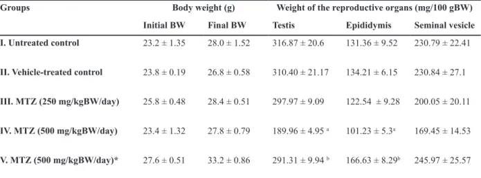
The testis of untreated and vehicle-treated controls (Fig 1A) showed normal histologi-cal features. MTZ at therapeutic dose induced mild regressive changes in the seminiferous tu-bules such as loosening of the germ cells only. However regressive changes in the seminifer-ous tubules appeared more pronounced in the testis of mice administered with high dose of MTZ. The changes included shrinkage of the seminiferous tubules, depletion,
Fig 1: (A-D) Transverse section (T.S.) of the Testis of control (A) showing normal appearance of seminiferous tubules. (B-D) MTZ (500 mg/kgBW/day)-treated mouse for 28 days where (B) shows the shrinkage of the seminiferous tubules, depletion, disorganization, vacuolization and sloughing of the germ cells and appearance of multinucleated giant cells in the seminifer -ous tubules; (C) shows the giant cell (arrow); (D) shows the recovery in spermatogenesis in animals sacriiced 42 days after cessation of the treatment.
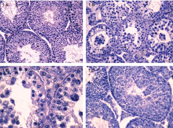
Quantitative study of the testis
Quantitative analysis of the spermatogenic cycle revealed no alterations in all the stages of the seminif-erous tubules in the testis of mice administered with therapeutic dose of MTZ as compared with the
con-trols. In contrast, a signiicant decrease was observed
in stages I-VIII after high dose of MTZ-treatment as compared with the controls (Table 2). Treatment with both doses of MTZ caused decrease in the germ cells in stage VII of the seminiferous tubules. However, reductions in the number of these cells in stage VII
of the seminiferous tubules were signiicant in the
testis of mice administered only with high dose of MTZ (Table 3). Forty two days after cessation of the
treatment, number of cells at various stages of the seminiferous tubules (Table 2) as well as the differ-ent types of germ cells of stage VII recovered to that of control values (Table 3).
Morphometric study of the seminiferous tubules
Therapeutic dose of MTZ treatment did not in-duce any alteration in the diameter of the
seminif-erous tubules while a signiicant decrease in the
same was noted in the testis of mice administered with high dose of the drug as compared with con-trols (Table 3). By 42 days after cessation of the treatment, tubular diameter recovered almost to the control value (Table 3).
1 A 1 B
Table 2: Effect of oral administration of MTZ on the percentage frequencies of stages of the spermatogenic cycle (values are mean ± SE of ive animals)
Weight of the reproductive organs (mg/100 gBW) Groups
Stage XI-XII Stage IX-X
Stage VII-VIII Stage V-VI
Stage I-IV
15.53 ± 2.13 13.20 ± 1.73
27.37 ± 1.22 17.80 ± 3.29
28.05 ± 1.56
I. Untreated control
17.67 ± 4.05 11.99 ± 2.02
25.19 ± 3.87 23.18 ± 2.5
25.32 ± 1.56
II. Vehicle-treated control
15.09 ± 1.65 11.64 ± 1.09
25.98 ± 1.43 21.61 ± 0.9
25.64 ± 1.97
III. MTZ (250 mg/kgBW/day)
37.27 ± 14.92 36.93 ± 9.5 a
05.99 ± 2.46 a
12.91 ± 7.09 a
06.85 ± 5.88 a IV. MTZ (500 mg/kgBW/day)
18.14 ± 1.73 10.69 ± 0.74 b
24.43 ± 1.26 b
23.05 ± 1.26 b
23.64 ± 1.85 b V. MTZ (500 mg/kgBW/day)*
*; Administration of MTZ for 28 days followed by sacriicing the animals 42 days after cessation of the treatment, a; As com -pared to Group I and II: p<0.05 and b; As com-pared to Group IV: p< 0.05.
Table 3: Effect of oral administration of MTZ on the diameter and number of various types of germ cells of stage VII of the seminiferous tubules (values are mean ± SE of ive animals)
Round Pachytene
Preleptotene Type A
Groups
spermatids spermatocytes
spermatocytes spermatogonia
Diameter (μm)
175.00 ±14.25 71.32 ± 4.22
51.32 ± 2.19 1.92 ± 0.19
215.55 ± 06.80
I. Untreated control
174.34 ±18.90 75.00 ± 8.08
49.44 ± 5.88 2.16 ± 0.31
223.28 ± 09.90
II. Vehicle-treated control
147.92 ± 06.53 56.96 ± 3.36
40.36 ± 1.55 1.92 ± 0.30
218.96 ± 07.84
III. MTZ (250 mg/kgBW/day)
024.48 ± 24.40 a
11.60 ± 11.6 a
09.68 ± 9.60 a
0.60 ± 0.60 a
179.71 ±11.20 a IV. MTZ (500 mg/kgBW/day)
159.36 ± 20.69 b
79.04 ± 7.49 b
47.36 ± 5.13 b
1.6 ± 0.28 b
198.72 ± 09.34
V. MTZ (500 mg/kgBW/day)*
*; Administration of MTZ for 28 days followed by sacriicing the animals 42 days after withdrawal of the treatment, a; As compared to Groups I and II: p<0.05 and b; As compared to Group IV: p<0.05.
Epididymis
The epididymis of the untreated and vehicle-treated controls exhibited normal histological
features. In the Swiss mice, ive segments (I-V)
were noticed in the epididymis. Segments I-III constituted the caput (Fig 2A-C); segment IV-cor-pus (Fig 2D) and segment V-cauda epididymides (Fig 2E). In mice treated with low dose of MTZ, these segments presented almost normal histology. High dose of MTZ-treatment caused no alteration
in the irst region of caput epididymidis (Fig 2F) while the same dose caused marked alterations in the lumina of second and third segments of caput (Fig 2G-2H) as well as in the corpus (Fig 2I) and cauda epididymides (Fig 2J), as indicated by presence of exfoliated germ cells and PAS-positive material with
sperm debris. Increase in the ibromuscular stroma
Fig 2: T.S. of various segments of the epididymis. (A-E) Segments of I-V of control to show normal histological features. (F-J) Segments of I-V of MTZ (500 mg/kgBW/day)-treated mouse for 28 days showing PAS-positive material, sperm debris and sloughed off germ cells in the lumina of segments II (Fig G), IV (Fig I) and V (Fig J).
2 A 2 F
2 G
2 H
2
I
2 J 2 B
2 C
2 D
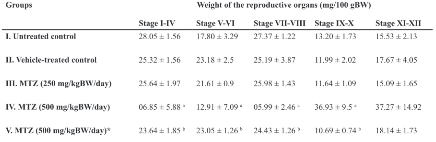
Seminal vesicle
Treatment with MTZ at any dose did not cause any alteration in the histoarchitecture of the semi-nal vesicle as compared with the control (Fig 3A and B).
Concentrations of sialic acid and fructose
Administration of MTZ at any dose did not
in-duce signiicant alterations in the concentrations
of sialic acid in the epididymis and fructose in the seminal vesicle (Table 4).
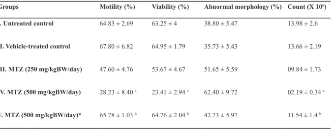
Epididymal sperm assessment
Therapeutic dose of MTZ did not cause signii
-cant reductions in the motility, viability and count of epididymal spermatozoa. By contrast these
sperm indices declined signiicantly in mice ad -ministered with high dose of MTZ. Percentage of abnormal spermatozoa increased in MTZ-treated
groups, though, the values were not signiicant.
Withdrawal of the treatment, however, resulted in marked recovery in motility, viability and count of spermatozoa in the epididymis comparable to that of control (Table 5).
Fig 3: T.S. of the Seminal vesicles of control (A) to show normal histological features (B) MTZ (500 mg/kgBW/day)-treated mouse for 28 days showing unaltered histology.
Table 4: Effect of the oral administration of MTZ on the concentrations of sialic acid in the epididymis and fructose in the seminal vesicle (values are mean ± SE of ive animals)
Concentration of fructose (μg/100 mg of tissue)
Concentration of sialic acid
(μmole/100 mg of tissue)
Groups
264.53 ±15.4 196.91 ± 46.08
I. Untreated control
259.41 ±15.18 195.71 ± 18.3
II. Vehicle-treated control
237.75 ±13.78 212.88 ± 41.26
III. MTZ (250 mg/kgBW/day)
235.09 ± 21.02 155.51 ± 22.55
IV. MTZ (500 mg/kgBW/day)
243.80 ± 15.73 214.21 ± 8.91
V. MTZ (500 mg/kgBW/day)*
*; Administration of MTZ for 28 days followed by sacriicing the animals 42 days after cessation of the treatment.
Table 5: Effect of the oral administration of MTZ on sperm motility, viability, morphology and count in the cauda epididymidis (values are mean ± SE of ive animals)
Count (X 106)
Abnormal morphology (%) Viability (%)
Motility (%) Groups
13.98 ± 2.6 38.80 ± 5.47
63.25 ± 4 64.83 ± 2.69
I. Untreated control
13.66 ± 2.19 35.73 ± 5.43
64.95 ± 1.79 67.80 ± 6.82
II. Vehicle-treated control
09.84 ± 1.73 51.65 ± 5.59
53.67 ± 4.67 47.60 ± 4.76
III. MTZ (250 mg/kgBW/day)
02.19 ± 0.34 a
62.40 ± 9.72 23.41 ± 2.94 a
28.23 ± 8.40 a IV. MTZ (500 mg/kgBW/day)
11.54 ± 1.4 b
42.73 ± 5.97 64.76 ± 2.04 b
65.78 ± 1.03 b V. MTZ (500 mg/kgBW/day)*
*; Administration of MTZ for 28 days followed by sacriicing the animals 42 days after cessation of the treatment, a; As com -pared to Groups I and II: p<0.05 and b; As com-pared to Group IV: p<0.05.
Serum testosterone level
No signiicant change was found in the level of
serum testosterone caused either by therapeutic or high dose of MTZ as compared with the control (Table 6).
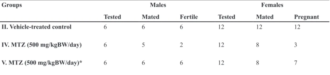
Mating ability and fertility
Mating ability of all the treated males re-mained almost unaffected comparable to that of the controls. Marked reduction was noted in the fertility of the males treated with high dose of MTZ; 67% of treated males became infertile af-ter the treatment. Fertility of the virgin females
impregnated with such males also declined by 75% (Table 7). In the remaining 25% fertile fe-males, the number of live implants decreased significantly. An insignificant increase in the number of pre- and postimplantation loss was also noticed in such females (Table 8). Forty two days after cessation of the treatment, fer-tility of all the males recovered, however, only 34% of females showed recovery in their fertil-ity when impregnated with such males (Table 7). The number of live implants as well as pre- and postimplantation loss was also recovered to some extent after withdrawal of the treatment (Table 8).
Table 6: Effect of the oral administration of MTZ on the level of serum testosterone (values are mean ± SE of ive animals)
Level of serum testosterone (ng/ml) Groups
2.44 ± 0.2
II. Vehicle-treated control
2.42 ± 0.39
III. MTZ (250 mg/kgBW/day)
2.14 ± 0.4
IV. MTZ (500 mg/kgBW/day)
2.32 ± 0.25
V. MTZ (500 mg/kgBW/day)*
Table 7: Effect of the oral administration of MTZ on the mating ability and fertility of the males and the females (values are mean ± SE of ive males and twelve females)
Females Males
Groups
Pregnant Mated
Tested Fertile
Mated Tested
12 12
12 6
6 6
II. Vehicle-treated control
3 8
12 2
5 6
IV. MTZ (500 mg/kgBW/day)
7 8
12 6
6 6
V. MTZ (500 mg/kgBW/day)*
*; Administration of MTZ for 28 days followed by sacriicing the animals 42 days after cessation of the treatment.
Table 8: Effect of the oral administration of MTZ on the number of live blastocysts and pre- and post-implantation loss (values are mean ± SE of twelve females)
Post-implantation loss Pre-implantation loss
Number of live blastocysts Groups
0.41 4.25
7.25
II. Vehicle-treated control
1.25 6.25
2.75 a IV. MTZ (500 mg/kgBW/day)
0.16 5.16
4.5
V. MTZ (500 mg/kgBW/day)*
*; Administration of MTZ for 28 days followed by sacriicing the animals 42 days after cessation of the treatment and a; As compared to Group II: p<0.05.
Discussion
Oral administration of MTZ at therapeutic and high doses did not affect body weight of all the animals. However, significant reduction was noticed in the weight of the testis in the mice treated with high dose of MTZ. This is consistent with the findings reported in rats and mice (3, 7, 9-11, 13). Reduction in the testicular weight may be attributed to the depletion of the germ cell population (24).
The histological study revealed that the thera-peutic dose (250 mg/kgBW) of the drug did not cause marked alterations in the seminiferous tubules when administered for 28 days while the drug at high dose (500 mg/kg BW) for the same duration induced noticeable regressive changes in the seminiferous tubules resulting in the suppression of spermatogenic activity.
dose of MTZ treatment. This is consistent with the finding reported in Balb/c mice (15). De-crease in the diameter is attributed to cell death or exfoliation of the germ cells resulting in the shrinkage of the seminiferous tubules (25). Also, in the present study, sloughing or exfolia-tion of the germ cells has often been noticed in the seminiferous tubules in the testis of mice treated with high dose of the MTZ. The multi-nucleated giant cells observed in some of the regressed seminiferous tubules in the testis af-ter treatment with high dose of MTZ is consist-ent with that observed in rat (13). These cells are considered to be an expression of germ cell degradation (26).
The spermatogenic inhibition as noticed in our study is reflected by alterations in the fre-quency of different stages as well as diminu-tion of the germ cells at stage VII of the sper-matogenic cycle. Earlier studies have shown that intraperitonial administration of 130 mg/ kgBW of MTZ for 7 days in CFW mice caused no alterations in the number of stages in the seminiferous tubules while the number of cells in stages I, V and XII was significantly increased (8). Quantitative study of Sohrabi and Mellati (10) has reported that oral admin-istration of MTZ at the doses of 200 mg/kgBW and 400 mg/kgBW for 60 days caused signifi-cant reductions in the number of preleptotene spermatocytes and step 7 spermatid of stage VII of seminiferous tubule cycle in rats. In the present study, a significant decrease in stages I-VIII has been also observed with significant decrease in the number of type A spermato-gonia, preleptotene spermatocytes, pachytene spermatocytes and round spermatids of stage VII seminiferous tubule of spermatogenic cy-cle in rat. The difference in the frequency of stages of the spermatogenic cycle is sugges-tive of alterations in the kinetics of spermato-genesis (27). The reductions in the germ cells of stage VII might be due to alterations in the hypothalamic-pituitary-gonadal axis feedback mechanism causing abnormal concentration of gonadotropins or testosterone (28) or due to the access of drug (Specify the drug) to the germ cells of seminiferous tubules through the
blood-testis barrier (29) thereby resulting in spermatogenic arrest. In contrast to the find-ings of others (7, 9-11, 13), in our study, no significant reduction was noticed in the level of serum testosterone, therefore, suggesting the direct action of the drug on the spermato-genic activity. Tolnidamine, an indazole car-boxylic acid, is also reported to cause direct effect on spermatogenesis without altering the androgen status in the Parkes mice (30). Ac-cording to Edward et al. (31) the drugs belong-ing to the nitroimidazole group act through reduction of the nitro group in the cell which further oxidizes DNA thereby causing strand breaks and subsequently the cell death. In our study it might be possible that the drug at the high dose would have crossed the blood-testis barrier causing the germ cell death without inducing significant alteration in the level of serum testosterone thus indicating its direct ef-fect on spermatogenesis. MTZ-induced oxida-tive stress in the testis has been reported by some other authors (11, 32). Our pilot studies (unpublished) have revealed significant al-terations in the testicular antioxidant enzymes after MTZ-treatment. Therefore, oxidative stress-induced degeneration of germ cells may also be considered as a possible factor in sper-matogenic inhibition.
product of the epididymis (33) and its secretion is testosterone-dependent (34). Therefore it ap-pears that the unaltered level of the serum tes-tosterone as noticed in the MTZ treated mice have not interfered in the secretory activity of the epididymis due to which the level of sialic acid remained unaffected.
MTZ is reported to inhibit the sperm motility at different doses in mice and rats (5, 7, 11). Likewise, in the present study significant low sperm motility was observed in a dose-depend-ent manner. Previous findings have suggested that the decrease in sperm motility by adminis-tration of ornidazole may be due to the inability of spermatozoa to obtain ATP through the gly-colytic pathway (35) or due to the inhibition of energetic transferase or non-protein substance in the epididymis (6). Raji et al. (11) has re-ported reduced sperm motility due to alteration in the level of testicular SOD after MTZ admin-istration. Therefore, based on our preliminary unpublished findings, we also cannot rule out the possibility of oxidative stress-induced de-crease in sperm motility and viability.
Significant reduction in the sperm count no-ticed after high dose of MTZ treatment is con-sistent with the earlier findings (3, 11, 12). Decrease in the sperm count is the outcome of spermatogenic arrest following MTZ ad-ministration. In the present study an increased percentage of abnormal spermatozoa has been noticed in Swiss mice following oral admin-istration of MTZ, though the values were not significant as compared with the controls. By contrast, findings of Mudry et al. (8) have re-ported a significant increase in the sperm cells abnormalities in CFW bred mice even with low-er dose of MTZ (130 mg/kgBW) administlow-ered intraperitonially for 7 days. The discrepancy between our findings and that of Mudry et al. (8) may be because of the different responses exhibited by these two strains of mice and mode of administration of the drug.
In contrast to the reports of Nahas and El-Ashmawy (7) and Sohrabi and Mellati (10) in-dicating a significant decrease in the weight of
the seminal vesicle in MTZ- treated rodents, the present study revealed no significant reduc-tion in the weight of the organ. Further, admin-istration of MTZ at any dose did not alter the histology as well as the level of seminal ve-sicular fructose as compared with the controls. Since the structural and functional integrity of the accessory sex glands in the males are an-drogen-dependent (36), insignificant decrease in the level of serum testosterone as noticed in our study is probably not sufficient to alter the histoarchitecture as well as the fructose content of the seminal vesicle markedly.
MTZ administration at any dose did not af-fect the mating ability of the mice. However, marked reduction was noticed in the fertility of the males administered only with high dose of MTZ resulting in decrease in the fertility of fe-males impregnated with such fe-males. A consist-ent finding is reported in the rat (3). Decrease in the fertility of the treated males and increase in the pre- and postimplantation loss noticed in the females impregnated with such males, are possibly due to poor sperm quality which might have caused significant reduction in the num-ber of live blastocysts. Recovery in fertility of the males and pre- and postimplantation loss in impregnated females 42 days after cessation of the treatment suggests that MTZ is not causing irreversible reproductive toxicity.
Conclusion
High dose of MTZ induced rrelatively reversible deleterious effects on male reproduction and fertil-ity, attributable to the direct action of MTZ on the spermatogenic activity rather than through serum testosterone depletion.
Acknowledgements
con-flict of interest regarding the relevant research and the present article.
References
1. Sigman M. Medications that impair male fertility. SRM. 2007; 5(2): 11-16.
2. Brezina PR, Yunus FN, Zhao Y. Effects of phar-maceutical medications on male fertility. J Reprod Infertil. 2012; 13(1): 3-11.
3. McClain RM, Downing JC, Edgcomb JE. Effect of metronidazole on fertility and testicular function in male rats. Fundam Appl Toxicol. 1989; 12(3): 386-396.
4. Oberländer G, Yeung CH, Cooper TG. Induction of reversible infertility in male rats by oral ornidazole and its effects on sperm motility and epididymal secretions. J Reprod Fertil. 1994; 100(2): 551-559. 5. Foote RH. Effects of metronidazole, ipronidazole,
and dibromochloropropane on rabbit and human sperm motility and fertility. Reprod Toxicol. 2002; 16(6): 749-755.
6. Pang XB, Zhu Y, Li HG, Zhou H, Zhu JW, Liao AH, et al. Effect of ornidazole on sperm in rats and its mechanism of action. Zhonghua Nan Ke Xue. 2005; 11(1): 26-28.
7. El-Nahas AF, El-Ashmawy IM. Reproductive and cytogenetic toxicity of metronidazole in male mice. Basic Clin Pharmacol Toxicol. 2004; 94(5): 226-231.
8. Mudry MD, Palermo AM, Merani MS, Carballo MA. Metronidazole-induced alterations in murine sper-matozoa morphology. Reprod Toxicol. 2007; 23(2): 246-252.
9. Grover JK, Vats V, Srinivas M, Das SN, Jha P, Gupta DK. Effect of metronidazole on spermato-genesis and FSH, LH and testosterone levels of pre-pubertal rats. Indian J Exp Biol. 2001; 39(11): 1160-1162.
10. Sohrabi D, Mellati AA. Effect of metronidazole on spermatogenesis, plasma gonadotrophins and tes-tosterone in male rats. IJPR. 2007; 6(4): 279-283. 11. Raji Y, Kunle-Alabi OT, Olaleye SB, Gbadegesin
MA, Awobajo FO, Osonuga, OA, et al. Impact of
α-tocopherol on metronidazole and tetracycline-in
-duced alterations in reproductive activities of male albino rats. J Bio Sci. 2007; 7(1): 41-46.
12. Karbalay-Doust S, Noorafshan A. Ameliorative ef-fects of curcumin on the spermatozoon tail length, count, motility and testosterone serum levelin met-ronidazole-treated mice. Prague Med Rep. 2011; 112(4): 288-297.
13. Oda SS. Histopathological and biochemical al-terations of metronidazole-induced toxicity in male rats. GV. 2012; 9(3): 303-310.
14. Reagan-Shaw S, Nihal M, Ahmad N. Dose trans-lation from animal to human studies revisited. FASEB J. 2008; 22(3): 659-661.
15. Noorafshan A, Karbalay-Doust S, Valizadeh A, Aliabadi E. Ameliorative effects of curcumin on the structural parameters of seminiferous tubules
and leydig cells in metronidazole-treated mice: a stereological approach. Exp Toxicol Pathol. 2010; 63(7-8): 627-633.
16. Hess RA, De Franca LR. Spermatogenesis and cy-cle of seminiferous epithelium. In: Cheng, CY, edi-tor. Molecular mechanisms in spermatogenesis. 1st
ed. Texas: Springer; 2008; 1-15.
17. Russell LD, Ettlin RA, Sinha Hikim AP, Clegg ED. Histological and histopathological evaluation of the testis. Int J Androl.1993; 16(1): 83.
18. Aminoff D. Methods for the quantitative estimation of N-acetylneuraminic acid and their application to hydrolysates of sialomucoids. Biochem J. 1961; 81(2): 384-392.
19. Linder HR, Mann T. Relationship between the con-tent of androgenic steroids in the testis and se-cretory activity of the seminal vesicle in the bull. J Endocrinol. 1960; 21: 341-360.
20. World Health Organization. WHO laboratory man-ual for the examination and processing of human semen. 5th ed. Geneva, Switzerland: WHO Press;
2010; 21-56.
21. Wyrobek AJ, Bruce WR. Chemical induction of sperm abnormalities in mice. Proc Natl Acad Sci USA. 1975; 72(11): 4425-4429.
22. Zaneveld L, Polakoski KL. Collection and physi-cal examination of the ejaculate. In: Hafez ESE, editor. Techniques of human andrology. New York: North-Holland Pub, Co; 1977; 147-172.
23. Narotsky MG, Brownie CF, Kavlock RJ. Critical period of carbon tetrachloride-induced pregnancy loss in Fischer-344 rats, with insights into the de-tection of resorption sites by ammonium sulfide staining. Teratology. 1997; 56(4): 252-261.
24. D’souza UJ, Narayana K. Induction of seminifer-ous tubular atrophy by single dose of 5-fluorouracil (5-FU) in Wistar rats. Indian J Physiol Pharmacol. 2001; 45(1): 87-94.
25. Gol’dberg ED, Borovskaya TG, Timina EA, Fomina TI, Gol’dberg VE. Spermatogenesis in rat after in-jection of the antitumour drug vepeside. Bull Exptl Biol Med. 1997; 12: 1194-1197.
26. Singh SK, Chakravarty S. Effect of nitrofurazone on the reproductive organs in adult male mice. Asian J Androl. 2001; 3(1): 39-44.
27. Hess RA, Schaeffer DJ, Eroschenko VP, Keen JE. Frequency of the stages in the cycle of the semi-niferous epithelium in the rat. Biol Reprod. 1990; 43(3): 517-524.
28. Nudell DM, Monoski MM, Lipshultz LI. Common medications and drugs: how they affect male fertil-ity. Urol Clin North Am. 2002; 29(4): 965-973. 29. Dixon RL, Lee IP. Possible role of the
blood-tes-ticular barrier in dominant lethal testing. 1973; 6: 59-63.
30. Singh P. Tolnidamine-induced changes in the tes-tis, sperm count, fertility and accessory sex glnds of the laboratory mouse. Int J Fertil Steril. 2008; 1(4): 159-164.
nitroimida-zole and benzotriazine drugs. In: Adams GE, Breccia A, Feilden M, Wardman P, editors. Selective activation of drugs by redox processes. 1st ed. Boston: Plenum
Press; 1990; 275-283.
32. Ligha AE, Paul CW. Oxidative effect of metronidazole on the testis of Wistar rats. Aus J Bas Appl Sci. 2011; 5(12): 1339-1344.
33. Hamilton DW. Structure and function of the epithelium lining the ductus epididymidis, and ductus deferens in the rat. In: Hamilton DW, Greep RO, editors. Handbook of Physiology. Washington DC: American Physiological Society; 1975; 259-302.
34. Rajalakshmi M. Physiology of the epididymis and sper-matozoa. J Biosci. 1985; 7(2): 191-195.
35. Bone W, Jones NG, Kamp G, Yeung CH, Cooper TG. Effect of ornidazole on fertility of male rats: Inhibition of glycolysis-related motility pattern and zona binding required for fertilization in vitro. J Reprod Fertil. 2000; 118: 127-135.