MESQUITA FILHO”
FACULDADE DE MEDICINA
Carina Guidi Pinto
Associação da suplementação de vitamina D3 e do
alcoolismo experimental em ratos: efeitos morfológicos e
comportamentais
Dissertação apresentada à Faculdade de Medicina, Universidade Estadual Paulista “Júlio de Mesquita Filho”, Câmpus de Botucatu, para obtenção do título de Mestre em Bases Gerais da Cirurgia.
Orientadora: Profa. Dra. Selma Maria Michelin Matheus
Associação da suplementação de vitamina D
3e do
alcoolismo experimental em ratos: efeitos
morfológicos e comportamentais
Dissertação apresentada à Faculdade de Medicina, Universidade Estadual Paulista “Júlio de Mesquita Filho”, Câmpus de Botucatu, para obtenção do título de Mestre em Bases Gerais da Cirurgia.
Orientadora: Profa. Dra. Selma Maria Michelin Matheus
Botucatu
Dedico este trabalho aos meus pais: Luiz e Silvia por todo apoio e simplesmente por sempre acreditarem nos meus projetos e sonhos, pois sem eles nada disso seria possível, eles foram à peça fundamental para a
ao longo dessa jornada.
Aos meus pais, Luiz e Silvia, meus maiores exemplos. Obrigada por cada palavra de incentivo e orientação e pelas orações em meu favor.
À minha irmã, Camila, que me ensinou que não importa o tamanho do obstáculo que à vida nos impõe, o primeiro passo é ter muita Fé em Deus. Obrigada por tudo.
À minha bebezona , minha avó Marli por todo amor, carinho e pela torcida de que tudo desse certo.
Ao meu namorado, Carlos Eduardo, por todo amor, carinho e pelo companheirismo em todos os momentos da minha vida, sempre me motivando e me apoiando à seguir adiante. Obrigada por tudo.
Ao programa de Pós-graduação em Bases Gerais da Cirurgia, ao Instituto de Biociências, UNESP de Botucatu e ao Laboratório de Farmacologia da Escola de Enfermagem-USP/Ribeirão Preto, pela estrutura e apoio para a realização deste trabalho, e a CAPES e PROPe/RENOVE pelo suporte financeiro.
Á todos os professores do departamento de Anatomia pelo auxílio durante esses dois anos de mestrado.
Principalmente, a minha orientadora, Professora Drª Selma M. M. Matheus, por confiar no meu potencial, me dar à oportunidade de crescer profissionalmente. Quero expressar o meu reconhecimento e admiração pela sua competência profissional e minha gratidão pela sua amizade, obrigada por tudo. Como disse Isaac Newton, ― Se eu vi mais longe, foi por estar sobre ombros de gigantes .
Aos colegas do laboratório de Anatomia, grata pela amizade de cada um, pelo apoio, auxílio e companheirismo no dia a dia.
Agradeço ao Professor José Eduardo Corrente pela realização e ajuda com todas as análisesestátiticas do meu trabalho.
Aos membros das bancas de qualificação e defesa pela disposição e
colaborações fundamentais para o trabalho.
Aos meus animais do experimento pelas vidas que engrandeceram a
ciência.
Este momento jamais seria o mesmo sem o apoio e incentivo dos meus colegas, amigos e familiares, que estão presentes, fisicamente ou virtualmente, nos momentos de dificuldades tornando-os mais leves e fazendo os meus dias mais alegres.
Um sorriso pode mudar um dia e por isso sou grata a todos que de alguma forma contribuíram para a realização desde sonho.
O SENHOR é o meu pastor: nada me faltará.
Ele me faz descansar em pastos verdes e me leva a águas tranquilas.
O SENHOR renova as minhas forças e me
guia por caminhos certos, como Ele mesmo prometeu.
Modelo Experimental...22
Justificativa e Objetivo...23
Referências...24
Capítulo 1. Behavior assessment of UChB and Wistar rats supplemented with vitamin D3...30
Abstract...31
Introduction...32
Objectives...33
Material and Methodos ...33
Results...36
Discussion...41
Conclusion...44
References...45
Capítulo 2. Effect of vitamin D3 supplementation in alcoholic myopathy and neuropathy: experimental study in rats...55
Abstract...56
Introduction...57
Objectives...58
Material and Methodos...59
Results...62
Discussion...64
Conclusion...68
References...69
Figures and Tables...74
Anexos Anexo 1. Aprovação do Comitê de Ética...82
Anexo 2. Alteração do Título...83
Lista de Abreviaturas
1,25(OH)2D3 - Vitamin D3/ Vitamina D3 ativa 25(OH)D3 - Vitamin D3/ Vitamina D3 circulante
Acb - Nucleus Acumbens/ Núcleo Acumbens
ASR- Acoustic Startle Reflex/ Reflexo acústico de sobressalto
Ca2+ - Calcium/Cálcio CAT –Catalase/ Catalase
CNS - Central Nervous System/ Sistema Nervoso Central
EDL –Muscle extensor digitorum longus/Músculo extensor longo dos dedos EROS - Espécies Reativas de Oxigênio
GABA - Gamma-aminobutyric acid/ Ácido gama-aminobutírico
GSH - Reduced Glutathione/Glutationa reduzida
HE – Hematoxilina - Eosina
O2- - Lucigenin/ Lucigenia-Ânion superóxido
OMS – Organização Mundial da Saúde
P - Phosphorus/Fósforo
PPI - Evaluate Prepulse Inhibition/ Inibição por estimulo prévio
PTH –Parathormone/Paratormônio
ROS –Reactive Oxygen Species
SOD - Superoxide Dismutase/Superóxido Dismutase
TBARS - Thiobarbiturate Acid/ Ácido Tiobarbitúrico
UCh - Chile University/Universidade do Chile
VDR - Vitamin D3 receptor/ Receptores para Vitamina D3
Introdução
O uso de substâncias psicoativas é tão antigo quanto à humanidade. Entre elas, destacam-se as bebidas alcoólicas. O consumo de etanol e problemas relacionados a ele afetam amplamente todo o mundo, sendo um grande problema de saúde pública. E é considerado o terceiro maior fator de risco para doenças e incapacidades no mundo (Who, 2011).
O alcoolismo é definido pela Organizaçã Mundial da Saúde (OMS) como uma doença que não constitui uma entidade nosológica definida, mas a totalidade dos problemas motivados pelo etanol, no indivíduo, vem causando perturbações da vida familiar, profissional e social, com repercussões econômicas, legais e morais (Mello et al., 2001). É considerada uma condição que se adquire pela repetida exposição ao etanol: quanto maior o consumo, maior o risco; tendo então diferentes causas entre elas a vulnerabilidade genética (Babor, 2009).
Em nosso país são observados mais dependentes de etanol do gênero masculino (Abreu et al., 2006; Mascarenhas et al., 2009), em número cerca de três vezes maior que do gênero feminino (Carlini et al.,2002), sendo em sua maioria jovens entre 21 a 30 anos de idade, com nível médio de escolaridade (Duailibi et al., 2007).
O uso indiscriminado do etanol está ligado a mais de 60 diferentes tipos de doenças e dados demográficos, também sugerem que aumento da compulsão por álcool está associado com uma razão de probabilidade mais elevada de mortalidade. O número de incapacitados provenientes do consumo de etanol é equivalente à soma dos casos de morte ou de doenças provocados pela hipertensão arterial e pelo fumo (Room et al., 2005; Courtne & Polich, 2009; Holahan et al., 2014).
O etanol é considerado uma substância tóxica, uma vez que tem efeitos diretos e indiretos sobre vários órgãos e sistemas (Babor, 2009; Jayasekara et al., 2014). O metabolismo do álcool resulta na formação de acetaldeído e de espécies reativas de oxigênio que danificam o tecido saudável (Jung et al., 2011).
vitaminas A, B, C, D e E, assim podendo resultar em uma perda de massa corporal (Lieber, 2003; Burke et al., 2003; Breslow et al., 2006; Fernandes et al., 2010, Arceles et al., 2011).
Após a ingestão, os órgãos que acumulam maiores concentrações de etanol são: sangue, cérebro, rins, pulmões, coração, parede intestinal, fígado e músculo esquelético (Steiner & Lang, 2015).
Nas primeiras 3 horas após a administração de etanol foi observado nível significativamente superior de carbonos derivados de etanol no músculo esquelético quando comparado a outros tecidos, demonstrando que esse tecido está diretamente relacionado com seu metabolismo, atuando possivelmente na sua oxidação final (Cornier et al., 2000).
A sua ingestão crônica leva à alterações musculares severas (Lynch,1969; Rubin et al., 1976; Levy et al., 1986; Levy,1991; Torrejais et al., 2002). Características comuns observadas em alcoólicos crónicos que inclui fraqueza e dificuldades na marcha, com sintomas de redução da força muscular esquelética e perda de até 30% da massa muscular esquelética total (Wang et al., 2012; Jung et al., 2011).
Ekbom et al. (1964) foram os primeiros a chamar a atenção para uma doença muscular crônica caracterizada por fraqueza e atrofia proximais que surgia como complicação do uso prolongado de etanol. Essa doença é comumente chamada miopatia alcoólica (Preedy et al., 2003).
A miopatia alcoólica crônica está presente em até 70% dos alcoólicos, sendo caracterizada histologicamente pela atrofia seletiva das fibras tipo II (glicolítica, contração rápida), especialmente fibras do tipo IIx enquanto que as fibras do tipo I geralmente não são afetadas (Preedy & Peters, 1990; Reilly et al.,2000). Além disso, aumento da apoptose das fibras musculares tem sido observado em alcoólatras (Fernández-Sola et al., 2003).
Alterações ultra-estruturais incluem edema intracelular, mitocôndrias
alargadas e distorcidas, formação de “type-grouping” (grupamentos alterando a
Vários mecanismos podem estar envolvidos na patogênese da miopatia alcoólica. Dados clínicos e experimentais justificam, que a desnutrição provavelmente contribui para a miopatia alcoólica crônica (Conde et al.,1992; Romero et al., 1994, Nicolas et al., 2003; Castellón et al., 2005). E é mais comum do que outras doenças induzidas pelo álcool, como cirrose (15-20% dos alcoólatras crônicos), neuropatia periférica (15-20%), doença intestinal (30 -50%) ou cardiomiopatia (15-35%) (Preedy et al., 2003; Wang et al., 2012).
Além disso, em pacientes alcoólicos crônicos é observado neuropatia periférica, que é uma complicação potencialmente incapacitante, caracterizada por dor e disestesias (enfraquecimento ou alteração na sensibilidade dos sentidos) principalmente nas extremidades inferiores, e é pouco aliviadas por terapias disponíveis (Ratcliff, 1979; Koike et al., 2003). Com isso constantemente levam a dificuldade em caminhar e alterações na coordenação de movimentos (Juntunen et al, 1978; Hodges et al., 1986).
Os pacientes que apresentam a neuropatia alcoólica periférica, apresentam degeneração axonal das fibras nervosas sensoriais e motoras, no entanto, com maior envolvimento dosnervos sensoriais presentes nos membros inferiores e, redução na mielinização dessas fibrasneurais (Ammendola et al., 2001).
Indivíduos que fazem o uso abusivo de etanol também tendem a consumir pequenas quantidades de nutrientes essenciais e vitaminas, e/ou possuem a absorção gastrointestinal prejudicada devido aos efeitos diretos do etanol (Mezey, 1980; Ryle & Thomson, 1984).
A etiologia da neuropatia induzida pelo etanol vem sendo debatida por mais de umséculo e, atualmente, considera - se que ela esteja associada a vários fatores de risco, tais como predisposição genética, má nutrição, doenças sistêmicas, deficiência de tiamina (vitamina B1) ou toxicidade dos metabólitos do etanol, como o acetaldeído que aumenta a concentração de espécies reativas de oxigênio (ERO), e histórico familiar de etilismo. Porém, ainda não está claro qual desses fatores possuio papel fundamental na indução desta patologia (Mellion et al., 2010).
O alcoolismo crônico também tem influência negativa na regeneração do nervo periférico, com uma diminuição significante no número de axônios e aumento da degeneração axonal (Aminoff, 2007; Ertem et al., 2009; Haes et al., 2010).
seja um fator de risco para a miopatia e neuropatia decorrente da deficiência de vitaminas (Koike et al., 2003).
Devido ao consumo de etanol, o qual é uma substância tóxica, ocorre o aparecimento dos radicais livres ou espécies reativas de oxigênio (EROS), que contribuem para o aparecimento e estão presentes em diversas doenças, levando ao estresse oxidativo que é definido como um distúrbio no estado de equilíbrio, no sistema de pró-oxidantes e antioxidantes, nas células intactas (Adachi et al., 2000; Kotidis et al., 2012; Fernandez-Sola et al., 2007).
Vários estudos concluíram que o aumento dos níveis pró-oxidantes no músculo esquelético e reduções da capacidade antioxidante podem exacerbar os sintomas relacionados com a miopatia alcoólica (Fernandez-Sola et al, 2002; Adachi et al, 2000; Koo-Ng et al, 2000; Mansouri et al, 2001; Preedy et al., 2001).
O etanol é um potente inibidor da síntese de proteínas musculares (Preedy et
al., 2001), o que também acontece em situações de desnutrição proteica (Svanberg et al., 2000).
A síntese de proteínas é intensamente diminuída em fibras do tipo II quando
comparadas a fibras do tipo I. Em contraste, o efeito de etanol na degradação de proteínas é menos conhecido. No entanto, demonstrou-se que o etanol reduz o catabolismo proteico (Koll, et al., 2002), um efeito também observado na desnutrição crônica e proteica (Mitch & Goldberg, 1996).
O consumo de etanol gera desnutrição por vários mecanismos. Em primeiro
lugar, substitui as calorias da dieta, mas suas calorias vazias, não parecem ser aproveitadas para o crescimento corporal e não são acompanhadas de vitaminas e sais minerais (Liber, 2003). Por outro lado a má nutrição assim gerada resulta em um prejuízo funcional gerando má absorção, má digestão e dano aos processos de detoxicação (Liber, 1991).
Muitos pesquisadores (Turner et al.,1988; Diamond et al., 1989; Laitinen &
Välimäki, 1991; Lindholm et al., 1991; Santori et al., 2008), verificaram o efeito do
etanol sobre tecido ósseo, e observara baixos níveis de vitamina D3, em indivíduos
alcoólicos e animais tratados com etanol.
Além dos possíveis efeitos sobre a ingestão, absorção ou síntese deficiente
de vitamina D3 (Manari et al., 2003), relacionado aos efeitos diretos e indiretos do etanol e ao estilo de vida peculiar do paciente alcoólico (Pits & Van Thiel, 1986), tem
síntese metabólica da vitamina D3 (Shankar et al., 2008). Este efeito está relacionado com o aumento do dano oxidativo induzido pelo etanol que acarreta
redução do nível plasmático da vitamina D3.
A vitamina D3 possui ação em vários locais do organismo. Sendo que
receptores para vitamina D3 (VDR) são encontrados em vários tecidos (Demay,
2003; Projednic & Ceglia, 2014), incluindo o músculo esquelético (Bischoff et al.,
2001). Há evidencias do efeito da vitamina D3 sobre o músculo esquelético na
miopatia de deficiência de vitamina D3, performance física e quedas, a qual é
importante para crescimento e homeostase da musculatura esquelética. (Ceglia, 2008).
A vitamina D3 é transportada para o fígado onde ele é hidroxilada a 25-hidroxivitamina D3 (25(OH)D3), a principal forma circulante de vitamina D3. A
25(OH)D3 é hidroxilada ainda mais pela, 1,25-di-hidroxivitamina D3 (1,25(OH)2D3), a
principal via de atividade da enzima 1-α-hidroxilase (Figura 1) (Projednic & Ceglia, 2014; Christakos et al., 2013).
Figura 1. Alvos clássicos e novos para a vitamina D3.
Camundongos “knockout” para VDR têm mostrado atrofia das fibras
musculares. Em cultura de células, tem sido demonstrado que vitamina D3 afeta a
diferenciação de mioblastos quanto de miotubos (Dirks & Lennon-Edwards, 2011).
Baixos níveis de vitamina D3 foram associados à redução da força de
preensão manual e redução de massa muscular em um estudo realizado em 90 alcoólatras (González-Reimers et al., 2011).
Já a suplementação com vitamina D3 reintegra o tecido muscular (Annweiler
et al., 2010) e tem sido associada a um aumento de diâmetro médio e porcentagens de fibras de tipo II e também exerce efeitos antioxidantes (Chatterjee, 2001; Ceglia, 2009).
Fica evidente então que o consumo de etanol leva a uma diminuição nos níveis de vitamina D3 e a deficiência de vitamina D3 exerce um efeito prejudicial
sobre o músculo, desse modo à deficiência de vitamina D3 está diretamente
relacionada com a miopatia alcoólica (González-Reimers et al., 2010).
Por outro lado, o etanol é considerado uma substância neurotóxica que afeta significativamente a estrutura e o metabolismo do sistema nervoso central (SNC) (Zimatkin & Phedina, 2015), e em roedores, apresenta – se com efeitos bifásicos sobre a atividade motora, mostrando-se estimulante ou depressor dependendo da dose e do tempo de uso da droga (Pohorecky, 1977; Little, 2000).
Estudo realizado por Izumi et al. (2015) constata que a intoxicação alcoólica é classificada como um fator de estresse em ratos, sendo que nessas condições há aumento nos níveis plasmáticos de corticosterona, resultando consequentemente em um aumento dos níveis desse glicocorticoide no cérebro (Chauveau et al., 2010). Atualmente os testes comportamentais para avaliação de substâncias tóxicas, como o etanol, que atuam no SNC, tem sido bastante utilizado em animais - ratos, entre eles destaca-se o teste de reflexo de sobressalto (Mejia-Toiber et al., 2014) e o teste de campo aberto (Teng et al., 2015).
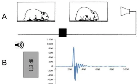
O reflexo auditivo de sobressalto (ASR) é uma reação motora rápida e intensa que culmina na contração da musculatura estriada esquelética da face e do corpo como um todo em resposta a um estímulo acústico inesperado e de alta intensidade O ASR é um reflexo acústico motor desencadeado por estruturas do tronco encefálico, que está presente em diversas espécies de mamíferos, inclusive no
Em roedores, esse reflexo se manifesta com o encurtamento do comprimento total do animal e aumento da pressão arterial e frequência cardíaca, mediadas pelo sistema nervoso autônomo (Baudrie et al., 1997; Koch,1999). Ele possui um caráter defensivo frente a uma provável agressão ou de alerta perante a acontecimentos inesperados. As contrações musculares em conjunto levam o animal a adotar uma postura de defesa, protegendo partes vitais do corpo, como a face, pescoço e abdome (Figura 2).
Figura 2. A -Esquema do reflexo auditivo de sobressalto no rato. Após ser estimulado por um som inesperado e de alta intensidade, o animal contrai a musculatura da face, pescoço e corpo para assumir uma postura defensiva. B- Gráfico da amplitude de resposta frente à apresentação de um estímulo sonoro de alta intensidade (113 dB). Modificado de Koch (1999).
Provas comportamentais envolvendo modulações do ASR têm despertado interesse em questões de diagnóstico clínico (Wilkins et al., 1986), tanto na clínica neurológica quanto psiquiátrica, por modificar-se diante de várias condições patológicas como doenças neurodegenerativas, esquizofrenia (Braff et al., 2001) depressão (Kaviani et al., 2004), transtorno do estresse pós traumático (Grillon et al., 1996), estresse (Andreski et al., 1998; Stam, 2007), ansiedade (Kaviani et al., 2004), medo (Anisman et al., 2000; Davis, 2006; Winslow et al., 2007), bem como frente a estados de dependência de drogas, como opiáceos (Mansbach et al., 1992; Borowski & Kokkinidis, 1994) e consumo de etanol (Grillon et al., 1994).
explicado pelo efeito bifásico da substância, que atua como estimulante inicialmente (Dudek et al., 1991), pela liberação de dopamina (Lewis & Gould, 2003), e, em seguida, como um depressor por ativação GABAérgica.
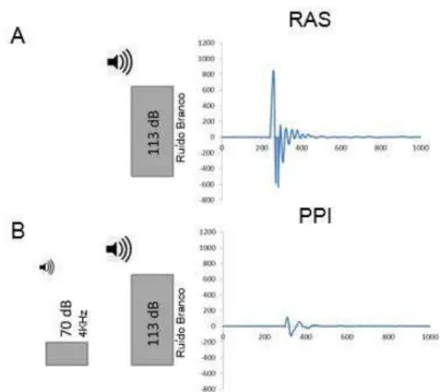
Entre as principais modulações do RAS, encontra-se a inibição por estimulo prévio (PPI), que é caracterizada pela diminuição ou completa abolição do reflexo quando o estímulo sonoro desencadeante é precedido por um estímulo sensorial (sonoro, visual ou tátil) de baixa intensidade, que isoladamente, não desencadearia o reflexo (RAS) (Figura 3).
Figura 3. Registro da resposta do reflexo auditivo de sobressalto (RAS) e da resposta de sobressalto após a inibição por estímulo prévio (PPI). A - apresentação de um estímulo sonoro de alta intensidade (113 dB) que desencadeia uma resposta de sobressalto no animal de grande amplitude. B – apresentação de um estímulo sonoro precedido por um estímulo de menor intensidade (70dB) (PPI), o qual a amplitude é menor. Modificado de Koch (1999).
ocorre uma diminuição desta variável, está associada com efeitos ansiogénico/sedativo (Prut & Belzung, 2003).
Para este teste é utilizado uma arena circular dividida em 12 setores, onde é analisada a atividade locomotora dos roedores, ou seja, os deslocamentos entre um ponto a outro da arena e seu comportamento exploratório não locomotor observando movimentos que o animal pode realizar sem a necessidade de deslocamento (De Lima et al., 2005).
O teste de campo aberto têm sido utilizado para verificar os efeitos do etanol em diversas situações: injeções intraventriculares (Correa et al., 2003) e intraperitoneais (Pardo et al., 2013), sua abstinência (Karadayiana et al., 2013); sensibilização comportamental (Bellot et al., 1996; Araujo et al, 2005), sua relação com exercício físico (Sosa, et al., 2015) e seus efeitos agudo e crônico (Fukushiro et al., 2012).
No SNC há evidência de que a vitamina D3 pode ser um importante
modulador do desenvolvimento do cérebro, atuando nas desordens neurológicas e neuropsiquiátricas (Harms et al., 2011). Atua ainda como neuroprotetor, sendo que em níveis baixos pode predispor ao desenvolvimento de depressão e déficit cognitivo (Groves et al., 2013).
Modelo Experimental
Muitos são os modelos animais utilizados em pesquisas relacionadas ao consumo de etanol entre eles destacam-se os modelos de preferência por etanol. As variedades de ratos UChA (baixo consumo de etanol) e UChB (alto consumo de etanol) (UCh=Universidade do Chile) constituem modelos importantes desse tipo de animais consumidores de etanol e, por isso, essas variedades são importantes para o estudo de características que podem ser associadas ao alcoolismo humano. Eles derivam de uma colônia original de ratos Wistar que tem sido criada seletivamente na Universidade do Chile por mais de 70 gerações (Quintanilla et al., 2006). Denominou-se UChA e UChB as linhagens de baixo e alto consumo de etanol respectivamente. Eles diferem quanto à atividade cerebral da aldeído-desidrogenase e na capacidade de desenvolver tolerância aguda ao etanol (Tampier et al., 1984,1994).
durante longo período, o que faz da linhagem UChB um adequado modelo experimental, pois possui predisposição genética para ingerir voluntariamente grandes quantidades de etanol a 10%. Há caminhos importantes para o enfoque dos estudos sobre o alcoolismo humano que podem ser investigados com o modelo animal UChB: a busca de marcadores genéticos que indiquem a predisposição ao alcoolismo, a busca de terapêuticas eficazes para o tratamento do alcoolismo e as alterações morfofuncionais provocados pela ingestão de etanol (Mardones, 1993; Martinez et al., 2000).
Considerando que o consumo crônico de etanol induz a miopatia (atrofia principalmente das fibras tipo II), neuropatia periférica e leva a uma diminuição nos
níveis de vitamina D3, que por sua vez desempenham um efeito prejudicial sobre o
músculo esquelético e SNC.
O objetivo deste trabalho foi avaliar se a administração de vitamina D3 durante o
Referências Bibliográficas
ABREU, A. M. M.; LIMA, J. M. B. L.; ALVES, T. A. O impacto do álcool na mortalidade em acidentes de trânsito: uma questão de saúde pública. Esc. Anna. Nery. R. Enferm, v.10, n.1, p.87 – 94, 2006.
ADACHI J. et al. 7alpha- and 7beta-hydroperoxycholest-5-en-3beta-ol in muscle as indices of oxidative stress: response to ethanol dosage in rats. Alcohol Clin Exp Res, v.24, p.675-81, 2000.
ADACHI, J. et al. Alcoholic muscle disease and biomembrane perturbations (review). The Journal of nutritional biochemistry. v.14, p. 616-625, 2003.
AMINOFF M.J. Neurology and General Medicine. 4th. Ed.: Churchill Livingstone; 2007. AMMENDOLA, A. et al. Peripheralneuropathy in chronicalcoholism: a retrospective crosssectional study in 76 subjects. AlcoholAlcohol, v. 36, p. 271–75, 2001.
ANDRESKI P, CHILCOAT H, BRESLAU N. Post-traumatic stress disorder and somatization symptoms: A prospective study. Psychiatry Research. v.79, p.131-138, 1998.
ANISMAN, H. et al. Acoustic startle and fear-potentiated startle in rats selectively bred for fast and slow kindling rates: Relation to monoamine activity. European Journal of Neuroscience, v.12, p. 4405-4416, 2000.
ANNWEILER, C. et al. Fall prevention and vitamin D in the elderly: an overview of the key role of the non-bone effects. Journal of Neuro Engineering and Rehabilitation, v.7, 2010. ARAUJO, N. et al. The importance of housing conditions on behavioral sensitization and tolerance to ethanol. Pharmacology Biochemistry and Behavior, v. 82, p. 40-45, 2005. ARCELES, M.L. et al. Morphometric study of duodenun myentric plexus in rats submitted to alcoholism. UNOPAR Científica Ciências Biológicas e da Saúde, v. 14, 2011.
BABOR, T. Álcool: Bem de consumo sui generis. Revistao Toxico dependências -Edição IDT, v.15, n.1,p. 77-86, 2009.
BAUDRIE, V. et al. Autonomic components of the cardiovascular responses to an acoustic startle stimulus in rats. Journal of autonomic pharmacology, v. 17, p. 303-309, 1997. BELLOT, R.G. et al. Monosialoganglioside attenuates the excitatory and behavioural sensitization effects of ethanol. European Journal of Pharmacology. v.313, p.175–179, 1996.
BISCHOFF, H.A.; BORCHERS, M.; GUDAT, F. In situ detection of 1,25-dihydroxyvitamin D3 receptor in human skeletal muscle tissue. Histochem J, v. 33, p.19–24, 2001.
BOROWSKI T.B. & KOKKINIDIS L. Cocaine preexposure sensitizes conditioned fear in a potentiated acoustic startle paradigm. Pharmacol. Biochem. Behav, v. 49, p. 935-942. 1994.
BRAFF, D. L.; GEYER, M. A.; SWERDLOW, N. R. Human studies of prepulse inhibition of startle: normal subjects, patient groups, and pharmacological studies.
Psychopharmacology, v. 156, p. 234-258, 2001.
BRESLOW, R. A., Guenther, P. M., & Smothers, B. A. Alcohol drinking patterns and diet quality: the 1999–2000 National Health and Nutrition Examination Survey. American journal of epidemiology, v.163, p. 359-366, 2006.
BURKE, L.M. et al. Effect of alcohol intake on muscle glycogen storage after prolonged exercise. J. Appl. Physiol. v. 95, p. 983-990, 2003.
CARLINI, E.A. et al. I Levantamento Domiciliar sobre o Uso de Drogas no Brasil – 2001.
São Paulo: CEBRID – Centro Brasileiro de Informações Sobre Drogas
Psicotrópicas:UNIFESP – Universidade Federal de São Paulo, 2002.
CASTELLÓN, M.C. D. et al. Alcoholic myopathy: lack of effect of zinc supplementation. Food and chemical toxicology, v. 43, p. 1333-1343, 2005.
CEGLIA, L. Vitamin D and Its Role in Skeletal Muscle. Curr Opin Clin Nutr Metab Care,
v.12, p. 628–633, 2009.
CEGLIA, L. Vitamin D and skeletal muscle tissue and function. Mol. Aspects Med, v.29, p. 407–14, 2008.
CHAUVEAU, F. et al. Rapid stress‐induced corticosterone rise in the hippocampus reverses serial memory retrieval pattern. Hippocampus, v. 20, p. 196-207, 2010.
CHRISTAKOS S. et al. Vitamin D: beyond bone. Ann. N Y Acad. Sci, v. 1287, p.45–
58,2013.
CONDE, A.; GONZÁLEZ-REIMERS, E.; GONZÁLEZ-HERNÁNDEZ, T. Relative and combined roles of ethanol and protein malnutrition on skeletal muscle. Alcohol Alcohol. v.27, p.159–63, 1992.
CORNIER, M.A; JACKMAN, M.R; BESSESEN, D.H. Disposition of Dietary Ethanol Carbons in Rats: Effects of Gender and Nutritional Status. Metabolism, v.49, p. 379-385, 2000. CORREA, M. et al.. Open field locomotor effects in rats after intraventricular injections of ethanol and the ethanol metabolites acetaldehyde and acetate. Brain Research Bulletin. v.62, p. 197–202 , 2003.
Courtney K.E. & Polich J. Binge drinking in young adults: Data, definitions, and determinants.
Psychol Bull,v.135, p.142–156, 2009.
DAVIS M. Neural systems involved in fear and anxiety measured with fear-potentiated startle. American Psychologist. v.61, p.741-756, 2006.
DE LIMA, M. M. et al. Recognition memory impairment and brain oxidative stress induced by postnatal iron administration. European Journal of Neuroscience, v. 21, p. 2521-2528, 2005.
DEMAY M. Muscle: a nontraditional 1,25-dihydroxyvitamin D target tissue exhibiting classic hormone-dependent vitamin D receptor actions. Endocrinology, v.144, p. 5135–7, 2003. DIAMOND, T. et al. Ethanol reduces boné formation and may cause osteoporosis. Am. J. Med, v.86, p. 282–8, 1989.
DIRKS-NAYLOR, A.J. & LENNON-EDWARDS, S. The effects of vitamin D on skeletal muscle function and cellular signaling. Journal of Steroid Biochemistry & Molecular Biology, v.125, p.159–16, 2011.
DUAILIBI, S.; PINSKY, I.; LARANJEIRA, R. Prevalência do beber e dirigir em Diadema, estado de São Paulo. Rev. Saúde Pública, v.41, n.5, p. 1058-61, 2007.
DUDEK, B.C.; PHILLIPS, T.J.; HAHN, M.E. Genetic analyses of the biphasic nature of the alcohol dose-response curve. Alcoholism, clinical & experimental research. v.15, p.262-269, 1991.
EKBOM K.; HED, R.; L.; ASTROM, K.E. Muscular Affections In Chronic Alcoholism. Arch. Neurol, v.10, p.449-458, 1964.
ERTEM, K. et al. Impairment of Peripheral Nerve Healing After Nerve Repair in Rats Chronically Exposed To Alcohol. Archives of Medical Research, v.40, p.325-330, 2009. FERNANDES, E.V. et al. Efeitos da ingestão alcoólica crônica e do exercício físico na massa corporal, no consumo alimentar e na ingestão líquida de ratos wistar. R. da Educação Física/UEM-Maringá, v. 21, p. 527-533, 2010.
FERNANDEZ-SOLA J. et al. Muscle anti-oxidant status in chronic alcoholism. Alcohol Clin Exp Res, v.26, p.1858–1862, 2002.
FERNÁNDEZ-SOLÀ, J. et al. Evidence of apoptosis in chronic alcoholic skeletal myopathy.
Human pathology, v. 34, p. 1247-1252, 2003.
FERNANDEZ‐SOLÀ, J. et al. Molecular and Cellular Events in Alcohol‐Induced Muscle Disease. Alcoholism: Clinical and Experimental Research, v. 31,p. 1953-1962, 2007. FUKUSHIRO, D.F. et al. Acute and chronic ethanol differentially modify the emotional significance of a novel environment: implications for addiction. International Journal of Neuropsychopharmacology. v.15, p.1109–1120, 2012.
GONZÁLEZ-REIMERS, E. et al. Alcoholic Myopathy: Vitamin D Deficiency is Related to Muscle Fibre Atrophy in a Murine Model. Alcohol & Alcoholism, v.45, p. 223–230, 2010. GRILLON, C. et al. Baseline startle amplitude and prepulse inhibition in Vietnam veterans with posttraumatic stress disorder. Psychiatry research, v. 64, p. 169-178, 1996.
GROVES, N. J. et al. Adult vitamin D deficiency leads to behavioural and brain neurochemical alterations in C57BL/6J and BALB/c mice. Behavioural brain research, v. 241, p. 120-131, 2013.
HAES, T.M. et al. Álcool e sistema nervoso central. Medicina (Ribeirão Preto), v.43,p. 153-63, 2010.
HARMS, L. R. et al. Vitamin D and the brain. Best Practice & Research Clinical Endocrinology & Metabolism. v. 25, p.657-669, 2011.
HODGES ,D.L.; KUMAR, V.N.; REDFORD, J.B. Effects of alcohol on bone, muscle and nerve. Am Fam Physician, v.34, p.149-56, 1986.
HOLAHAN C.J. et al. Episodic heavy drinking and 20-year total mortality among late-life moderate drinkers. Alcohol Clin Exp Res 38: 1432–1438, 2014
IZUMI, Y. et al. Corticosterone enhances the potency of ethanol against hippocampal long-term potentiation via local neurosteroid synthesis. Frontiers in cellular neuroscience, v. 9, 2015.
JAYASEKARA H. et al. Alcohol consumption over time and risk of death: a systematic review and meta-analysis. Am J Epidemiol, v.179, p.1049 –1059, 2014
JUNG, M. K. et al. Alcohol Exposure and Mechanisms of Tissue Injury and Repair. Alcoholism, Clinical and Experimental Research, v.35, p.392–399, 2011.
JUNTUNEN, J. et al. Experimental alcoholic neuropathy in the rat: Histological and electrophysiological study on the myoneural junctions and the peripheral nerves. Acta Neuropathologica, v.41, p. 131-137, 1978.
KARADAYIANA, A.G. et al. Alterations in affective behavior during the time course of alcohol hangover. Behavioural Brain Research. v.253, p.128– 138, 2013.
KAVIANI H. et al. Affective modulation of the startle response in depression: Influence of the severity of depression, anhedonia, and anxiety. Journal of Affective Disorders, v. 83, p.21-31, 2004
KOCH M. The neurobiology of startle. Prog. Neurobiol. v.59, n.2, p.107-128,1999.
KOIKE H. et al. Alcoholic neuropathy is clinicopathologically distinct from thiamine-deficiency neuropathy. Ann Neurol, v.54, p.19–29, 2003.
KOLL, M. et al. Effect of Acute and Chronic Alcohol Treatment and Their Superimposition on Lysosomal, Cytoplasmic, and Proteosomal Protease Activities in Rat Skeletal Muscle In Vivo.
Metabolism, v. 51, p. 97-104, 2002.
KOLL. M.; AHMED, S.; MANTLE, D. Effect of acute and chronic alcohol treatment and their superimposition on lysosomal, cytoplasmic, and proteosomal protease activities in rat skeletal muscle in vivo. Metabolism, v.51,p.97–104, 2002.
KOO-NG R. et al. Carbonyl levels in type I and II fiber-rich muscles and their response to chronic ethanol feeding in vivo and hydroxyl and superoxide radicals in vitro. Alcohol Clin Exp Res, v.24, p.1862–1868; 2000.
KOTIDIS E. et al. Can chronic intra-abdominal hypertension cause oxidative stress to the abdominal wall muscles? An experimental study. J Surg Res, v. 176, p. 102-7; 2012.
LAITINEN, K. & VÄLIMÄKI, M. Alcohol and bone. Calcif. Tissue Int. v.49, p.S70–3, 1991. LEVY, J.A. Neuromiopatias. Arq. Neuro-Psiquiatr, v. 44, 1986.
LEVY, J.A. Polineuropatias Alcoólicas. In: FORTES, J.R.A.; CARDO, W.N. Alcoolismo, diagnóstico e tratamento. São Paulo, Sarvier, p.151-6. 1991.
LEWIS, M.C. & GOULD, T.J. Nicotine & ethanol enhancements of acoustic startle reflex are mediated in part by dopamine in C57BL/6J mice. Pharmacol. Biochem. Behav, v.76, p.179-186, 2003.
LIBER, C.S. Hepatic, Metabolic and Toxic Effects of Ethanol. Alcohol Clin Exp Res, v.15, p.573-92, 1991.
LIEBER, C.S. Relationships between nutrition, alcohol use, and liver disease. Alcohol Research & Health, v.27, p.220-31, 2003.
LINDHOLM, J.; STEINICHE, T.; RASMUSSEN, E. Bone disorders in men with chronic alcoholism: a reversible disease? J. Clin. Endocrinol. Metab, v.73, p.118–24, 1991.
LYNCH, P. G. Alcoholic Myopathy. Journal of the Neurological Sciences. v.9, p. 449–462, 1969.
MANSBACH, R.S.; GOLD, L.H.; HARRIS, L.S. The acoustic startle response as a measure of behavioral dependence in rats. Psychopharmacology (Berl), v.108, p.40-46, 1992. MANSOURI A. et al. Acute ethanol administration oxidatively damages and depletes mitochondrial DNA in mouse liver, brain, heart and skeletal muscles: protective effects of anti-oxidants. J Pharmacol Exp Ther, v. 298, p.737–743, 2001.
MARDONES J.; SEGOVIA-RIQUELME N. Thirty-Two Years Of Selection Of Rats By Ethanol Preference: UchA And UchB Strain. Neur.Tox. And Terat. v.5, p.171-8, 1983.
MARTINEZ, F.E.; MARTINEZ, M.; PADOVANI, C.R.; BUSTOS-OBREGON, E. Morphology Of Testis And Epididymis In An Ethanol-Drinking Rat Strain (UCha And UChb). J. Submicrosc. Cytol. Pathol, v. 32, p. 175-184, 2000.
MASCARENHAS, M.D.M. et al. Consumo de álcool entre vítimas de acidentes e violências atendidas em serviços de emergência no Brasil, 2006 e 2007. Ciência & Saúde Coletiva,
v.14, n.5, p.1789-1796, 2009.
MEJIA-TOIBER, et al. Impulsive choice and anxiety-like behavior in adult rats exposed to chronic intermittent ethanol during adolescence and adulthood. Behavioural brain research, v. 266, p. 19-28, 2014.
MELLION, M. L. et al. Experimental model of alcohol – related peripheral neuropathy.
Muscle Nerve, v. 48, p. 204–211, 2013.
MELLO, M.L.M.; BARRIAS, J.C.; BREDA, J.J. Álcool e problemas ligados ao álcool em Portugal, Lisboa, Direção Geral de Saúde, p. 121, 2001.
MEZEY, E. Alcoholicliverdisease: roles of alcohol and malnutrition. American Journal of ClinicalNutrition, v. 33, p. 2709–2718, 1980
MITCH, W.E. & GOLDBERG, A.L. Mechanisms of muscle wasting. The role of the ubiquitin-proteasome pathway. N. Engl. J. Med, v. 335, p.1897–905, 1996.
NICOLAS, J.M.; GARCIA, G.; FATJO, F. Influence of nutritional status on alcoholic myopathy. Am J Clin Nutr, v.78, p.326–33, 2003.
PARDO, M. et al. Acetate as an active metabolite of ethanol: studies of locomotion, loss of righting reflex, and anxiety in rodents. Frontiers in Behavioral Neuroscience. v.7, 2013. PEREIRA, K.F & CONEGERO, C.I. Interface músculo-tendínea de ratos induzidos a ingestão alcóolica. Acta Scientiarum. Biological Sciences Maringá, v. 26, p. 361-364, 2004.
PITTS, T.O. & VAN-THIEL, D.H. Disorders of divalent ions and vitamin D metabolism in chronic alcoholism. Recent Dev. Alcohol, v.4, p.357–77, 1986.
POHORECKY, L.A. Biphasic action of ethanol. Biobehavioral Reviews, v. 1, p. 231–240, 1977.
PREEDY, V.R & PETERS, T.J. Alcohol and skeletal muscle disease. Alcohol and Alcoholism. v.25, p.177-187, 1990.
PREEDY, V.R. et al. Alcoholic myopathy: biochemical mechanisms. Drug Alcohol Depend,
v.63, p.199–205, 2001.
PREEDY, V.R. et al. The importance of alcohol-induced muscle disease. Journal of Muscle Research & Cell Motility. v.24, p. 55-63, 2003.
PROJEDNIC, R.M & CEGLIA, L. The emerging biomolecular role of vitamin D in skeletal muscle. Exerc Sport Sci Rev, v.42, p. 76-81, 2014.
PRUT, L.; BELZUNG, C. The open field as a paradigm to measure the effects of drugs on anxiety-like behaviors: a review. European Journal of Pharmacology, v.463, 28, p. 3–33, 2003.
QUINTANILLA, M.E.; ISRAEL, Y.; SAPAG, A.; TAMPIER, L. The Ucha and Uchb Rat Lines: Metabolic And Genetic Differences Influencing Ethanol Intake. Addict. Biol., v. 11, p.310-23, 2006.
RATCLIFF, E.V. Alcoholic neuropathies. Aust Fam Physician, v.8, p.171–7, 1979.
ROMERO, J.C.; SANTOLARIA, F.; GONZÁLEZ-REIMERS, E. Chronic alcoholic myopathy and nutritional status. Alcohol, v.11, p. 549–55, 1994.
ROOM, R.; BABOR, T.; REHM, J. Alcohol And Public Health. Lancet. v. 365, n. 9458, p. 519-30, 2005.
RUBIN E. et al. Muscle damage produced by chronic alcohol consumption. Am J Pathol, v.83, p.499–515, 1976.
RYLE, P. R. & THOMSON, A. D. Nutrition and vitamins in alcoholism. ContemporaryIssues in ClinicalBiochemistry, v. 1, p. 188–224, 1984.
SANTORI, C.; CECCANTI, M.; DIACINTI, D. Skeletal turnover, bone mineral density, and fractures in male chronic abusers of alcohol. J. Endocrinol. Invest, v. 31, p.321–6, 2008. SESTOFT L. et al. Working capacity and expression of myosin heavy chain isoforms in skeletal muscle of chronic alcoholic men without liver disease after 1 day and 4 weeks of alcohol abstinence. Clin Sci (Lond). v.86, p.433-40, 1994.
SHANKAR, K. et al. Chronic etanol consumption leads to disruption of vitamin D3 homeostasis associated with induction of renal 1,25 dihydroxyvitamin D3-24-hydroxylase (CYP24A1). Endocrinology, v.149, p.1748–56, 2008.
SOSA, P. M. et al. Physical exercise prevents motor disorders and striatal oxidative imbalance after cerebral ischemia-reperfusion. Brazilian Journal of Medical and Biological Research, v. 48, p. 798-804, 2015.
STAM R. PTSD and stress sensitisation: A tale of brain and body. Part 1: Human studies.
Neuroscience and Biobehavioral Reviews. v.31, p.530-557, 2007.
STEINER, J.L. & LANG, C.H. Dysregulation of skeletal muscle protein metabolism by alcohol. Am J Physiol Endocrinol Metab, v. 308, p.E699–E712, 2015.
SVANBERG, E. et al. Semi-starvation alters myofibrillar mRNA concentrations to expedite rapid recovery of muscle protein stores following feeding. Eur J Clin Invest. v.30, p.722–8, 2000.
TAMPIER, L. et al. Biological Similarities And Differences Between Rats Genetically Different In Alcohol Preference. Alcohol Alcohol., v. 19, p. 203-209, 1984.
TAMPIER, L.; QUINTANILLA, M.E.; MARDONES, J. Acetaldehyde Metabolism: Differences Between UchA And UchB Rats. Alcohol Alcohol. v.29, p.751-755, 1994.
TENG, et al. Alcohol exposure after mild focal traumatic brain injury impairs neurological recovery and exacerbates localized neuroinflammation. Brain, behavior, and immunity, v. 45, p. 145-156, 2015.
TORREJAIS, M.M. et al. Histochemical and SEM evaluation of the neuromuscular junctions from alcoholic rats. Tissue & Cell. v. 34, p.117–123, 2002.
TURNER, R.T. et al. Chronic alcohol treatment results in disturbed vitamin D metabolism and skeletal abnormalities in rats. Alcohol Clin. Exp. Res, v. 12, p. 159–62, 1988.
WANG, J. et al. Effects of increased matrix metalloproteinase-9 expression on skeletal muscle fibrosis in prolonged alcoholic myopathies of rats. Molecular Medicine Reports, v.5, p.60-65, 2012.
WILKINS DE, HALLETT M, WESS MM. Audiogenic startle reflex of man & its relationship to startle syndromes. A review. Brain, v.109, p.561-573,1986.
WINSLOW J.T.; NOBLE P.L.; DAVIS M. Modulation of Fear-Potentiated Startle and Vocalizations in Juvenile Rhesus Monkeys by Morphine, Diazepam, and Buspirone.
Biologicalpsychiatry. v.61, p.389-395, 2007.
WORLD HEALTH ORGANIZATION. Global status report on alcohol and health. Geneva, Switzerland: WHO Press, World Health Organization, p.85, 2011.
Behavior assessment of UChB and Wistar rats supplemented with vitamin D3
Carina Guidi Pinto1, Marina Galleazzo Martins2, André de Souza Mecawi3, José de
Anchieta de Castro e Horta-Júnior2, Selma Maria Matheus Michelin2*
1 Graduation Program on the General Bases of Surgery, Botucatu Medical School,
Department of Anatomy, Institute of Biosciences, UNESP - UNIV Estadual Paulista, Botucatu Campus, Travessa da Rua Prof. Dr. Gilberti Moreno, Zip Code: 18618-689, Botucatu, São Paulo, Brazil. E-mail: carina_guidi@hotmail.com
2 Department of Anatomy, Institute of Biosciences, UNESP - UNIV Estadual Paulista,
Botucatu Campus, Travessa da Rua Prof. Dr. Gilberti Moreno, Zip Code: 18618-689,
Botucatu, São Paulo, Brazil. E-mail: marina.gmartins@gmail.com;
anchieta@ibb.unesp.br; micmath@ibb.unesp.br
3 Department of Physiological Sciences - Institute of Biology Federal Rural University
of Rio de Janeiro BR465, Km7, Zip Code: 23897-970, Seropedica, RJ, Brazil. E-mail: mecawi.as@gmail.com
Corresponding author: Carina Guidi Pinto, Botucatu Medical School, Department of Anatomy, Institute of Biosciences, UNESP - UNIV Estadual Paulista, Botucatu Campus, Travessa da Rua Prof. Dr. Gilberti Moreno, Zip Code: 18618-689, Botucatu,
São Paulo, Brazil. E-mail: carina_guidi@hotmail.com. Telephone: +55 (014) 3880-0025. Email: carina_guidi@hotmail.com
ABSTRACT
Ethanol intake compromises brain structure, presenting biphasic effects over motor activity, acting as a stimulant or depressor depending on the dose or duration of use.
It interferes in vitamin D3 absorption and metabolism, what correlates to some
neurologic and neuropsychiatric disorders. There are reports on the association of
ethanol with bone alterations, including low levels of vitamin D3. Based on that, the
objective of this study was to evaluate the effects in behavior tests of isolated administration of vitamin D3 or its administration in association with ethanol, during chronic alcoholism. In order to achieve that, two experimental groups were used: male Wistar rats (n=20), and UChB lineage male rats (n=20) (volunteer ethanol
drinkers). Both groups were divided in two subgroups: Vitamin D3 – 12.5µg/kg/day
(500 UI) of cholecalciferol (WV, n=10, and UV, n=10), and Control (WC, n=10, and UC, n=10), for a period of 75 days. Body weight analyses and behavior tests (acoustic startle reflex and open field) were conducted at 90 and 165 days of age. In addition to that, corticosterone plasma levels were measured at 165 days, with no statistical difference between the experimental groups. The Wistar group presented lower ASR values in the final moment (Control and supplemented), while the PPI percentages were higher in the initial group. In the UChB group there was no difference in PPI percentages with the pre-stimuli used. When the ASR responses were compared between groups (Wistar and UChB), the UChB group presented lower ASR amplitudes as compared to the animals of the Wistar group. In relation to PPI percentages in the three pre stimuli used in the UChB group, the percentages were higher as compared to the animals in the Wistar group, the ASR values been smaller in the UChB group. The presence of feces and urine was similar both in the initial and final moments, in the open field test, for all the experimental groups. Considering the other parameters of open field, decreased locomotion was noted in
the UChB group in the vitamin D3 subgroup, as well as in the final control group as
compared to the initial control. When the Wistar and UChB groups were compared, the latter presented higher rates in locomotion. The results obtained are consonant to the depressive/sedative effect of ethanol over the CNS, reducing ASR amplitude, with a likely enhancement over the inhibitory circuits which mediate PPI, determining the increase in inhibition percentage in the UChB group, as well as an increase in locomotion presented by the UChB group in the open field test, characterized by a decrease in the aversive effect to the new environment. In addition we emphasize that the dose of vitamin D3 used did not have effect over the behavior parameter analyzed, differences observed in this group seeming to be related to getting used to the gavage. This study describes for the first time in literature the differences in responses to the acoustic startle reflex test between Wistar and UChB rats, the latter genetically predisposed to ethanol intake, a lineage adequate to be used in experiments comprising analyses of behavior in face of ethanol intake.
1.0 Introduction
Ethanol is among the most common substances of abuse in the world, leading to many problems. Ethanol is considered a neurotoxic substance that significantly affects the structure and metabolism of the central nervous system (CNS) [1].
In rodents CNS, ethanol presents biphasic effects over motor activity, stimulating or depressing it depending on the dose and duration of use [2;3].
In rats, alcohol intoxication is classified as a stress factor, and under that condition there are increased plasma levels of corticosterone [4], resulting in increased levels of that steroid in the brain [5].
Ethanol attenuates the co-release of gamma-aminobutyric acid (GABA), inhibiting its biosynthesis, a mechanism by which exposure to ethanol may modulate the activity of GABAergic synapses [6].
In order to evaluate the action of drugs over the CNS, among them ethanol, many behavioral assessments are conducted, including the acoustic startle reflex (ASR) [7;8;9;10;11;12]
ASR is an acoustic-motor reflex triggered by structures of the encephalic trunk, present in many mammal species, including man. It is a quick, intense motor reaction that culminates in the contraction of skeletal muscles from the face and body as a response to an unexpected and highly intense acoustic stimulus (Koch, 1999). This reflex may be altered under many physiological conditions such as fear [13;14;15], anxiety [16], and stress [17;18].
The effects of ethanol administration given intracerebroventricular [19] or intraperitoneal [20] have been studied in the open field test, as well as behavioral sensitization [21;22], relation to physical exercise [23], acute and chronic effects [24;25;26;27;28;29;30] and effects after abstinence [31].
Ethanol also interferes in the absorption and metabolism of essential nutrients [32;33;34;35;36].
Many researchers have analyzed the relation of ethanol with bone
abnormalities, in which low levels of vitamin D3 in alcoholics or animals treated with
ethanol have been reported [37;38;39;40;41].
Ethanol has therefore proved to have a profound effect in calcium
absorption, and synthesis refers to the direct or indirect effect of ethanol over the body, and may also occur as a consequence of life style in alcoholic subjects [43].
Besides the well-known role in calcium homeostasis, vitamin D3 has been
related to studies comprising the CNS [44].
There are evidences that vitamin D3 may be an important modulator in brain
development, acting in neurologic and neuropsychiatric disorders [45]. This vitamin also acts as a neuroprotective substance, and in low levels may lead to the development of depression and cognitive deficit [46].
Many are the animal models used in researches related to ethanol intake, among which stand out the models of ethanol preference as the UChA and UChB rats lineages (UCh = Chile University), which are important for translational studies [47;48].
ASR as well as the open field test are behavior tests sensitive to alcohol intoxication that alters calcium and vitamin D3 metabolism, what in its turn under certain circumstances exerts neuroprotective effect over the CNS. The objective of this study was therefore to assess the effects of isolated administration or the
association of vitamin D3 and ethanol for chronic alcoholism and analyze the
differences between Wistar rats and UChB front of behavioral tests.
2.0 Materials and Methods
2.1 Experimental groups
40 male adult 90-day-old rats were divided in two experimental groups: 20 UChB rats (voluntary ethanol drinkers, group U), originated from the animal research laboratory of the Anatomy Department of IBB/UNESP, and 20 Wistar rats (group W) originated from the animal research laboratory of the Biomedical Sciences Institute of USP São Paulo-SP). The animals were kept in polyethylene boxes (40x30x15cm), with solid floors, covered with wood shavings, under controlled conditions of luminosity (12h light, 12h dark), and temperature (20 to 25 ºC), receiving filtered
water and industrialized food (Provence®) ad libitum. All the experimental procedures
were approved by the Committee on Animal Research and Ethics of the Biosciences Institute, UNESP, Botucatu (Protocol number 531).
water and industrialized food ad libitum. At 55 days of age they were isolated and, from 60 days on, filtered water and a 10% ethanol solution were offered, both periodically alternated, ad libitum. After 15 days evaluating the intake of the 10% ethanol solution, the selection and standardization of the UChB lineage was carried out [49]. The animals that had average intake higher than 2.0mL 10% ethanol/100g of body weight/day were selected for the UChB lineage. After selection and during
the whole experiment 10% ethanol solution was offered to the UChB animals ad
libitum.
Each experimental group (Wistar and UChB) was divided in two subgroups: Vitamin D3 subgroup (n = 10 per lineage), which received 12.5 µg/kg (500 UI) of Cholecalciferol (C9756-Sigma) diluted in olive oil, given daily by gavage ; and Control subgroup (n = 10 per lineage), which received the vehicle (olive oil), with the same periodicity, both for 75 days.
Groups Subgroups Voluntary intake Animals (nº) Assessment moments
UChB
Control (UC) Ethanol 10% 10 Initial - 90 days
Final - 165 days
Vitamin D3 (UV) Ethanol 10% 10
Final - 165 days
Wistar
Control (WC) Water 10 Initial - 90 days
Final - 165 days
Vitamin D3 (WV) Water 10
Final - 165 days
2.2 Behavior assessment
The animals were individually weighted and the acoustic startle reflex and open field activity measured in two moments: initial, at 90 days (Wistar Initial and UChB Initial), and final, at 165 days of age (WC final; UC final; WV and UV). The initial behavior assessment comprised the animals of groups Wistar and UChB, the animals of the Vitamin D3 subgroups, since they belonged initially to the groups of
origin, were evaluated after the experimental period of Vitamin D3 supplementation,
2.2.1 Measuring the Acoustic Startle Reflex
For the evaluation of the acoustic startle reflex, an assessment system made of an acoustically isolated box, a measuring platform, 9 cm from the loud-speakers, and a system that generated sound stimuli controlled by a software and connected to a computer (Insight Ltda.) was used. The ASR quantification was made by a sensor set under the test cage, which registered the animal‟s weight variation, during and after acoustic testing, considering that the response is a ballistic movement.
All the behavior tests were conducted between 1pm and 4pm. Each ASR session encompassed four different attempts: isolated sound stimulus, and sound stimulus preceded by a pre-stimulus of 65, 70 or 75 dB, in order to evaluate prepulse inhibition (PPI). During the whole session, the chambers had constant ventilation and background noise (white 60dB noise); the animals were monitored by means of a video camera. Every session began with a 5-minute acclimatization period. ASR/PPI measurements were then conducted using a sound stimulus, 113 dB SPL white noise, lasting for 20 ms. In the other attempts, a pre-stimulus of 4 KHz pure tone signal of 65dB SPL, 70dB SPL or 75 dB SP and lasting 20 ms preceded the stimulus
in 50 ms. The interval between the attempts was 30 s with 33% random variation.
ASR amplitudes and the PPI percentage, calculated as PPI= [(reflex amplitude) –
(reflex amplitude with pre stimulation)] x 100/ (reflex amplitude) were evaluated in the attempts with isolated stimulus.
2.2.2 Open field test
Open field evaluation rendered possible the measurement of variables concerning the modifications in the exploratory activity and locomotion. The apparatus used was a round arena with a 90-centimeters diameter, with 12 radial divisions painted on the floor, and acrylic walls 50 cm high.
2.3 Euthanasia
After the 75 days of treatment with Vitamin D3, on the day following the last session on behavior assessment, the animals were fasted for 12 hours, weighted, anesthetized anesthetized by intraperitoneal injection of ketamine (Dopalen - 90 mg / kg) and Xylazine (Rompun - 10mg / kg). The blood was obtained by cardiac puncture and stored in a container with heparin. Plasma was then extracted centrifuging the blood at 2,000 RPM per 10 min and the corticosterone plasma concentration was determined by specific radioimmunoassay (RIA), after ethanol extraction as previously described [50]. RIA sensitivity and the intra and inter-assay variation of coefficients were 0.16 µg/dl and 5.1%, and 8.1%, respectively. The results were expressed as µg/dl.
3.0 Results
3.1 Body weight
Body weight at 165 days (final) was greater than at 90 days of age (initial) in both lineages (p < 0.05). When we evaluated animals of the same age, there were no significant differences between the subgroups (Figure 1).
3.2 Behavior assessment
3.2.1 Measuring Acoustic Startle Reflex (ASR)
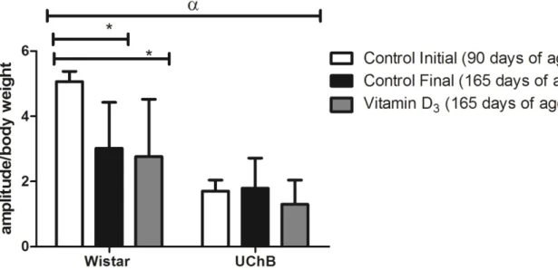
Considering that the amplitude of ASR is proportional to body weight [51], data concerning response amplitudes were normalized according to the animal‟s body weight, avoiding possible distortions in the results (ASR amplitude/body weight). ASR amplitude in the Wistar group was smaller in the control-final subgroups (3.0 ± 0.4; p < 0.001), and in the group supplemented with vitamin D3 (2.7 ± 0.5; p < 0.001) at 165 days, as compared to the subgroup control-initial (5.0 ± 0.3). There was no difference in the ASR amplitudes in any of the UChB lineage subgroups (p > 0.05) (Figure 2).
Figure 2. Graphic for normalized ASR amplitude. The Wistar final and Wistar Vitamin D3 groups presented smaller startle amplitude as compared to the initial Wistar subgroup (p < 0.001). In regard to the comparison of lineages, the UChB group presents smaller troughs as compared to the Wistar group. The data are presented as average and standard error of the mean, and two consecutive factors were analyzed by ANOVA followed by Bonferroni‟s post test (p < 0.05) * = p < 0.05 and α = p < 0.0001.
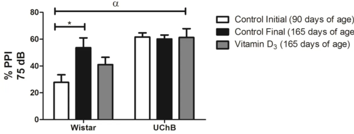
The three pre-stimuli intensities used in the sessions (65, 70, and 75dB) were efficient in triggering PPI in all experimental groups.
intensities of pre-stimulus used, 65dB (22.8 ± 4.1; p < 0.0001), 70 dB (27.5 ± 5.3; p < 0.005), and 75 dB (27.7 ± 5.6; p < 0.01) (Figure 3). In the subgroup supplemented with vitamin D3 there was no significant difference (p > 0.05).
In the UChB group there was no difference in the PPI percentages between the initial and final moments, as well as between the three intensities of pre-stimulus used (p > 0.05) (Figure 3). This same behavior was sustained in the UChB group
supplemented with vitamin D3.
Figure 3. Graphic for the PPI percentage with the 65, 70 and 75 dB pre stimuli. The PPI percentages were smaller in the initial Wistar subgroup as compared to the final Wistar, regardless of the pre stimulus used: * = 65dB (p < 0.0001), 70 dB (p < 0.005) and 75 dB (p < 0.01). In relation to the statistical analyses of the lineages, in the UChB group the values were higher in all pre stimuli used: α = 65 dB (p = 0.0002), 70 and 75 dB (p < 0.0001). The data are presented as average and standard error of the mean, and two consecutive factors were analyzed by ANOVA followed by Bonferroni‟s post test (p < 0.05).
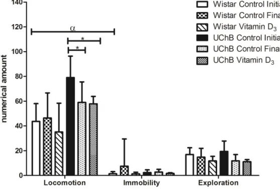
3.2.2 Open field test
During the open field tests, both in the initial and final moments there were similar amounts of feces and urine on the floor of the device for both groups, (p > 0.05) (Figure 4).
The Wistar group was similar in relation to the behaviors analyzed in both moments, no difference being noted between the experimental groups (p > 0.05). In
the UChB group there was a decrease in locomotion in the vitamin D3 subgroup (57.8
± 6.0; p < 0.001) and in the control-final subgroup (59.0 ± 5.2; p < 0.01) as compared to the control-initial subgroup (79.0 ± 5.4) (Figure 5).
When comparing the groups (Wistar and UChB) there was a significant difference between them in relation to the open field test (p < 0.0001).
Considering those differences observed, there was statistical difference only in locomotion, where the animals from the UChB group (Control-Initial: 79.0 ± 5.4;
Control-Final: 59.0 ± 5.2 and Vitamin D3: 57.8 ± 6.0) had higher locomotion pattern
as compared to the animals of the Wistar group (Initial: 43.6 ± 4.5; Control-Final: 46.4 ± 5.8; and Vitamin D3: 35.0 ± 6.7) (p < 0.001) (Figure 5).
Figure 5. Graphic of the parameters evaluated in the open field test of the experimental groups, where the control animals of the UChB group and the Vitamin D3 subgroup demonstrated decreased locomotion on the final moment at 165 days [(* = p < 0.01) and (* = p < 0.001), respectively], in comparison to the initial moment. In relation to lineage they
diverge (α = p < 0.0001), and in the parameters analyses, there was statistical difference
3.3 Biochemical analyses
3.3.1 Corticosterone plasma concentration
There was no statistical difference in cortisone plasma concentration (µg/dL) between the groups treated (WV=6.06 ± 4.03; UV=7.3 ± 3.5) and not treated with
vitamin D3 (WC=8.3 ± 3.2; UC=7.5 ± 4.8) (p > 0.05) (Figure 6).
Figure 6. Graphic for the corticosterone plasma concentration (µg/dL) in the groups studied. Data are presented as average and standard deviation and were analyzed by the parametric
Student‟s t-test (p > 0.05).
4.0 Discussion
Excessive use of alcohol may cause structural and functional abnormalities in the brain [52], besides damage to other organs, such as skeletal muscles [53].
The generation of reactive oxygen species (ROS), consequent of oxidative stress from acetaldehyde, radical that is a byproduct of ethanol metabolization [53;54;55;56] has been proposed as the cause for neuronal damage.
Behavior assessment contributes for the study of neural mechanisms associated to dependence, among them ethanol [11;12;29;30]
All animals evaluated presented acoustic startle reflex after sound stimulus, consonant to previous reports [59]. The previous presentation of the pre-stimuli was also able to reduce the amplitude of response, following the PPI paradigm [59].
The control animals in the Wistar lineage at 165 days of age (final moment) had a smaller ASR amplitude and higher PPI percentage than the control animals at
90 days of age (initial moment). Vitamin D3 supplementation did not change ASR
amplitude or PPI percentage in any of the groups evaluated.
Studies correlate ASR amplitude to the level of anxiety in man [16;60] and rodents [61].
ASR amplitude may be increased in situations potentiated by fear [13;14;15], anxiety [16,62], and stress [17;18]. We therefore believe that the differences observed in ASR and PPI in the Wistar rats in the final moment are related to the decrease of anxiety in those animals. Anxious patients, for example, have larger PPI amplitude than subjects with low levels of anxiety [16].
Considering that the animals were submitted to daily gavage from 90 to 165 days of age, that being considered a stress factor for the animal [63], we assume that they got habituated to this stressor, what would lead to a reduction in anxiety levels.
No statistical difference was noted in ASR amplitude or PPI percentage in the animals of the UChB lineage, in any experimental subgroup.
ASR amplitude was smaller in the UChB as compared to the Wistar rats. Additionally, the UChB group presented higher PPI percentages. This result is in accordance to the depressor effect of ethanol over the CNS, in reducing ASR amplitude [8], likely stimulating the inhibitory circuits that mediate PPI, determining an increase in the percentage of the inhibition in UChB animals.
The deficiency in GABAergic transmission has been connected to the etiology of alcoholism and anxiety disorders, and has been proposed as being responsible for individual differences in ASR [64;65].
Many evidences demonstrate that drugs of abuse converge towards a common circuit in the limbic system. The main investigated pathway initiates in the
ventral tegmental area (VTA) and sends dopaminergic projections to the nucleus
Our results are consonant to the findings of Jones et al. [8], who demonstrated a reduction in ASR amplitude in rats with preference for 10% ethanol solution (UChB).
Slawecki & Ehlers [9], studying the effect of ethanol in ASR and PPI in young and adult rats exposed to ethanol vapor, concluded that ASR amplitude was smaller, but the percentage of PPI increased significantly only in young rats.
On the other hand, the increase in ASR amplitude in rats with high preference for ethanol intake has also been reported [11;67], with no PPI alterations [11].
We believe that those differences in our results may be explained by the biphasic effect of ethanol [2], that in low doses induces a quick excitatory effect, acting as stimulant [68] as a consequence of the dopaminergic activation [69]. In high doses, however, or during chronic use, ethanol has a depressor effect over CNS, through GABAergic activation [70;71;72].
The open field test has been used in behavior trials on anxiety and for motor evaluation in rats, as well as in order to evaluate sedation or activation, providing indicators for neurologic alterations, for instance, after exposure to neurotoxic substances [27;31;73].
In relation to this test, four parameters were evaluated: locomotion, exploration, immobility, and presence of feces or urine. In animal models, the evaluation of these behavior parameters has been used in order to understand the effects of different psychostimulant and anxiolytic drugs [73], among others.
Our results demonstrated that in the UChB groups there was less locomotion when the final UChB subgroup was compared to the initial UChB subgroup. These data are in accordance to those found in ASR amplitude and PPI percentage, most likely due to the sedative/depressor effect of ethanol over the CNS.
Chronic ethanol intoxication during puberty lead to significant motor deficits in rats in the adult phase, with impairment in locomotion, coordination, and muscle strength, followed by neuronal death, increased levels of nitrite and of lipid peroxidation in the brain cortex [74;75].