Staphylococcus epidermidis
with Normal and
Small-Colony-Variant Phenotype Is Mouse Strain Dependent
Gunnar Sander1*, Tina Bo¨rner1, Andre´ Kriegeskorte2, Christof von Eiff2¤, Karsten Becker2, Esther Mahabir1
1Comparative Medicine, Center for Molecular Medicine, University of Cologne, Cologne, Germany,2Institute of Medical Microbiology, University Hospital of Mu¨nster,
Mu¨nster, Germany
Abstract
Coagulase-negative staphylococci (CoNS) form a thick, multilayered biofilm on foreign bodies and are a major cause of nosocomial implant-associated infections. Although foreign body infection models are well-established, limitedin vivodata are available for CoNS with small-colony-variant (SCV) phenotype described as causative agents in implant-associated infections. Therefore, we investigated the impact of theStaphylococcus epidermidisphenotype on colonization of implanted PVC catheters and abscess formation in three different mouse strains. Following introduction of a catheter subcutaneously in each flank of 8- to 12-week-old inbred C57BL/6JCrl (B6J), outbred Crl:CD1(ICR) (CD-1), and inbred BALB/cAnNCrl (BALB/c)
male mice, doses ofS. epidermidisO-47 wild type, itshemBmutant with stable SCV phenotype, or its complemented mutant
at concentrations of 106to 109colony forming units (CFUs) were gently spread onto each catheter. On day 7, mice were
sacrificed and the size of the abscesses as well as bacterial colonization was determined. A total of 11,500 CFUs of the
complemented mutant adhered to the catheter in BALB/c followed by 9,960 CFUs and 9,900 CFUs fromS. epidermidiswild
type in BALB/c and CD-1, respectively. SCV colonization was highest in CD-1 with 9,500 CFUs, whereas SCVs were not detected in B6J. The minimum dose that led to colonization or abscess formation in all mouse strains was 107or 108CFUs of
the normal phenotype, respectively. A minimum dose of 108or 109CFU of thehemBmutant with stable SCV phenotype led
to colonization only or abscess formation, respectively. The largest abscesses were detected in BALB/c inoculated with wild
type bacteria or SCV (64 mm2 vs. 28 mm2). Our results indicate that colonization and abscess formation by different
phenotypes ofS. epidermidisin a foreign body infection model is most effective in inbred BALB/c followed by outbred CD-1 and inbred B6J mice.
Citation:Sander G, Bo¨rner T, Kriegeskorte A, von Eiff C, Becker K, et al. (2012) Catheter Colonization and Abscess Formation Due toStaphylococcus epidermidis with Normal and Small-Colony-Variant Phenotype Is Mouse Strain Dependent. PLoS ONE 7(5): e36602. doi:10.1371/journal.pone.0036602
Editor:Vance G. Fowler, Duke University Medical Center, United States of America
ReceivedJanuary 12, 2012;AcceptedApril 9, 2012;PublishedMay 7, 2012
Copyright:ß2012 Sander et al. This is an open-access article distributed under the terms of the Creative Commons Attribution License, which permits
unrestricted use, distribution, and reproduction in any medium, provided the original author and source are credited.
Funding:This study was supported by intramural funding from the Center for Molecular Medicine, University of Cologne (CMMC) to EM and GS and in part by a grant to KB from the Bundesministerium fu¨r Bildung und Forschung (BMBF), Germany (01KI1009A ‘‘Skin Staph’’ [Susceptibility and Resistance towards Infections]). The funders had no role in study design, data collection and analysis, decision to publish, or preparation of the manuscript. No additional external funding was received for this study.
Competing Interests:The authors have declared that no competing interests exist.
* E-mail: [email protected]
¤ Current address: Pfizer Pharma GmbH, Berlin, Germany
Introduction
The opportunistic pathogenStaphylococcus epidermidis, member of
the group of coagulase-negative staphylococci (CoNS) and usually colonizing the human skin and mucous membranes, is one of the most frequently isolated pathogens involved in nosocomial device-associated infection. Clinical experience with such infections shows that often neither host defense mechanisms nor antibacterial therapy are able to cure these bacteria, probably due to the ability ofS. epidermidisto form a thick, multi-layered biofilm on surfaces of implanted or inserted foreign bodies [1]. Consequently, when treatment fails, catheters or prostheses have to be replaced.
The bacterial genetic and regulatory factors leading to biofilm formation has been investigated in several studies and the concept of cell-density-dependent quorum sensing has been identified as an important factor for bacterial communication and induction of the
formation of biofilm communities. In contrast to S. aureus, S.
epidermidis produces only a limited amount of toxins and
degradative exoenzymes. As such, investigations of S. epidermidis
biofilms and its potential as a virulence factor were intensively performed in animal models in the past three decades, but resulted in conflicting data with respect to biofilm formation. Reports differed depending on the bacterial strain, the presence of foreign bodies such as catheters, tissue damaging caused by insertion or removal of foreign bodies, and the choice of suitable mouse strains [2,3,4,5,6].
and recurrent infections, and it was suggested that this property was linked to the ability of SCVs to survive intracellularly, thereby being protected from the host immune system [11,12]. Compared to its normal phenotype counterpart, an augmented expression of
polysaccharide intercellular adhesin, the main component of S.
epidermidis biofilms, was detected in a hemB mutant with SCV phenotype [13]. The influence of the bacterial phenotype on biofilm formation, virulence, and on the potential to cause chronic
and recurrent infection has not been investigatedin vivoto date.
In order to elucidate the role of the bacterial phenotype on infection, the importance of a critical infectious dose and the strain of mouse as host used in such studies, we established a bacterial
phenotype-, dose-, and host-dependentS. epidermidis
foreign-body-infection model.
Materials and Methods
Bacterial strains, growth conditions, and growth curves
Wild-typeS. epidermidisO-47 with normal phenotype, its hemB
mutant with SCV phenotype, and its complemented mutant displaying the wild-type phenotype were grown on tryptic-soy-agar (TSA, Sigma Aldrich, Germany) or in tryptic-soy-broth (TSB,
Sigma Aldrich) at 37uC and aerated at 180 rpm [13]. Bacteria
from overnight cultures were inoculated 1:100 in a medium-to-flask ratio of 1:10 and grown to cell densities appropriate for the bacterial doses required. Antibiotics were purchased from Sigma
and were added to the medium at final concentrations of 5mg/ml
erythromycin to thehemBand complemented mutant, because of
introduced resistance cassettes [13]. The latter was also
supple-mented with 10mg/ml chloramphenicol [13]. Growth curves were
determined by measuring the optical density atl= 578 nm over
12 hours. Live-cell determination was performed by plating adequate dilutions of growing cultures on TSA each hour and counting the number of counting colony-forming units (CFU) after at least 24 h of incubation as described previously [14].
Mice and Husbandry
Inbred C57BL/6JCrl (B6J), outbred Crl:CD1(ICR) (CD-1), and inbred BALB/cAnNCrl (BALB/c) mice were introduced via embryo transfer and bred in a full barrier unit at the CMMC animal facility. Breeding colonies were kept in individually ventilated cages (IVCs, Tecniplast, Italy) at a temperature of 20
to 24uC, humidity of 50 to 60%, 60 air exchanges per hour and a
12/12-hour light/dark cycle. Wood shavings (Ssniff, Germany) were provided as bedding. Mice were fed a standardized mouse
diet (1314, Altromin, Germany) and provided drinking water ad
libitum. All materials, including IVCs, lids, feeders, bottles, bedding, and water were autoclaved before use. Sentinel mice were investigated and monitored negative for all murine infectious
agents includingS. epidermidis. Experimental and control mice were
kept in IVCs under negative pressure and the conditions stated above. All animal manipulations were performed in a class II laminar flow biological safety cabinet (Tecniplast).
Foreign-body-infection model
A foreign-body-infection model was performed as described previously with modifications [6]. Briefly, male mice, aging 8 to 12 weeks, were anaesthetized, shaved dorsally, and the skin was disinfected with Cutasept (Bode, Germany). A 0.5-cm incision in each flank was made and two 1-cm long sterile PVC catheter segments were implanted subcutaneously (Figure 1A, I). Doses
from PBS-washed overnight cultures ofS. epidermidisO-47, itshemB
mutant with stable SCV phenotype, or its complemented mutant
at concentrations of 106, 107, 108or 109CFUs per 50ml in 0.9%
NaCl were gently spread onto each catheter (one dose per mouse; two catheters per mouse). Wounds were closed with absorbable sutures and wound clips. For each infection dose and bacterial strain, four mice were inoculated (overall 48 mice per strain). Each
of additional four negative controls received 50ml of 0.9% NaCl
per catheter. An aliquot of the bacterial suspension used was subsequently streaked out in appropriate dilutions on TSA to confirm doses. On day 7, mice were sacrificed by cervical dislocation and the abscesses were measured (Figure 1A, II; 1B V). To determine the number of adherent bacteria, catheters were removed and washed twice with PBS (n = 7 for groups with bacteria; n = 3 for negative controls). Tween-EDTA buffer was added prior to 3 minutes of sonication and vortexing. The supernatant and adequate dilutions were streaked out on TSA,
incubated at 37uC for at least 48 hours and CFUs were counted. A
drop of blood, wound (approx. 5 mm63 mm skin biopsy
surrounding incision), and abscess samples were incubated in 10 ml TSB for at least 48 h. Single colonies from positive cultures were isolated on TSA. Identification and confirmation of subcultured bacteria were performed by susceptibility tests for
thehemBmutant and the complemented mutant, and additionally
16S ribosomal RNA gene sequencing. The biofilm formation on catheters (n = 1 for each dose) was determined by safranin staining as described previously [15,16,17]. Briefly, catheters were air-dried overnight, stained in 0.1% safranin for 30 s, air-dried, and the staining intensity was monitored.
Statistical analysis
Statistical analysis was performed using the unpaired Studentt
-test. Values of p,0.05 were considered as significant.
Ethic statement
All animal experiments were conducted and approved by local authorities (‘‘Landesamt fu¨r Natur, Umwelt und Verbrau-cherschutz’’, North Rhine Westphalia; reference number 87-51.04.2010.A353) in accordance with German law of animal
protection (18th of May 2006 (BGBI.I S. 1206 1313) which was
amended on the 18thof December 2007 (BGBI I S. 3001; 2008,
47).
Results
All mice included in this study showed no symptoms of systemic infection during the 7-day-period of the investigation. Wound healing in infected mice was neither affected nor retarded compared to controls. However, mice lost 7% (B6J, CD-1) to 9% (BALB/c) body weight until the end of experiment when
infected withS. epidermidisnormal phenotype in doses of 109(data
not shown, p.0.05).
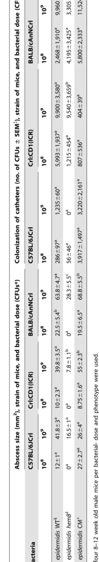
No abscess formation was observed on day 7 at doses of 106and
107CFUs per catheter. Data for abscess formation on day 7 for all
three mouse strains at doses of 108and 109CFUs ofS. epidermidis
O-47 or the complemented mutant are presented in Table 1. At this dose, abscesses detected from O-47 infection were significantly
larger in BALB/c, measuring 22.5 mm2, compared to the
abscesses from CD-1 and B6J. Figure 1B, III-V shows
represen-tative abscess formation with S. epidermidis O-47 in the three
different mouse strains inoculated with doses of 107–109bacteria
per catheter. Abscess formation was not observed in all mice below
a dose of 108 hemB mutant bacteria displaying the
SCV-phenotype. Starting at a dose of 109, the hemB mutant resulted
in abscess formation in all mouse strains with sizes being significantly different among all three strains, but smaller
B6J and CD-1, the largest abscesses were detected in BALB/c with
an infectious dose of 109of the complemented mutant (69 mm2,
p,0.05).
As shown in Table 1, an inoculation dose of 109CFUs per
catheter resulted in a recovery of 11,524 CFUs of the comple-mented mutant from BALB/c followed by 9,960 CFUs and
9,900 CFUs of S. epidermidis O-47 from BALB/c and CD-1,
respectively. Colonization with the hemB mutant displaying the
SCV phenotype was most effective in CD-1 with 9,540 CFUs at a
dose of 109CFUs per catheter, whereas no S. epidermidis hemB
mutant bacteria were detected in B6J (p,0.05). The number of
bacteria recovered at lower doses of 106 and 107ranged from a
minimum of zero (CD-1 including all three bacterial strains and
BALB/c with the hemB mutant at a dose of 106 bacteria per
catheter) to a maximum colonization of 1,230 CFUs (BALB/c infected with the complemented mutant, see Table S1).
In Figure 1B, VI–VIII, safranin staining of representative catheters is shown. Biofilm formation by bacteria was detected at
doses of 108 and 109 bacteria per catheter, whereas none was
observed at doses of 106and 107(data not shown). With increasing
number of adherent bacteria, the red staining became more intensive. Negative controls had the same intensity of staining as shown in Figure 1B, VI.
The expected bacteria were solely confirmed in all samples of abscess and in wound cultures where catheter colonization was observed. All blood cultures remained negative.
Discussion
Although studies investigating the virulence ofS. epidermidisin
foreign-body-infection models in different mouse hosts have been performed [2,3,4,5,6], we report for the first time an attenuation of colonization and abscess formation in mice infected with an SCV
phenotype ofS. epidermidiscompared to the normal phenotype in
three different immunocompetent mouse strains. As it is still
unknown whether SCV formation is solely due to bacterial adaption to host or intracellular conditions, and/or is the result of mutations, we performed this model of acute foreign-body-infection for an observation period of seven days. [12,18,19,20].
The results reveal anS. epidermidisdose-dependent colonization
of catheters in immunocompetent mice. Neither the dose of 106
nor 107led to significant replication of bacteria in all three mouse
strains probably due to the fact that adherence to abiotic surfaces is mainly mediated through biofilm formation and expression of adhesins, which lead to better survival in the host [21]. Quorum sensing, which depends on cell density, might explain colonization
of catheters at a minimum dose of 108bacteria, which seems to be
the initial dose required for inducing effective biofilm formation in
this model. Compared to doses of 106, which often have been used
inS. aureusanimal models, the high dose ofS. epidermidisused in our
model is probably due to the fact thatS. epidermidislacks mass of
virulence factors, which are present inS. aureus, and its versatile
potential to evade host defense mechanisms [22,23].
As described in several studies for S. aureus, thehemB mutant
with SCV phenotype differs in virulence compared to the normal
phenotype depending on the in vitro or in vivo model used
[12,24,25]. S. aureus hemB mutants have been shown to survive
intracellularly and display reduced virulencein vitro[12]. Although
theS. epidermidis hemBmutant is described to produce more biofilm
than the normal phenotypein vitro, in the present study it does not
colonize catheters at amounts comparable to that of the normal
phenotype with the exception of CD-1 mice and at a dose of 108
bacteria in BALB/c mice. This is confirmed by the observation
that abscesses fromS. epidermidisdisplaying the SCV phenotype are
smaller than those formed by the normal phenotype. As such, our
in vivoresults do not confirm previousin vitrofindings [13]. This is most likely due to a combination of defects in electron transport,
resulting in growth retardation which in vivo leads to reduced
biofilm formation and might also lead in consequence to a reduced
survival in the mouse. A possible intracellular survival of the S.
Figure 1. Post-operative view of mice, abscesses, and biofilm staining of catheters.Figure 1A, I: Implantation of a 1-cm long sterile PVC catheter segment subcutaneously subsequent to anesthetizing, shaving, and making a small incision in each flank of the mouse. Figure 1A, II. Abscess formation in a mouse 7 days post inoculation withS. epidermidisO-47. Figure 1B: Subcutaneous abscesses from mice 7 days after inoculation ofS.
epidermidisO-47; III) BALB/c mouse with a dose of 107, IV) CD-1 mouse with a dose of 108, and V) B6J mouse with a dose of 109CFUs and
epidermidis hemBmutant, as shown for theS. aureus hemBmutant, is not excluded but needs further studies [11,25]. However,
standardized in vitro conditions differ not only in nutrition
availability, oxygen- and salt concentration, pH, but also in the
influence and attack of the immune system to growing bacteriain
vivo. Thus, the difference betweenin vitroandin vivofindings is not
quite surprising.
The observation that the complemented mutant does not
behave exactly as S. epidermidis O-47 (see Table 1) in terms of
catheter colonization and abscess formation may be due to the fitness-related fact that this mutant expresses two additional resistance genes, namely, erythromycin and chloramphenicol.
In general, all mice showed no symptoms of systemic infection. Bacteria were not detected in blood indicating that mice were infected only locally. We can not exclude that a possible dissemination to other tissues or blood might occur afterwards. It is possible that a critical cell density in the biofilm community leads to detachment and spreading of bacteria which was not achieved during 7 days of experiment [26,27,28]. In addition, the area of the skin in the periphery of the abscess did not show any pathological lesions, and wound healing was not delayed.
Our results indicate that colonization of catheters and abscess
formation by different phenotypes ofS. epidermidisin a
subcuta-neous foreign-body-infection model is most effective in inbred BALB/c followed by outbred CD-1 and inbred B6J mice. Different results with immunocompetent strains of mice have been reported previously [2,3,4,5,6,29] but the underlying molecular mechanisms are mostly unknown. Nevertheless, the
different potential ofS. epidermidis strains to produce biofilm and
the different methods for determination of colonization also contribute to the differences in results obtained by different authors [2,3,4,5,6]. Furthermore, the route and site of infection in addition to host-dependent factors play a major role [29,30,31]. In a subcutaneous infection model without a foreign body where
26107CFUs ofS. aureusSH1000 were inoculated in the left hind
footpad of 8–12-week old mice, Nippe et al. showed that, in
contrast to our findings, B6J mice were more susceptible to S.
aureusthan BALB/c mice, most probably due to strain-dependent differences in granulocyte recruitment during infection [29].
Granulocyte recruitment andS. epidermidisbiofilm formation may
be the reason for faster abscess formation in BALB/c mice than in B6J mice in our study (see Table 1). Surrounded by the abscess the bacteria might be protected from other immune components and may therefore have a better survival chance compared to the situation in B6J at least during the seven day observation period. Thus, when immunocompetent strains are needed for subcutane-ous foreign-body-infection models, we recommend using BALB/c
mice at a dose of at least 109S. epidermidisO-47 per catheter.
In conclusion, the present study provides further information on choice of mouse strain with regard to bacterial phenotype variants
in anS. epidermidis infection model. An elucidation of molecular
differencesin vivobetween normal and SCV phenotype, immune
modulation of the host during infection, and differences between the susceptibility in different mouse strains will be helpful in future investigations involving bacterial and host factors in establishing infections.
Supporting Information
Table S1 Colonization of catheters according to dose of three
Staphylococcus epidermidis strains in inbred C57BL/6JCrl, outbred Crl:CD1(ICR), and inbred BALB/cAnNCrl mice
Author Contributions
Conceived and designed the experiments: GS CvE KB EM. Performed the experiments: GS TB. Analyzed the data: GS AK EM. Contributed reagents/materials/analysis tools: CvE KB. Wrote the paper: GS EM.
References
1. von Eiff C, Peters G, Heilmann C (2002) Pathogenesis of infections due to
coagulase-negative staphylococci. Lancet Infect Dis 2: 677–685.
2. Christensen GD, Baddour LM, Simpson WA (1987) Phenotypic variation of
Staphylococcus epidermidisslime production in vitro and in vivo. Infect Immun 55: 2870–2877.
3. Deighton MA, Borland R, Capstick JA (1996) Virulence of Staphylococcus
epidermidisin a mouse model: significance of extracellular slime. Epidemiol Infect 117: 267–280.
4. Patrick CC, Plaunt MR, Hetherington SV, May SM (1992) Role of the
Staphylococcus epidermidisslime layer in experimental tunnel tract infections. Infect Immun 60: 1363–1367.
5. Rupp ME, Ulphani JS, Fey PD, Bartscht K, Mack D (1999) Characterization of
the importance of polysaccharide intercellular adhesin/hemagglutinin of Staphylococcus epidermidisin the pathogenesis of biomaterial-based infection in a mouse foreign body infection model. Infect Immun 67: 2627–2632.
6. Vuong C, Kocianova S, Yu J, Kadurugamuwa JL, Otto M (2008) Development
of real-time in vivo imaging of device-relatedStaphylococcus epidermidisinfection in
mice and influence of animal immune status on susceptibility to infection. J Infect Dis 198: 258–261.
7. Baddour LM, Christensen GD (1987) Prosthetic valve endocarditis due to
small-colony staphylococcal variants. Rev Infect Dis 9: 1168–1174.
8. Seifert H, Oltmanns D, Becker K, Wisplinghoff H, von Eiff C (2005)
Staphylococcus lugdunensis pacemaker-related infection. Emerg Infect Dis 11: 1283–1286.
9. von Eiff C, Vaudaux P, Kahl BC, Lew D, Emler S, et al. (1999) Bloodstream
infections caused by small-colony variants of coagulase-negative staphylococci following pacemaker implantation. Clin Infect Dis 29: 932–934.
10. Proctor RA, von Eiff C, Kahl BC, Becker K, McNamara P, et al. (2006) Small colony variants: a pathogenic form of bacteria that facilitates persistent and recurrent infections. Nat Rev Microbiol 4: 295–305.
11. von Eiff C, Heilmann C, Proctor RA, Woltz C, Peters G, et al. (1997) A
site-directedStaphylococcus aureus hemBmutant is a small-colony variant which persists
intracellularly. J Bacteriol 179: 4706–4712.
12. Tuchscherr L, Medina E, Hussain M, Volker W, Heitmann V, et al. (2011) Staphylococcus aureusphenotype switching: an effective bacterial strategy to escape host immune response and establish a chronic infection. EMBO Mol Med 3: 129–141.
13. Al Laham N, Rohde H, Sander G, Fischer A, Hussain M, et al. (2007) Augmented expression of polysaccharide intercellular adhesin in a defined Staphylococcus epidermidis mutant with the small-colony-variant phenotype. J Bacteriol 189: 4494–4501.
14. Seggewiss J, Becker K, Kotte O, Eisenacher M, Yazdi MR, et al. (2006) Reporter metabolite analysis of transcriptional profiles of a Staphylococcus aureus strain with normal phenotype and its isogenic hemB mutant displaying the small-colony-variant phenotype. J Bacteriol 188: 7765–7777.
15. Christensen GD, Simpson WA, Younger JJ, Baddour LM, Barrett FF, et al. (1985) Adherence of coagulase-negative staphylococci to plastic tissue culture plates: a quantitative model for the adherence of staphylococci to medical devices. J Clin Microbiol 22: 996–1006.
16. Heilmann C, Gerke C, Perdreau-Remington F, Go¨tz F (1996) Characterization
of Tn917 insertion mutants of Staphylococcus epidermidis affected in biofilm
formation. Infect Immun 64: 277–282.
17. Pfaller M, Davenport D, Bale M, Barrett M, Koontz F, et al. (1988) Development of the quantitative micro-test for slime production by coagulase-negative staphylococci. Eur J Clin Microbiol Infect Dis 7: 30–33.
18. Lannergard J, Cao S, Norstrom T, Delgado A, Gustafson JE, et al. (2011) Genetic Complexity of Fusidic Acid-Resistant Small Colony Variants (SCV) in Staphylococcus aureus. PLoS One 6: e28366.
19. Lannergard J, von Eiff C, Sander G, Cordes T, Seggewiss J, et al. (2008) Identification of the genetic basis for clinical menadione-auxotrophic
small-colony variant isolates ofStaphylococcus aureus. Antimicrob Agents Chemother 52:
4017–4022.
20. Tuchscherr L, Heitmann V, Hussain M, Viemann D, Roth J, et al. (2010) Staphylococcus aureussmall-colony variants are adapted phenotypes for intracel-lular persistence. J Infect Dis 202: 1031–1040.
21. Wang R, Khan BA, Cheung GY, Bach TH, Jameson-Lee M, et al. (2011) Staphylococcus epidermidis surfactant peptides promote biofilm maturation and dissemination of biofilm-associated infection in mice. J Clin Invest 121: 238–248. 22. Hart E, Azzopardi K, Taing H, Graichen F, Jeffery J, et al. (2010) Efficacy of antimicrobial polymer coatings in an animal model of bacterial infection associated with foreign body implants. J Antimicrob Chemother 65: 974–980.
23. Otto M (2011) Molecular basis ofStaphylococcus epidermidisinfections. Semin
Immunopathol.
24. Jonsson IM, von Eiff C, Proctor RA, Peters G, Ryden C, et al. (2003) Virulence
of ahemBmutant displaying the phenotype of aStaphylococcus aureussmall colony
variant in a murine model of septic arthritis. Microb Pathog 34: 73–79. 25. Sifri CD, Baresch-Bernal A, Calderwood SB, von Eiff C (2006) Virulence of
Staphylococcus aureussmall colony variants in theCaenorhabditis elegansinfection model. Infect Immun 74: 1091–1096.
26. O’Toole G, Kaplan HB, Kolter R (2000) Biofilm formation as microbial development. Annu Rev Microbiol 54: 49–79.
27. Vuong C, Gerke C, Somerville GA, Fischer ER, Otto M (2003) Quorum-sensing
control of biofilm factors inStaphylococcus epidermidis. J Infect Dis 188: 706–718.
28. Vuong C, Kocianova S, Yao Y, Carmody AB, Otto M (2004) Increased colonization of indwelling medical devices by quorum-sensing mutants of Staphylococcus epidermidis in vivo. J Infect Dis 190: 1498–1505.
29. Nippe N, Varga G, Holzinger D, Lo¨ffler B, Medina E, et al. (2011)
Subcutaneous infection withS. aureusin mice reveals association of resistance
with influx of neutrophils and Th2 response. J Invest Dermatol 131: 125–132. 30. Hume EB, Cole N, Khan S, Garthwaite LL, Aliwarga Y, et al. (2005) A Staphylococcus aureusmouse keratitis topical infection model: cytokine balance in different strains of mice. Immunol Cell Biol 83: 294–300.
31. von Kockritz-Blickwede M, Rohde M, Oehmcke S, Miller LS, Cheung AL, et al. (2008) Immunological mechanisms underlying the genetic predisposition to
severeStaphylococcus aureusinfection in the mouse model. Am J Pathol 173: