ÍTALO ANTÔNIO COTTA COUTINHO
ANATOMIA FOLIAR COMO SUBSÍDIO PARA TAXONOMIA DE CHAMAECRISTA MOENCH COM ÊNFASE NA SEÇÃO APOUCOUITA
(LEGUMINOSAE - CAESALPINIOIDEAE)
Tese apresentada à
Universidade Federal de Viçosa, como parte das exigências do Programa de Pós-Graduação em Botânica, para obtenção do título de Doctor Scientiae.
VIÇOSA
A ignorância gera mais frequentemente confiança do que o conhecimento: são os que sabem pouco, e não aqueles que sabem muito, que afirmam de uma forma tão categórica que este ou aquele problema nunca será resolvido pela ciência.
“Once we have sequenced all of the relevant plant genomes and have come to realize that such sequence data leaves many questions in organismal biology, unanswered, we may finally appreciate that organisms are valid and fundamental biological units worthy of our attention. Then, morphology may finally be appreciated and respected as a key to
the understanding of plant organismal biology”.
Donald R. Kaplan
E o Espírito de Deus o encheu de sabedoria, entendimento, ciência... para criar
invenções... Também lhe dispôs o coração para ensinar a outros... e encheu-os de
sabedoria do coração, para fazer toda a obra de mestre, até a mais engenhosa... fazendo
toda a obra, e criando invenções.
Àquela que há 13 anos decidiu me orientar
Toc toc toc toc toc... sempre na passada rápida e cada vez mais alto. Ela está chegando, e logo começa a metralhar perguntas:
Que cara é essa meu filho? O que você estava fazendo até agora? Por que ainda não fez isso ou aquilo? Ainda temos dinheiro de projeto?
Já mandou os documentos par FUNARBE/CNPq/FAPEMIG/CAPES/SISBio? Está tudo pronto? Tudo dominado?
E a clássica: TEM CAFÉ???
Ou mesmo atendendo ao telefone: “O que eu tenho que falar mesmo”? Ué Renata: Alô?
Sem dúvida a que mais sentirei falta é: Como você está? Com um tom sério de preocupação, de alguém que realmente se importa.
Ou pode ser ainda uma chuva de exclamações, que são mais preocupantes que as perguntas:
Você não está me convencendo disso! (Talvez mais umas 20 exclamações aqui!!!!!) Que demora é essa meu filho! Vou cortar seu ponto, hein!
Isso está errado! Faz de novo! Não é assim, não! Me liga! (mensagem recebida às 6h da matina)
Ítalo, passa na minha sala! (essa sim, gelava a espinha mais que Antártica Subzero!!!!)
Assim defino uma pessoa que é mais que uma orientadora, uma pessoa que durante esses anos tornou-se mais que uma amiga.
Renata, infelizmente você não é perfeita, mas absolutamente ninguém o é! No entanto, saiba que sem sombra de dúvidas, quaisquer de seus defeitos nunca irão apagar o brilhantismo de sua dedicação como orientadora, de seu potencial como pesquisadora e de sua sincera amizade.
Se fosse para recomeçar do zero, o faria tudo de novo, com muito prazer. Não escolheria nenhuma outra pessoa para me orientar.
Que os próximos anos sejam de colheitas ainda melhores do que já tivemos. No mais, ficará sempre a saudade de dividir a sala com você.
AGRADECIMENTOS
Chego ao final de mais uma fase, e não há como não agradecê-Lo por isso. Agradeço a Deus, pelo dom da vida, pelo sustento, pela compreensão, pela paciência, pelo cuidado, pela provisão, pelo consolo, pelo renovo e pelo amor dispensado.
À Universidade Federal de Viçosa e ao Departamento de Biologia Vegetal, onde me fiz bacharel, mestre a agora doutor.
Ao CNPq, CAPES, FAPEMIG e ao projeto Floresta Escola/UNESCO-HidroEx pela concessão das bolsas e/ou auxílio financeiro à pesquisa.
A minha família, meus pais, Coutinho e Penha e meus irmãos, Marcos, Sara e Tiago pelo amor e pelas orações. Em especial a Sarinha, que nesses últimos anos tem sido benção na minha vida.
Aos meus familiares que mesmo longe, estão sempre perto. Não tenho tios ou tias, tenho pais e mães adotivos, não tenho primos, tenho irmãos. Agradeço pelo convívio e pela amizade.
Às professoras Aristéa, Luzimar Campos e Marília Ventrella, pela amizade, companheirismo e ensino.
Aos amigos do laboratório, da IPV (Igreja Presbiteriana de Viçosa) e do REC (Real English Center), os quais fizeram toda diferença durante esses anos, em especial a Pamela Torres, companheira de república.
Agradeço em especial à amizade de duas grandes amigas, Vanessa Terra e Valdnéa Dalvi, que foram um achado precioso. E também Carlota (Carla Feio) que tem sido companheira nessa fase tão complicada da vida.
Às companheiras de coleta Isabel Silva e Valéria Fernandes e ao amigo Marc Guesdon, pelas infindáveis risadas em campo.
A todos os que riram ao choraram comigo no campo.
SUMÁRIO
RESUMO... ix
ABSTRACT... xi
1. INTRODUÇÃO GERAL... 1
2. REFERÊNCIAS BIBLIOGRÁFICAS…..... 4
CAPÍTULO I: New records of colleters in Chamaecrista (Leguminosae, Caesalpinioideae s. l.): structural diversity, secretion, functional role and taxonomic importance... 12
Abstract... 14
Introduction ... 16
Material and Methods... 18
Results... 20
Discussion... 23
Conclusion... 28
Acknowledgments………... 29
Literature Cited…... 29
Appendix... 34
Table... 38
Figures... 41
CAPÍTULO II: Structural diversity of extrafloral nectaries in Chamaecrista sect. Apoucouita... 46
Abstract... 48
Material and Methods... 50
Results... 52
Discussion... 55
Conclusion... 60
Acknowledgments………... 60
References…...…... 61
Appendix... 66
Table... 69
Figures... 71
CAPÍTULO III: A study of the morphoanatomical characters in Chamaecrista Moench sect. Apoucouita... 76
Abstract... 78
Introduction ... 79
Material and Methods... 81
Results... 82
Discussion... 87
Conclusion... 91
References…...…... 92
Table... 97
Appendix... 100
Figures... 103
RESUMO
COUTINHO, Ítalo Antônio Cotta, D. Sc., Universidade Federal de Viçosa, fevereiro de 2015. Anatomia foliar como subsídio para taxonomia de Chamaecrista Moench com ênfase na seção Apoucouita (Leguminosae - Caesalpinioideae). Orientadora: Renata Maria Strozi Alves Meira. Coorientadora: Adilva de Sousa Conceição.
Chamaecrista Moench (Leguminosae, Caesalpinioideae, Cassieae, Cassiineae) inclui
mais de 330 espécies. A presença de estruturas secretoras é um dos parâmetros
utilizados para a circunscrição das seis seções do gênero, no entanto, tal circunscrição
não é corroborada pelos estudos filogenéticos. A anatomia foliar de Chamaecrista tem
se mostrado uma importante ferramenta, corroborando inclusive as novas circunscrições
taxonômicas baseadas em dados moleculares. Dentre as várias estruturas secretoras
descritas para o gênero Chamaecrista, coléteres foram mencionados em algumas das
espécies estudadas. Entretanto um estudo comparativo da diversidade e importância
taxonômica dessas estruturas no gênero Chamaecrista ainda não foi realizado. Dentre as
seções de Chamaecrista, poucos estudos têm investigado a seção Apoucouita que inclui
espécies com glândulas no pecíolo/raque e no racemo denominadas de nectários. No
entanto, tal caracterização demanda estudos complementares como análise da presença
de açúcar e análises histoquímicas. Embora estudos moleculares tenham se mostrado
úteis para a separação e delimitação de grupos taxonômicos, caracteres morfológicos
são necessários para reconhecimento das espécies tanto em campo quanto em herbário e
nestes casos a morfoanatomia foliar é uma alternativa promissora. Os materiais foram
processados conforme metodologia usual de microscopia de luz e varredura utilizados
em estudos anatômicos. O trabalho foi organizado em três capítulos. No capítulo I a
presença e a diversidade de coléteres em folhas e flores em desenvolvimento de
espécies do gênero Chamaecrista, foram avaliadas. Foram descritos seis tipos de
“long digitiform” e “short digitiform”. As análises histoquímicas confirmaram a
presença de proteínas totais, polissacarídeos totais, mucilagens e lipídeos na secreção
dos coléteres. O tipo e posição dos coléteres corroboram as novas circunscrições com
base em estudos moleculares propostas para Chamaecrista. No capítulo II procedeu-se
o estudo anatômico das glândulas do pecíolo/raque para verificar se tais estruturas
correspondem a nectários como descrito na literatura. Foram observados 13 tipos de
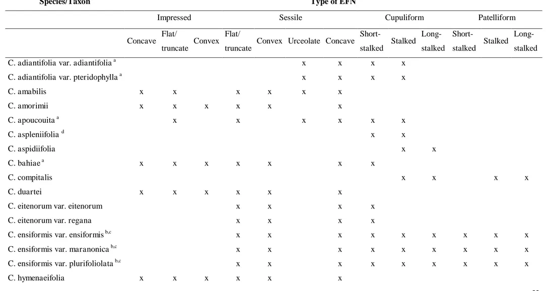
nectários extraflorais (NEF) para Chamaecrista, os quais podem ser impressos, sésseis
ou pedunculados, com superfície secretora côncava, plano/truncada ou convexa.
Embora variações morfológicas tenham sido observadas, a estrutura anatômica e a
composição química da secreção foram similares para todos os tipos de NEF. A
formação de periderme de cicatrização em NEFs mais velhos é um fato inédito para
Chamaecrista, uma vez que não foi relatada para as outras seções do gênero. O capítulo
III aborda a morfoanatomia das folhas de espécies de C. seção Apoucouita com vistas
verificar a utilidade dessas características para fins taxonomicos. O arranjo do sistema
vascular no pecíolo/raque, tipo de mesofilo, idioblastos mucilaginosos na face adaxial e
abaxial da epiderme, contorno das paredes periclinais das células epidérmicas (reto na
adaxial e sinuoso na abaxial) e posição e tipo de estômato, (hipoestomática e
paracítico-laterocíclico, respectivamente) foram caracteres comuns a todas as espécies da seção
Apoucouita. A presença e posição de papilas na epiderme e a posição e tipo de NEF são
ABSTRACT
COUTINHO, Ítalo Antônio Cotta, D. Sc., Universidade Federal de Viçosa, February
2015. Leaf anatomy as an additional tool to the taxonomy of Chamaecrista Moench
with emphasis on section Apoucouita (Leguminosae - Caesalpinioideae). Advisor: Renata Maria Strozi Alves Meira. Co-advisor: Adilva de Sousa Conceição.
Chamaecrista Moench (Leguminosae, Caesalpinioideae, Cassieae, Cassiineae) includes
more than 330 species. The presence of secretory structures is one of the parameters
used for the circumscription of the six sections of the genus. However, such
circumscription is not supported by the phylogenetic studies. Leaf anatomy of
Chamaecrista has proved an important tool to the new taxonomic circumscription based
on molecular data. Among the secretory structures described for Chamaecrista,
colleters have been mentioned in some species. However, a comparative study of the
diversity and taxonomic importance of such structures for Chamaecrista has not yet
been performed. Among the sections of Chamaecrista, few studies have focused on
sect. Apoucouita, a section with species displaying glands on the petiole/rachis and on
the raceme. Such glands have been called extrafloral nectaries (EFN), even though, no
additional studies such as the analysis of the presence of sugars and histochemical tests
have been performed to confirm if such glands are in fact EFNs. Although molecular
studies have proved useful in the circumscription of taxonomic groups, morphological
characters are necessary for recognizing species in the field and also when dried
material is analyzed and in both cases, leaf anatomy is a promising alternative for the
recognition of species. For this study, standard anatomical techniques for light and
scanning electron microscopy were carried out. The present study is organized in three
chapters. The presence and diversity of colleters on developing leaves and flowers of
club-shaped”, “racket-shaped”, “long bottle-shaped”, “short bottle-shaped”, “long
digitiform” and “short digitiform”. The histochemical analyses confirmed the presence
of total proteins, total polysaccharides, mucilage and lipids in the secretion of the
colleters. The type and position of colleter supported new circumscriptions based on
molecular studies proposed for the Chamaecrista. The anatomical study of the
petiole/rachis glands in order to verify if such structures are in fact nectaries, as
described in the literature, is given in Chapter II. It was observed 13 types of ENFs for
Chamaecrista. Such EFNs may be impressed, sessile or stalked, with secretory surface
concave, flat/truncate or convex. Although morphological variations were observed, the
anatomical structure and chemical composition of the secretion was similar to all types
of EFNs. The formation of a wound-healing periderm in older EFNs is a novelty for
Chamaecrista, as such was not reported for the other sections of the genus. The leaf
morphoanatomy of C. sect. Apoucouita with the intent of using such characters in the
taxonomy is given in Chapter III. The arrangement of the vascular system in the
petiole/rachis, type of mesophyll, mucilage idioblasts in the adaxial and abaxial
epidermis, outline of the anticlinal walls of the epidermal cells (straight on the adaxial
side and sinuous on the abaxial side) and the position and type of stomata (hipostomatic
leaves and paracytic laterocyclic, respectively) were characters common to all species
from sect. Apoucouita. The presence of papillae on the epidermis as well as the position
and type of extrafloral nectaries are promising characters regarding the taxonomy of
1. INTRODUÇÃO GERAL
Leguminosae é uma das famílias mais importantes dentre as plantas com flores,
amplamente distribuídas nas regiões tropicais, considerada a terceira maior família
dentre as Angiospermas, compreendendo mais de 720 gêneros e 19.300 espécies
(Lewis, 2005). Está família é tradicionalmente composta por três subfamílias:
Papilionoideae, Caesalpinioideae e Mimosoideae (Polhill et al., 1981; Lewis, 1987),
embora a filogenia molecular não corrobore a monofilia desses três grupos (Lewis,
2005).
O gênero Chamaecrista Moench (Leguminosae-Caesalpinioideae) está inserido
na tribo Cassieae subtribo Cassiineae. A consolidação de Chamaecrista como um
gênero foi feita por Irwin e Barneby (1982), quando dividiram o gênero Cassia em três:
Cassia, Senna e Chamaecrista, os quais atualmente compõem a subtribo Cassiineae
(Irwin e Barneby, 1982). Chamaecrista inclui mais de 330 espécies (Conceição et al.,
2001, 2008; Gereau and Walters, 2003; Rando et al., 2013) com cerca de 1/3 do gênero
endêmico ao Brasil, sendo 7% das espécies registradas para a região Amazônica (Lewis,
2005).
As espécies de Chamaecrista estão organizadas em seis seções: Apoucouita,
Absus, Caliciopsis, Chamaecrista, Grimaldia e Xerocalyx (Irwin and Barneby, 1982). A
presença de estruturas secretoras, como nectários extraflorais e tricomas, é um dos
parâmetros utilizados para a circunscrição destas seções (Irwin e Barneby, 1982;
Conceição et al., 2009; Francino, 2010; Coutinho et al., 2012). Tal tratamento
taxonômico das seções com base apenas em caracteres morfológicos, não é corroborada
pelos estudos filogenéticos baseados em dados moleculares (Conceição et al., 2009), os
Nos últimos dez anos, o gênero Chamaecrista tem sido foco de vários trabalhos
abordando a anatomia (Francino et al., 2006; Silva, 2012; Coutinho et al., 2012, 2013,
2015; Meira et al., 2014, biologia molecular (Conceição et al., 2008, 2009), taxonomia
(Camargo and Miotto, 2004; Rando, 2009; Rando and Pirani, 2011; Rando et al., 2013;
Torres et al., 2011; Dantas e Silva, 2013), ecologia (Nascimento e Del-Claro, 2007,
2010) ou importância econômica (Morris, 2012).
A anatomia foliar de Chamaecrista tem se mostrado uma importante ferramenta
na identificação das espécies, corroborando inclusive as novas circunscrições
taxonômicas baseadas em dados moleculares (Coutinho et al. 2013; Silva, 2012;
Francino et al. 2015). Dentre as várias estruturas secretoras descritas para o gênero
Chamaecrista (Coutinho et al. 2012; 2013, Silva 2012; Francino et al. 2015), coléteres
foram mencionados em algumas das espécies estudadas.
Coléteres são estruturas encontradas geralmente na face adaxial de estípulas e
brácteas e produzem uma substância pegajosa composta de mucilagem ou uma mistura
de mucilagem e resina, a qual recobre gemas foliares e florais, exercendo função
protetora, principalmente contra dessecação (Foster, 1942; Fahn and Benouaiche, 1979;
Paiva and Machado, 2006a; Barreiro and Machado, 2007; Sheue et al., 2012; Mayer et
al., 2013). Poucos estudos em Leguminosae, assim como no gênero Chamaecrista,
reportaram a presença de coléteres (Paiva and Machado, 2006a; b; De-Paula and
Oliveira, 2007; Paiva, 2009; Coutinho et al., 2013), de forma que um estudo
comparativo da diversidade e importância taxonômica dessas estruturas no gênero
Chamaecrista ainda não foi realizado.
No que se refere à seção Apoucouita, nenhuma atenção nenhuma atenção tem
sido dada desde a última revisão para a seção, feita na forma de um estudo suplementar
táxons, incluindo as variedades) da seção Apoucouita, caracteres como tamanho do
pecíolo/raque, número de pares de folíolos, forma do folíolo e posição dos nectários
foliares são amplamente utilizados (Irwin e Rogers, 1967; Irwin e Barneby, 1977, 1982;
Barneby, 1999). A identificação das variedades é uma tarefa difícil, sendo as chaves
baseadas em parâmetros arbitrários e que são plásticos facilmente modificados pelo
ambiente, tais como tamanho do pulvínulo, a forma do ápice do foliólulo e
presença/ausência de tricomas tectores. Dessa forma, são necessários estudos
complementares que dêem suporte à taxonomia clássica, isto é, aquela baseada apenas
em caractéres macro morfológicos.
A seção Apoucouita difere das demais seções por compreender
predominantemente espécies arbóreas das Florestas Amazônica e Atlântica (Irwin e
Barneby, 1977; Conceição et al. 2009). Nestas espécies, glândulas situadas no
pecíolo/raque e no racemo, foram denominadas como nectários com base apenas na
morfologia e na topografia. Estruturas secretoras de compostos lipofílicos podem ser
morfológica e topograficamente semelhantes aos nectários (Curtis e Lersten, 1978,
1980; Durkee et al., 1984; Jáuregui et al., 2002) e, nestes casos, a caracterização precisa
demanda estudos complementares como análise da presença de açúcar e análises
histoquímicas.
Nectários são estruturas secretoras especializadas na secreção de néctar, uma
solução açucarada composta principalmente por glicose, frutose e sacarose (Fahn, 1979;
Bentley e Elias, 1983; Roshchina e Roshchina, 1993; Nicolson et al., 2007). As
glândulas presentes no pecíolo/raque das espécies da seção Baseophyllum foram
descritas como NEFs por meio de estudos anatômicos minuciosos (Coutinho et al.,
das glândulas, fornecendo dado que corroborem estudos sobre a evolução das estruturas
secretoras em Chamaecrista.
Dessa forma o presente estudo se propõe (1) a investigar a presença de coléteres
no gênero Chamaecrista bem como proceder o estudo morfoanatômicos dessas
estruturas e sua a utilidade como caracteres que auxiliem estudos taxonômicos e
filogenéticos; (2) a avaliar se as glândulas foliares presentes no pecíolo/rachis de
espécies da seção Apoucouita correspondem a nectários extraflorais, procedendo sua
caracterização morfoanatomica; (3) a proceder o estudo morfoanatômica das folhas de
espécies da seção Apoucouita e averiguar se tais caracteres podem ser utilizados na
separação de espécies e variedades.
O presente trabalho encontra-se organizado sob a forma de artigos científicos,
como disposto nas normas de redação de teses da Universidade Federal de Viçosa. Cada
artigo segue a formatação da revista a que será submetido ou publicado.
2. REFERÊNCIAS BIBLIOGRÁFICAS
Arambarri, A.M., S.E. Freire, M.N. Colares, N.D. Bayón, M.C. Novoa, C. Monti, and S. a Stenglein. 2006. Leaf anatomy of medicinal shrubs and trees from gallery forests of the Paranaense Province (Argentina). Part 1. Boletin de la Sociedade Argetina de Botanica 41: 233–268.
Baker, H.G. 1977. Non-sugar chemical constituents of nectar. Apidologie 8: 349–356. Barneby, R.C. 1999. Increments to genus Chamaecrista (Caesalpiniaceae: Cassiinae)
from Bolivia and from Atlantic and Planaltine Brazil. Brittonia 51: 331–339. Barreiro, D.P., and S.R. Machado. 2007. Coléteres dendróides em Alibertia sessilis
(Vell.) K. Schum., uma espécie não-nodulada de Rubiaceae. Revista Brasileira de Botânica 30: 387–399.
Bentley, B., and T. Elias. 1983. The biology of nectaries. Columbia University Press, New York, USA.
Bentley, B.L. 1977. Extrafloral nectaries and protection by pugnacious bodyguards. Annual Review of Ecology and Systematics 8: 407–427.
Camargo, R.A., and S.T.S. Miotto. 2004. O gênero Chamaecrista Moench (Leguminosae-Caesalpinioideae) no Rio Grande do Sul. Iheringia 59: 131–148. Carvalho, D.M.G. Anatomia foliar de Cassia ensiformis Vell. (Leguminosae -
Caesalpinioideae). Arquivos do Jardim Botânico do Rio de Janeiro 27: 157–169. Caspary, R. 1848. De nectariis. Elverfeld, Bonnae.
Conceição, A. de S. 2006. Filogenia do gênero Chamaecrista (Leguminosae-Caesalpinioideae) e taxonomia do grupo Baseophyllum. Universidade Estadual de Feira de Santana.
Conceição, A.S., L.P. Queiroz, S.M. Lambert, A.C.S. Pereira, and E.L. Borba. 2008. Biosystematics of Chamaecrista sect. Absus subsect. Baseophyllum (Leguminosae-Caesalpinioideae) based on allozyme and morphometric analyses. Plant Systematics and Evolution 270: 183–207.
Conceição, A.S., L.P. Queiroz, and G.P. Lewis. 2001. Novas espécies de Chamaecrista Moench (Leguminosae-Caesalpinioideae) da Chapada Diamantina, Bahia, Brasil. Sitientibus, Série Ciências Biológicas 1: 112–119.
Conceição, A.S., L.P. Queiroz, G.P. Lewis, M.J.G. de Andrade, P.R.M. de Almeida, A.S. Schnadelbach, and C. van den Berg. 2009. Phylogeny of Chamaecrista Moench (Leguminosae-Caesalpinioideae) based on nuclear and chloroplast DNA regions. Taxon 58: 1168–1180.
Costa, F.M.C.B., A.T. Oliveira-Filho, and P.S. Oliveira. 1992. The role of extrafloral nectaries in Qualea grandiflora (Vochysiaceae) in limiting herbivory: an experiment of ant protection in cerrado vegatation. Ecological Entomology 17: 363–365.
Coutinho, Í.A.C., D.M.T. Francino, A.A. Azevedo, and R.M.S.A. Meira. 2012. Anatomy of the extrafloral nectaries in species of Chamaecrista section Absus subsection Baseophyllum (Leguminosae, Caesalpinioideae). Flora 207: 427–435. Coutinho, Í.A.C., D.M.T. Francino, and R.M.S.A. Meira. 2013. Leaf anatomical studies
of Chamaecrista subsect. Baseophyllum (Leguminosae, Caesalpinioideae): new evidence for the up-ranking of the varieties to the species level. Plant Systematics and Evolution 299: 1709–1720.
Coutinho, Í.A.C., and R.M.S.A. Meira. 2015. Structural diversity of extrafloral nectaries in Chamaecrista sect. Apoucouita. Botanyin press.
Coutinho, Í.A.C., V.M.M. Valente, and M.R.M.S. Alves. 2010. Ontogenetic, anatomical and histochemical study of the extrafloral nectaries of Sapium biglandulosum (Euphorbiaceae). Australian Journal of Botany 58: 224–232.
Cowan, R.S. 1981. Caesalpinioideae. In Advances in Legume Systematics, Part 1., 57– 64. Royal Botanical Gardens, Kew, Kew.
Curtis, J.D., and N.R. Lersten. 1978. Heterophylly in Populus grandidentata (Salicaceae) with emphasis on resin glands and extrafloral nectaries. American Journal of Botany 65: 1003–1010.
Curtis, J.D., and R.N. Lersten. 1980. Morphology and anatomy of resin glands in Salix lucida (Salicaceae). American Journal of Botany 67: 1289–1296.
Dantas, M.M., and M.J. Silva. 2013. O gênero Chamaecrista (Leguminosae, Caesalpinioideae, Cassieae) no Parque Estadual da Serra Dourada, Goiás, Brasil. Rodriguesia 64: 581–595.
Delpino, F. 1873. Ulteriori osservazioni e considerazioni sulla Dicogamia nel regno vegetale. Delle piante zooifile. Atti della Societá Italiana di Scienze Naturali 16: 151–349.
De-Paula, O., and D. Oliveira. 2007. Ocorrência de coléteres em embriões de três espécies de Chamaecrista Moench (Fabaceae: Caesalpinioideae). Revista Brasileira de Biociências 5: 348–350.
Dickson, W.C. 2000. Integrative plant anatomy. Academic Press, USA.
Durkee, L.T., C.W. Baird, and P.F. Cohen. 1984. Light and electron microscopy of the resin glands of Passiflora foetida (Passifloraceae). American Journal of Botany 71: 596–602.
Elias, T.S. 1972. Morphology and anatomy of foliar nectaries of Pithecellobium macradenium (Leguminosae). Botanical Gazette 133: 38–42.
Ellis, B., D.C. Daly, L.J. Hickey, K.R. Johnson, J.D. Mitchell, P. Wild, and S.L. Wing. 2009. Manual of leaf architecture. Cornell University Press, Ithaca, New York, USA.
Evert, R.F. 2006. Esau’s Plant Anatomy: Meristems, Cells, and Tissues of the Plant
Body: Their Structure, Function, and Development. John Wiley & Sons, Inc., Hoboken, New Jersey, USA.
Fahn, A. 1979. Secretory Tissues in Plants. Academic Press, London.
Fahn, A., and D.F. Cutler. 1992. Xerophytes. Gebrüder Bomtraeger, Berlin.
Fernandes, J.M. 2011. Ingea Benth. (Leguminosae. Mimosoideae) no Estado de Minas Gerais, Brasil: Taxonomia, morfoanatomia de nectários extraflorais e padrões de distribuiçao geográfica. Universidade Federal de Viçosa, Brazil.
Foster, A.S. 1942. Practical plant anatomy. D. van Nostrand Company, Inc., New York.
Francino, D.M.T. 2010. Anatomia foliar de Chamaecrista Moench (Leguminosae-Caesalpinioideae) como subsídio à taxonomia e à filogenia. Univseridade Federal de Viçosa, Brazil.
Francino, D.M.T., B.F. Sant’Anna-Santos, K.L.F. Silva, M. Thadeo, R.M.S.A. Meira, and A.A. Azevedo. 2006. Anatomia foliar e caulinar de Chamaecrista trichopoda (Caesalpinioideae) e histoquímica do nectário extrafloral. Planta Daninha 24: 695–705.
Freitas, L., G. Bernardello, L. Galetto, and A.A.S. Paoli. 2001. Nectaries and reproductive biology of Croton sarcopelatus (Euphorbiaceae). Botanical Journal of the Linnean Society 136: 267–277.
Gereau, R.E., and G.M. Walters. 2003. Chamaecrista mwangokae (Fabaceae, Caesalpinioideae ), a new species from the Southern Highlands of Tanzania. Novon 13: 438–442.
Heil, M., and D. McKey. 2003. Protective ant-plant interactions as model systems in ecological and evolutionary research. Annual Review of Ecology, Evolution, and Systematics 34: 425–553.
Howard, R.A. 1979. The petiole. In C. R. Metcalfe, and L. Chalk [eds.], Anatomy of Dicotyledons, volume I, 88–96. Clarendon Press, Oxford.
Irwin, H.S., and R.C. Barneby. 1985. A new arborescent Chamaecrista (Caesalpiniaceae: Cassiinae). Brittonia 37: 14–16.
Irwin, H.S., and R.C. Barneby. 1977. Monographic studies in Cassia (Leguminosae: Caesalpinioideae). IV. Supplementary notes on section Apoucouita Bentham. Brittonia 29: 277–290.
Irwin, H.S., and R.C. Barneby. 1982. The American Cassiinae: A synoptical revision of Leguminosae tribe Cassieae subtribe Cassiinae in the New World. Memoirs of the New York Botanical Garden 35: 1–918.
Irwin, H.S., and R.C. Barneby. 1979a. Three new Brazilian species of Chamaecrista Moench (Leguminosae: Caesalpinioideae). Brittonia 31: 149–155.
Irwin, H.S., and D.J. Rogers. 1967. Monographic studies in Cassia (Leguminosae-Caesalpinioideae). II. A taximetric study of section Apoucouita. Memoirs of the New York Botanical Garden 16: 71–118.
Jáuregui, D., M. García, and D. Pérez. 2002. Morfoanatomia de las glándulas en cuatro especies de Passiflora L. (Passifloraceae) de Venezuela. Caldasia 24: 33–40. Johansen, D.A. 1940. Plant microtechnique. McGraw-Hill Book, New York.
Keeler, K.H., and R.B. Kaul. 1979. Morphology and distribution of petiolar nectaries in Ipomoea (Convolvulaceae). American Journal of Botany 66: 946–952.
Koteyeva, N.K. 2005. A novel structural type of plant cuticle. Doklady Biological Sciences 403: 283–285.
Lackey, J. 1978. Leaflet anatomy of Phaseoleae (Leguminosae, Papilionoideae) and its relation to taxonomy. Botanical Gazette 139: 436–446.
Lanza, J., E.L. Vargo, S. Pulim, and Y.Z. Chang. 1989. Preferences of the fire ants Solenopsis invicta and S. geminata (Hymenbptera: Formicidae) for amino acid and sugar components of extrafloral nectars. Environmental Ecology 22: 411–417. Leitão, C.A.E., R.M.S.A. Meira, A.A. Azevedo, J.M. de Araújo, K.L.F. Silva, and R.G.
Collevatti. 2005. Anatomy of the floral, bract, and foliar nectaries of Triumfetta semitriloba (Tiliaceae). Canadian Journal of Botany 286: 279–286.
Lersten, N.R., and C.L. Brubaker. 1987. Extrafloral nectaries in Leguminosae: review and original observations in Erythrina and Mucuna (Papilionoideae; Phaseoleae). Bulletin of the Torrey Botanical Club 114: 437–447.
Lewis, G. 2005. Tribe Cassieae. In G. Lewis, B. Schrire, B. Mackinder, and M. Lock [eds.], Legumes of the World, 111–161. Royal Botanic Gardens, Kew.
Luckow, M.A. 2002. Anatomical features of the leaves in the Dichrostachys group (Leguminosae, Mimosoideae) and their utility for phylogenetic studies. Systematic Botany 27: 29–40.
Marazzi, B., P.K. Endress, L.P. de Queiroz, and E. Conti. 2006. Phylogenetic relationships within Senna (Leguminosae, Cassiinae) based on three chloroplast DNA regions: patterns in the evolution of floral symmetry and extrafloral nectaries. American Journal of Botany 93: 288–303.
Mayer, J.L.S., P. Cardoso-Gustavson, and B. Appezzato-da-Glória. 2011. Colleters in monocots: new record for Orchidaceae. Flora 206: 185–190.
Mayer, J.L.S., S.M. Carmello-Guerreiro, and P. Mazzafera. 2013. A functional role for the colleters of coffee flowers. AoB Plants 5: 1–13.
Meira, R.M.S.A., D.M.T. Francino, and L. Ascensão. 2014. Oleoresin trichomes of Chamaecrista dentata (Leguminosae): structure, function, and secretory products. International Journal of Plant Sciences 175: 336–345.
Melo, Y., S.R. Machado, and M. Alves. 2010. Anatomy of extrafloral nectaries in Fabaceae from dry seasonal forest in Brazil. Botanical Journal of the Linnean Society 163: 87–98.
Metcalfe, C.F., and L. Chalk. 1979. Anatomy of the dicotyledons: systematic anatomy of leaf and stem with a brief history of the subject, vol I. 2nd ed. Clarendon Press, Oxford.
Metcalfe, C.R., and L. Chalk. 1950. Anatomy of the Dicotyledons: leaves, stem and wood in relation to taxonomy with notes on economic uses, vol I. Oxford Clarendon, Oxford.
Morris, J.B. 2012. Showy partridge pea [Chamaecrista fasciculata (Michx.) Greene] with potential for cultivation as a multi-functional species in the United States. Genetic Resources and Crop Evolution 59: 1577–1581.
Nascimento, E.A., and K. Del-Claro. 2010. Ant visitation to extrafloral nectaries decreases herbivory and increases fruit set in Chamaecrista debilis (Fabaceae) in a Neotropical savanna. Flora 205: 754–756.
Nascimento, E.A., and K. Del-Claro. 2007. Floral visitors of Chamaecrista debilis (Vogel) Irwin & Barneby (Fabaceae - Caesalpinioideae) at Cerrado of Estação Ecológica de Jataí, São Paulo State, Brazil. Neotropical Entomology 36: 619–624. Nicolson, S.W., M. Nepi, and E. Pacini. 2007. Nectaries and nectar. Springer,
Dordrecht, The Netherlands.
O’Brien, T.P., and M.E. McCully. 1981. The study of plant structure principles and
selected methods. Termarcarphi Ptey. Ltd., Melbourne, Australia.
Oliveira, P.S., and H.F. Leitão-Filho. 1987. Extrafloral nectaries their taxonomic distribution and abundance in the woody flora of Cerrado vegetation in Southeast Brazil. Biotropica 19: 140–148.
Oliveira, P.S., V. Rico-Gray, C. Díaz-Castelazo, and C. Castillo-Guevara. 1999. Interaction between ants, extrafloral nectaries and insect herbivores in Neotropical coastal sand dunes: herbivore deterrence by visiting ants increases fruit set in Opuntia stricta (Cactaceae). Functional Ecology 13: 623–631.
Paiva, É.A.S. 2009. Occurrence, structure and functional aspects of the colleters of Copaifera langsdorffii Desf. (Fabaceae, Caesalpinioideae). Comptes Rendus Biologies 332: 1078–1084.
Paiva, É.A.S., and S.R. Machado. 2006b. Ontogênese , anatomia e ultra-estrutura dos nectários extraflorais de Hymenaea stigonocarpa Mart . ex Hayne (Fabaceae – Caesalpinioideae). Acta Botanica Brasilica 20: 471–482.
Pascal, L.M., E.F. Motte-Florac, and D.B. McKey. 2000. Secretory structures on the leaf rachis of Caesalpinieae and Mimosoideae (Leguminosae): Implications for the evolution of nectary glands. American Journal of Botany 87: 327–338.
Pearse, A.G.E. 1980. Histochemistry theoretical and applied. 4th ed. Churchill Livingston, Edinburgh.
Radford, A.E., W.C. Dickison, J.R. Massey, and C.R. Bell. 1974. Vascular Plant Systematics. Harper and Row, New York.
Rando, J.G. 2009. Chamaecrista Moench seções Apoucouita, Chamaecrista e Xerocalyx (Leguminosae - “Caesalpinioideae”) na Serra do Cipó, Minas Gerais, Brasil. Instituto de Biociências da Universidade Federal de São Paulo.
Rando, J.G., B. Loeuille, and J.R. Pirani. 2013. Taxonomic novelties in Chamaecrista (Leguminosae: Caesalpinioideae) from Brazil. Phytotaxa 97: 17–25.
Rando, J.G., and J.R. Pirani. 2011. Padrões de distribuição geográfica das espécies de Chamaecrista sect. Chamaecrista ser. Coriaceae (Benth.) H. S. Irwin & Barneby, Leguminosae - Caesalpinioideae. Revista Brasileira de Botânica 34: 499–513. Rizzini, C.T. 1976. Contribuicao ao conhecimento das floras Nordestinas. Rodriguésia
28: 173.
Roshchina, V. V, and V.D. Roshchina. 1993. The excretory function of higher plants. Springer-Verlag, Berlin, Germany.
Saheed, S.A., and H.C. Illoh. 2010. A Taxonomic study of some species in Cassiinae (Leguminosae) using leaf epidermal characters. Notulae Botanicae Horti Agrobotanici Cluj-Napoca 38: 21–27.
Sheue, C.-R., Y.-J. Chen, and Y.-P. Yang. 2012. Stipules and colleters of the mangrove Rhizophoraceae: morphology , structure and comparative significance. Botanical Studies 53: 243–254.
Silva, M.S. 2012. Anatomia foliar como subsídio à taxonomia de espécies de Chamaecrista Moench (Leguminosae-Caesalpinioideae). Dissertação de Mestrado, Universidade do Estado da Bahia (BA), Brazil.
Smith, F.H., and E.C. Smith. 1942. Anatomy of the inferior ovary of Darbya. American Journal of Botany 29: 464–471.
Solereder, H. 1908. Systematic anatomy of the dicotyledons. A handbook for laboratories of pure and applied Botany. Clarendon Press, Oxford.
Stpiczyńska, M., C. Milanesi, C. Faleri, and M. Cresti. 2005. Ultrastructure of the nectary spur of Platanthera chlorantha (Custer) Rchb. (Orchidaceae) during successive stages of nectar secretion. Acta Biologica Cracoviensia111–119.
Torres, D.C., J.P.M.S. Lima, A.G. Fernandes, E.P. Nunes, and T.B. Grangeiro. 2011. Phylogenetic relationships within Chamaecrista sect. Xerocalyx (Leguminosae, Caesalpinioideae) inferred from the cpDNA trnE-trnT intergenic spacer and nrDNA ITS sequences. Genetics and Molecular Biology 34: 244–251.
Wagner, D., and A. Kay. 2002. Do extrafloral nectaries distract ants from visiting flowers? An experimental test of an overlooked hypothesis. Evolutionary Ecology Research 4: 293–305.
Weber, M.G., and K.H. Keeler. 2013. The phylogenetic distribution of extrafloral nectaries in plants. Annals of botany 111: 1251–1261.
CAPÍTULO I
NEW RECORDS OF COLLETERS IN CHAMAECRISTA (LEGUMINOSAE, CAESALPINIOIDEAE S. L.): STRUCTURAL DIVERSITY, SECRETION,
FUNCTIONAL ROLE AND TAXONOMIC IMPORTANCE
Ítalo Antônio Cotta Coutinho1, Dayana Maria Teodoro Francino2, Renata Maria Strozi Alves Meira1*
1
Universidade Federal de Viçosa – Departamento de Biologia Vegetal – Anatomia Vegetal. Minas Gerais. Viçosa, 36.570–900, Brazil.
2
Universidade Federal dos Vales do Jequitinhonha e Mucuri, Campus JK–Minas Gerais
– Departamento de Ciências Biológicas – Anatomia Vegetal. Diamantina, 39.100-000, Brazil.
*
Author for correspondence; email: rmeira@ufv.br
Abstract
Premise of the research: Colleters are structures that secrete a sticky product
that covers and protects the shoot apex and floral buds. In Chamaecrista,
colleters have been reported in the cotyledons of three species and on the leaves
of all species belonging to sect. Absus subsect. Baseophyllum. Anatomical
studies using taxonomic and phylogenetic approaches are necessary to evaluate
the presence, diversity and importance of colleters for Chamaecrista.
Methodology: We analyzed 55 species of Chamaecrista belonging to five out
of the six sections of the genus. Samples from both herbarium- and
field-collected material of young vegetative and reproductive meristems were used.
The material was subjected to standard anatomical study by light microscopy
and SEM, and the secretion was evaluated using histochemical analyses.
Pivotal results: Histochemical analyses for the total proteins, total
polysaccharides, acid mucopolysaccharides, pectins/mucilage and lipids
generated positive results. Six types of colleters are described here:
club-shaped, racket-club-shaped, long bottle-club-shaped, short bottle-club-shaped, long digitiform
and short digitiform. Section Apoucouita showed the short digitiform and
club-shaped types and was the only section with colleters on the sepal margins. Most
species of sect. Absus subsect. Absus presented the short bottle-shaped type
while all species from subsect. Baseophyllum the short digitiform and
club-shaped types. Although the short bottle-club-shaped was the most common type
among species from sect. Chamaecrista, the short digitiform and club-shaped
were also observed. The short bottle-shaped colleters were also found in sect.
Conclusions: The topography and components identified in the secretion of the
colleters suggests that such structures may be involved in the protection of
developing leaves and flowers. Five out of the six types described in our study
are novelties for Chamaecrista. The distribution of colleter structural diversity
provides an important source of new data that may help to clarify the taxonomy
Introduction
The genus Chamaecrista Moench (Leguminosae, Caesalpinioideae s.l.) includes
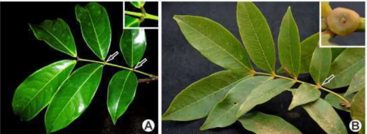
more than 330 species (Lewis 2005). Chamaecrista species are typically herbs (fig. 1A)
and shrubs (fig. 1B) with yellow flowers (fig. 1A, C and E) that are found in rocky
outcrops (fig. 1A–B) and in other open sunny areas. Woody species also appear in rainforests, especially in the Amazon and Brazilian Atlantic forests (Irwin and Barneby
1982; Lewis 2005; Conceição et al. 2009; Rando et al. 2013). Previously, Chamaecrista
species were grouped into six sections: Chamaecrista sect. Absus, Apoucouita,
Caliciopsis, Chamaecrista, Grimaldia and Xerocalyx (Irwin and Barneby 1982).
Molecular studies have suggested the definition of new sectional and species boundaries
(Conceição et al. 2008, 2009). According to the circumscription proposed by such
authors, the clades Apoucouita and Xerocalyx were recognized as sections, sect. Absus
subsect. Baseophyllum should be elevated to the sectional status and sect. Grimaldia
must now be included in sect. Absus. Sect. Caliciopsis and Chamaecrista were found to
be paraphyletic. Although these studies demonstrate the need for taxonomic revision
within the genus, taxon sampling was limited and more data are necessary before
amending taxon circumscriptions and adopting nomenclatural changes.
In the subfamily Caesalpinioideae s.l., secretory structures have been shown to
be meaningful additional characters in taxonomical evaluations (Irwin and Barneby
1978, 1982; Lersten and Curtis 1993, 1994, 1996; Rudall et al. 1994; Conceição et al.
2008, 2009; Coutinho et al. 2013). In several families, colleters have stood out in
taxonomical and phylogenetic interpretations (Lersten 1974; Thomas 1991; Klein et al.
2004; Silva et al. 2012; Dalvi et al. 2013, 2014). Colleters are structures that are usually
found on the adaxial side of stipules and bracts and secrete a sticky product composed
the shoot apex and floral buds (Foster 1942; Thomas 1991; Paiva and Machado 2006b;
Barreiro and Machado 2007; Mayer et al. 2011; Sheue et al. 2012).
Instead of the term colleter, other terms have been given to these structures, such
as “glandular shaggy hair” (Solereder 1908; Metcalfe and Chalk 1950), “resin glands”
(Curtis and Lersten 1980), “glandular trichomes” (Pascal et al. 2000), “extrafloral
nectary” (Freitas et al. 2001), and “filamentous structures” (De-Paula et al. 2011),
among others (Thomas 1991). Such nomenclatural diversity and/or misinterpretations
are primarily based on anatomical or morphological similarities with other secretory
structures. The term colleter is more appropriate to describe structures that secrete
substances responsible for protecting developing leaves and flowers mainly against
dehydration and pathogens (Thomas 1991; Mayer et al. 2011).
For Leguminosae, few studies have reported the presence of colleters (Paiva and
Machado 2006a; De-Paula and Oliveira 2007, 2012; Paiva 2009; Coutinho et al. 2013).
To our knowledge, there are no comparative studies on the diversity and taxonomic
importance of colleters for this family. In Chamaecrista, colleters have been reported in
the cotyledons of three species (De-Paula and Oliveira 2007, 2012) and on the leaves of
all of the species of Absus subsect. Baseophyllum (Coutinho et al. 2013). However, the
authors do not provide detailed information on the structural (morphological and
anatomical) variation and chemical nature of the secretion. Due to the importance of
colleters in taxonomy (Lersten 1974; Thomas 1991; Klein et al. 2004; Silva et al. 2012;
Dalvi et al. 2013, 2014), the following questions were raised: Are colleters common
secretory structures for Chamaecrista? Are there structural and topographical variations
among the species/sections? Could such variations be used to solve taxonomic problems
within this group? Is there variation in the chemical nature of the exudates that are
structural characterization and the histochemical analysis of the colleters found on both
vegetative and reproductive meristems of Chamaecrista.
Material and Methods Plant material
A total of 55 Chamaecrista species (65 taxa) belonging to five of the six sections
were studied (appendix). The proportion of the total taxa of the five sections of
Chamaecrista studied is indicated in table 1. Samples of the shoots (fig. 1C–D) and flower buds (fig. 1C, E) were obtained from herbarium material and field-collected
plants. For most species, at least three specimens were used as replicates.
Samples from herbarium material were microwaved in distilled water for 7 min
and left to rest overnight. Samples were then treated with 2% potassium hydroxide for 1
h at room temperature (Smith and Smith 1942), rinsed with tap water three times,
dehydrated in an ethanol series (10, 30, 50 and 70%, 10 min each) and stored in 70%
ethanol before being subjected to standard anatomical procedures. Samples from species
that were collected from the field (appendix) were fixed in FAA (formaldehyde, acetic
acid and 50% ethanol; 1:1:18, v/v) for 48 h and stored in 70% ethanol. Voucher
specimens were deposited at the herbarium of the Universidade Federal de Viçosa
(VIC), and duplicates were sent to the herbarium of The New York Botanical Garden
(NY), the United States National Herbarium – Smithsonian Institution (US) and the herbarium of the Universidade de São Paulo (SPF).
Slide preparation
Stipules, bracts and bracteoles from the samples of both herbarium material and
hydroxide and 20% hypochlorite solutions, stained with 50% ethanol-diluted fuchsin
and mounted in glycerinated gelatin (Johansen 1940).
The samples from both herbarium material and field-collected species that were
stored in 70% ethanol were embedded in methacrylate (Historesin Leica, Leica
Microsystems Nussloch GmbH, Heidelberg, Germany) following the manufacturer’s recommendation. Cross and longitudinal sections that were 4-µ m thick were made in an
automatic rotary microtome (Leica RM2155, Deerfield, IL, USA). For the structural
characterization, the sections were stained with toluidine blue at pH 4.4 (O’Brien and McCully 1981), dried at room temperature and mounted in resin (Permount, Fisher
Scientific, NJ, USA). Some of the sections were also used in histochemical tests.
Some of the field-collected species were also embedded in histological paraffin.
The material stored in 70% ethanol was dehydrated through a tert-butanol series and
embedded in histological paraffin (Histosec®, Merck, Germany) (Johansen 1940).
Cross and longitudinal 7-µ m thick serial sections were obtained from blocks using a
rotary microtome (Spencer 820 American Optical Corporation, Buffalo, NY, USA). The
sections were deparaffinized in xylene and rehydrated through an ethanol series. For
structural characterization, the sections were stained with safranin and astra blue,
dehydrated through an ethanol/xylene series (Gerlach 1969) and mounted in resin
(Permount®, Fisher Scientific, USA). Some of the sections were also used in
histochemical tests.
The histochemical tests were performed on fixed samples of Chamaecrista
brachystachya, C. cytisoides, C. debilis, C. decora, C. desvauxii var. latistipula, C.
ensiformis var. ensiformis, C. myrophenges and C. vauthiere. The following
histochemical tests were carried out on paraffin- and/or methacrylate-embedded
polysaccharides, periodic acid Schiff (PAS) (O’Brien and McCully 1981); for acid mucopolysaccharides, Alcian blue (Pearse 1980); and for pectins/mucilage, ruthenium
red (Johansen 1940). The paraffin-embedded sections of C. debilis. C. desvauxii var.
latistipula and C. myrophenges were tested for lipid compounds with Sudan IV and
Sudan Black B (Pearse 1980).
Paraffin- and/or resin-embedded material from six species were chosen for the
ontogenetic study of the colleters as follows: Chamaecrista brachystachya, C.
cytisoides, C. debilis, C. decora, C. desvauxii var. latistipula, and C. myrophenges.
Observations and photographs were collected using a light microscope (model
AX70TRF, Olympus Optical, Tokyo, Japan) that was equipped with a U-Photo system
and digital camera (AxioCam HRc – Carl Zeiss – Gottingen, Germany).
Scanning Electron Microscopy (SEM)
Samples from herbarium and field-collected material stored in 70% ethanol were
critical point-dried with CO2 in a Bal-Tec 020 CPD dryer (Bal-Tec, Balzers,
Liechtenstein). The samples were mounted onto stubs and coated with gold using an
FDU 010 sputter coater (Bal-Tec). The examinations and image captures were carried
out using a Leo 1430VP SEM (Zeiss, Cambridge, UK) at the Centro de Microscopia e
Microanálises at the Universidade Federal de Viçosa.
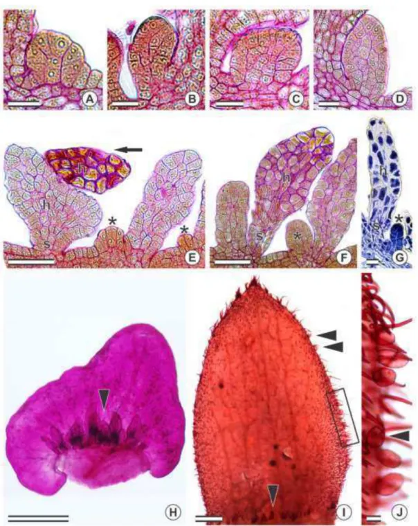
Results Ontogeny
There is no participation of any internal meristematic tissues in the origin of the
colleters. Such structures are formed from protodermal cell initials that could easily be
A single protodermal cell initial expands and goes through successive anticlinal
divisions (fig. 2E). The cells that originated from such anticlinal divisions begin
dividing in different planes (fig. 2A–D), giving rise to a stalk and a secretory (fig. 2E– F). The protodermal cell divisions are not synchronic as fully-formed colleters may be
observed while young ones are still going through cell divisions (fig. 2E–G).
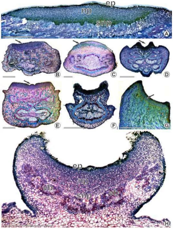
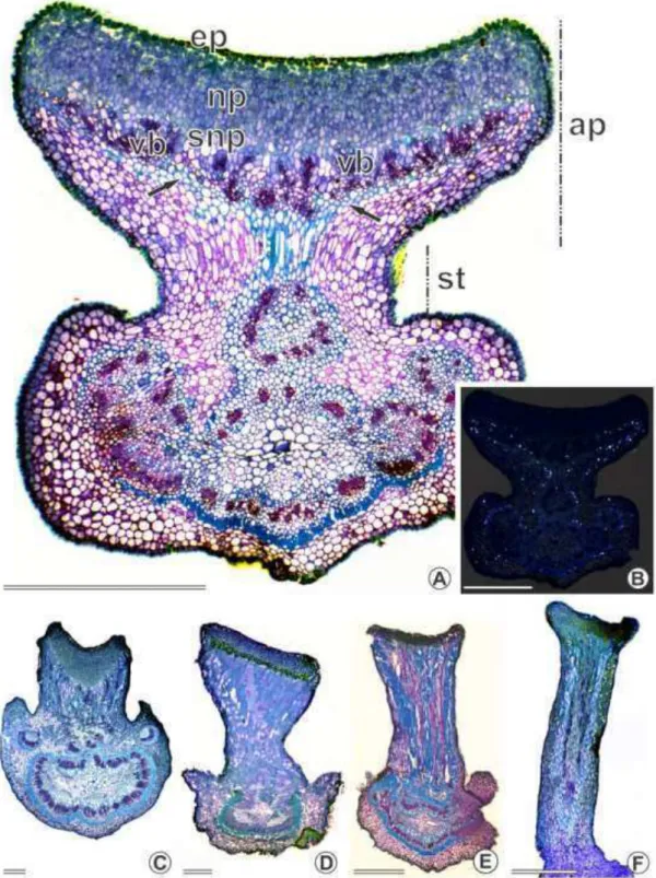
Structure and Distribution
No vascularization was observed in the colleters of the Chamaecrista species
studied (fig. 2F). The fully-formed colleters are composed of a stalk and a secretory
head (fig. 2E–G). The stalk cells are highly vacuolated with thick outer walls while those forming the head show a densely stained cytoplasm. Secretory pores were not
observed, and both light and SEM indicated that cuticle rupture was rare. The secretory
phase begins when the leaves and flowers are still being formed. When secretion ends,
the colleters wither, become dark brown or black and usually drop off.
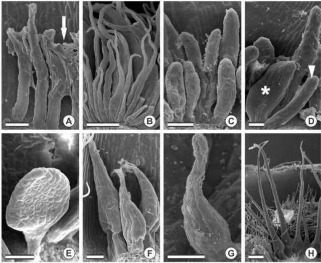
Colleters were always found in patches (figs. 2–4) at the adaxial side of different plant structures (table 1): at the base of bracts/bracteoles (figs. 2H, 3D–F, 4A, F, H), at the base and margins of sepals (fig. 2I–J), at the base of petals, on the rachides at the insertion of the leaflets (fig. 3A) and at the base of stipules (fig. 4B–D). They were also found around extrafloral nectaries (fig. 3B–C) and at the extension of the rachides (fig. 3C) but not in patches. The number of colleters is not fixed for each species or plant
organ/structure. All of the studied species showed at least one type of colleter, except
for Chamaecrista dentata, C. fagonioides, C. glaziovii and C. semaphora, in which
such structures could not be found.
4H), short bottle-shaped (< 300 µ m) (figs. 3F, 4F–G), long digitiform (> 500 µ m) (fig. 4B) and short digitiform (< 400 µm) (figs. 3C, 4A, D).
On petals, only the short digitiform type was found and occurred at the base of at
least one of the petals (table 1). The same types of colleters which occur on the
vegetative and reproductive structures (table 1) could also be associated to the
extrafloral nectaries (fig. 3B–C). On petals and at the base of the extrafloral nectaries, these structures were randomly found, usually less than four (table 1).
The species from sect. Apoucouita showed short digitiform and club-shaped
types on both vegetative and floral organs (table 1). Apoucouita stood out for being the
only section showing colleters on the sepal margins, wherein all
species of that section had the club-shaped type.
Five types of colleters were observed in sect. Absus (table 1). Most of the
species of subsect. Absus showed the short bottle-shaped type of colleters on both
vegetative and reproductive organs, except for C. monticola, C. setosa and C. speciosa,
which showed the long bottle-shaped type. Sticky glandular hairs, another type of
trichomes, were also present in species from subsect. Absus (figs. 1D–E, 3E, G). However, all of the species from subsect. Baseophyllum showed the short digitiform
and club-shaped type and in addition to these, C. coriacea also presented the
racket-shaped type. C. debilis, the only species in subsect. Otophyllum, displayed both
club-shaped and racket-club-shaped types.
Although the short bottle-shaped was the most common type among the species
from sect. Chamaecrista, the short digitiform and club-shaped types were also observed
(table 1). C. absus var. absus and C. absus var. meonandra, the only two taxa from sect.
Grimaldia, displayed the short bottle-shaped type (table 1). All of the species from sect.
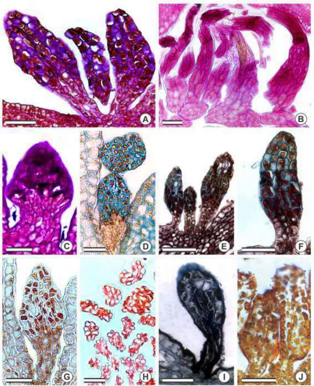
Histochemistry
Histochemical analyses for the total polysaccharides (fig. 5A–C), acid mucopolysaccharides (fig. 5D), pectins/mucilage (fig. 5E–F), total proteins (fig. 5G–H), and lipids (fig. 5I–J) generated positive results. Only the head of the colleters reacted to the histochemical analyses. The stalk cells did not secrete any of the analyzed metabolic
groups. The outer of stalk cells were densely stained by Sudan black B (fig. 5I),
demonstrating the presence of lipids in the cell wall composition.
Discussion
Classification, secretion and function of the colleters
The ontogenetic study of the colleters shows that such structures derive from the
protodermis only and, therefore, are a kind of secretory trichomes (Fahn 1990).
According to the topography, structure (micromorphology and anatomy), early
secretory activity and the compounds that are present in the secreted exudates, these
trichomes correspond to colleters (Foster 1942; Fahn 1990; Thomas 1991).
Although colleters that are found in Chamaecrista display a stalk and a secretory
head, a central axis and a secretory epidermis were not observed. Such colleters do not
match any of the colleter descriptions of Lersten (1974). Colleters consisting of
homogenous cells have been reported (Paiva and Machado 2006a; De-Paula and
Oliveira 2007; Silva et al. 2012), but a classification for this type of colleter has yet not
been proposed. We believe that such colleters should be called homogenous colleters
based on the homogeneity of the cellular composition. The homogenous colleters could
then be divided into different types according to the micromorphology of such
Secretory pores, which release the secretion to the exterior, were not observed,
and cuticle rupture was rare. Such observations suggest an involvement of the outer cell
wall in the secretion process and the releasing of the exudates via micropores or via
cuticle permeability (Ascensão et al. 1999; Klein et al. 2004; Paiva 2009; Mayer et al.
2011; Silva et al. 2012). The thickness and lipidic composition of the outer walls of the
stalk cells may contribute to the symplastic transport of the secretion precursors towards
the secretory head. Such process is common in secretory trichomes (Fahn 1979, 1990;
Leitão et al. 2005).
The histochemical tests revealed that the secreted exudates are composed of a
mixture of hydrophilic (total polysaccharides, acid mucopolysaccharides, pectins,
mucilages and total proteins) and lipophilic (lipids) compounds. Polysaccharides,
pectins and mucilages may play a role in water retention due to their considerable
ability to absorb water (Christodoulakis et al. 1990; Fahn and Cutler 1992; Al-Tardeh et
al. 2008). This ability allows these hydrophilic compounds to lubricate both the
vegetative and reproductive meristem, thereby protecting the developing organs against
dehydration (Foster 1942; Fahn 1990; Thomas 1991; Mayer et al. 2011; Martins 2012;
Silva et al. 2012; Chin et al. 2013; Mayer et al. 2013). According to Paiva (2009, 2012),
the hydrophilic material on the leaf surface reduces water loss to the external
environment, helping to maintain adequate humidity levels for leaf development, as
xylem transport is not yet efficient in developing organs. In addition to helping to
lubricate the meristems, derivates of the lipophilic fraction along with proteins present
in the secretion could act as an inhibitor of fungal plant pathogens (Barnes et al. 1997,
Miguel et al. 2006). In coffee flower buds, the secreted exudates produced by colleters
act as an adhesive, sealing the bud until flower development is complete. As a
The closeness of the colleters to the developing leaf and flower, their early
differentiation, the secretion period and the chemical nature of the exudates indicate a
protective function of such structures in Chamaecrista.
De-Paula and Oliveira (2007) did not observe secretion in the colleters of
Chamaecrista desvauxii var. latistipula. In our study, however, this species displayed
secreting colleters. It is possible that De-Paula and Oliveira (2007) performed the
histochemical tests when the colleters in the embryos were not yet active.
The colleters in Chamaecrista, especially those found on the stipules and
rachides, usually fall off after completing their activity. Paiva (2012) suggests that rapid
senescence may avoid possible problems with the growth of pathogens.
Of all of the studied species, Chamaecrista dentata, C. fagonioides, C. glaziovii
and C. semaphora are the only ones that do not possess colleters. These species display
high amounts of sticky glandular hairs all over the plant body. Meira et al. (2014)
reported the presence of oleoresin secretory trichomes in C. dentata and correlated the
presence of such secretion to protection against abiotic and biotic adverse factors. It is
possible that in all of the Chamaecrista species that do not have colleters but instead
have only sticky glandular hairs, the oily part of the trichome plays a role in lubricating
the young developing structures, similar to colleters.
Taxonomic importance of colleters
In Chamaecrista, colleters have been reported for a few species (De-Paula and
Oliveira 2007, 2012; Coutinho et al. 2013). However, 51 of the 55 studied species
displayed colleters, demonstrating how common these structures are for this genus. As
the colleters on the fully expanded leaves and flowers may have withered or dropped
Colleters of exclusively protodermal origin were observed in the five species of
Chamaecrista that were ontogenetically studied and in Hymenaea stigonocarpa (Paiva
and Machado 2006a). A protodermal origin for colleters seems to be a rare pattern of
development as colleters are commonly formed from both protodermal and ground
meristem and sometimes even from the procambial activity (Thomas 1991; Klein et al.
2004; Gonzalez and Tarragó 2009).
In Chamaecrista, six types of homogenous colleters could be distinguished. The
anatomical and morphological variations observed in colleters have been used as
taxonomic characteristics for several families (Thomas 1991): Apocynaceae (Simões et
al. 2006), Aquifoliaceae (Gonzalez and Tarragó 2009), Myrtaceae (Silva et al. 2012),
Rubiaceae (Lersten 1974; Klein et al. 2004), Salicaceae (Curtis and Lersten 1980) and
Rhizophoraceae (Sheue et al. 2012) but not yet in Leguminosae.
Colleters associated with the extrafloral nectaries, the adaxial base of petals and
along the rachis extension varied among the taxa (species and sections). However, we
could not demonstrate a useful pattern to delimitate sections or species groups.
Although colleters in Chamaecrista are found in groups in both vegetative and
reproductive organs, the number of colleters is not fixed. On the other hand, the
variation in their position is noteworthy. For instance, species from the sect. Apoucouita
are the only studied species to display colleters on the margin of the sepals. Conceição
et al. (2009) noted that sect. Apoucouita actually forms a monophyletic group. The
presence of club-shaped colleters on the margins of the sepals of species belonging to
sect. Apoucouita is a synapomorphy for this section.
Section Absus subsect. Baseophyllum stood out for showing the short digitiform
and club-shaped types of colleter. The same types of colleters were found in the sect.
(Conceição et al., 2009). The absence of the short bottle-shaped type of colleter,
characteristic of the sect. Absus subsect. Absus, in all the species of subsect.
Baseophyllum supports the elevation of subsect. Baseophyllum as a section distinct from
sect. Absus, as shown by molecular and anatomical studies (Conceição et al. 2009,
Coutinho et al. 2013).
Chamaecrista debilis is the only species in the sect. Absus subsect. Otophyllum.
This species displayed both the club-shaped and racket-shaped types of colleters. The
racket-shaped type appeared only in C. debilis and C. coriacea (subsect. Baseophyllum),
bringing C. debilis close to the species of subsect. Baseophyllum. The inclusion of C.
debilis in future molecular studies is necessary to confirm this possible affinity.
Most of the species of sect. Absus subsect. Absus had the short bottle-shaped
type of colleter on both vegetative and reproductive organs. The only two taxa that
belong to sect. Grimaldia, C. absus var. absus and C. absus var. meonandra, also
display the same type of colleter (short bottle-shaped). The similarity in the type of
colleters of these species reinforces the affinity between these two groups, supporting
the recent molecular data which included sect. Grimaldia and sect. Absus subsect. Absus
in a single clade (Table 1). Therefore, sect. Grimaldia should be treated as a synonym of
sect. Absus subsect. Absus in a future taxonomic revision of Chamaecrista.
The most common type of colleter in sect. Chamaecrista was the short
bottle-shaped. However, other types of colleters (club-shaped and short digitiform) also
appeared. Section Chamaecrista is paraphyletic (Conceição et al. 2009) (Table 1) and it
is possible that the diversity of colleters in this section reflects such paraphyletism.
Chamaecrista calycioides of sect. Caliciopsis is well supported within sect.
Chamaecrista while the position of C. supplex is poorly supported. Unfortunately, our
these two species are necessary to evaluate the importance of colleters in providing
better resolution to phylogenetic analyses. Future anatomical and phylogenic studies
need to include broader taxon sampling of sect. Chamaecrista before any taxonomical
considerations are taken into account.
Section Xerocalyx is a monophyletic group with poorly supported branches that
is placed in the clade formed by three sections (Table 1): Caliciopsis, Chamaecrista and
Xerocalyx (Conceição et al. 2009; Torres et al. 2011). Interestingly, species from sect.
Xerocalyx shows only the digitiform type of colleter (short and long) with the long
digitiform unique for C. desvauxii var. desvauxii and C. desvauxii var. latistipula. The
uniformity in the type of colleter for sect. Xerocalyx is additional evidence supporting
the monophyly of this section. The variation from long to short digitiform may be
related to the size of stipules/bracts. Species with larger stipules/bracts (C. desvauxii
var. desvauxii and C. desvauxii var. latistipula) displayed both long and short digitiform
types while species with smaller stipules/bracts (C. desvauxii var. graminea, C. ramosa
and C. ramosa var. parvifoliola) displayed the short digitiform only.
Conclusion
Based on the topography, structural characters, ontogeny and components that
were identified in the secretion, the secretory structures found in young leaves and
flowers of Chamaecrista species are colleters. Such structures may be involved in the
protection of developing leaves and flowers. The presence of colleters on leaves and
flowers is a new report for Chamaecrista. Five of the six types of colleters described in
our study are novelties in the genus.
The structural diversity of colleters in Chamaecrista provides characters that
recent phylogenetic study (Conceição et al., 2009). However, taxon sampling for both
phylogeny and anatomical studies is inadequate to justify a revised classification. Future
research will focus on broadening the taxon sampling. In addition, future taxon
sampling should give high priority to sampling the same species in the phylogenetic and
anatomical analyses.
Acknowledgments
The authors thank CNPq, CAPES, FAPEMIG and Floresta-Escola
(SECTES/UNESCO/HidroEX/FAPEMIG) for financial support; CAPES and CNPq for
granting a scholarship to ÍAC Coutinho and RMSA Meira; the Smithsonian Institution
for granting the first author with the Cuatrecassas Award; the herbaria HUEFS, NY,
RB, SPF and US for allowing sampling of voucher specimens; JG Rando along with A
Nogueira for presenting us with four fixed samples of Chamaecrista sect. Apoucouita;
AS Conceição for determining the species of a few collections; Instituto Chico Mendes
for permission to collect specimens; and the anonymous reviewers along with PS
Herendeen for helpful and critical comments that improved the manuscript.
Literature Cited
Al-Tardeh S, Sawidis T, Diannelidis B-E, Delivopoulos S 2008 Water content and reserve allocation patterns within the bulb of the perennial geophyte red squill (Liliaceae) in relation to the Mediterranean climate. Botany 86:291–299.
Ascensão L, Mota L, Castro MM 1999 Glandular trichomes on the leaves and flowers of Plectranthus ornatus: morphology, distribution and histochemistry. Ann Bot 84:437–447.