Neutrophils Contribute to the Protection
Conferred by ArtinM against Intracellular
Pathogens: A Study on
Leishmania major
Rafael Ricci-Azevedo, Aline Ferreira Oliveira, Marina C. A. V. Conrado, Fernanda Caroline Carvalho, Maria Cristina Roque-Barreira*
Departamento de Biologia Celular e Molecular e Bioagentes Patogênicos, Faculdade de Medicina de Ribeirão Preto, Universidade de São Paulo, Ribeirão Preto, São Paulo, Brasil
*mcrbarre@fmrp.usp.
Abstract
ArtinM, a D-mannose binding lectin fromArtocarpus heterophyllus, has immunomodulatory activities through its interaction with N-glycans of immune cells, culminating with the estab-lishment of T helper type 1 (Th1) immunity. This interaction protects mice against intracellu-lar pathogens, includingLeishmania majorandLeishmania amazonensis. ArtinM induces neutrophils activation, which is known to account for both resistance to pathogens and host tissue injury. Although exacerbated inflammation was not observed in ArtinM-treated ani-mals, assessment of neutrophil responses to ArtinM is required to envisage its possible application to design a novel immunomodulatory agent based on carbohydrate recognition. Herein, we focus on the mechanisms through which neutrophils contribute to ArtinM-induced protection againstLeishmania, without exacerbating inflammation. For this pur-pose, human neutrophils treated with ArtinM and infected withLeishmania majorwere ana-lyzed together with untreated and uninfected controls, based on their ability to eliminate the parasite, release cytokines, degranulate, produce reactive oxygen species (ROS), form neutrophil extracellular traps (NETs) and change life span. We demonstrate that ArtinM-stimulated neutrophils enhancedL.majorclearance and at least duplicated tumor necrosis factor (TNF) and interleukin-1beta (IL-1β) release; otherwise, transforming growth factor-beta (TGF-β) production was reduced by half. Furthermore, ROS production and cell degranulation were augmented. The life span of ArtinM-stimulated neutrophils decreased and they did not form NETs when infected withL.major. We postulate that the enhanced leishmanicidal ability of ArtinM-stimulated neutrophils is due to augmented release of inflammatory cytokines, ROS production, and cell degranulation, whereas host tissue integ-rity is favored by their shortened life span and the absence of NET formation. Our results reinforce the idea that ArtinM may be considered an appropriate molecular template for the construction of an efficient anti-infective agent.
OPEN ACCESS
Citation:Ricci-Azevedo R, Oliveira AF, Conrado MCAV, Carvalho FC, Roque-Barreira MC (2016) Neutrophils Contribute to the Protection Conferred by ArtinM against Intracellular Pathogens: A Study on
Leishmania major. PLoS Negl Trop Dis 10(4): e0004609. doi:10.1371/journal.pntd.0004609
Editor:Rodrigo Correa-Oliveira, FIOCRUZ - Minas, BRAZIL
Received:July 14, 2015
Accepted:March 14, 2016
Published:April 8, 2016
Copyright:© 2016 Ricci-Azevedo et al. This is an open access article distributed under the terms of the
Creative Commons Attribution License, which permits unrestricted use, distribution, and reproduction in any medium, provided the original author and source are credited.
Data Availability Statement:All the relevant data are within the paper and in the Supporting Information files, with only one exception regarding the "Accession number/ID" for the ArtinM lectin, which is ID: Q7M1T4_ARTIN, available on UniProtKB database.
Author Summary
Vaccination is a successful way to eliminate infectious diseases. The generated antibodies neutralize the invading microbe and avoid the establishment of infection. Vaccines are efficient to prevent infections by pathogens living outside rather than inside the host`s cells. This occurs because protection against intracellular pathogens requires the engage-ment of T lymphocytes. The discovery of receptors on innate immunity cells opened new perspectives in trying manners to stimulate effective response against intracellular patho-gens. Frequently the microbial sensing by Toll-like receptors (TLRs) besides triggering immediate defense also orchestrates adaptative immunity towards T-cell response. There-fore, TLR ligands started to be assayed in new anti-infective approaches. Our laboratory has been investigating the immunomodulation induced by lectins, which are ubiquitous sugar-binding proteins. Our primary model is ArtinM, from the seeds of jackfruit, a lectin that binds to TLR2 sugar chains on macrophages and dendritic cells and promotes pro-duction of cytokines that engages T lymphocytes in a process that culminate with elimina-tion of intracellular pathogens. Concomitantly, ArtinM activates other immune cells, including neutrophils, which contributes to the pathogen elimination, but may also account for tissue damage. This last possibility led us to investigate the lectin effects on neutrophils deeply. We analyzed neutrophils treated with ArtinM and infected with Leish-mania major. We concluded that the leishmanicidal ability of ArtinM-stimulated neutro-phils was due to augmented release of inflammatory cytokines, ROS production, and cell degranulation. Otherwise, host tissue integrity is favored by shortened cells lifespan and absence of NET formation. This work reinforces the idea that ArtinM can be an appropri-ate molecular templappropri-ate for the construction of an efficient anti-infective agent.
Introduction
Global immunization regimes have eradicated smallpox and controlled a large number of other infections [1,2]. Indeed, vaccines have been successful against infections caused by extra-cellular pathogens or those whose pathogenesis is mediated by toxins. Under these circum-stances, vaccines confer protection by inducing antibodies that neutralize the inoculum and prevent the establishment of infections [3]. This effective formula is not applied to prevent infections with intracellular pathogens [4] because they require T-cell mediated immunity for elimination. Therefore, a research field has emerged that concerns the development of alterna-tive prophylactic or therapeutic agents to enhance host cellular response. In this context, ago-nists of innate immunity receptors, especially Toll-like receptors (TLRs), provide promising approaches [5]. The interaction of agonists with TLRs triggers cell signaling and production of inflammatory and anti-inflammatory mediators [6]. This process, beyond inducing early mechanisms of host defense, primes and orchestrates antigen-specific adaptive responses [7]. The ability of activated TLRs to modulate adaptive immunity motivates ongoing trials of new drugs based on natural or synthetic TLR ligands for infectious diseases in humans [8].
ArtinM, a D-mannose-binding lectin obtained from the seeds ofArtocarpus heterophyllus, binds to TLR2 N-glycans and functions as a TLR2 agonist that exerts immunomodulatory properties [9]. The ArtinM interaction with TLR2 on macrophages and dendritic cells results in high levels of IL-12 production, driving immunity towards the T helper (Th) 1 axis [10]. This ability accounts for the protection conferred by ArtinM administration against Leish-mania major[11],Leishmania amazonensis[12],Paracoccidioides brasiliensis[10,13], Neos-pora caninum[14], andCandida albicans[15] infections in mice. Beyond acting on antigen
study design, data collection and analysis, decision to publish, or preparation of the manuscript.
presenting cells, ArtinM exerts activities on lymphocytes [16], mast cells [17,18], and neutro-phils [19,20]. This pleiotropic activity on immune cells is considered to account for the ArtinM property of conferring resistance against intracellular pathogens [21].
Concerning neutrophils, the cell type focused in this work, they are known to participate in the protection against intracellular pathogens, through mechanisms that involve phagocytosis, cell degranulation, ROS production, release of lipid mediators, and formation of neutrophil extracellular traps (NETs) [22]. Further mechanisms known to favor host defense are the release of cytokines combined with changes in cell life span [23]. Our previous work showed that ArtinM induces neutrophil migration by haptotaxis [24], due to the concomitant interac-tions of ArtinM CRDs with N-glycans on neutrophil surface receptors, such as those linked to C-X-C chemokine receptor 2 (CXCR2), and glycoproteins of the extracellular matrix, such as laminin [25,26]. Also, ArtinM activates neutrophils, causing tyrosine phosphorylation, L-selec-tin shedding, and interleukin-8 (IL-8) and leukotriene B4 (LTB4) secretion. These responses result in the enhancement of phagocytic and microbicidal abilities of neutrophils [19,20] and indicate that ArtinM activates neutrophils hugely.
Although effective against pathogens, neutrophils also account for exacerbated inflamma-tion and tissue injury [27], a fact that caused concerns regarding the possibility of using ArtinM to design a novel class of immunomodulatory agents acting through carbohydrate recognition. Although exacerbated inflammation was never observed in the ArtinM-treated animals, we always had concerns regarding the occurrence of inflammatory tissue injury. Indeed, it is unclear how ArtinM may take advantage of neutrophil activation to eliminate pathogens, with-out promoting tissue damage. Therefore, in this study, we focused on understanding the mech-anisms through which neutrophils contribute to the protective effect of ArtinM against intracellular pathogens and how this process occurs without exacerbating inflammation.
Materials and Methods
Ethics statement
The Ethics Committee of the Clinical Hospital of the Faculty of Medicine of Ribeirão Preto, University of São Paulo, approved this study (Doc. Number: 10012/2009) and all the adult vol-unteers signed an informed consent form prior to blood and/or urine donation.
ArtinM preparations and treatment
ArtinM (ID: Q7M1T4_ARTIN, available on UniProtKB database) was extracted from Artocar-pus heterophyllusseeds and purified by sugar affinity chromatography as previously described by Santos-de-Oliveira et al. (1994) [24]. The concentration of ArtinM lectin used to treat the neutrophils in this study was 2.5μg/mL, as previously utilized [20], except in some assays, as
specified.
Neutrophil isolation
Leishmania major
culture and infection
L.major(LV39 strain) promastigotes were grown in Schneider’s medium supplemented with 1% antibiotic-antimycotic, 10% heat-inactivated fetal calf serum (FCS), 2 mM L-glutamine (all from Invitrogen, USA), and 2% human urine, pH 7.2. Neutrophils were pre-treated with ArtinM, and after 30 min, they were infected with stationary-phase promastigotes (5–7 day) at a parasite-PMN ratio of 3:1. Control neutrophils were not treated with ArtinM and not infected. Plates/tubes were centrifuged (300 ×g/ 10 min) and incubated at 37°C in RPMI 1640 or Hank’s Balanced Salt Solution (HBSS) in a humidified atmosphere containing 5% CO2. Some assays were performed by infecting neutrophils with green-fluorescentL.majorforms (mβT3-LV39), which were kindly provided by Prof. Angela Kaysel Cruz and Dr. Mônica Cris-tina Terrão.
Leishmania major
uptake by neutrophils
Human neutrophils (2 × 10⁶cells/mL) were previously treated with ArtinM for 30 min, and infected withL.major. Infected/untreated neutrophils were used as controls. After 3 and 20 h post-infection, the cultures were cytocentrifuged (50 × g, 5 min) and Diff-Quik-stained (Labor-clin, Brazil). The number of neutrophils with intracellular parasites was determined by count among 200 cells/condition/time, using an Olympus BX50 microscope coupled to a Nikon DXM-1200 photographic system.
Parasite viability
To analyze the intracellular leishmanicidal activity, we assessed the parasite viability, as described by Tavares et al. (2014) [28], with slight modifications. Briefly, neutrophils that were treated with ArtinM and untreated controls were infected and cultured for 3 h with the para-sites. Uninternalized parasites were discarded from cultures by washing twice for 5 min at 100 ×g, and the culture was immediately fed with supplemented Schneider’s medium. The cells were then cultured at 26°C for an additional 48 h. The intracellular leishmanicidal activity was determined by assessing the number of extracellular motile promastigotes produced.
To analyze if some degranulated content had leishmanicidal activity, we performed a free cell assay based on methodology previously demonstrated by Mikus and Steverding (2000) [29]. Neutrophils treated with ArtinM for 1 h and untreated controls were pelleted, and the supernatants of the cells were collected. The supernatants were incubated with antibodies to myeloperoxidase (anti-MPO; ab62141; Abcam; 1/100) or neutrophil elastase (anti-NE; ab21595; Abcam; 1/100) for 30 min. Control supernatants were not incubated with the anti-bodies. After the incubation, 1 × 10⁵parasites were added to the supernatants for 1 h, and then assayed for viability in Schneider’s medium containing Alamar Blue (Life Technologies, USA; 1/10 dilution) for an additional incubation of 24 h. Fluorescence measurement was performed on a FLx800 Fluorescence Microplate Reader (BioTek Instruments, USA; excitation, 590 nm; emission, 635 nm). Fluorescence count data from unchallenged, serially diluted parasites were used to obtain a standard curve of viable parasites.
ELISA
Freshly isolated human neutrophils (2 × 10⁶cells/mL) were previously treated with ArtinM for 30 min and infected withL.major. Neutrophils without ArtinM treatment and uninfected with
USA), and TGFβ-1 (R&D Systems, USA) were measured by sandwich ELISA, according to the manufacturer’s instructions.
Measurement of ROS production
The neutrophil chemiluminescence assay was performed in 96-well microplates using the pro-cedure described by Lucisano-Valim et al. (2002) [30], with slight modifications. Freshly iso-lated human PMNs (2 × 10⁵cells/well in HBSS) were mixed with the chemiluminescent probes luminol (0.1 mM) or lucigenin (0.1 mM). The mixture was incubated at 37°C for 3 min, and the reaction was initiated after adding ArtinM, phorbol myristate acetate (PMA, 0.1μM),
for-myl-Met-Leu-Phe (fMLP, 1μM), or HBSS. Some assays were performed by first incubating the
cells with ArtinM during 30 minutes, and then by adding fMLP, PMA orL.major(MOI 3:1). The luminol- and lucigenin-enhanced chemiluminescence was measured in a microplate luminometer (LB 960 Centro, Berthold Technologies, Germany), and light emission was recorded in photon counts per second (CPS) for 30–60 min, at 37°C. AUC represents the area under the time–course curve, which was used to determine the total amount of measured ROS.
Evaluation of Neutrophil Elastase (NE) activity
Neutrophil elastase activity was evaluated as previously described [31], with some modifica-tions. Briefly, 1 × 10⁵neutrophils were treated for 30 min with different concentrations of ArtinM (2.5μg/mL to 312 ng/mL), fMLP (1μM), or medium only, and NE activity was
detected by using the substrate N-succinyl-alanine-alanine-valine-p-nitroanilide (SAAVNA) (1 mM), which is cleaved by the enzyme released in the supernatant, forming p-nitroaniline as one of the products, which was spectrophotometrically quantified, using the microplate reader PowerWaveX (BioTek Instruments, USA; 405 nm).
Intracellular Myeloperoxidase (MPO) detection
Myeloperoxidase, as well as elastase, are azurophilic granule contents of neutrophils. Their intracellular levels were measured in neutrophils after 20 h of incubation with or without ArtinM, and challenged or not withL.major. To detach ArtinM-treated neutrophils, it was necessary to use EDTA-glucose (10μM/5μM) containing PBS (pH 7.2). Then, the cells were
washed twice with PBS, and immediately fixed and permeabilized using the Cytofix/Cytoperm kit (BD—Biosciences, USA), following the manufacturer’s instructions. Next, the cells were incubated with anti-MPOPEor anti-Hamster IgG1PEisotype control antibodies (BD— Biosci-ences, USA), for 20 min. Immunofluorescent staining was analyzed by flow cytometry using Guava EasyCyte Mini (Millipore, USA).
NET-DNA release measurement
Neutrophils (2 × 10⁵cells/well in HBSS) were incubated with or without ArtinM, PMA (50 nM), or both together (ArtinM+PMA) in a 96-well plate for 2 and 4 h at 37°C, in 5% CO2. NET-DNA was quantified using a modified version of a previously published method [32]. Bacterial endonucleaseEcoRI(200 unit/mL) was added for 10 min to partially digest any released NETs. Next, the cells and debris were pelleted by centrifugation at 600 ×gfor 10 min. A sample of the supernatant was added to SYTOX green nucleic acid stain (Invitrogen; 1μM)
NET immunofluorescence detection
Neutrophils (2 × 10⁶cells/mL in HBSS) were incubated on poly-L-lysine-treated glass cover-slips in a 24-well plate and treated or not with ArtinM or PMA, followed by incubation at 37°C for 6 h. Samples were collected, by gently removing coverslips, fixed with 3% paraformaldehyde (20 min at room temperature), and blocked in PBS supplemented with 3% FCS for 1 h at room temperature. Coverslips incubated overnight at 4°C with anti-neutrophil elastase antibody (ab21595; Abcam; 1:200) were washed and incubated for 1 h at room temperature with Alexa Fluor488 goat anti-rabbit IgG secondary antibody (Molecular Probes; 1:1000), and then washed and mounted with ProLong Antifade containing DAPI (Molecular Probes). Images were obtained using a Leica CTR 6000 fluorescence microscope, with Leica Application Suite software (Wetzlar, Germany).
Assessment of neutrophil morphology
To assess cellular morphology, freshly isolated human neutrophils (2 × 10⁶cells/mL in RPMI-1640) were treated with ArtinM, IL-8 (25 nM), Lysis Buffer (ammonium chloride 70 mM, Tris 10mM), or non-treated, and incubated during 3 and 20 h at 37°C in 5% CO2. After incubation, the cells were detached, as described above for MPO detection, cytocentrifuged onto a microscope slide using a Cytospin 3 cytocentrifuge (Thermo Shandon, USA), Diff-Quick-stained (Laborclin, Brazil) and examined using light microscopy (Olympus BX50—Olympus América INC, USA). Photomicrographs were taken with a Nikon DXM-1200 camera (Nikon instruments, USA).
DNA fragmentation analysis
DNA electrophoresis was performed to determine the effects of ArtinM treatment on neutro-phils DNA degradation. The analysis was performed using a kit, the Wizard SV genomic DNA purification system (Promega Corporation, USA). Briefly, after 24 and 48 h of incubation, 4 × 10⁶cells were washed twice with PBS and lysed, and genomic DNA was isolated. The extracted DNA was quantified using the NanoVue Plus spectrophotometer (GE Healthcare, USA). A sample of 1μg of DNA was analyzed using 1.5% agarose gel electrophoresis and
stained with ethidium bromide. The DNA was then visualized under UV light on ChemDoc MP Imaging System and photographed using ImageLab software v.4.0 (both from Bio-Rad Laboratories, USA). Regarding the analysis of DNA fragmentation, the band area was quanti-fied using ImageJ Software, and represented graphically in pixels2.
Phosphatidylserine exposure assessment
Neutrophil apoptosis was assessed by Annexin V staining [33]. Human neutrophils (2 × 10⁶ cells/mL in RPMI-1640), were treated with ArtinM, IL-8, Lysis Buffer, or non-treated, and co-incubated or not withL.majorduring 3 and 20 h at 37°C in 5% CO2. After incubation, the cells were detached, as described above (MPO detection), washed once and suspended in 100μL of
annexin V binding buffer (140 mM NaCl, 2.5 mM CaCl2, 1.5 mM MgCl2, and 10 mM HEPES, pH 7.4), containing annexin V-FITC or PE(1μg/mL) for 15 min. Some assays were performed
on neutrophils infected with green-fluorescentL.majorforms (mβT3-LV39 strain), in order to detect if the dying cells are the ones infected. Immunofluorescence staining was analyzed by flow cytometry, using a Guava EasyCyte Mini instrument (Millipore, USA).
Disruption of mitochondrial transmembrane potential
solution (10μM) was then added to the wells and incubated at 37°C for 15 min. The fluorescent
intensity for monomeric forms of JC-1 was measured using the FLx800 Fluorescence Micro-plate Reader (BioTek Instruments, USA; excitation 485 nm, emission 528 nm).
Statistical analysis
Statistical analyses were performed by Student t test, one way ANOVA followed by Bonferro-ni's post-test, and two way ANOVA followed by BonferroBonferro-ni's post-test (all using GraphPad Prism software version 6; GraphPad), as indicated at the graph. The p values<0.05 were deemed statistically significant.
Results
ArtinM enhances uptake and killing of
L
.
major
by neutrophils
To evaluate whether neutrophil activation by ArtinM contributes to its protective effect against intracellular pathogens [21], we compared the internalization and elimination ofL.majorby neutrophils that were pre-treated or were not pre-treated with ArtinM.
We assessed the effect of ArtinM treatment on the frequency of neutrophils with internal-ized parasites. At 3 h afterin vitroinfection, the number (15±0%) of ArtinM-treated neutro-phils with internalized parasites was 88% higher than the number (7.5±0.7%) verified in untreated neutrophils (Fig 1A). The difference increased 140% (14.5±0.7%vs34±2.8%) when the same assay was performed 20 h after infection (Fig 1A).
We also evaluated the leishmanicidal activity of the ArtinM-treated neutrophils. Cells were washed at 3 h post-infection and cultured in Schneider`s medium for additional 48 h. At this point, the number of mobile parasites (2.9x10⁵±0.7x10⁵) recovered from the culture of ArtinM-treated neutrophils was, on average, 50% lower than the number of viable parasites (5.8x10⁵±1.3x10⁵) recovered from untreated neutrophils (Fig 1B).
In conclusion, our data show that ArtinM treatment increased the ability of neutrophils to eliminateL.major, a fact that favors the idea that neutrophils can contribute to the protective effect of ArtinM against infection.
ArtinM alters the pattern of cytokine production by
L
.
major
-infected
neutrophils
The prominent production of inflammatory or anti-inflammatory cytokines by neutrophils contributes to an appropriate microenvironment forL.majorelimination or survival, respec-tively [34,35]. We quantified the TNF, TGF-β, and IL-1βcytokines in the supernatant of non-treated or ArtinM-non-treated neutrophils that were infected or not withL.major. Twenty h after ArtinM-treatment, the supernatant contained 7-fold higher concentration of TNF (241.7±5.59
ρg/mL) than the supernatant of untreated neutrophils (65.65±5.47ρg/mL). A similar level was
detected when the assay was performed withL.majorinfected neutrophils (245.77±3.93ρg/mL
and 93.77±0.68ρg/mL—Fig 2A). On the other hand, TGF-βproduction was 3-fold augmented
inL.major-infected neutrophils (169.6±32.01ρg/mL), while ArtinM-treated cells, infected
(56.99±16.73ρg/mL) or not (27.51±3.14ρg/mL), released levels that were similar to the ones
verified for untreated cells (54.94±15.27ρg/mL—Fig 2B). IL-1βwas not detected in the
super-natant of untreated orL.majorinfected neutrophils. In contrast, we detected IL-1βproduction by ArtinM-treated neutrophils (14.37±2.19ρg/mL), which increased by 6-fold when these
ArtinM-treated cells were infected withL.major(94.67±7.01ρg/mL—Fig 2C). Taken together,
-infected neutrophils, the ArtinM pre-treated neutrophils released higher levels of TNF and IL-1β.
ArtinM induces neutrophil degranulation
Releasing of the contents of neutrophil granules contributes to elimination of pathogens [36]. In the case ofL.majorinfection, elastase released by azurophilic granules of neutrophils plays an outstanding protective role in host responses [37]. Here, we examined the release of neutro-phil elastase (NE) and myeloperoxidase (MPO) by cells that were treated or not with ArtinM. The occurrence of neutrophil degranulation was verified by detection of (1) decreased
Fig 1. ArtinM enhancesL.majoruptake and killing by human neutrophils.Human neutrophils were treated or were not treated with ArtinM, and infected withL.majorpromastigotes (MOI 3:1).A–L.major uptake.After incubation for 3 and 20 h, the cells were centrifuged on slides and stained for evaluation by light microscopy. Arrows indicate internalized parasites. In the graphic, data are expressed as the mean of neutrophils with intracellular parasites±SD.*p<0.05;**p<0.01 in comparison to untreated cells at the same period. One way ANOVA followed by Bonferroni's post-test.B–L.majorkilling.After 3 h of infection
the cultures were washed to discard uninternalized parasites, fed with Schneider`s medium and cultured for additional 48 h. Motile promastigotes were counted, and the data are expressed as mean of viable parasites±SD.**p<0.002: compared to untreated, Studentttest. Each assay was carried out in triplicate. The shown data are representative from three different experiments.
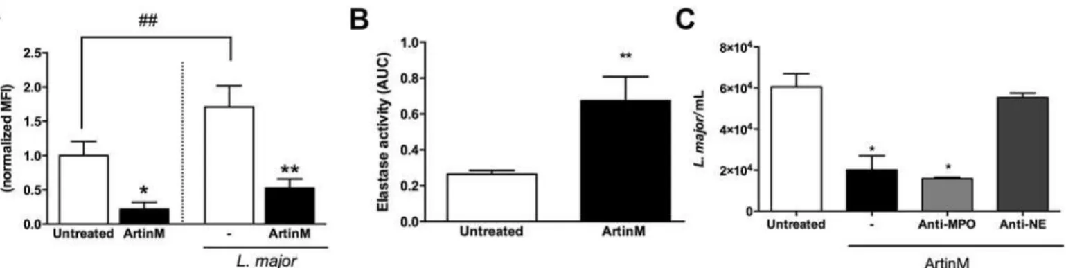
intracellular content of MPO, using flow cytometry analysis, and (2) by augmented levels of NE in the cell supernatant, assessed by enzymatic activity quantification.
The intracellular MPO content in ArtinM-treated neutrophils was 4-fold lower than that verified in untreated cells (Fig 3A). Consistent with this, 2-fold higher levels of NE activity were detected in the supernatant of ArtinM-treated neutrophils in comparison to that observed in the supernatant of untreated cells (Fig 3B). This last assay was actually performed to verify the period necessary for occurrence of neutrophil degranulation in response to ArtinM, and the highest NE activity was detected shortly (5–60 min) following treatment, even when the concentrations used were as low as 312 ng/mL of lectin, as demonstrated by a time-course assay (S2 Fig).
We also examined the neutrophil degranulation afterL.majorinfection. Regarding MPO intracellular content, we verified that the infectionper sewas able to inhibit the degranulation
Fig 2. ArtinM stimulates the production of inflammatory cytokines byL.major-infected or uninfected neutrophils.Human neutrophils were treated with or without ArtinM or PMA, and either infected or not infected withL.majorpromastigotes (MOI 3:1). At 20 h post-infection, the supernatant of these cells was collected for cytokine measurement by ELISA.A–TNF, B–TGFβ-1 and C–IL-1βconcentrations are
expressed as mean±SD.*p<0.05;****p<0.001 in comparison to untreated cells, or as indicated. One way ANOVA followed by Bonferroni's post-test. ND stands for“not detected”. ELISA assays were carried out in triplicate. The shown data are representative from three different experiments.
of untreated cells (70%—Fig 3A). In contrast, the intracellular levels of MPO decreased drasti-cally in the ArtinM-treated neutrophils (3-fold), showing that the lectin promotes degranula-tion even inL.majorinfected neutrophils (Fig 3A). Therefore, ArtinM induces degranulation of both uninfected and infected neutrophils.
Elastase contributes to the augmented leishmanicidal activity induced
by ArtinM on neutrophils
To assess the leishmanicidal activity of the neutrophil secreted products, we incubated parasites with the supernatant of cells that were pre-stimulated with ArtinM. The number of viableL.
majorwas 66% lower following incubation with the supernatant of ArtinM-treated neutrophils (2.0x10⁴±0.6x10⁴) than with the supernatant of untreated cells (6.0x10⁴±6.4x10⁴). In order to evaluate the specific contribution of MPO and NE, released by azurophilic granules, to the observed leishmanicidal activity, the supernatant of ArtinM-treated neutrophils was pre-incu-bated with antibodies specific to MPO or NE, and then added to the parasite suspensions. Our results show that anti-MPO had no effect on the number of viableL.major
(1.5x10⁴±0.07x10⁴), whereas anti-NE antibodies inhibited the leishmanicidal activity provided by the supernatant of ArtinM-treated neutrophils once the number of parasites recover in this condition (5.5x10⁴±0.2x10⁴) was similar to that found on the supernatant coming from untreated neutrophils (Fig 3C). These data suggest that the leishmanicidal activity of ArtinM-treated neutrophils is due, at least partially, to an NE-mediated mechanism.
ArtinM inhibits the formation of Neutrophil Extracellular Traps induced by
Leishmania major
Besides killing microbes through its direct enzymatic activity [38–40], NE also contributes to the formation of neutrophil extracellular traps (NET) [41], which constitutes a mechanism for capture and kill microorganisms [42] and for host tissue damage [43]. To investigate whether neutrophil treatment with ArtinM could result in NET formation, the DNA concentration was measured in the supernatant of neutrophils, 2 and 4 h after ArtinM-treatment. The DNA levels
Fig 3. ArtinM stimulates the degranulation ofL.major-infected or uninfected neutrophils. A–Intracellular levels of myeloperoxidase.Human
neutrophils were incubated for 20 h with ArtinM or medium only (untreated), and infected or not infected withL.majorpromastigotes (MOI 3:1); cells were permeabilized and reacted with anti-MPOPE. Cells were analyzed by flow cytometry and data were expressed as normalized mean±SD of the median fluorescence intensity.*p<0.02,**p<0,01. Student'sttest.B–Elastase releasing.Human neutrophils were stimulated with ArtinM or medium only
(untreated); Cell supernatants were monitored for 30 min for enzymatic activity, using the substrate N-succinyl-Ala-Ala-Val-p-nitroanilide. The NE activity was expressed as the area under curve (AUC)±SD.**p<0.05 in comparison to untreated cells. Student'sttest.C–Elastase leishmanicidal activity.
Supernatants from neutrophils treated for 1 h or not treated with ArtinM were incubated for 30 min (or not incubated) with anti-MPO or anti-NE antibodies (both 1:500). Next, supernatants were added toL.majorculture (1x10⁵), in Schneider
’s medium. After 24 h, live parasites were quantified by reaction with Alamar Blue. Data are expressed as the number of viable parasites±SD.*p<0.05 in comparison to the supernatant from untreated cells. One way ANOVA, followed by Bonferroni's post-test. Each assay was carried out in duplicate. The shown data are representative from three different experiments.
detected in the supernatant of ArtinM-treated cells were as low as those found in untreated neutrophils. In contrast, neutrophil stimulation with PMA (positive control) resulted in the detection of high DNA levels (>5- fold,Fig 4A), at both periods analyzed. When we assayedL.
major-infected neutrophils, we observed that untreated cells formed NET, in concordance with previous reports [44], but, in contrast, ArtinM-treated cells do not form NET (Fig 4A). The microscopic observation of the assayed cells was consistent with the DNA measurement in the neutrophil supernatant (Fig 4B). Therefore, we concluded that the ArtinM treatment does not induce NET formation and inhibits the formation of NET that followsL.majorinfection.
ArtinM induces ROS production only by infected neutrophils
Once we had verified that ArtinM triggers intense neutrophil activation not associated with NET formation, we evaluated whether lectin induces the production of ROS, which is directly implicated in NET formation [45]. We monitored the levels of ROS following exposure to the ArtinM stimulus, through reactions with luminol or lucigenin, which are oxygenated by H2O2 (and its derived species), or by superoxide anion, respectively [46].
As shown inFig 5, ArtinM-treated neutrophils, as well as untreated control cells, did not produce ROS since low and stable levels were demonstrated by both luminol- and lucigenin-chemiluminescence detection. In contrast, neutrophils stimulated with PMA or fMLP (positive controls) triggered high ROS production (Fig 5A and 5B).
Although ArtinM did not induce ROS production, it did not inhibit the production induced by other agents, a fact that was demonstrated by the observation that following the incubation with ArtinM, neutrophils responded to PMA- or fMLP-stimulus, with ROS levels similar to the ones measured in cells that were not pre-incubated with ArtinM (Fig 5CandS3 Fig).
The luminol-monitored ROS detection revealed that a higher amount (26-fold augmented) was produced by infected (3.93x10⁷±1.0x10⁶AUCxCPS) compared to uninfected neutrophils (1.49x10⁶±0.2x10⁶AUCxCPS). When the infected cells were pre-treated with ArtinM, the ROS production increased by 25% (5.3x10⁷±1.1x10⁶AUCxCPS—Fig 5C). Taken together, in spite of not inducing uninfected cells to produce ROS, ArtinM enhances ROS production byL.
majorinfected neutrophils, providing a mechanism that rapidly eliminates the parasite.
ArtinM treatment prolongs neutrophil survival
The sustained neutrophil activation induced by ArtinM, which was detected even 20 h after treatment (S4 Fig), as well as the absence of NET formation and ROS production, motivated us to evaluate the survival rate of ArtinM-treated neutrophils. Several methods were used to assess the occurrence of cell death, such as the analysis of neutrophil morphological changes, frag-mentation of genomic DNA, phosphatidylserine (PS) exposure, and disruption of the mito-chondrial transmembrane potential.
The morphology of neutrophils was evaluated by optical microscopy. At 3h post-treatment, the microscopic features of ArtinM- or IL-8-treated cells, as well as of untreated cells, were typ-ical of live neutrophils. At the same time point, cells treated with lysis buffer, as expected, showed remarkable nuclear condensation, as usually observed in apoptotic neutrophils [5,6]. After 20 h, nuclear condensation was verified in untreated cells, while the ArtinM- or IL-8-treated ones preserved their original morphology (Fig 6A).
(45.89±3.9%) and IL-8 (38.8±1.4%) -treated cells, respectively. The analysis of neutrophils 20 h after treatment with lysis buffer was barred due to the scarce number of remaining cells (Fig 6B).
ROS production, mostly mitochondrial, is implicated in the initiation of cell apoptosis [47]. Since disruption of the mitochondrial trans-membrane potential (ΔC) is one of the earliest intracellular events occurring in apoptotic cells, we examined mitochondrial instability through the detection of JC-1 monomer.Fig 6Cshows that the treatment of neutrophils with ArtinM (5091±276.5 MFI) or IL-8 (4446±800.8 MFI) favored mitochondrial stability, since at 3 h post-treatment, the levels of JC1 monomer were at least half of those detected in untreated (10020 ±105.4 MFI) or lysis buffer-treated neutrophils (12186±1037 MFI). At 20 h, although JC1 monomer levels increased in ArtinM (9331±211.2 MFI) or IL-8 (8066±635 MFI) treated neu-trophils, they remained significantly lower (-15%) than those detected in untreated neutrophils (11035±891 MFI). JC1 monomers were not detected in the rare neutrophils remaining 20 h after treatment with lysis buffer.
The electrophoresis analysis of DNA showed that ArtinM-treated neutrophils displayed at least two-fold less fragmented DNA than untreated cells at 24 h of incubation. At 48 h of incuba-tion, treated and untreated neutrophils displayed similar DNA fragmentation. (Fig 6D and 6E).
To summarize, these data demonstrate that ArtinM treatment of human neutrophils post-poned apoptosis, as shown by delayed disruption of the mitochondrial trans-membrane poten-tial, DNA fragmentation, nuclear condensation and PS exposure. Altogether, these results revealed that ArtinM treatment prolongs neutrophil survival.
ArtinM triggers early apoptosis of
L
.
major
infected neutrophils
We found that ArtinM prolongs neutrophil survival, a fact that is considered to facilitateL.
majorinfection [48]. Then, we analyzed the PS exposure on ArtinM-treated neutrophils that were infected withL.major. The ArtinM effect, exerted 3 hours after stimulation on the
Fig 4. ArtinM inhibits NET formation induced byL.major.Human neutrophils were incubated with ArtinM, PMA, ArtinM+PMA, or medium (untreated), and were either infected or not infected withL.majorpromastigotes (3:1).A–DNA quantitation.DNA released at 2 and 4 h after treatment/infection was
quantified in the cell supernatants by fluorescence detection (Ex./Em. 480/520 nm) after reaction with SYTOX green. Data are expressed as a mean of fluorescence intensity±SD.**p<0.01;***p<0.001;****p<0.0001. Two way ANOVA followed by Bonferroni's post-test. DNA quantitation assays were
performed in triplicate and, the data shown are representative of four independent experiments.B–NET immunofluorescence detection.Neutrophils were
plated onto poly-l-lysine coated slides, treated or untreated with PMA or ArtinM, and either infected or not infected withL.majorpromastigotes (MOI 3:1). After 6 h, the cells were stained with DAPI (DNA blue stain) and with anti-NE antibody (green). Merged images confirmed NETs by colocalization of staining. NET immunofluorescence detection assays were performed in duplicate and the data shown are representative of three independent experiments.
doi:10.1371/journal.pntd.0004609.g004
Fig 5. ArtinM augments ROS production byL.major-infected neutrophils.Human neutrophils were treated with PMA, fMLP, ArtinM or medium (untreated). ROS production was quantified by reaction with luminol (A) and lucigenin (B), producing chemiluminescent photons (CPS). Data on PMA stimulation orL.majorpromastigotes infected neutrophils (3:1), which were treated or not treated with ArtinM, are shown in panelC. The amounts of ROS released are expressed as mean AUCxCPS±SD.**p<0.01;***p<0.001;****p<0.0001. One way ANOVA followed by Bonferroni's post-test. Each assay was carried out in triplicate. The data shown are representative of three independent experiments.
Fig 6. ArtinM treatment postpones apoptosis of uninfected neutrophils.Human neutrophils were incubated (indicated period) with medium (untreated), lysis buffer, ArtinM or IL-8.A–Neutrophil morphology.Neutrophils were cytocentrifuged and stained for evaluation by light microscopy. Arrows indicate
neutrophils with nuclear condensation.B–Phosphatidylserine exposure.Neutrophils were labeled with Annexin V-FITCand analyzed by flow cytometry.
Data are expressed as mean of percentage of AnnexinV+neutrophils±SD.***p<0.001 in comparison with the untreated cells, two way ANOVA followed by
Bonferroni's post-test. ND stands for“not detected”.C–JC-1 monomer detection.Neutrophils were incubated with JC-1 probe and green fluorescence detection (Ex/Em = 485/528) was performed at 3 and 20 h after treatment. Data are expressed as mean of fluorescence intensity±SD.***p<0.01 comparing with untreated curve, two way ANOVA followed by Bonferroni's post-test.D and E—Electrophoretic detection of DNA fragmentation.
Neutrophils were incubated for 24 and 48 h with ArtinM or medium (untreated). Their genomic DNA was analyzed by gel electrophoresis. Images are shown on panelD.PanelErepresents the band area in pixels2, regarding the electrophoretic detection of DNA fragmentation (ImageJ Software). Each assay was carried out in triplicate. The shown data are representative from three different experiments.
neutrophils PS exposure, at least doubled when the cells were infected withL.major, compared to uninfected cells (34.8±2.07%vs11.97±0.74%,Fig 7). In addition, these infected/ArtinM-treated neutrophils exhibited 40% higher PS exposure than the uninfected/ArtinM-treated/infected neutrophils (34.8±2.07%vs21.41±3.92%,Fig 7). Uninfected cells, treated or not with ArtinM, were used as control, at the same time point. The obtained results (11.97±0.74% and 11.55±0.61%) were close to those shown inFig 6B(3h). Therefore, ArtinM stimulus delays the death of non-infected neutrophils (at 20 hours’time point,Fig 6B), and accelerates the death ofL.major -infected neutrophils (Fig 7), favoring the parasite elimination.
Some assays performed by infecting neutrophils with fluorescentL.majorforms allowed verifying that 91% of the AnnexinV labeled cells were infected. A close proportion (89%) was verified among ArtinM-treated cells as well (S5 Fig). We therefore concluded that apoptotic neutrophils were most frequentlyL.majorinfected cells, and that ArtinM did not change this distribution.
Discussion
The immunomodulatory lectin ArtinM induces Th1 immune response and confers resistance to intracellular pathogens [21], without causing apparent tissue damage. The response to ArtinM is induced by its interaction with glycoconjugates on several immune cells, namely, macrophages, dendritic cells, neutrophils, mast cells and lymphocytes [10,11,13,16,17,24– 26,49]. Considering that neutrophils constitute a two-edged sword, accounting for resistance to pathogens and also for tissue injury, a full exploration of the neutrophil responses is required to envisage a possible application of ArtinM, or its analogues, as immunomodulatory agent. In this study, we verified that ArtinM enhances the neutrophils ability of eliminating the intracel-lular pathogenL.major. The parasite killing was associated with strong neutrophil activation, not accompanied by NET formation, which is known to cause tissue damage.
Although macrophages are the definitive refuge forLeishmaniaspecies in the host, neutro-phils are considered by many as transitional shelters for the few invader parasites that survive
Fig 7. ArtinM induces early apoptosis ofL.majorinfected neutrophils.Human neutrophils were incubated with medium (untreated), lysis buffer, ArtinM, or IL-8, and infected or not withL.major
promastigotes (MOI 3:1). At 3 h post infection, cells were labeled with Annexin V-FITCand analyzed by flow cytometry. Data are expressed as mean of percentage of AnnexinV+neutrophils±SD.
**p<0.01;*** p<0.001 in comparison to untreated cells, or as indicated. Two way ANOVA followed by Bonferroni's
post-test. Each assay was carried out in triplicate. The shown data are representative from three different experiments.
the toxic extracellular milieu [50]. This idea is based on the observation that in the early stages ofL.majorinfection, caused by the bite of an infected sandfly, large amounts of neutrophils are attracted to the site. Since a low number of macrophages reside in the invaded tissue and the recruited neutrophils failed to killL.major, the authors concluded that the invading parasites depended on the rapidly recruited neutrophils to survive [51]. Once inside the neutrophils,L.
majorpromastigotes postpone neutrophil apoptosis until 2 days [48] and are silently trans-ferred to macrophages, without activating the immune response [51]. In opposition to this
“Trojan horse”mechanism of parasite evasion, ArtinM accelerates the death ofL.
major-infected neutrophils, favoring the process of parasite elimination.
The production of the anti-inflammatory cytokine TGF-βfavors the silent uptake of apo-ptotic cells by macrophages, whereas the production of pro-inflammatory mediators like TNF and IL-1βdecreases after the uptake of apoptotic cells [52]. Consequently, the prominence of TGF-βcorrelates with permissibility toL.majorinfection, whereas high production of TNF is associated with resistance to infection [35]. Our observation thatL.major-infected neutrophils largely augmented TGF-βproduction and diminished the secretion of pro-inflammatory medi-ators is consistent with previous demonstrations that neutrophils are important contributors toward providing the required microenvironment for parasite survival [34,53,54]. Notably, the treatment of neutrophils with ArtinM inverted this pattern by significantly diminishing TGF-β production and augmenting the secretion of pro-inflammatory cytokines. We found that ArtinM promoted IL-1βsecretion by human neutrophils, which was maximum when the lectin was added toL.majorinfected neutrophils. This is a relevant finding because IL-1βmaturation, as recently demonstrated, results from activation of the NLRP3 inflammasome, which is an innate platform that crucially restricts parasite replication. The NLRP3 inflammasome triggers inducible nitric oxide synthase (NSO2)-mediated production of NO, a potent leishmanicidal factor [55]. In addition, NLRP3 inflammasome activation is associated with ROS production [56], which is a major factor forLeishmaniakilling by human cells [57]. Indeed, macrophages derived from human monocytes do not produce NO after classical activation [58] or upon infection withLeishmania[59]. Thus, neutrophils, monocytes, and macrophages can control the parasites via ROS that are produced during the respiratory burst process [60]. By promot-ing ROS production and activatpromot-ing the NLRP3 inflammasome, ArtinM facilitatesLeishmania
killing, as shown in this work. Considering that the importance of IL-1β, derived from inflam-masome, for conferring resistance to Leishmania infection was demonstrated for other species thanL.major[55], we plan performing further experiments withL.braziliensis-infected neu-trophils, to better assess the IL-1βrelevance for the ArtinM-induced protection against the parasite.
elimination ofL.major[67]. In the current work, we did not investigate the signaling through TLR4, nor the relationship with macrophages, but we demonstrated that the ArtinM-induced NE release was followed by elimination ofL.majorby human neutrophils (Figs1and2). The released NE accounts for the ArtinM leishmanicidal effect, once the NE presence in the extra-cellular space correlates withL.majorelimination, as indicated by experiments using anti-NE neutralizing antibodies. Although NE is a serine protease that promotes microbe killing, our study apparently constitutes the first demonstration of its leishmanicidal activity, a finding that surely deserves further investigation.
ArtinM-treated neutrophils, in sterile conditions, did not produce ROS, while other neutro-phil activators such as fMLP and PMA induced rapid and intense responses. TheL.major
infection of neutrophils increased ROS production, which was even higher in ArtinM-treated neutrophils. It is well established that ROS provides an important mechanism to combatL.
major[68], but is also able to cause tissue injury. The ROS production induced by ArtinM was restricted to infected cells whose life span was shortened by ArtinM treatment, allowing us to
Fig 8. ArtinM increases the leishmanicidal capacity of human neutrophils: a model.Once infected withL.major, non-stimulated neutrophils produce high levels of TGF-β, and low levels of TNF, and IL-1β, which are associated with decreased cell degranulation and postponed cell death. In contrast, ArtinM-treated neutrophils become activated, produce high levels of TNF, and IL-1β, while TGF-b is reduced, and enhance degranulation. Collectively, these ArtinM induced responses augments the leishmanicidal capacity of neutrophils.
postulate that the ability of ROS to cause tissue damage could be reduced. The effect of ROS on parasite elimination could be preserved, since it is effective in the early stages of infection [69].
ROS production accounts for NET formation, as demonstrated by the observation that neu-trophils treated with NADPH oxidase, a pharmacological inhibitor of ROS production, do not form NET in response to conventional stimuli [70]. We showed that ArtinM-stimulated neu-trophils, either uninfected or infected withL.major, did not form NET. This means that ArtinM inhibits the NET formation induced by the infection itself [44] and additionaly by PMA The inhibition occurred in spite of NE release and ROS production, which favor NET formation and were verified to occur in ArtinM-treated neutrophils. NET formation consti-tutes a mechanism whereby neutrophils eliminate pathogens, includingL.amazonensis pro-mastigotes [71]. OtherLeishmaniaspp, namely,L.donovani,L.major,L.infantum, andL.
mexicana, escape from their toxicity despite NET formation and trapping by these web-like structures [44,72,73]. The fact that ArtinM, although inhibiting NET formation, facilitatesL.
majorclearance by human neutrophils reinforces the idea that NET formation is not required forL.majorelimination. On the other hand, the absence of NETs can favor host tissue integ-rity, since NET-associated proteases and granular proteins have been shown to damage host tissue [74].
We conclude that ArtinM treatment of human neutrophils enhances the clearance of the intracellular pathogenL.major, through mechanisms that include: (1) production of inflam-matory cytokines, i.e., high TNF and IL-1β, and virtual absence of TGF-β; (2) increased neutro-phil degranulation; the released elastase promotesL.majorelimination; (3) increased ROS production by infected neutrophils (4) shortened neutrophil survival. On the other hand, the host tissue integrity is favored by (1) the short period of ROS production and (2) the absence of NET formation.Fig 8delineates our model of ArtinM effects on neutrophils duringL.major
infection, based on our results and assumptions.
Supporting Information
S1 Fig. Neutrophil purity analysis.Layered polymorphonuclear cells were analyzed for neu-trophil purity.A—Cells were labeled with anti-CD16bPEor isotype controlPEantibodies and
analyzed by flow cytometry.B–Cells were cytocentrifuged and stained for morphology
ana-lyzes on light microscopy. (TIF)
S2 Fig. Time-course assay for detection of NE activity in the cell supernatants.Human neu-trophils were treated with ArtinM (312,5–2.500μg/mL), fMLP or medium (untreated). Cell
supernatants were monitored for 30 min for enzymatic activity by using the substrate N-succi-nyl-Ala-Ala-Val-p-nitroanilide.
(TIF)
S3 Fig. ArtinM does not block ROS production.Human neutrophils were pre-treated with ArtinM for 30 min and then treated with PMA or fMLP. ROS production was quantified by reaction with Luminol producing chemiluminescent photons (CPS). The kinetics of ROS pro-duction is shown. No significant differences were detected between pre-treated or not treated with ArtinM.
(TIF)
S4 Fig. ArtinM promotes neutrophil polarization.Human neutrophils were 3 and 20 h incu-bated with ArtinM, IL8, or medium (untreated).A—Images from the wells of the plates were
obtained by inverted light microscopy coupled with an image capturing system.B–The
mean of percentage of polarized cells analyzed in 3 different fields ± SD.p<0.05:p<0,01: p<0.001 in comparison to untreated cells at the respective time. Two way ANOVA, fol-lowed by Bonferroni's post-test.
(TIF)
S5 Fig. AnnexinVPhycoerythrinlabelling of a human neutrophils culture incubated with green-fluorescent forms ofL.majorpromastigotes (MOI 3:1).Flow cytometry analysis showed that about 90% of cells were double labelled, regardless the ArtinM treatment. (TIF)
Acknowledgments
We are grateful to Dra. Maria Aparecida de Souza and MSc Thiago Aparecido da Silva for the discussions regarding this study. Micássio Andrade contributed to the ROS measurement experiments. The technical assistance of Sandra Maria de Oliveira Thomaz and Patrícia Edivâ-nia Vendrusculo was essential for this study, which respectively, afford the lectin purification and the parasite maintenance. Erica Vendrusculo was responsible for the paperwork.
Author Contributions
Conceived and designed the experiments: RRA AFO FCC MCRB. Performed the experiments: RRA AFO MCAVC FCC. Analyzed the data: RRA AFO MCRB. Contributed reagents/materi-als/analysis tools: MCRB. Wrote the paper: RRA AFO MCRB.
References
1. Andre FE, Booy R, Bock HL, Clemens J, Datta SK, et al. (2008) Vaccination greatly reduces disease, disability, death and inequity worldwide. Bull World Health Organ 86: 140–146. doi:10.2471/BLT.07. 040089PMID:18297169
2. Levine MM, Robins-Browne R (2009) Vaccines, global health and social equity. Immunol Cell Biol 87: 274–278. doi:10.1038/icb.2009.15PMID:19308074
3. Robbins JB, Schneerson R, Szu SC (1995) Perspective: hypothesis: serum IgG antibody is sufficient to confer protection against infectious diseases by inactivating the inoculum. J Infect Dis 171: 1387–
1398. PMID:7769272
4. Griffiths KL, Khader SA (2014) Novel vaccine approaches for protection against intracellular patho-gens. Curr Opin Immunol 28: 58–63. doi:10.1016/j.coi.2014.02.003PMID:24608070
5. Mifsud EJ, Tan ACL, Jackson DC (2014) TLR agonists as modulators of the innate immune response and their potential as agents against infectious disease. Front Immunol 5. doi:10.3389/fimmu.2014. 00079
6. Kumar H, Kawai T, Akira S (2011) Pathogen recognition by the innate immune system. Int Rev Immunol 30: 16–34. doi:10.3109/08830185.2010.529976PMID:21235323
7. Pandey S, Kawai T, Akira S (2015) Microbial Sensing by Toll-Like Receptors and Intracellular Nucleic Acid Sensors. Cold Spring Harb Perspect Biol 7. Available:http://cshperspectives.cshlp.org/content/7/ 1/a016246.abstract.
8. Junquera EC, Mateos-Hernández L, Fuente J de la, Lastra JMP de la (2014) Recent Advances in the Development of Anti-Infective Prophylactic and/or Therapeutic Agents Based on Toll-Like Receptor (TLRs). Recent Pat Antiinfect Drug Discov 9: 14–24. Available:http://www.eurekaselect.com/122772/ article. PMID:25164058
9. Mariano VS, Zorzetto-Fernandes AL, da Silva TA, Ruas LP, Nohara LL, et al. (2014) Recognition of TLR2 N-glycans: critical role in ArtinM immunomodulatory activity. PLoS One 9: e98512. Available: http://dx.plos.org/10.1371/journal.pone.0098512. Accessed 2 August 2014. doi:10.1371/journal.pone. 0098512PMID:24892697
11. Panunto-Castelo A, Souza MA, Roque-Barreira M-C, Silva JS (2001) KM+, a lectin from Artocarpus integrifolia, induces IL-12 p40 production by macrophages and switches from type 2 to type 1 cell-medi-ated immunity against Leishmania major antigens, resulting in BALB/c mice resistance to infection. Gly-cobiol 11: 1035–1042. Available:http://glycob.oxfordjournals.org/content/11/12/1035.abstract.
12. Teixeira CR, Cavassani KA, Gomes RB, Teixeira MJ, Roque-Barreira MC, et al. (2006) Potential of KM + lectin in immunization against Leishmania amazonensis infection. Vaccine 24: 3001–3008. doi:10. 1016/j.vaccine.2005.11.067PMID:16455170
13. Coltri KC, Oliveira LL, Ruas LP, Vendruscolo PE, Goldman MH, et al. (2010) Protection against Para-coccidioides brasiliensis infection conferred by the prophylactic administration of native and recombi-nant ArtinM. Med Mycol 48: 792–799. doi:10.3109/13693780903501671PMID:20392144
14. Cardoso MRD, Mota CM, Ribeiro DP, Santiago FM, Carvalho J V., et al. (2011) ArtinM, a d-mannose-binding lectin from Artocarpus integrifolia, plays a potent adjuvant and immunostimulatory role in immu-nization against Neospora caninum. Vaccine 29: 9183–9193. doi:10.1016/j.vaccine.2011.09.136 PMID:22001880
15. Loyola W, Custodio LA, Felipe I, Conchon-Costa I, Carvalho PG De, et al. (2012) Artin M enhances TNF-αproduction and phagocytosis of Candida albicans mediated by dectin-1 and mannose receptors. Int Immunopharmacol 12: 378–383. doi:10.1016/j.intimp.2011.12.010PMID:22207010
16. da Silva T, de Souza M, Cecílio N, Roque-Barreira M (2014) Activation of spleen cells by ArtinM may account for its immunomodulatory properties. Cell Tissue Res: 1–12. Available:http://dx.doi.org/10. 1007/s00441-014-1879-8.
17. Moreno AN, Jamur MC, Oliver C, Roque-Barreira MC (2003) Mast cell degranulation induced by lectins: Effect on neutrophil recruitment. Int Arch Allergy Immunol 132: 221–230. doi:10.1159/000074303 PMID:14646383
18. Barbosa-Lorenzi VC, Buranello PA de A, Roque-Barreira MC, Jamur MC, Oliver C, et al. (2011) The lectin ArtinM binds to mast cells inducing cell activation and mediator release. Biochem Biophys Res Commun 416: 318–324. doi:10.1016/j.bbrc.2011.11.033PMID:22108054
19. Pereira-Da-Silva G, Moreno AN, Marques F, Oliver C, C??lia Jamur M, et al. (2006) Neutrophil activa-tion induced by the lectin KM+ involves binding to CXCR2. Biochim Biophys Acta—Gen Subj 1760: 86–94. doi:10.1016/j.bbagen.2005.09.011
20. Toledo KA, Scwartz C, Oliveira AF, Conrado MCA V, Bernardes ES, et al. (2009) Neutrophil activation induced by ArtinM: Release of inflammatory mediators and enhancement of effector functions. Immunol Lett 123: 14–20. doi:10.1016/j.imlet.2009.01.009PMID:19428547
21. Souza MA, Carvalho FC, Ruas LP, Ricci-Azevedo R, Roque-Barreira MC (2013) The immunomodula-tory effect of plant lectins: A review with emphasis on ArtinM properties. Glycoconj J 30: 641–657. doi: 10.1007/s10719-012-9464-4PMID:23299509
22. Borregaard N (2010) Neutrophils, from Marrow to Microbes. Immunity 33: 657–670. doi:10.1016/j. immuni.2010.11.011PMID:21094463
23. Witko-Sarsat V, Pederzoli-Ribeil M, Hirsh E, Sozzani S, Cassatella MA (2011) Regulating neutrophil apoptosis: New players enter the game. Trends Immunol 32: 117–124. doi:10.1016/j.it.2011.01.001 PMID:21317039
24. Santos-de-Oliveira R, Dias-Baruffi M, Thomaz SM, Beltramini LM, Roque-Barreira MC (1994) A neutro-phil migration-inducing lectin from Artocarpus integrifolia. J Immunol 153: 1798–1807. PMID:8046246
25. Ganiko L, Martins AR, Espreáfico EM, Roque-Barreira MC (1998) Neutrophil haptotaxis induced by the lectin KM+. Glycoconjugate Journal. Vol. 15. pp. 527–530. doi:10.1023/A:1006947306260PMID: 9881756
26. Ganiko L, Martins AR, Freymüller E, Mortara RA, Roque-Barreira MC (2005) Lectin KM +-induced neu-trophil haptotaxis involves binding to laminin. Biochim Biophys Acta—Gen Subj 1721: 152–163. doi: 10.1016/j.bbagen.2004.10.012
27. Kruger P, Saffarzadeh M, Weber ANR, Rieber N, Radsak M, et al. (2015) Neutrophils: Between Host Defence, Immune Modulation, and Tissue Injury. PLOS Pathog 11: e1004651. Available:http://dx.doi. org/10.1371/journal.ppat.1004651. Accessed 14 March 2015. doi:10.1371/journal.ppat.1004651 PMID:25764063
28. Tavares NM, Araújo-Santos T, Afonso L, Nogueira PM, Lopes UG, et al. (2014) Understanding the Mechanisms Controlling Leishmania amazonensis Infection In Vitro: The Role of LTB4 Derived From Human Neutrophils. J Infect Dis: 1–11. Available:http://www.ncbi.nlm.nih.gov/pubmed/24634497.
30. Lucisano-Valim YM, Kabeya LM, Kanashiro A, Russo-Carbolante EM, Polizello AC, et al. (2002) A sim-ple method to study the activity of natural compounds on the chemiluminescence of neutrophils upon stimulation by immune complexes. J Pharmacol Toxicol Methods 47: 53–58. Available:http://www. ncbi.nlm.nih.gov/pubmed/12387939. PMID:12387939
31. Johansson S, G??ransson U, Luijendijk T, Backlund A, Claeson P, et al. (2002) A neutrophil multitarget functional bioassay to detect anti-inflammatory natural products. J Nat Prod 65: 32–41. doi:10.1021/ np010323oPMID:11809061
32. Palmer LJ, Cooper PR, Ling MR, Wright HJ, Huissoon A, et al. (2012) Hypochlorous acid regulates neutrophil extracellular trap release in humans. Clin Exp Immunol 167: 261–268. doi: 10.1111/j.1365-2249.2011.04518.xPMID:22236002
33. Rieger AM, Nelson KL, Konowalchuk JD, Barreda DR (2011) Modified annexin V/propidium iodide apo-ptosis assay for accurate assessment of cell death. J Vis Exp. doi:10.3791/2597
34. Launois P, Swihart KG, Milon G, Louis JA (1997) Early production of IL-4 in susceptible mice infected with Leishmania major rapidly induces IL-12 unresponsiveness. J Immunol 158: 3317–3324. PMID: 9120289
35. van Zandbergen G, Bollinger A, Wenzel A, Kamhawi S, Voll R, et al. (2006) Leishmania disease devel-opment depends on the presence of apoptotic promastigotes in the virulent inoculum. Proc Natl Acad Sci U S A 103: 13837–13842. doi:10.1073/pnas.0600843103PMID:16945916
36. Kumar V, Sharma A (2010) Neutrophils: Cinderella of innate immune system. Int Immunopharmacol 10: 1325–1334. doi:10.1016/j.intimp.2010.08.012PMID:20828640
37. Ribeiro-Gomes FL, Moniz-de-Souza MCA, Alexandre-Moreira MS, Dias WB, Lopes MF, et al. (2007) Neutrophils activate macrophages for intracellular killing of Leishmania major through recruitment of TLR4 by neutrophil elastase. J Immunol 179: 3988–3994. doi:10.4049/jimmunol.179.6.3988PMID: 17785837
38. Odeberg H, Olsson I (1976) Microbicidal mechanisms of human granulocytes: synergistic effects of granulocyte elastase and myeloperoxidase or chymotrypsin-like cationic protein. Infect Immun 14: 1276–1283. PMID:12111
39. Belaaouaj a. a. (2000) Degradation of Outer Membrane Protein A in Escherichia coli Killing by Neutro-phil Elastase. Science (80-) 289: 1185–1187. Available:http://www.sciencemag.org/cgi/doi/10.1126/ science.289.5482.1185. Accessed 4 August 2014. PMID:10947984
40. Jena P, Mohanty S, Mohanty T, Kallert S, Morgelin M, et al. (2012) Azurophil Granule Proteins Consti-tute the Major Mycobactericidal Proteins in Human Neutrophils and Enhance the Killing of Mycobacte-ria in Macrophages. PLoS One 7: e50345. Available:http://www.pubmedcentral.nih.gov/articlerender. fcgi?artid=3522671&tool=pmcentrez&rendertype=abstract. Accessed 17 July 2014. doi:10.1371/ journal.pone.0050345PMID:23251364
41. Papayannopoulos V, Metzler KD, Hakkim A, Zychlinsky A (2010) Neutrophil elastase and myeloperoxi-dase regulate the formation of neutrophil extracellular traps. J Cell Biol 191: 677–691. Available:http:// www.pubmedcentral.nih.gov/articlerender.fcgi?artid=3003309&tool=pmcentrez&rendertype=abstract. Accessed 29 July 2014. doi:10.1083/jcb.201006052PMID:20974816
42. Brinkmann V, Zychlinsky A (2007) Beneficial suicide: why neutrophils die to make NETs. Nat Rev Microbiol 5: 577–582. doi:10.1038/nrmicro1710PMID:17632569
43. Liu F-C, Chuang Y-H, Tsai Y-F, Yu H-P (2014) Role of neutrophil extracellular traps following injury. Shock 41: 491–498. Available:http://www.ncbi.nlm.nih.gov/pubmed/24837201. doi:10.1097/SHK. 0000000000000146PMID:24837201
44. Gabriel C, McMaster WR, Girard D, Descoteaux A (2010) Leishmania donovani Promastigotes Evade the Antimicrobial Activity of Neutrophil Extracellular Traps. J Immunol 185: 4319–4327. Available: http://www.jimmunol.org/content/185/7/4319.abstract. doi:10.4049/jimmunol.1000893PMID: 20826753
45. Kirchner T, Mller S, Klinger M, Solbach W, Laskay T, et al. (2012) The impact of various reactive oxygen species on the formation of neutrophil extracellular traps. Mediators Inflamm 2012. doi:10.1155/2012/ 849136
46. Alves CMOS, Marzocchi-Machado CM, Carvalho IF, Lucisano Valim YM (2003) Application of the chemiluminescence systems to evaluate the role of Fcgamma and complement receptors in stimulating the oxidative burst in neutrophils. Talanta 60: 601–608. Available:http://www.sciencedirect.com/ science/article/pii/S0039914003001887. Accessed 29 June 2015. doi:10.1016/S0039-9140(03) 00188-7PMID:18969082
48. Aga E, Katschinski DM, van Zandbergen G, Laufs H, Hansen B, et al. (2002) Inhibition of the spontane-ous apoptosis of neutrophil granulocytes by the intracellular parasite Leishmania major. J Immunol 169: 898–905. PMID:12097394
49. Buranello PA de A, Moulin MRI, Souza DA, Jamur MC, Roque-Barreira MC, et al. (2010) The lectin ArtinM induces recruitment of rat mast cells from the bone marrow to the peritoneal cavity. PLoS One 5. doi:10.1371/journal.pone.0009776
50. Charmoy M, Auderset F, Allenbach C, Tacchini-Cottier F (2010) The prominent role of neutrophils dur-ing the initial phase of infection by leishmania parasites. J Biomed Biotechnol 2010. doi:10.1155/2010/ 719361
51. van Zandbergen G, Klinger M, Mueller A, Dannenberg S, Gebert A, et al. (2004) Cutting edge: neutro-phil granulocyte serves as a vector for Leishmania entry into macrophages. J Immunol 173: 6521–
6525. doi:173/11/6521 [pii]. PMID:15557140
52. Fadok VA, Bratton DL, Konowal A, Freed PW, Westcott JY, et al. (1998) Macrophages that have ingested apoptotic cells in vitro inhibit proinflammatory cytokine production through autocrine/paracrine mechanisms involving TGF-??, PGE2, and PAF. J Clin Invest 101: 890–898. doi:10.1172/JCI1112 PMID:9466984
53. Safaiyan S, Bolhassani A, Nylen S, Akuffo H, Rafati S (2011) Contribution of human neutrophils in the development of protective immune response during in vitro Leishmania major infection. Parasite Immu-nol 33: 609–620. doi:10.1111/j.1365-3024.2011.01321.xPMID:21793857
54. Keyhani A, Riazi-Rad F, Pakzad SR, Ajdary S (2014) Human polymorphonuclear leukocytes produce cytokines in response to Leishmania major promastigotes. APMIS. doi:10.1111/apm.12252
55. Lima-Junior DS, Costa DL, Carregaro V, Cunha LD, Silva ALN, et al. (2013) Inflammasome-derived IL-1βproduction induces nitric oxide-mediated resistance to Leishmania. Nat Med 19: 909–915. Avail-able:http://www.ncbi.nlm.nih.gov/pubmed/23749230. doi:10.1038/nm.3221PMID:23749230
56. Sutterwala FS, Haasken S, Cassel SL (2014) Mechanism of NLRP3 inflammasome activation. Ann N Y Acad Sci 1319: 82–95. Available:http://www.pubmedcentral.nih.gov/articlerender.fcgi?artid= 4074217&tool=pmcentrez&rendertype=abstract. doi:10.1111/nyas.12458PMID:24840700
57. Novais FO, Nguyen BT, Beiting DP, Carvalho LP, Glennie ND, et al. (2014) Human classical mono-cytes control the intracellular stage of Leishmania braziliensis by reactive oxygen species. J Infect Dis 209: 1288–1296. Available:http://www.pubmedcentral.nih.gov/articlerender.fcgi?artid=3969552&tool= pmcentrez&rendertype=abstract. doi:10.1093/infdis/jiu013PMID:24403561
58. Schneemann M, Schoedon G, Hofer S, Blau N, Guerrero L, et al. (1993) Nitric oxide synthase is not a constituent of the antimicrobial armature of human mononuclear phagocytes. J Infect Dis 167: 1358–
1363. doi:10.1093/infdis/167.6.1358PMID:7684756
59. Gantt KR, Goldman TL, McCormick ML, Miller M a, Jeronimo SM, et al. (2001) Oxidative responses of human and murine macrophages during phagocytosis of Leishmania chagasi. J Immunol 167: 893–
901. doi:10.4049/jimmunol.167.2.893PMID:11441096
60. Brüne B, Dehne N, Grossmann N, Jung M, Namgaladze D, et al. (2013) Redox control of inflammation in macrophages. Antioxid Redox Signal 19: 595–637. Available:http://www.ncbi.nlm.nih.gov/pubmed/ 23311665. doi:10.1089/ars.2012.4785PMID:23311665
61. Klebanoff SJ (1967) Iodination of bacteria: a bactericidal mechanism. J Exp Med 126: 1063–1078. doi: 10.1084/jem.126.6.1063PMID:4964565
62. Sengeløv H, Kjeldsen L, Borregaard N (1993) Control of exocytosis in early neutrophil activation. J
Immunol 150: 1535–1543. PMID:8381838
63. Sengeløv H, Follin P, Kjeldsen L, Lollike K, Dahlgren C, et al. (1995) Mobilization of granules and secre-tory vesicles during in vivo exudation of human neutrophils. J Immunol 154: 4157–4165. PMID: 7535822
64. Borregaard N, Sørensen OE, Theilgaard-Mönch K (2007) Neutrophil granules: a library of innate immu-nity proteins. Trends Immunol 28: 340–345. doi:10.1016/j.it.2007.06.002PMID:17627888
65. Soehnlein O, Weber C, Lindbom L (2009) Neutrophil granule proteins tune monocytic cell function. Trends Immunol 30: 538–546. doi:10.1016/j.it.2009.06.006PMID:19699683
66. Soehnlein O (2009) Direct and alternative antimicrobial mechanisms of neutrophil-derived granule pro-teins. J Mol Med 87: 1157–1164. doi:10.1007/s00109-009-0508-6PMID:19641860
67. Faria MS, Reis FCG, Azevedo-Pereira RL, Morrison LS, Mottram JC, et al. (2011) Leishmania inhibitor of serine peptidase 2 prevents TLR4 activation by neutrophil elastase promoting parasite survival in murine macrophages. J Immunol 186: 411–422. doi:10.4049/jimmunol.1002175PMID:21098233
69. Horta MF, Mendes BP, Roma EH, Noronha FSM, MacDo JP, et al. (2012) Reactive oxygen species and nitric oxide in cutaneous leishmaniasis. J Parasitol Res 2012. doi:10.1155/2012/203818
70. Fuchs T a, Abed U, Goosmann C, Hurwitz R, Schulze I, et al. (2007) Novel cell death program leads to neutrophil extracellular traps. J Cell Biol 176: 231–241. Available:http://www.pubmedcentral.nih.gov/ articlerender.fcgi?artid=2063942&tool=pmcentrez&rendertype=abstract. Accessed 9 July 2014. PMID: 17210947
71. Guimarães-Costa AB, Nascimento MTC, Froment GS, Soares RPP, Morgado FN, et al. (2009)
Leish-mania amazonensis promastigotes induce and are killed by neutrophil extracellular traps. Proc Natl Acad Sci U S A 106: 6748–6753. doi:10.1073/pnas.0900226106PMID:19346483
72. Guimarães-Costa AB, DeSouza-Vieira TS, Paletta-Silva R, Freitas-Mesquita AL, Meyer-Fernandes
JR, et al. (2014) 3’-nucleotidase/nuclease activity allows Leishmania parasites to escape killing by neu-trophil extracellular traps. Infect Immun 82: 1732–1740. doi:10.1128/IAI.01232-13PMID:24516114
73. Hurrell BP, Schuster S, Grün E, Coutaz M, Williams RA, et al. (2015) Rapid Sequestration of Leish-mania mexicana by Neutrophils Contributes to the Development of Chronic Lesion. PLoS Pathog 11: e1004929. Available:http://dx.doi.org/10.1371/journal.ppat.1004929. Accessed 1 June 2015. doi:10. 1371/journal.ppat.1004929PMID:26020515