Bats from the Czech Republic and Slovakia
Nata´lia Martı´nkova´1,2*, Peter Bacˇkor3, Toma´sˇ Bartonicˇka4, Pavla Blazˇkova´5,6, Jaroslav Cˇerveny´7, Luka´sˇ Falteisek8, Jirˇı´ Gaisler4, Vladimı´r Hanzal9, Daniel Hora´cˇek10, Zdeneˇk Huba´lek1, Helena Jahelkova´8, Miroslav Kolarˇı´k11, L’ubosˇ Koryta´r12, Alena Kuba´tova´11, Blanka Lehotska´13, Roman Lehotsky´14, Radek K. Lucˇan8, Ondrˇej Ma´jek2, Jan Mateˇju˚8,15, Zdeneˇk Rˇeha´k4, Jirˇı´ Sˇafa´rˇ16, Prˇemysl Ta´jek5, Emil Tkadlec1,17, Marcel Uhrin18, Josef Wagner19, Dita Weinfurtova´8, Jan Zima1, Jan Zukal1,4, Ivan Hora´cˇek8
1Institute of Vertebrate Biology, Academy of Sciences of the Czech Republic, Brno, Czech Republic,2Institute of Biostatistics and Analyses, Masaryk University, Brno, Czech Republic,3Department of Biology and Ecology, Matej Bel University, Banska´ Bystrica, Slovakia,4Department of Botany and Zoology, Masaryk University, Brno, Czech Republic,5Agency for Nature Conservation and Landscape Protection of the Czech Republic, Maria´nske´ La´zneˇ, Czech Republic,6Department of Zoology, University of South Bohemia, Cˇeske´ Budeˇjovice, Czech Republic,7Department of Forest Protection and Game Management, Czech University of Life Sciences Prague, Praha, Czech Republic,8Department of Zoology, Faculty of Science, Charles University in Prague, Praha, Czech Republic,9Agency for Nature Conservation and Landscape Protection of the Czech Republic, Praha, Czech Republic,10Sametova´ 721/18, Liberec, Czech Republic,11Department of Botany, Faculty of Science, Charles University in Prague, Praha, Czech Republic,12University of Veterinary Medicine and Pharmacy, Kosˇice, Slovakia,13Department of Landscape Ecology, Comenius University, Bratislava, Slovakia,14Miniopterus - Principal Organization of the Slovak Union for Nature and Landscape Conservators, Bratislava, Slovakia,15Agency for Nature Conservation and Landscape Protection of the Czech Republic, Karlovy Vary, Czech Republic,16Agency of Nature Conservation and Landsape Protection of the Czech Republic, Olomouc, Czech Republic,17Department of Ecology and Environmental Science, Palacky University, Olomouc, Czech Republic,18Institute of Biology and Ecology, P. J. Sˇafa´rik University, Kosˇice, Slovakia,19ZO CˇSS 7-01 ORCUS Bohumı´n, Czech Speleological Society, Bohumı´n, Czech Republic
Abstract
Background:White-nose syndrome is a disease of hibernating insectivorous bats associated with the fungus Geomyces destructans. It first appeared in North America in 2006, where over a million bats died since then. In Europe,G. destructans was first identified in France in 2009. Its distribution, infection dynamics, and effects on hibernating bats in Europe are largely unknown.
Methodology/Principal Findings:We screened hibernacula in the Czech Republic and Slovakia for the presence of the fungus during the winter seasons of 2008/2009 and 2009/2010. In winter 2009/2010, we found infected bats in 76 out of 98 surveyed sites, in which the majority had been previously negative. A photographic record of over 6000 hibernating bats, taken since 1994, revealed bats with fungal growths since 1995; however, the incidence of such bats increased inMyotis myotisfrom 2% in 2007 to 14% by 2010. Microscopic, cultivation and molecular genetic evaluations confirmed the identity of the recently sampled fungus asG. destructans, and demonstrated its continuous distribution in the studied area. At the end of the hibernation season we recorded pathologic changes in the skin of the affected bats, from which the fungus was isolated. We registered no mass mortality caused by the fungus, and the recorded population decline in the last two years of the most affected species,M. myotis, is within the population trend prediction interval.
Conclusions/Significance:G. destructanswas found to be widespread in the Czech Republic and Slovakia, with an epizootic incidence in bats during the most recent years. Further development of the situation urgently requires a detailed pan-European monitoring scheme.
Citation:Martı´nkova´ N, Bacˇkor P, Bartonicˇka T, Blazˇkova´ P, Cˇerveny´ J, et al. (2010) Increasing Incidence ofGeomyces destructansFungus in Bats from the Czech Republic and Slovakia. PLoS ONE 5(11): e13853. doi:10.1371/journal.pone.0013853
Editor:Dee A. Carter, University of Sydney, Australia
ReceivedJune 24, 2010;AcceptedOctober 17, 2010;PublishedNovember 5, 2010
Copyright:ß2010 Martı´nkova´ et al. This is an open-access article distributed under the terms of the Creative Commons Attribution License, which permits unrestricted use, distribution, and reproduction in any medium, provided the original author and source are credited.
Funding:This study was supported by grants MSM0021620828 and LC06073 provided by Ministry of Education, Youth and Sports of the Czech Republic. The funders had no role in study design, data collection and analysis, decision to publish, or preparation of the manuscript.
Competing Interests:The authors have declared that no competing interests exist.
* E-mail: martinkova@ivb.cz
Introduction
White-nose syndrome (WNS) is an emerging infectious disease, affecting hibernating insectivorous bats [1]. Since its first known appearance in 2006, WNS has spread with each year into the underground hibernacula in the USA and Canada, and over one million deaths within populations of several bat species have been attributed to the disease [1,2]. The decline is severe enough to
warrant a prediction that once common Myotis lucifugus might become locally extinct in less than two decades [2].
these animals [1,3]. The fungus invades the hair follicles and associated glands, or it breaks the epidermis of naked skin on the ears, muzzle, and wing membranes [1,4]. The specific etiology of the fungal infection is unknown, but the bats awaken from hibernation. As arousal from hibernation is energetically demand-ing, it is believed that WNS leads to a more rapid disappearance of fat reserves [1,4,5,6], deteriorating the body condition, and often to increased mortality due to starvation [7]. The bats prematurely emerge from the hibernacula and attempt to forage, which in winter conditions causes frostbite and subsequent necrosis of the wing membranes [8]. The fungal lesions co-infected with Gram-negative bacteria exhibit necrosis and pustules [5], furthering wing membrane damage and compromising flight abilities. Although not all of the details of the epizootics are fully understood [4,5,7,8,9,10], it is widely accepted that WNS poses a severe threat for the bat populations in North America [2,11]. The threat is likely to increase in the future, leading to local extinctions of the bats [2], and suggesting altering ecosystem dynamics [7,10].
The problem may expand onto one on a global scale, asG. destructanswas reported in France in March 2009 [12], and from Germany, Switzerland, and Hungary in the winter of 2008/2009 [13]. Here, we show that the occurrence ofG. destructansin Europe is not episodic, but it is locally widespread and could be associated with skin lesions. We believe that G. destructanshas been present historically within Europe, but that the epizootic is currently (re-)starting, with marked local differences in both the intensity and the dynamics of the disease.
Results
Historical Record of Geomycosis
The compilation of photographs of hibernating bats revealed a white patch on the muzzle of aM. myotisindividual on March 4, 1995 from the Zbojnı´cka Cave, Male´ Karpaty, Slovakia (Table 1). A photograph from January 25, 1997 from the Javorˇı´cˇske´ Caves in
northern Moravia, Czech Republic depicts aM. myotisindividual with a fungal growth typical of theG. destructansinfection (Fig. 1A). Further records show sporadic images of randomly photographed affected M. myotis until 2007/2008, when the incidence of bats with white patches started to increase in several species (Table 1).
Recent Presence of Geomycosis
Targeted on-site inspections of WNS-like clinical signs (white fungal growths on a bat, loss of sheen on wing membranes, emaciated forearms or the whole body if the hair was wet - worded as and regarded as ‘WNS-suspect’ throughout the remainder of this text) commenced in 2008/2009, as a part of the regular bat census. Bats exhibiting white fungal growths on their muzzle and/ or wings were found at 7 sites. In total, 6 bat species were affected;
M. myotis(24 individuals),M. blythii(1),M. brandtii(1),M. dasycneme
(1), M. emarginatus (1) and M. mystacinus (1). During regular monitoring through the most recent winter of 2009/2010 (January/February), WNS-suspect bats were found at 33 sites out of over 800 hibernacula in the Czech Republic (CZ) and Slovakia (SK), combined. Additionally, 98 sites were inspected again in late February and March, and the fungus was then sampled for cultivation, microscopic, and genetic analyses. During that time period, the incidence of WNS-suspect bats increased to 76 localities across CZ and SK (Fig. 2). Most often the WNS-suspect bats wereM. myotis(375 individuals), but also includedM. blythii(19),M. dasycneme(2),M. bechsteinii(1),M. mystacinus(1), and
M. nattereri(1). Specific regions differed in the prevalence of WNS-suspect bats. The highest levels of infestations were concentrated in submountain humid to mesic regions, where 11 to 100% ofM. myotis were WNS-suspect. Infestation was less frequent in the hibernacula within mountainous zones (Sˇumava Mts.: 5% WNS-suspectM. myotis; the majority of SK localities: 0–5%), and also in limestone regions (Bohemian: 3%, Moravian: 2%, and Slovak: 3% karsts).
Table 1.Summary of WNS-suspect bats from those photographed randomly (number of all photographed bats/number of photographed WNS-suspect bats) during the periods 1994–1998 and 2003–2010.
Winter period 1994/95 1995/96 1996/97 1997/98 2003/04 2004/05 2005/06 2006/07 2007/08 2008/09 2009/10
Species
R. hipposideros - 3/0 - 2/0 - 4/0 11/0 26/0 82/0 45/0 153/15
M. bechsteinii - - - - 1/0 2/0 - - 3/0 3/0 8/0
M. brandtii - 1/0 - - - 3/0 2/0 5/1 5/0
M. daubentonii - 10/0 2/0 7/0 14/0 9/0 14/0 38/0 18/0 42/0 55/0
M. emarginatus - - - 6/0 2/0 6/0 42/0
M. myotis 7/1 117/1 37/0 238/4 555/0 452/2 577/7 434/1 738/16 500/13 612/86
M. mystacinus - - 2/0 3/0 4/0 - 4/0 8/0 - 18/1 3/0
M. nattereri - 3/0 6/0 3/0 5/0 7/0 2/0 15/0 28/0 25/1 20/0
E. nilssonii - 3/0 - 12/0 4/0 5/0 6/0 11/1 7/0 4/0 10/1
E. serotinus - 1/0 - - - 2/0 - - - -
-P. pipistrellus - - - 2/0 - - - 437/0
N. noctula - - - 2/0
V. murinus - - - - 1/0 - - 1/0 - -
-B. barbastellus - 148/0 - 5/0 8/0 11/0 12/0 122/0 55/0 205/0 22/0
P. auritus - 4/0 2/0 7/0 15/0 4/0 8/0 9/0 12/0 16/0 26/0
P. austriacus - 2/0 - - 5/0 1/0 - 9/0 1/0 - 14/0
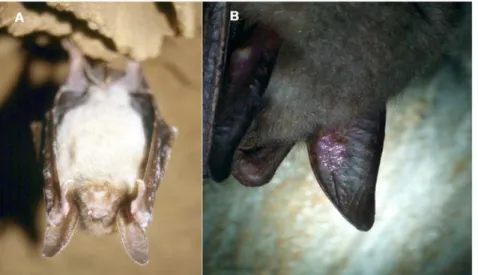
Figure 1. White-nose syndrome symptoms in the Czech Republic and Slovakia.(A) HibernatingM. myotisin the Javorˇı´cˇske´ Caves, Czech Republic, photographed on 25 January 1997. Fungal growth was not identified. (Photo by Jirˇı´ Sˇafa´rˇ) (B) Skin lesions onM. myotisfrom the Mala´ Amerika Mines, Karlsˇtejn, Czech Republic, photographed on 16 March 2010.G. destructans, isolate number CCF3942, was isolated from the sample taken from the lesion. (Photo by Ivan Hora´cˇek).
doi:10.1371/journal.pone.0013853.g001
Figure 2. Occurrence ofGeomyces destructansin the Czech Republic and Slovakia.(A) Distribution of WNS on the background of localities targeted for WNS screening. Some circles represent more than one hibernaculum. White circles localities censused in 2009 and 2010; black circles -localities with WNS-suspect bats; stars - -localities with photographic evidence of WNS in 2007 and 2008. (B) Prevalence of WNS-suspect individuals fromMyotis myotispopulations. Data pooled according to region; circle size is proportional to the population size.
Four localities with WNS-suspect bats in Central Bohemia were visually checked every two weeks between late February and March 2010. We found decreasing percentages of individuals with fungal growth on muzzle and wings towards the end of their hibernation.
Occurrence ofGeomyces destructans
We collected the fungus on swabs and transparent adhesive tape between February 2, 2010 and March 26, 2010. In total, we collected the fungus from 90 bats, where 58 samples were collected onto cotton swabs, 10 onto nylon swabs, and 20 onto adhesive tape, one on both a nylon swab and adhesive tape and one sample consisted of shed hair (Table S1). Direct microscopic observation of the adhesive tape samples and nylon swabs from the WNS-suspect bats (M. myotis) confirmed the presence of conidia and mycelia with morphology consistent withG. destructanson 22 bats (Fig. 3A). Out of the 48 cultures, we isolatedG. destructansfrom 16 (Table S1, Fig. 3B, C); and of these, 6 originated from the nylon swabs, 9 from the cotton swabs, and 1 from the adhesive tape sample. The isolates showed microscopic features typical of G. destructans (according to [3]), i. e. branched conidiophores with intercalary, lateral and terminal arthroconidia, conidia with a truncate base, mostly 5.8–7.762.7–3.4mm, young conidia ob-ovoid or cymbiform, mature conidia asymmetrical, crescent-like, curved (Fig. 3B). Colonies grow best on either malt extract or yeast and malt extract agars at 15uC (Fig. 3D). They grow slowly, reaching 18 mm after one month. The colonies were initially white, later pale brown to grey; the reverse uncoloured to brown or grey. These characteristics are similar to those previously described for isolates ofG. destructans[3,12].
We isolated DNA from 59 fungus samples, and 32 sequences, 933 base-pairs in length, were deposited into GenBank (Accession Numbers: HM584948 - HM584979; Table S1). Twenty-eight sequences were identical to previously sequenced G. destructans
isolates [3,9,12]. Four other sequences, 3 from samples collected fromM. myotis, and 1 fromM. bechsteinii, exhibited a single ARG substitution in the sequenced region, namely, at position 144 of the internal transcribed spacer 1 gene (ITS1); additionally, one of those samples contained both the A and G alleles. Other samples
did not amplify in the PCR reaction, or the sequences represented different taxa (Table S1).
At least 6 individuals were without an apparent mycelia cover, but had conspicuous lesions on either their auricles or wing membranes (Fig. 1B).G. destructansisolate CCF3942, was isolated from a sample taken from the lesion, and identified both by direct microscopy and cultivation (Fig. 3A).
Population Size Trend ofMyotis myotis
Both the CZ and SK populations of M. myotis have been continuously growing during the studied period (Fig. 4). The average annual realized growth rate per capita of the CZ population is 0.058 (95% CI -0.008 to 0.122), corresponding to an increase of about 6% per year. In SK, the average annual growth rate is 0.008 (95% CI -0.087 to 0.103), corresponding to an increase of about 1% per year. Since 2008, the numbers of CZ and SK populations have declined by 6% and 11%, respectively (the joint numbers declined by 8%). However, the declining numbers fall well within the prediction intervals calculated for 2009 and 2010 (Fig. 4). Hence, there is as yet no evidence for a change in the population trend (CZ:p= 0.88, SK:p= 0.81). These conclusions remained unchanged after input of the missing data (CZ:p= 0.82, SK:p= 0.82).
Discussion
We demonstrated that the fungus G. destructans is present in Central Europe, and that it is accompanied by aspects that might be suggestive of WNS (specifically white fungal growth on muzzle and wings, the skin lesions, loss of sheen on wing membranes, emaciated forearms or the whole body if the hair was wet). We have not conducted a histopathologic examination [4,5], as no animals were euthanized in the course of this study; however,G. destructanswas isolated from a scarred ear of aM. myotisindividual without any apparent fungal growth. The presence ofG. destructans
has been previously demonstrated [12,13], but the bat examined by Puechmaille et al. [12] was healthy, and Wibbelt et al. [13] reported a bat from Hungary with G. destructansgrowth to have survived until its next hibernation without any subsequent
Figure 3. Spores and colonies ofG. destructans.(A) Adhesive tape sample from the lesion ofM. myotisphotographed in Figure 1B, locality Mala´ Amerika Mines, Karlsˇtejn, Czech Republic (Phase contrast). (B)G. destructansCCF3937. Conidiophores and arthroconidia (SDA, 14 days, 15uC, phase contrast). (C) Primary isolation ofG. destructansCCF3942 (SDA, 1 month, 15uC). (D) Growth characteristics ofG. destructanson four agar media at c. 15uC.
manifestation of the fungus. Neither study affirmed the presence of the disease, due to the absence of mass mortality in European bats; this contrasting with the disastrous population declines that have been seen in North America [1,2]. We have shown that the G. destructansinfection in our study exhibited a marked difference in the possible impacts on the bat populations compared to the North American case. Long-term population census data indicate an increase in population size inM. myotisin the Czech Republic and Slovakia, followed by a minor decline in 2009 and 2010, but well within the prediction interval for new data. Consequently, future observations are necessary in order to decide on the causality between possible WNS and bat population trends in Europe. An association of this population size fluctuation with the emergence of G. destructans infection cannot be ruled out, however, at the moment, we treat the result with caution. Our population trend analysis showed that the decline likely either represents a natural population fluctuation. Further monitoring will be necessary for a more complete evaluation of this trend.
The incidence of white fungal patches, a clinical sign of WNS, in hibernating bats in CZ and SK, increased markedly in 2010, suggestive of an epizootic spread of the fungus. Seasonally, more WNS-suspect bats were found late in their hibernation; although, the fungal growths disappeared prior to their leaving the hibernacula. This is in accord with previous information thatG. destructansgrows slowly, and that visually apparent mycelia mostly develop in the late winter and early spring [1,9,12]. Direct observations of arousing bats suggested that the infected bats tend
to groom and remove surface mycelia immediately after arousal. According to our data, sampling the fungus onto nylon swabs enabled successful cultivations, even from lesions without visible mycelia. Previous studies have shown that isolations ofG. destructans
cultures were relatively rare, despite the presence of fungal spores in the samples that were revealed microscopically [9,14]. Our results on a small sample size might help improve future sampling methodology to better facilitate the culture diagnostics of the pathogen.
Sequences of the ITS1 gene showed for the first time to our knowledge polymorphism in the gene ofG. destructans. In general, the ITS region has been used in WNS-related studies as a conservative marker that facilitates molecular identification of fungal species, similar in principle to DNA barcoding [1,3,9,12,13]. There are 33 sequences of the G. destructans ITS
region in GenBank (retrieved on June 4, 2010), and all are identical. We have found four samples with a new allele. Genes encoding ribosomal RNA exhibit a low variability across large areas in fungi [15,16], so we can speculate that occurrence ofG. destructansin Europe predates its presence in North America, as was suggested by [13]. Our inspection of the photographed bats with fungal growths since 1995 further supports this assumption. If
G. destructanswas historically present in Europe, why has it never been detected on a large scale before (on the other hand, see [17])? During more than four decades of continuous monitoring in CZ, we have only detected faint fungal-like growths on hibernating bats since the 1990s. Our microscopic and genetic analyses showed that such a faint sheen might represent a wide spectrum of organisms, including nematodes. While some photographs might be debatable, we believe that Figure 1A shows an infection ofG. destructans. In Javorˇı´cˇske´ caves in north-eastern part of CZ, where the earliest photographic record of infectedM. myotisoriginated, the species is recently rare. Later photographs from the north-western part of CZ coincide with regions with multiple positive records from the winter 2008/2009, as well as the highest infestation in 2010.
These facts indirectly support the hypothesis, presented above, thatG. destructanswas a resident element in Europe prior to its first appearance in North America [13]. If that is the case, why does WNS not, and why in the recent past did it not, cause mass mortality in Europe? At the moment, we lack the data that would answer these questions unequivocally, but we agree with the hypothesis of Wibbelt et al. [13] that differences in clustering behaviour in the most affected species (M. lucifugus vs.M. myotis) during hibernation might play an important role.
Until now, no other agent except G. destructans has been consistently associated with WNS [1,3,4,5,9], and we can further assume that the proximate effects of the fungus result in increased arousal frequency, flight activity in and outside of the hibernacula, and secondary infections. The mass mortality accompanying WNS is present in North America, but not in Europe. Different strategies of hibernation in the European underground hibernac-ula and those in North America could magnify the final effects of a yet undefined causality chain ofG. destructansinfection that leads to fatal consequences. While in Europe bats tend to hibernate isolated or to form small clusters, in North America, some hibernacula are characterized by very large aggregations of hibernating bats, amounting to thousands of individuals. Within such large clusters, multiple appearances of infected bats, their repeated arousals, grooming, and temperature increase would lead to the disturbance of neighbouring animals, potentially spreading across the cluster, as in a shock wave. In addition to the behavioural disturbances, large clusters would be influenced by density-dependent disease transmission [18]. Seen from an
Figure 4. Upward population trends of hibernatingM. myotis.In the Czech (A) and Slovak Republic (B) the trends were modelled over the period 1995–2010 by fitting Poisson regression allowing for over-dispersion in the data. The point prediction (solid line) and 95% prediction intervals (shaded area) are based on observations up to 2008 (solid symbols) and then extrapolated to 2009 and 2010. The open symbols represent observed data for 2009 and 2010.
evolutionary perspective, WNS may act as a strong selection force that drives a change in hibernation strategy from hibernation in large clusters to a preference for less-populated hibernacula. This is the prevailing hibernation strategy in European Myotis. The hypothesis that this strategy was possibly selected for by previous mass mortality events, and the history of fungus-bat co-evolution [13] is indirectly supported by data on the historical occurrence of
M. bechsteinii. In contrast to M. myotis, which first appeared in Central Europe in the Late Holocene, M. bechsteinii has been a constant element of the Mid-European interglacial communities since Early Pliocene. Mass accumulations of bat skeletal remains in European cave deposits of the Pleistocene and Pliocene age were often dominated by this species [19]. Currently,M. bechsteiniiis a rare species that mostly avoids hibernation in caves and mines [20]. This suggests its regular hibernation in caves in the past with occasional mortality events. Assuming that some of the past mass mortality events in hibernacula could have been a result of a disease is not beyond the realm of possibility.
Unfortunately, the idea as to whether the disappearance ofM. bechsteinii from caves was caused by recurring G. destructans
infection, or a similar agent, is as yet merely speculation, and it might not be possible to reveal any hard facts supporting it. Nevertheless, the history of outburst events of G. destructans, environmental factors which could cause the outbursts, as well as the interactions between the fungus and hibernating bats are worthy of very detailed study. Further research of the ecological and genetic differentiation of hosts and pathogens might well provide crucial information for an assessment of the impacts of WNS (cf. [2]).
Conclusions
We have shown thatGeomyces destructans, the suspected infectious agent of WNS, is present across CZ and SK, without distinctive areas of prevalence. The reported incidence of its occurrence has increased since 2008, but it has likely been present since 1995, at the very least. To date, mass mortality has not been recorded, and the population fluctuation ofM. myotisobserved in 2009 and 2010 cannot be causally linked to the emergence of the disease. Nevertheless, we assume that white-nose syndrome is present in Europe. Future research should be aimed at establishing the precise effects of the disease on bats in Europe, as well as to elucidate the possible reasons for its less-severe impacts on the continent, whether it turns out to be immunological resistance, disparity in hibernating behaviour, genetic differences and associated virulence between European and American isolates of the pathogen, or environmental factors affecting the fungal growth.
Materials and Methods
Material
We used nylon swabs (microRheologics, Brescia, Italy), cotton swabs, or transparent adhesive tape to collect the 90 samples of fungi from the muzzle and wing membranes of hibernating bats. The nylon swabs were used according to the manufacturer’s recommendations. The cotton swabs were placed directly into 1.5 ml plastic tubes as per [12], and the adhesive tape was stuck onto microscopic slides as per [13]. In total, we collected 10 samples using the nylon swabs, 58 samples using the cotton swabs, 20 using the adhesive tape, and 1 using both the nylon swab and tape. One other sample consisted of shed hair.
We examined photographs of hibernating bats, taken prior to 2009, for the presence of white fungal patches. The database consisted of photographs from 1994–1998 and from 2003–2010.
Hibernacula Monitoring
The bat populations had been monitored in their underground hibernacula once a year, since 1969 [21]. The program currently consists of almost 900 sites [22,23,24]. In 2010, besides the standard census monitoring, 98 sites were repeatedly inspected in March. The animals were illuminated for a short time. The research adhered to the conditions of Permit#00356/KK/2008/ AOPK for CZ, certificate of competency per Law No. 114/1992; for SK we employed Licence#2598/715/03-5.1pil, 5376/2009-2.1/jam, from the Ministry of Environment of the Slovak Republic, certificate of competency per Law No. 543/2002.
Fungal Cultures
We conducted a mycological examination of 48 nylon swabs, cotton swabs and adhesive tape samples from the WNS-suspect bats, from 19 localities in CZ and SK (Table S1). Of those, 15 samples exhibited distinctive spores ofG. destructans under direct microscopic observation of the nylon swabs and adhesive tapes (Fig. 1A). We inoculated the fungal material from the swabs and tapes onto Sabouraud dextrose agar plates (SDA, [25]) and incubated them in the dark at two temperatures (c. 7uC and 15uC). After 14 or more days, we isolated the outgrowing colonies ofG. destructans and any other organisms. We identified the isolates according to [3], based on their phenotypic characteristics. Seven isolates are deposited at the Culture Collection of Fungi (CCF), Charles University in Prague, and 3 additional isolates in the Czech Collection of Microorganisms (CCM) at Masaryk Univer-sity in Brno (Table S1).
To assess the basic growth characteristics, we studied three isolates ofG. destructans(CCF 3937, 3938, 3939) at c. 15uC on four different agar media: malt extract agar (MEA), corn meal agar (CMA), yeast and malt extract agar (YMA), and creatine sucrose agar (CREA; [25]). We measured the colonies after 10, 20, and 30 days.
DNA Sequencing
We isolated the fungal DNA directly from the 33 cotton swabs which were not used for the mycological examination, using a ZR Genomic DNA II kit (Zymo research, Orange, CA, USA), and 26 from adhesive tape and culture isolates, using a DNeasy Tissue Kit (Qiagen, Halden, Germany). In the initial screening, we amplified the genes encoding the partial SSU, the complete SSU intron, ITS1, 5.8S rRNA, ITS2, and the partial LSU, using universal fungal primers ITS-myko-F (59 -CAAACTTGGTCATTTAGAG-GAA-39) and ITS-myko-R (59 -CCTCCGCTTATTGATATG-CT-39). The PCR reactions, in a volume of 50ml, consisted of 16 La buffer, 100mMdNTPs, 50 pMof each primer, 1U LA DNA polymerase (Top-bio), and 2ml DNA. Cycling conditions were 94uC for 5 min, followed by 30 cycles of 94uC for 20 s, 55uC for 20 s, and 68uC for 1 min. We purified the PCR products from an agarose electrophoresis gel using a Zmoclean gel DNA recovery kit (Zymo Research).
Data Analyses
We assembled the contigs in Aligner 3.5.6 (CodonCode, Dedham, MA, USA), and we identified the resulting sequences by comparing them to GenBank, using BLASTN 2.2 [26].
For the population trend analyses, we selected 106 hibernacula, with the most complete continuous records since 1995. The average annual realized growth rate per capita was estimated by regression through the origin, according to [27]. To test the hypothesis of significant changes in population size, we used data collected over years 1995 to 2008 from 106 hibernacula, and extrapolated the time trends to 2009 and 2010. We computed the prediction intervals, considering both the uncertainty about future count expectation and the random error of Poisson-distributed observations [28]. We included an estimation of the dispersion parameter to address the unexplained extra-Poisson variance. The model fitting and prediction was performed using Stata/IC 10.1 statistical software (StataCorp, College Station, TX, USA). To test the effect of the missing data, we reanalysed the dataset with the missing values input as a combination of the last observation carried forward and the next observation carried backward methods.
Supporting Information
Table S1 Material examined for Geomyces destructans presence found in the Czech Republic and Slovakia, host species, localities, direct microscopic examination, sequence Accession Numbers, and isolate numbers.
Found at: doi:10.1371/journal.pone.0013853.s001 (0.13 MB PDF)
Acknowledgments
We are grateful for contributions of Martin Celˇuch, Jaroslav Hromas, Peter Kanˇuch, Ondrˇej Koukol, Miroslav Kovarˇı´k, and Peter Pjencˇa´k who assisted with the project. Jaroslav Barva, Ludeˇk Bufka, Jaroslav Vogeltanz, Oldrˇich Vojteˇch and Petr Wolf provided their photographs for assessing the presence of fungal growth. The comments of Se´bastien Puechmaille, Gudrun Wibbelt, Dee Carter, and an anonymous reviewer helped to improve a previous version of the manuscript, and we thank Winifred Frick, Marianne Moore, Paul Racey and Emma Teeling for their support.
Author Contributions
Conceived and designed the experiments: NM J. Zima IH. Performed the experiments: NM P. Blazˇkova´ TB LF ZH MK L´ K AK. Analyzed the data: NM TB JCˇ AK OM ET MU IH. Contributed reagents/materials/analysis tools: NM PB TB PB JCˇ LF JG VH DH HJ BL RL RKL JM ZRˇ JSˇ PT MU JW DW JZ JZ IH. Wrote the paper: NM JG AK OM ET IH.
References
1. Blehert DS, Hicks AC, Behr M, Meteyer CU, Berlowski-Zier BM, et al. (2009) Bat white-nose syndrome: An emerging fungal pathogen? Science 323: 227–227. 2. Frick WF, Pollock JF, Hicks AC, Langwig KE, Reynolds DS, et al. (2010) An emerging disease causes regional population collapse of a common North American bat species. Science 329: 679–682.
3. Gargas A, Trest MT, Christensen M, Volk TJ, Blehert DS (2009) Geomyces destructanssp. nov. associated with bat white-nose syndrome. Mycotaxon 108: 147–154.
4. Meteyer CU, Buckles EL, Blehert DS, Hicks AC, Green DE, et al. (2009) Histopathologic criteria to confirm white-nose syndrome in bats. J Vet Diagn Invest 21: 411–414.
5. Courtin F, Stone WB, Risatti G, Gilbert K, Van Kruiningen HJ (2010) Pathologic findings and liver elements in hibernating bats with white-nose syndrome. Vet Pathol 47: 214–219.
6. Boyles JG, Brack V (2009) Modeling survival rates of hibernating mammals with individual-based models of energy expenditure. J Mamm 90: 9–16. 7. Boyles JG, Willis CKR (2010) Could localized warm areas inside cold caves
reduce mortality of hibernating bats affected by white-nose syndrome? Front Ecol Environ 8: 92–98.
8. Reichard JD, Kunz TH (2009) White-nose syndrome inflicts lasting injuries to the wings of little brown myotis (Myotis lucifugus). Acta Chiropterol 11: 457–464. 9. Chaturvedi V, Springer DJ, Behr MJ, Ramani R, Li XJ, et al. (2010) Morphological and molecular characterizations of psychrophilic fungusGeomyces destructansfrom New York bats with white nose syndrome (WNS). PLoS ONE 5(5): e10783. doi:10.1371/journal.pone.0010783.
10. Frick WF, Reynolds DS, Kunz TH (2010) Influence of climate and reproductive timing on demography of little brown myotisMyotis lucifugus. J Anim Ecol 79: 128–136.
11. White-Nose Syndrome Consortium (2010) The potential threat of white-nose syndrome to European bats: An action plan 10.
12. Puechmaille SJ, Verdeyroux P, Fuller H, Gouilh MA, Bekaert M, et al. (2010) White-nose syndrome fungus (Geomyces destructans) in bat, France. Emerg Infect Dis 16: 290–293.
13. Wibbelt G, Kurth A, Hellmann D, Weishaar M, Barlow A, et al. (2010) White-nose syndrome fungus (Geomyces destructans) in bats, Europe. Emerg Infect Dis 16: 1237–1242.
14. Lorch JM, Gargas A, Meteyer CU, Berlowski-Zier BM, Green DE, et al. (2010) Rapid polymerase chain reaction diagnosis of white-nose syndrome in bats. J Vet Diagn Invest 22: 224–230.
15. Fernandes EKK, Moraes AML, Pacheco RS, Rangel DEN, Miller MP, et al. (2009) Genetic diversity among Brazilian isolates of Beauveria bassiana: comparisons with non-Brazilian isolates and other Beauveria species. J Appl Microbiol 107: 760–774.
16. Fournier A, Widmer F, Enkerli J (2010) Development of a single-nucleotide polymorphism (SNP) assay for genotyping ofPandora neoaphidis. Fungal Biology 114: 498–506.
17. Feldmann R (1984) Teichfledermaus - Myotis dasycneme (Boie, 1825). Die Sa¨ugetiere Westfalens. Mu¨nster: Westfa¨lisches Museum fu¨r Naturkunde. pp 107–111.
18. Fenton MB, Davison M, Kunz TH, McCracken GF, Racey PA, et al. (2006) Linking bats to emerging diseases. Science 311: 1098–1099.
19. Hora´cˇek I, Hana´k V, Gaisler J (2000) The bats of the Palearctic region: A taxonomic and biogeographic review. In: Woloszyn BW, ed. Proceedings of the 8th European Bat Research Conference. Krakow: PAN. pp 11–157. 20. Hutson AM, Spitzenberger F, Tsytsulina K, Aulangier S, Juste J, et al. (2008)
Myotis bechsteinii. In: IUCN 2010. IUCN Red List of Threatened Species. Version 2010.1.,www.iucnredlist.org.. Downloaded on 24 June 2010.
21. Gaisler J (1975) A quantitative study of some populations of bats in Czechoslovakia (Mammalia: Chiroptera). Acta Sci Nat Brno 9(5): 1–44. 22. Bauerova´ Z, Gaisler J, Kovarˇı´k M, Zima J (1989) Variation in numbers of
hibernating bats in the Moravian Karst: Results of visual censuses in 1983-1987. In: Hana´k V, Hora´cˇek I, Gaisler J, eds. European Bat Research 1987. Praha: Charles University Press. pp 499–505.
23. Hora´cˇek I (2001) Censusing bats in the underground hibernacula of the Czech Republic. Vespertilio 5: 3–5.
24. Uhrin M, Benda P, Obuch J, Urban P (2010) Changes in abundance of hibernating bats in central Slovakia (1992-2009). Biologia 65: 349–361. 25. Samson RA, Hoekstra ES, Frisvad JC (2004) Introduction to food- and airborne
fungi. 7th Ed. Utrecht: CBS.
26. Zhang Z, Schwartz S, Wagner L, Miller W (2000) A greedy algorithm for aligning DNA sequences. J Comput Biol 7: 203–214.
27. Dennis B, Munholland PL, Scott JM (1991) Estimation of growth and extinction parameters for endangered species. Ecol Monogr 61: 115–143.