Delta-Opioid Receptor Analgesia Is Independent of
Microglial Activation in a Rat Model of Neuropathic Pain
Joanna Mika*., Katarzyna Popiolek-Barczyk., Ewelina Rojewska, Wioletta Makuch, Katarzyna Starowicz,
Barbara Przewlocka*
Department of Pain Pharmacology, Institute of Pharmacology, Polish Academy of Sciences, Krakow, Poland
Abstract
The analgesic effect of delta-opioid receptor (DOR) ligands in neuropathic pain is not diminished in contrast to other opioid receptor ligands, which lose their effectiveness as analgesics. In this study, we examine whether this effect is related to nerve injury-induced microglial activation. We therefore investigated the influence of minocycline-induced inhibition of microglial activation on the analgesic effects of opioid receptor agonists: morphine, DAMGO, U50,488H, DPDPE, Deltorphin II and SNC80 after chronic constriction injury (CCI) to the sciatic nerve in rats. Pre-emptive and repeated administration of minocycline (30 mg/kg, i.p.) over 7 days significantly reduced allodynia and hyperalgesia as measured on day 7 after CCI. The antiallodynic and antihyperalgesic effects of intrathecally (i.t.) administered morphine (10–20mg), DAMGO (1–2mg) and
U50,488H (25–50mg) were significantly potentiated in rats after minocycline, but no such changes were observed after
DPDPE (10–20mg), deltorphin II (1.5–15mg) and SNC80 (10–20mg) administration. Additionally, nerve injury-induced
down-regulation of all types of opioid receptors in the spinal cord and dorsal root ganglia was not influenced by minocycline, which indicates that the effects of opioid ligands are dependent on other changes, presumably neuroimmune interactions. Our study of rat primary microglial cell culture using qRT-PCR, Western blotting and immunocytochemistry confirmed the presence of mu-opioid receptors (MOR) and kappa-opioid receptors (KOR), further we provide the first evidence for the lack of DOR on microglial cells. In summary, DOR analgesia is different from analgesia induced by MOR and KOR receptors because it does not dependent on injury-induced microglial activation. DOR agonists appear to be the best candidates for new drugs to treat neuropathic pain.
Citation:Mika J, Popiolek-Barczyk K, Rojewska E, Makuch W, Starowicz K, et al. (2014) Delta-Opioid Receptor Analgesia Is Independent of Microglial Activation in a Rat Model of Neuropathic Pain. PLoS ONE 9(8): e104420. doi:10.1371/journal.pone.0104420
Editor:Bradley Taylor, University of Kentucky Medical Center, United States of America ReceivedFebruary 24, 2014;AcceptedJuly 9, 2014;PublishedAugust 8, 2014
Copyright:ß2014 Mika et al. This is an open-access article distributed under the terms of the Creative Commons Attribution License, which permits unrestricted use, distribution, and reproduction in any medium, provided the original author and source are credited.
Funding:This work was supported by MNiSzW/NCN grant NN405375937, grant OPUS NCN 2011/03/B/NZ4/00042, grant PRELUDIUM NCN 2012/07/N/NZ3/00379, grant MAESTRO NCN 2012/06/A/NZ4/00028 and statutory funds. Katarzyna Popiolek-Barczyk is a Ph.D. student and has a scholarship from the KNOW sponsored by the Ministry of Science and Higher Education, Republic of Poland. The funders had no role in study design, data collection and analysis, decision to publish, or preparation of the manuscript.
Competing Interests:The authors have declared that no competing interests exist. * Email: przebar@if-pan.krakow.pl (BP); joamika@if-pan.krakow.pl (JM)
.These authors contributed equally to this work.
Introduction
Neuropathic pain has been shown in clinical and animal studies to be resistant to alleviation by morphine [1,2,3,4,5], but the mechanism of this effect is unclear. The classical opioid system modulates nociception through three G-protein-coupled receptors: delta-opioid receptors (DOR) [6,7] kappa-opioid receptors (KOR) [8,9] and mu-opioid receptors (MOR) [8]. Opioid receptors do not necessarily function independently and can exist as heterodi-mers that modulate their pharmacology [10,11,12]. Several experimental studies have emphasised that the effects of DOR agonists are distinctively more potent than those of MOR and KOR receptors [13,14,15] in neuropathic pain. The field of DOR analgesia has been widely studied [16,17,18] and the DOR agonists seems to be a good drugs that would be effective in neuropathic pain, but still some of the aspects of DOR ligands interactions have to be clarified.
Reduction of morphine antinociceptive potency has been postulated to be a consequence of changes in the activity of opioid systems or opioid-specific signalling [3,19]. Although a reduction in the number of receptors may be a major factor in the
reduced efficacy of opioids, it has become clear that many other factors affect the efficacy of morphine. Such factors include heterologous desensitisation between opioid and proinflammatory chemokine receptors via shared G-protein-coupled systems [20], down-regulation of glutamate transporters in glial cells [21] or release of such substances as NO, ATP, excitatory amino acids, prostaglandins, and proinflammatory cytokines from activated glia [22,23,24,25,26,27]. In a previous study, we used minocycline, which is a tetracycline derivative with pleiotropic biological effects, to clarify whether the analgesic opioid effect is associated with the activation of microglia. Minocycline is a potent inhibitor of the microglial activation [22,28,29,30] that impairs microglial viability and migration [31,32]. Minocycline also up-regulates a tissue inhibitors of matrix metalloproteinases (TIMPs) [33], inhibits MMP-9 [34] and has been shown in many studies to have neuroprotective effects [35,36]. Some authors have suggested that minocycline may also reduce pain by inhibiting Ca2+ and Na+
co-localized with activated microglia [38,39]. It was shown [40,41,42] that the level of phospho-p38 MAPK in microglia was reduced after minocycline treatment, which suggests that this kinase is a minocycline target. Little is known about its influence on neurons, but some authors suggest its neuroprotective effects in dopami-nergic and glutamatergic neurons [43,44]. We demonstrated in our previous study that chronic administration of minocycline attenuated the development of neuropathic pain and enhanced morphine analgesia [45,46,47]. Filipovic and Zecevic [48] demonstrated inin vitrostudies that minocycline protects neurons from LPS-induced inflammation by inhibiting the activation of microglia. Activated microglia may weaken morphine analgesia by releasing pronociceptive factors, and the administration of minocycline antagonises this weakening [23,46,47].
The aim of the present study was to determine the extent to which the activation of microglia induced by injury to the sciatic nerve (by chronic constriction injury, CCI) in rats changes the analgesic effect of opioid receptors agonists. Minocycline, a well characterized drug for inhibiting microglial activity [49], was intraperitoneally (i.p.) administered pre-emptively and repeatedly to CCI-exposed rats, subsequently its influence on allodynia and hyperalgesia was measured. On day 7 after CCI, vehicle-treated and minocycline-treated rats received the opioid receptor agonists morphine, DAMGO, U50,488H, DPDPE, deltorphin II and SNC80 intrathecally. We conducted behavioural experiments and simultaneously analysed molecular changes in the transcription of
MOR,KOR, andDORmRNA in the dorsal horn of the lumbar spinal cord and in DRG (L4–L6) at day 7 after injury in CCI-exposed rats after minocycline or vehicle administration. We also analysed the presence of MOR, KOR and DOR in rat primary microglial cell cultures by using qRT-PCR, Western blotting and immunocytochemistry.
Methods
Animals
Male Wistar rats (200–350 g) were housed in cages that were lined with sawdust under a standard 12/12 h light/dark cycle (lights on at 08:00 h), with food and water availablead lib. Care was taken to reduce the number of animals used, and all experiments were performed according to the recommendations of IASP [50] and the NIH Guide for the Care and Use of Laboratory Animals and were approved by the II Local Bioethics Committee branch of the National Ethics Committee for Experiments on Animals based at the Institute of Pharmacology, Polish Academy of Sciences (Cracow, Poland).
Surgical preparations
Chronic constriction injury (CCI) was produced in rats by tying four ligatures around the sciatic nerve under sodium pentobarbital anaesthesia (60 mg/kg; i.p.). The biceps femoris and thegluteus superficialis were separated, and the right sciatic nerve was exposed. The ligatures (4/0 silk) were tied loosely around the nerve distal to the sciatic notch with 1-mm spacing until they elicited a brief twitch in the respective hind limb. The procedure has been described in detail by Bennett and Xie, [51]. After the surgery, all rats developed long-lasting neuropathic pain symptoms such as allodynia and hyperalgesia. Because we have shown in earlier studies that there are no differences between the nociceptive responses of naı¨ve and sham rats [52], we used naı¨ve animals for the behavioural experiments in the present study.
Intrathecal (i.t.) injection
Rats were prepared for intrathecal (i.t.) injection by implanting catheters under pentobarbital anaesthesia (60 mg/kg i.p.). The intrathecal catheter consisted of polyethylene tubing that was 12 cm long (PE 10, Intramedic; Clay Adams, Parsippany, NJ) with an outside diameter of 0.4 mm and a dead space of 10ml that had been sterilised by immersion in 70% (v/v) ethanol and fully flushed with sterile water before insertion. Rats were placed on a stereotaxic table (David Kopf Instruments, Tujunga, CA), and an incision was made in the atlanto-occipital membrane. The catheter (7.8 cm of its length) was carefully introduced into the subarachnoid space at the rostral level of the spinal cord lumbar enlargement (L4–L5) according to the method of Yaksh and Rudy [53]. After the implantation, the first injection of 10ml of water was performed slowly and the catheter was tightened. One day after catheter implantation, the rats were monitored for physical impairments. Those showing motor deficits were excluded from further study. Animals were allowed a minimum 1 week of recovery after the surgery before the experiment began. Water for injection or respective drugs were delivered slowly (1–2 min) in a volume of 5ml through the i.t. catheter and were followed by 10ml of water for injection, which flushed the catheter.
Behavioural tests
Tactile allodynia (von Frey test). Allodynia was measured in rats subjected to CCI by the use of an automatic von Frey apparatus (Dynamic Plantar Aesthesiometer Cat. No. 37400, Ugo Basile Italy). Rats were placed in plastic cages with a wire net floor 5 min before the experiment. The von Frey filament was applied to the midplantar surface of the hind foot, and measurements were taken automatically [23]. The strength of the von Frey stimuli ranged from 0.5 to 26 g in rats.
Cold hyperalgesia (cold plate test). Hyperalgesia was assessed using the cold plate test (Cold/Hot Plate Analgesia Meter No. 05044 Columbus Instruments, USA) as has been described previously [23,45]. The temperature of the cold plate was maintained at 5uC, and the cut-off latency was 30 s. The rats were placed on the cold plate, and the time until lifting of the hind foot was recorded. The injured foot was the first to react in every case.
Drug administration
carried out after vehicle or opioid agonist administration (Figure 1).
Microglial cell cultures and treatments
Primary cultures of microglial cells were prepared from 1-day-old Wistar rat pups as has been previously described [54] Briefly, cells were isolated from the rats’ cerebral cortices and were plated at a density of 36105cells/cm2in a culture medium that consisted of DMEM/Glutamax/high glucose (Gibco, USA) supplemented with heat-inactivated 10% foetal bovine serum (Gibco, USA), 100 U/ml penicillin, and 0.1 mg/ml streptomycin (Gibco, USA) on poly-L-lysine coated 75 cm2culture flasks and were maintained at 37uC and 5% CO2. The culture medium was changed after 4
days. The loosely adherent microglial cells were recovered after 9 days by mild shaking and centrifugation. Microglial cells were suspended in a culture medium and plated at a final density of 26105 cells onto 24 well plates and 1.26106 cells onto 6 well plates. Adherent cells were incubated for 48 h in a culture medium before being used for the analyses. Cell specificity was determined using an antibody to OX-42 (microglial marker) in cultures of primary microglia. Levels of mRNA forC1q(a microglial marker) andGFAP(an astroglial marker) were also investigated. Cultured primary microglia were more than 95% positive for OX-42 and C1q.
We have also analyzed the level of MOR, DOR and KOR mRNA level after LPS-stimulation (data not shown). Cells were prepared as described above and then incubated for 6 h with LPS [100 ng/ml] [55] or vehicle (PBS). We have not observed any significant changes in expression of MOR and KOR transcripts in comparison with vehicle-treated cells. In the LPS-stimulated microglial cells the presence of DOR mRNA also haven’t been detected. Lipopolysaccharides from Escherichia coli 0111:B4 (Sigma-Aldrich, USA).
qRT-PCR analysis of gene expression
Total RNA was extracted according to the method Chomc-zynski and Sacchi [56] using the TRIzol reagent (Invitrogen) as previously described [57]. The RNA concentration was measured
using a NanoDrop ND-1000 Spectrometer (NanoDrop Technol-ogies). Reverse transcription was performed on 500 ng (from cell cultures) or 1000 ng (from tissue) of total RNA using Omniscript reverse transcriptase (Qiagen Inc.) at 37uC for 60 min. cDNA was diluted 1:10 with H2O. qRT-PCR was performed using
Assay-On-Demand TaqMan probes according to the manufacturer’s protocol (Applied Biosystems) and run on a Real-Time PCR iCycler device (BioRad, Hercules, CA, USA). Rn00561699_m1
(Oprd1), Rn01430371_m1 (Oprm1), and Rn00567737_m1
(Oprk1) were used as TaqMan primers and probes. The expression of HPRT (a housekeeping gene) was quantified to control for variation in cDNA amounts. Cycle threshold values were calculated automatically by iCycler IQ 3.0 software with default parameters. Abundance of RNA was calculated as 22(threshold cycle)
.
Western blot analysis
Cell lysates were collected in a RIPA buffer with a protease inhibitor cocktail and cleared by centrifugation (140006g for 30 min, 4uC). Samples containing 20mg of protein were heated in a loading buffer (50 mM Tris–HCl, 2% SDS, 2%b -mercapto-ethanol, 4% glycerol and 0.1% bromophenol blue) for 5 min at 70uC and resolved by SDS–PAGE on 12% polyacrylamide gels. Following gel electrophoresis, the proteins were electrophoretically transferred to Immune-Blot PVDF membranes (Bio-Rad). The blots were blocked for 30 min using 5% non-fat dry milk (Bio-Rad) in Tris-buffered saline with 0.1% Tween 20 (TBST). The blots were incubated with primary antibodies (rabbit polyclonal anti-MOR, 1:500 [58]; rabbit polyclonal anti-DOR, 1:500 [59]; rabbit polyclonal anti-KOR 1:500, Abcam [58] that had been diluted in a SignalBoost Immunoreaction Enhancer Kit (Merck Millipore) for 24 h at 4uC and then incubated with a goat polyclonal antibody that had been conjugated to horseradish peroxidase (goat anti-rabbit IgG, BioRad) at a dilution of 1:1000 for 1 h at room temperature. After four 5-minute washes in TBST, immunocom-plexes were detected using a Lumi-Light Western Blotting Kit and visualised using a Fujifilm LAS-4000 fluorimager system. The blots were washed 4 times for 5 minutes each in TBST and reprobed with a mouse antibody against GAPDH (1:5000, Millipore) as a Figure 1. Drug administration.
doi:10.1371/journal.pone.0104420.g001
loading control. The relative levels of immunoreactivity were quantified using Fujifilm Image Gauge software.
Immunocytochemical analysis
We used commercially available specific anti-MOR, anti-KOR and anti-OX/42 antibodies. Cells were fixed for 20 minutes in 4% paraformaldehyde in a 0.1 M phosphate buffer (pH 7.4) and incubated with primary antibodies (rabbit anti-MOR, 1:400 [60], rabbit anti-KOR, 1:400 [61], rabbit anti-DOR, 1:400, [62] Neuromics; mouse anti-OX/42, 1:500, Serotec) for 2 days at 4uC. After three washes in PB, double immunofluorescence was revealed by incubation for 2 h in the appropriate fluorochrome-conjugated secondary antibody, Alexa Fluor546 donkey anti-rabbit and Alexa Fluor488 donkey anti-mouse, diluted 1:500 in 5% NDS. Sections were then washed with PB and coverslipped with an Aquatex mounting medium (Merck, Darmstadt, Ger-many). Sections without primary antibodies were used as negative controls.
Data analysis
The behavioural data are presented as the mean6S.E.M of 8– 16 rats per group. The results of the experiments were statistically evaluated using one-way analysis of variance (ANOVA). All of the differences between the treatment groups were further analysed with Bonferroni’s post-hoc tests. Significant differences in com-parisons with vehicle-treated CCI-exposed rats are indicated by *P
(,0.05), **P(,0.01) and ***P (,0.001). Significant differences between vehicle-treated CCI-exposed rats that had received a single dose of opioid receptor ligands and minocycline-treated CCI-exposed rats that had received a single dose of opioid receptor ligands are indicated by#P(,0.05),##P(,0.01) and
###P
(,0.001).
The qRT-PCR analyses were performed in three groups: naı¨ve, CCI-exposed and minocycline-treated CCI-exposed rats. The results are presented as fold changes compared with the naı¨ve rats in the ipsilateral dorsal lumbar spinal cord and DRG. The qRT-PCR data are presented as the mean6S.E.M and represent the normalised averages that were derived from the threshold qRT-PCR cycles from four to eight samples for each group. Intergroup differences were analysed using ANOVAs followed by Bonferro-ni’s multiple comparison tests. Significant differences resulting from comparison with naı¨ve rats are indicated by *P(,0.05), **P
(,0.01) and ***P(,0.001). The data from 3 to 10 cell cultures are presented.
Results
Repeated administration of minocycline significantly diminished allodynia and hyperalgesia in neuropathic pain in rats
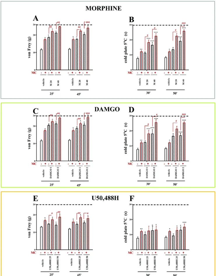
All vehicle-treated CCI rats exhibited neuropathic pain symptoms in the behavioral tests. The rats exhibited strong allodynia on the seventh day after ligation as measured by the von Frey test (11.6 g60.6 vs. 25.8 g60.2 for naı¨ve rats) (Fig. 2A,C,E; 3A,C,E) and potent hyperalgesia as measured by the cold plate test (7.6 s60.9 vs. 29.7 s60.3 for naı¨ve rats) (Fig. 2B,D,F; 3B,D,F). Repeated administration of minocycline (MC; 30 mg/kg; i.p.) attenuated allodynia (17.4 g60.5 vs. 11.6 g60.6 for the vehicle-treated CCI rats) (Fig. 2A,C,E; 3A,C,E) and also hyperalgesia (12.1 s60.9 vs. 7.6 s60.9 for the vehicle-treated CCI rats) (Fig. 2B,D,F; 3B,D,F) to a similar extent at both time points.
Repeated administration of minocycline significantly influenced the effects of morphine in neuropathic pain in rats
The doses of morphine effective in neuropathic pain (M; 20 and 40mg; i.t.) were used. Morphine was injected one hour after the last morning dose of minocycline (30 mg/kg; i.p. repeatedly) or vehicle, and allodynia and hyperalgesia were measured (see Figure 1). Morphine at a dose of 20mg significantly diminished allodynia 25 and 45 minutes after injection compared with vehicle-treated animals (18.60 g60.4 vs. 11.6 g60.6 and 17.57 g60.39 vs. 11.9 g60.5, respectively; Fig. 2A). In minocy-cline-treated animals, the antiallodynic effect of morphine at a dose of 20mg was potentiated by minocycline at both times as shown in the von Frey test (25 minutes: 22.04 g60.52 vs. 18.60 g60.4 for morphine-treated rats, 45 minutes: 22.3 g60.34 vs. 17.57 g60.39 for morphine-treated rats; Fig. 2A). Morphine at a dose of 20mg did not influence thermal hyperalgesia but in the group which was injected repeatedly with minocycline, the antihyperalgesic effect of morphine was demonstrated (30 min-utes: 18.30 s61.6 vs. 11.3 g60.8 for morphine-treated rats, 50 minutes: 22.5 s61.7 vs. 13.5 s61.5 for morphine-treated rats; Fig. 2B). A higher dose of morphine (40mg) was effective in reducing mechanical allodynia 25 and 45 minutes after injection compared with vehicle-treated animals (25 minutes: 21.1 g60.48 vs. 11.6 g60.6, 45 minutes: 23.9 g60.6 vs. 11.9 g60.5; Fig. 2A). In minocycline-treated rats, the antiallodynic effect of morphine at a dose of 40mg was potentiated at both times (25 minutes: 24.84 g60.27 vs. 21.1 g60.48, 45 minutes: 23.98 g60.51 vs. 20.25 g60.6; Fig. 2A). In opposite to the lower dose, morphine at a dose of 40mg showed antihyperalgesic effect at both time points (30 minutes: 16.5 s61.9 vs. 7.6 s60.9, 50 minutes: 19.1 s62.2 vs. 8.2 s60.8; Fig. 2B). The increased effect of morphine at a dose of 40mg in minocycline-treated group was observed at both time points, however, this effect was more significant after 50 minutes (25.8 s61.0 vs. 19.1 s62.2; Fig. 2B).
Repeated administration of minocycline significantly influenced the effects of MOR (DAMGO) and KOR (U50,488H) agonists in neuropathic pain in rats
group 30 minutes after opioid injection, however, this effect was more significant after 50 minutes (25.3 s61.1 vs.16.9 s61.5; Fig. 2D).
During neuropathy, the antiallodynic effect of U50,488H appeared after doses of 25mg i.t. only 45 minutes after injection (14.6 g60.6 vs. 11.9 g60.5; Fig. 2E). The effect of this dose of U50,488H was potentiated in minocycline-treated group as shown in the von Frey test at both times, but at 45 minutes after injection the effect was more pronounced (18.4 g60.9 vs. 14.6 g60.6; Fig. 2E). U50,488H at a dose of 25mg did not influence hyperalgesia and minocycline did not change this action (Fig. 2F). The higher dose (50mg; i.t.) diminished allodynia at both times (25 min: 14.1 g60.8 vs. 11.6 g60.6, 45 min: 15.4 g60.6 vs. 11.9 g60.5; Fig. 2E). In minocycline-treated rats, the antiallody-nic effect of U50,488H at a dose of 50mg was potentiated at both times, but it potentiation was higher after 25 minutes (18.7 g61.3 vs. 14.1 g60.8; Fig. 2E). This dose of U50,488H slightly diminished hyperalgesia at 30 (12.7 s62.9 vs. 7.6 s60.9; Fig. 2F) and 50 (13.0 s62.1 vs. 8.2 s60.8; Fig. 2F) minutes after injection. Repeated minocycline administration did not change the action of U50,488H at a dose of 50mg (Fig. 2F).
Repeated administration of minocycline did not
influence the effects of DOR agonists (DPDPE, Deltorphin II and SNC80) in neuropathic pain in rats
DPDPE at a dose of 10mg diminished allodynia 25 and 45 minutes after injection compared with vehicle-treated animals (25 min: 19.8 g62.1 vs. 11.6 g60.6, 45 min: 16.6 g62.4 vs. 11.9 g60.5; Fig. 3A) and minocycline treatment did not change this effect (Fig. 3A). Higher dose of DPDPE (20mg) was effective in reducing mechanical allodynia only 25 minutes after injection compared with vehicle-treated animals (20.3 g61.9 vs. 11.6 g60.6, Fig. 3A) and this antiallodynic effect was not changed by minocycline (Fig. 3A). As shown in the cold plate test, DPDPE significantly diminished thermal hyperalgesia after both doses of 10 and 20mg (30 min: 16.1 s62.3 vs. 7.6 s60.9; 50 min: 17.50 s61.9 vs. 8.2 s60.8 and 30 min: 20.3 s61.9 vs. 7.6 s60.9; 50 min: 23.6 s61.7 8.2 s60.8; respectively; Fig. 3B) and minocycline did not modulate this action (Fig. 3B).
Deltorphin II at a dose of 1.5mg slightly diminished allodynia 25 and 45 minutes after injection compared with vehicle-treated animals (25 min: 17.761.7 vs. 11.6 g60.6, 45 min: 17.4 g61.4 vs. 11.9 g60.5; Fig. 3C) and no changes in deltorphin II action in minocycline-treated group were observed (Fig. 3C). The higher dose of Deltorphin II (15mg) was effective in reducing mechanical allodynia 25 (17.8 g61.5 vs. 11.6 g60.6; Fig. 3C) and 45 (17.2 g62.1 vs. 11.9 g60.5; Fig. 3C) minutes after injection compared with vehicle-treated animals. Repeated minocycline injection did not change the antiallodynic effect of deltorphin II (Fig. 3D). Deltorphin II at a dose of 1.5mg did not influence hyperalgesia in CCI-exposed rats and minocycline also did not change this effect (Fig. 3D). As shown in the cold plate test, deltorphin II at a dose of 15mg diminished thermal hyperalgesia
only 50 minutes after injection (11.3 s62.8 vs. 8.2 s60.8; Fig. 3D) and minocycline did not change the antihyperalgesic action of the higher dose of Deltorphin II (Fig. 3D).
SNC80 at a dose of 10mg slightly diminished allodynia 25 and 45 minutes after injection compared with vehicle-treated animals (25 min: 16.4 g60.8 vs. 11.6 g60.6, 45 min: 17.9 g61.7 vs. 11.9 g60.5; Fig. 3E). The higher dose of SNC80 (20mg) was effective in reducing mechanical allodynia 25 (19.9 g60.6 vs. 11.6 g60.6; Fig. 3E) and 45 (19.0 g61.2 vs. 11.9 g60.5; Fig. 3E) minutes after injection compared with vehicle-treated animals. Repeated minocycline injection did not change the antiallodynic effect of both doses of SNC80 (Fig. 3E). As shown in the cold plate test, SNC80 at a dose of 10mg influenced hyperalgesia in CCI-exposed rats (30 min: 19.5 s62.8 vs. 7.6 s60.9; 50 min: 13.0 s62.3 vs. 8.2 s60.8; Fig. 3F) SNC80 at a dose of 20mg diminished thermal hyperalgesia 30 (24.2 s62 vs. 7.6 s60.9; Fig. 3F) and 50 (20.8 s64.0 vs. 8.2 s60.8; Fig. 3F) minutes after injection. Minocycline did not change the antihyperalgesic action of both doses of SNC80 (Fig. 3F).
Repeated administration of minocycline did not
influenceMOR,DORandKORmRNAs during neuropathic pain in rats
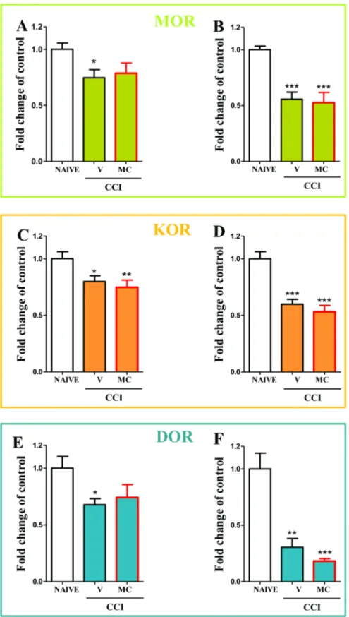
In the spinal cord the downregulation of MORmRNA from 160.05 to 0.760.07 (Fig. 4A) and in the DRG from 160.03 to 0.560.06 (Fig. 4B) was observed compared to the naı¨ve rats. Minocycline not influence the level ofMORmRNA in the spinal cord and in the DRG (Fig. 4A and B, respectively).
In the spinal cord the downregulation of KORmRNA from 160.06 to 0.860.05 (Fig. 4C) and in the DRG from 160.06 to 0.660.04 (Fig. 4D) was observed compared to the naı¨ve rats. Minocycline not influence the level ofKORmRNA in the spinal cord and in the DRG (Fig. 4C and D, respectively).
In the spinal cord the downregulation of DORmRNA from 160.1 to 0.6760.05 (Fig. 4E) and in the DRG from 160.1 to 0.360.07 (Fig. 4F) was observed compared to the naı¨ve rats. Minocycline not influence the level ofKORmRNA in the spinal cord and in the DRG (Fig. 4E and F, respectively).
Opioid receptor expression in primary microglial cell cultures
Using a reverse transcriptase-polymerase chain reaction, we found that mRNA for MOR and KOR, but not for DOR, is expressed in rat primary microglial cell cultures (Fig. 5A). In the Western blot analysis, we found that protein for MOR and KOR is present in microglia (Fig. 5B). The protein for DOR was undetectable (Fig. 5B). The expression of MOR and KOR, but not DOR, in microglial cells was confirmed by immunocyto-chemistry (Fig. 5C).
administration by the cold plate test (B, D, F). Minocycline (MC; 30 mg/kg; i.p.) was administered intraperitoneally pre-emptively 16 h and 1 h before CCI, and then repeatedly twice daily for 7 days. Vehicle-treated and minocycline-treated rats received intrathecal morphine (M; 20; 40mg/5ml), DAMGO (1; 2mg/5ml) or U50,488H (25; 50mg/5ml) one hour after the last morning administration on day 7 after CCI. The data are presented as the mean response6S.E.M. (8–16 rats per group). The results of the experiments were statistically evaluated using One-way Analyses of Variance (ANOVA). The differences between the treatment groups throughout the study were further analysed with Bonferroni’spost-hoctests. *P,0.05, **P, 0.01 and ***P,0.001 indicate significant differences compared with vehicle-treated CCI-exposed rats;#P,0.05,##P,0.01 and###P,0.001 indicate significant differences between vehicle-treated exposed rats that received a single dose of morphine and minocycline-treated CCI-exposed rats that received a single dose of morphine, DAMGO or U50,488H. Dotted line is a value for naı¨ve animals (for von Frey test 25.8 g; for cold plate test 29.7 s).
Discussion
Opioids are fundamental to the treatment of pain, but their efficacy is limited by side effects, including tolerance and hyperalgesia [63]. Some authors suggest that reduced sensitivity to opioids and the increases in their dosages that are necessary to obtain adequate analgesia heighten the risk of side effects [4,5,64]. It remains unclear why morphine is a less potent analgesic in clinical [65,66] and experimental [15,46,67] studies of neuropath-ic pain in contrast to inflammatory pain [15,67]. In many studies it was shown that selective DOR agonists do not lose their effectiveness in the treatment of neuropathic pain [13,15,68]. It has been suggested that the lesser effects of morphine on neuropathic pain are due to the reduced number of presynaptic opioid receptors that results from nerve damage-induced degen-eration of primary afferent neurons [64,69] besides other effects, like upregulation of the anti-opioid system CCK, and the NMDA receptor-dependent central sensitization, between others. Obaraet al. [15] used ED50 analysis to demonstrate in 2009 that much
higher doses of MOR and KOR agonists injected intraplantarly are required to produce antinociception in neuropathic than in inflammatory pain. However, it remains unclear why the ED50of
DOR agonists, but not those of MOR and KOR agonists, is comparable in both neuropathic and inflammatory pain [15]. Our study showed that the expression of all types of opioid receptor mRNA in the spinal cord and DRG was decreased in neuropathic pain. Also Stone et al. [70], using three animals models of peripheral nerve injury (CCI, SNL and SNT), observed decreases in spinal DOR expression at day 10thafter operations at the side of injury. Herradonet al.[71] compared two strains of rats, Fischer 344 and Lewis rats, in neuropathic pain model and found significant down-regulation of DOR mRNA 28 days after CCI in the DRG of Lewis rats and the same trend was observed in F344 rats. Those data correspond well with our results obtained at day 7th after CCI in Wistar rats. In the study of Obara et al.[15] biochemical experiments were conducted at 3rdand 14thday (but not day 7th) after CCI and showed a non-significant, downward trend in the expression of mRNA forMOR,DORandKORat the spinal cord level. At the DRG level of neuropathic rats these authors observed down-regulation of mRNA level for MORin L4–L5 DRGs (at day 3rd) and in L5 DRG (at day 14th), forDOR
in L5 DRG (at both days) and forKORin L4 and L5 DRGs (at day 3rd) and in L5 DRG (at day 14th) in comparison with naive animals. Those data correspond well with our results obtained at day 7thafter CCI, however, in our experiments we pooled L4–L6 DRGs [15]. In the paper of Kabli and Cahill [68] the up-regulation of DOR protein was observed 14 days after sciatic nerve injury at the DRG level. The authors suggest that the increase in DOR protein is correlated with DOR trafficking to the site of injury, which may explain the lower level of mRNA as shown by our results. Should be noted, however, that in some papers no decrease in opioid receptor expression was observed [71,72]. Those discrepant data may result from different animal
species used in experiments or time of the tissue collection after injury as it is shown in the mentioned works.
However, minocycline does not affect the reduced levels of all opioid receptor mRNAs, although it potentiates analgesia after such opioid agonists as morphine, DAMGO and U50,488H. Therefore, we should consider other mechanisms of weakened analgesia in neuropathic pain that can be restored by minocycline administration. These mechanisms may be important for sustained analgesia after administration of DOR ligands.
Several studies have suggested that the activation of glia and the enhancement of proinflammatory cytokine levels in the spinal cord and DRG are responsible for the development of both neuropathic pain and morphine tolerance [73,74,75]. We have previously observed an attenuation of the development of neuropathic pain when we administered minocycline, which is a substance that inhibits microglial activation in CCI-exposed rats [45,46]. Although minocycline can influence some neuronal functions, its ability to reduce microglial activation [76] as well as its selectivity of the action has been recently demonstrated [49]. We show in the present paper that the antiallodynic and antihyperalgesic effects of morphine, DAMGO and U50,488H, but not of DPDPE, deltorphin II or SNC80, were significantly potentiated with minocycline in CCI-subjected rats. In many studies we may found pharmacological evidence of distinct subtypes of DOR [77]. It is known that DOR1is activated by DPDPE, DOR2by deltorphin II
and SNC80 is a a highly selective agonist for DOR. We had demonstrated in our previous studies the occurrence of strong antiallodynic and antihyperalgesic effects of DOR1 and DOR2
agonists after their acute and chronic i.t. administration in a rat neuropathic pain model [13]. Analgesic effects of i.t. injected SNC80 was also shown in many pain models [77,78,79]. In the present paper we have shown that the effectiveness of DPDPE, deltorphin II and SNC80 is not enhanced by minocycline treatment. Our results suggest that DOR analgesia is not dependent on injury-induced microglial activation. DORs are therefore a promising target for the development of analgesics. Indeed as previously reported by Holdridge et al. [80,81] the prolonged morphine treatment-induced incensement in microglial cell size was not functionally relevant in DOR analgesia, which confirms our research concerning the presence of DOR on microglia.
In the in vitro study, we used qRT-PCR, Western blot and immunocytochemistry assays to confirm the presence of MOR and KOR in microglia, and we have shown for the first time that those microglial cells do not express DOR. Our results are in agreement with other studies that have shown that microglia express MOR and KOR [82,83,84]. Chaoet al.[85] first reported in 1996 that KOR was present in human microglia using qRT-PCR and a ligand-binding assay. The expression of KOR in microglial cells was also confirmed by the membrane binding of selective ligand [3H]U69,593. Additionally, Chaoet al.1997 [86]
have shown that morphine and DAMGO suppressed human microglia chemotaxis in a dose-dependent fashion and this effect is significantly attenuated by b-funaltrexamine (a MOR selective Figure 3. The effects of DPDPE, deltorphin II and SNC80 on vehicle- and minocycline-treated CCI-exposed rats.The response to DPDPE, deltorphin II and SNC80 was measured 25 and 45 minutes after administration by the von Frey test (A, C, E) and 30 and 50 minutes after administration by the cold plate test (B, D, F). Minocycline (MC; 30 mg/kg; i.p.) was administered intraperitoneally pre-emptively 16 h and 1 h before CCI, and then repeatedly twice daily for 7 days. Vehicle-treated and minocycline-treated rats received intrathecal DPDPE (10; 20mg/5ml), deltorphin II (del II; 1.5; 15mg/5ml) or SNC80 (10; 20mg/5ml) one hour after the last morning administration on day 7 after CCI. The data are presented as the mean response6S.E.M. (8–16 rats per group). The results of the experiments were statistically evaluated using ANOVAs. The differences between the treatment groups throughout the study were further analysed with Bonferroni’s post-hoc tests. *P,0.05, **P,0.01 and ***P,0.001 indicate significant differences compared with vehicle-treated CCI-exposed rats. Dotted line is a value for naı¨ve animals (for von Frey test 25.8 g; for cold plate test 29.7 s).
Figure 4.MOR,DOR,KORmRNAs in spinal cord and DRGs in vehicle- and minocycline-treated CCI-exposed rats.Minocycline (MC; 30 mg/kg; i.p.) was administered intraperitoneally pre-emptively 16 h and 1 h before CCI, and then repeatedly twice daily for 7 days. On the seventh day, spinal cords (L4–L6) and DRG were collected for the qRT-PCR analysis ofMOR(A, B),KOR(C, D) andDOR(E, F) gene expression. The data are presented as the means6SEM and represent the normalised averages derived from the threshold qRT-PCR cycles from four to eight samples for each group. Intergroup differences were analysed using ANOVAs followed by Bonferroni’s multiple comparison tests. *P,0.05; **P,0.01; ***P,0.001 indicate significant differences compared with naı¨ve rats.
doi:10.1371/journal.pone.0104420.g004
antagonist), which is consistent with our confirmation of the presence of MOR in microglial cells. The occurrence of DOR in microglial cells is still controversial. Our results strongly suggest
Figure 6. The possible influence of minocycline on analgesia after opioid receptors ligands.In our opinion, activated spinal microglia are key factors in the development of neuropathic pain and play a major role in the antagonizing of some opioids effectiveness. The results of our paper show for the first time that that DOR, in contrast to MOR and KOR, is not present in microglial cells. This phenomenon might be responsible for the different analgesic effects of MOR, KOR and DOR ligands (Fig. 2 and 3). We provide evidence that minocycline (a potent inhibitor of microglial activation and proliferation) enhances the effects of selective MOR (DAMGO; Fig. 2C,D) and selective KOR (U50,488H; Fig. 2E,F) agonists by inhibition of microglial cell activation. The effectiveness of DOR agonists (DPDPE, deltorphin II and SNC80) is not changed by minocycline (Fig. 3A–F). Our results indicate that an important element of the effectiveness of opioid drugs in neuropathic pain is the activation of microglia. The lack of DOR receptors in these cells causes that DOR receptor-mediated analgesia is not weaker under neuropathic pain, in which there is a strong activation of microglia. Earlier inhibition of microglial activation by minocycline administration therefore did not influence the effect of DOR selective agonists. The above results indicate not only that minocycline potentiates analgesia after MOR and KOR agonists but also that DOR is a potentially important target in the search for new drugs that would be effective against neuropathic pain.
doi:10.1371/journal.pone.0104420.g006
Green), which is not sufficiently selective and can give false positive results. The TaqMan assay that we used is standardised and highly specific, and we confirmed the lack of DOR in microglia cell cultures by using Western blot and immunocytochemistry, therefore, our results are not consistent with those that were obtained by immunostaining by Turchan-Cholewoet al.[87] and Thorlinet al.[88]. Moreover studies by Turchan-Cholewoet al.
[87] used commercial antibodies against DOR, which are no longer recommended for such studies. Due to the lack of selective DOR antibody a unique genetic mouse model was developed in order to investigate the distribution of DOR in the nervous system [16,89]. Opioid receptors have high degrees of homology, and antibodies may recognise other subtypes within the same family. DOR expression has also been observed on glial-like cells in the dentate gyrus [90] and rat cervical spinal cord [91], but none of these authors distinguished between astrocytes and microglia. Our results concerning the absence of DOR in microglia are consistent with recently published results of pharmacological studies that were conducted by Merighiet al.[84]. These authors have shown in primary microglia cell cultures that DPDPE, a DOR agonist, does not change the level of protein for PKC after LPS stimulation, while morphine and DAMGO up-regulate this kinase. The ineffectiveness of DPDPE in this study may be explained by the lack of DOR receptors in microglial cells. Horvath and DeLeo, [92] showed that selective agonists of DOR1DPDPE had no effect
on microglial migration, which confirms our results that DOR, in contrast to MOR and KOR, is not present in microglial cells. DOR shows a functional profile that is distinct from that of MOR or KOR [93] and plays an important role in chronic pain, for example DOR knockout mice showed augmented neuropathic pain [94,95]. DOR agonists are poor analgesics in acute pain [96], but are highly effective following inflammatory or neuropathic pain [13,96,97,98,99].
Happel et al. [100] suggested in 2008 that morphine and DAMGO influence the immune system. For example, opioids alter macrophage functions and they modulate cytokine produc-tion and chemokine and chemokine receptor expression. Activa-tion of proinflammatory chemokine receptors is known to down-regulate the analgesic functions of opioid receptors, and this enhances the perception of pain [101]. Horvath et al. [102] showed that morphine increases microglial migration by means of an interaction betweenMORand P2X4 receptors. This
interac-tion is dependent on PI3K/Akt pathway activainterac-tion [102]. Under neuropathic pain, the phosphorylation of p38 MAPK in microglia results in increased synthesis of the proinflammatory cytokines IL-1b, IL-6, and TNF-a. Spinal blockade of these cytokines is known to attenuate neuropathic pain and morphine tolerance [74,103]. Therefore, in cases where MOR and KOR agonists target
microglial signalling by inhibiting the actions of chemokines (fractalkine, CCL2), ATP receptors (P2X4, P2X7), MMP-9, p38 MAPK, or/and proinflammatory cytokines (IL-1b, IL-6, and TNF-a) improve their effectiveness. However, this is not the case with DOR agonists. This difference deserves future exploration.
MOR agonists, especially morphine, still remain the drugs of choice for the treatment of neuropathic pain, despite the side effects and limited efficacy. It was shown that DOR agonists were effective in persistent pain [13,68,81,104,105] and the mechanisms underlying this analgesic action was probably linked with trafficking to the cell membrane or better receptor coupling to signalling effectors [104]. Nadal et al. [95] demonstrated that neuropathic pain was enhanced in delta-opioid receptor knockout mice. Some authors suggest a delta opioid agonists as a promising alternative to mu analgesics in the treatment of chronic pain [13,95,104,106]. Currently, research is being conducted on analgesic effects of new DOR ligands [107] and some of these substances are being tested in clinical trials [108].
The results of the present study document for the first time that DOR, in contrast to MOR and KOR, are not present in microglial cells (Figure 6). In conclusion, we provide evidence that minocycline not only diminishes neuropathic pain-related behav-iour but also enhances the effectiveness of morphine and selective MOR and KOR opioid ligands under neuropathic pain condi-tions. Our findings lend support to the view that neuroimmuno-logical changes in the spinal cord and DRG are important for opioid effectiveness in neuropathic pain. In our opinion, activated spinal microglia are key factors in not only the development of neuropathic pain but also in the different efficacies of opioid analgesics. Our results also suggest that DOR analgesia is not dependent on injury-induced microglial activation. We therefore suggest that DOR is an interesting target for the development of new drugs that would be effective against neuropathic pain.
Acknowledgments
This work was supported by MNiSzW/NCN grant NN405375937, grant OPUS NCN 2011/03/B/NZ4/00042, grant PRELUDIUM NCN 2012/ 07/N/NZ3/00379, grant MAESTRO NCN 2012/06/A/NZ4/00028 and statutory funds. Katarzyna Popiolek-Barczyk, is a Ph.D. student and has a scholarship from the KNOW sponsored by the Ministry of Science and Higher Education, Republic of Poland.
Author Contributions
Conceived and designed the experiments: JM KP-B ER KS BP. Performed the experiments: JM KP-B ER WM KS BP. Analyzed the data: JM KP-B ER WM KS BP. Contributed reagents/materials/analysis tools: JM KP-B ER WM KS BP. Wrote the paper: JM KP-B ER KS BP.
References
1. Hirsch SJ, Dickenson AH (2014) Morphine sensitivity of spinal neurons in the chronic constriction injury neuropathic rat pain model. Neurosci Lett 562: 97– 101.
2. Przewlocki R, Przewlocka B (2001) Opioids in chronic pain. Eur J Pharmacol 429:79–91.
3. Przewlocki R, Przewlocka B (2005) Opioids in neuropathic pain. Curr Pharm Des 11: 3013–3025.
4. Eisenberg E, McNicol ED, Carr DB (2005) Efficacy and safety of opioid agonists in the treatment of neuropathic pain of non- malignant origin: Systematic review and meta-analysis of randomized controlled trials. JAMA; 293:3043–3052.
5. McQuay HJ (2002) Neuropathic pain: evidence matters. Eur J Pain.;6:11–8. 6. Evans CJ, Keith DE Jr, Morrison H, Magendzo K, Edwards RH (1992)
Cloning of a delta opioid receptor by functional expression. Science 258: 1952– 1955.
7. Kieffer BL, Befort K, Gave´riaux-Ruff C, Hirth CG (1992) The delta-opioid receptor: isolation of a cDNA by expression cloning and pharmacological characterization. Proc Natl Acad Sci U S A 89: 12048–12052.
8. Chen Y, Mestek A, Liu J, Yu L (1993) Molecular cloning of a rat kappa opioid receptor reveals sequence similarities to the mu and delta opioid receptors. Biochem J 295: 625–628.
9. Minami M, Hosoi Y, Toya T, Katao Y, Maekawa K, et al. (1993) In situ hybridization study of kappa-opioid receptor mRNA in the rat brain. Neurosci Lett 162: 161–164.
10. George SR, Fan T, Xie Z, Tse R, Tam V, et al. (2000) Oligomerization of mu-and delta-opioid receptors. Generation of novel functional properties. J Biol Chem 275: 26128–26135.
11. Gomes I, Jordan BA, Gupta A, Trapaidze N, Nagy V, et al. (2000) Heterodimerization of mu and delta opioid receptors: a role in opiate synergy. J Neurosci 20: RC110.
12. Gomes I, Gupta A, Filipovska J, Szeto HH, Pintar JE, et al. (2004) A role for heterodimerization of mu and delta opiate receptors in enhancing morphine analgesia. Proc Natl Acad Sci USA 101: 5135–5139.
14. Nozaki C, Le Bourdonnec B, Reiss D, Windh RT, Little PJ, et al. (2012)d -Opioid mechanisms for ADL5747 and ADL5859 effects in mice: analgesia, locomotion, and receptor internalization. J Pharmacol Exp Ther 342: 799– 807.
15. Obara I, Parkitna JR, Korostynski M, Makuch W, Kaminska D, et al. (2009) Local peripheral opioid effects and expression of opioid genes in the spinal cord and dorsal root ganglia in neuropathic and inflammatory pain. Pain 141: 283– 291.
16. Gave´riaux-Ruff C, Kieffer BL (2011) Delta opioid receptor analgesia: recent contributions from pharmacology and molecular approaches. Behav Pharma-col 22: 405–414.
17. Fenalti G, Giguere PM, Katritch V, Huang XP, Thompson AA, et al. (2014) Molecular control ofd-opioid receptor signalling. Nature 506: 191–196. 18. Somvanshi RK, Kumar U (2014) d-Opioid Receptor and Somatostatin
Receptor-4 Heterodimerization: Possible Implications in Modulation of Pain Associated Signaling. PLoS One 9: e85193.
19. Lee YW, Chaplan SR, Yaksh TL (1995) Systemic and supraspinal, but not spinal, opiates suppress allodynia in a rat neuropathic pain model. Neurosci Lett 199: 111–114.
20. Steele AD, Szabo I, Bednar F, Rogers TJ (2002) Interactions between opioid and chemokine receptors: heterologous desensitization. Cytokine Growth Factor Rev 13: 209–222.
21. Osikowicz M, Mika J, Przewlocka B (2013) The glutamatergic system as a target for neuropathic pain relief. Exp Physiol 98: 372–384.
22. Cui Y, Liao XX, Liu W (2008) A novel role of minocycline: attenuating morphine antinociceptive tolerance by inhibition of p38 MAPK in the activated spinal microglia. Brain Behav Immun 22: 114–123.
23. Makuch W, Mika J, Rojewska E, Zychowska M, Przewlocka B (2013) Effects of selective and non-selective inhibitors of nitric oxide synthase on morphine- and endomorphin-1-induced analgesia in acute and neuropathic pain in rats. Neuropharmacology 75: 445–447.
24. Mika J, Rojewska E, Makuch W, Przewlocka B (2010) Minocycline reduces the injury-induced expression of prodynorphin and pronociceptin in the dorsal root ganglion in a rat model of neuropathic pain. Neuroscience 165: 1420–1428. 25. Mika J, Zychowska M, Popiolek-Barczyk K, Rojewska E, Przewlocka B (2013)
Importance of glial activation in neuropathic pain. Eur J Pharmacol 716: 106– 119.
26. Watkins LR, Maier SF (2003) Glia: a novel drug discovery target for clinical pain. Nat Rev Drug Discov 2: 973–985.
27. Wieseler-Frank J, Maier SF, Watkins LR (2005) Central proinflammatory cytokines and pain enhancement. Neurosignals 14: 166–174.
28. Amin AR, Attur MG, Thakker GD (1996) A novel mechanism of action of tetracyclines: effects on nitric oxide synthases. Proc Natl Acad Sci U S A 93: 14014–14019.
29. Colovic M, Caccia S (2003) Liquid chromatographic determination of minocycline in brain-to-plasma distribution studies in the rat. Life Sci 791: 337–343.
30. Ledeboer A, Sloane EM, Milligan ED, Frank MG, Mahony JH, et al. (2005) Minocycline attenuates mechanical allodynia and proinflammatory cytokine expression in rat models of pain facilitation. Pain 115: 71–83.
31. Pinkernelle J, Fansa H, Ebmeyer U, Keilhoff G (2013) Prolonged minocycline treatment impairs motor neuronal survival and glial function in organotypic rat spinal cord cultures. PLoS One 8: e73422.
32. Tikka T, Fiebich BL, Goldsteins G, Keinanen R, Koistinaho J (2001) Minocycline, a tetracycline derivative, is neuroprotective against excitotoxicity by inhibiting activation and proliferation of microglia. J Neurosci 21: 2580– 2508.
33. Niimi N, Kohyama K, Matsumoto Y (2013) Minocycline suppresses experimental autoimmune encephalomyelitis by increasing tissue inhibitors of metalloproteinases. Neuropathology 33: 612–616.
34. Machado LS, Kozak A, Ergul A, Hess DC, Borlongan CV, et al. (2006) Delayed minocycline inhibits ischemia-activated matrix metalloproteinases 2 and 9 after experimental stroke. BMC Neurosci 7: 56.
35. Liu X, Su H, Chu TH, Guo A, Wu W (2013) Minocycline inhibited the pro-apoptotic effect of microglia on neural progenitor cells and protected their neuronal differentiation in vitro. Neurosci Lett 542: 30–36.
36. Garcia-Martinez EM, Sanz-Blasco S, Karachitos A, Bandez MJ, Fernandez-Gomez FJ, et al. (2010) Mitochondria and calcium flux as targets of neuroprotection caused by minocycline in cerebellar granule cells. Biochem Pharmacol 79: 239–250.
37. Kim TH, Kim HI, Kim J, Park M, Song JH (2011) Effects of minocycline on Na+currents in rat dorsal root ganglion neurons. Brain Res 1370: 34–42. 38. Jin SX, Zhuang ZY, Woolf CJ, Ji RR (2003) p38 mitogen-activated protein
kinase is activated after a spinal nerve ligation in spinal cord microglia and dorsal root ganglion neurons and contributes to the generation of neuropathic pain. J Neurosci 23: 4017–4022.
39. Zhuang ZY, Wen YR, Zhang DR, Borsello T, Bonny C, et al. (2006) A peptide c-Jun N-terminal kinase (JNK) inhibitor blocks mechanical allodynia after spinal nerve ligation: respective roles of JNK activation in primary sensory neurons and spinal astrocytes for neuropathic pain development and maintenance. J Neurosci 26: 3551–3560.
40. Chang YW, Waxman SG (2010) Minocycline attenuates mechanical allodynia and central sensitization following peripheral second-degree burn injury. J Pain 11: 1146–1154.
41. Hains BC, Waxman SG (2006) Activated microglia contribute to the maintenance of chronic pain after spinal cord injury. J Neurosci 26: 4308– 4317.
42. Hua XY, Svensson CI, Matsui T, Fitzsimmons B, Yaksh TL, et al. (2005) Intrathecal minocycline attenuates peripheral inflammation-induced hyperal-gesia by inhibiting p38 MAPK in spinal microglia. Eur J Neurosci 22: 2431– 2440.
43. Radad K, Moldzio R, Rausch WD (2010) Minocycline protects dopaminergic neurons against long-term rotenone toxicity. Can J Neurol Sci 37: 81–85 44. Gonza´lez JC, Egea J, Del Carmen Godino M, Fernandez-Gomez FJ,
Sa´nchez-Prieto J, et al. (2007) Neuroprotectant minocycline depresses glutamatergic neurotransmission and Ca(2+) signalling in hippocampal neurons. Eur J Neur-osci 26: 2481–2495.
45. Mika J, Osikowicz M, Makuch W, Przewlocka B (2007) Minocycline and pentoxifylline attenuate allodynia and hyperalgesia and potentiate the effects of morphine in rat and mouse models of neuropathic pain. Eur J Pharmacol 560: 142–149.
46. Mika J, Wawrzczak-Bargiela A, Osikowicz M, Makuch W, Przewlocka B (2009) Attenuation of morphine tolerance by minocycline and pentoxifylline in naive and neuropathic mice. Brain Behav Immun 23: 75–84.
47. Zychowska M, Rojewska E, Kreiner G, Nalepa I, Przewlocka B, et al. (2013) Minocycline influences the anti-inflammatory interleukins and enhances the effectiveness of morphine under mice diabetic neuropathy. J Neuroimmunol 262: 35–45.
48. Filipovic R, Zecevic N (2008) Neuroprotective role of minocycline in co-cultures of human fetal neurons and microglia. Exp Neurol 211: 41–51. 49. Kobayashi K, Imagama S, Ohgomori T, Hirano K, Uchimura K, et al. (2013)
Minocycline selectively inhibits M1 polarization of microglia. Cell Death Dis 4: e525.
50. Zimmermann M (1983) Ethical guidelines for investigations of experimental pain in conscious animals. Pain 16: 109–110.
51. Bennett GJ, Xie Y (1988) A peripheral mononeuropathy in rat that produces disorders of pain sensation like those seen in man. Pain 33: 87–107. 52. Osikowicz M, Mika J, Makuch W, Przewlocka B (2008) Glutamate receptor
ligands attenuate allodynia and hyperalgesia and potentiate morphine effects in a mouse model of neuropathic pain. Pain 139: 117–126.
53. Yaksh TL, Rudy TA (1976) Chronic catheterization of the spinal subarachnoid space. Physiol. Behav 17: 1031–10.
54. Zawadzka M, Kaminska B (2005) A novel mechanism of FK506-mediated neuroprotection: downregulation of cytokine expression in glial cells. Glia 49: 36–51.
55. Przanowski P, Dabrowski M, Ellert-Miklaszewska A, Kloss M, Mieczkowski J, et al. (2014) The signal transducers Stat1 and Stat3 and their novel target Jmjd3 drive the expression of inflammatory genes in microglia. J Mol Med (Berl) 92: 239–254.
56. Chomczynski P, Sacchi N (1987) Single-step method of RNA isolation by acid guanidinium thiocyanate-phenol-chloroform extraction. Anal Biochem 162: 156–159.
57. Mika J, Obara I, Przewlocka B (2011) The role of nociceptin and dynorphin in chronic pain: implications of neuro-glial interaction. Neuropeptides 45: 247– 261.
58. Kerros C, Brood I, Sola B, Jauzac P, Allouche S (2010) Reduction of cell proliferation and potentiation of Fas-induced apoptosis by the selective kappa-opioid receptor agonist U50 488 in the multiple myeloma LP-1 cells. J Neuroimmunol 220: 69–78.
59. Abcam website. Available: http://www.abcam.com/delta-opioid-receptor-antibody-ab66317.html. Accessed 2014 May 20.
60. Neuromics website. Available: http://www.neuromics.com/ittrium/visit/ A1x66x1y1x85b1x1x9cy1x6217x1x96y1x4fax1x82y1x510x1x7f. Accessed 2014 May 20.
61. Mansson E, Bare L, Yang D (1994) Isolation of a human kappa opioid receptor cDNA from placenta. Biochem Biophys Res Commun 202: 1431–1437. 62. Ko JL, Arvidsson U, Williams FG, Law PY, Elde R, et al. (1999) Visualization
of time-dependent redistribution of delta-opioid receptors in neuronal cells during prolonged agonist exposure. Brain Res Mol Brain Res 69: 171–185. 63. Mayer DJ, Mao J, Holt J, Price DD (1999) Cellular mechanisms of neuropathic
pain, morphine tolerance, and their interactions. Proc Natl Acad Sci U S A 96: 7731–7736.
64. Ossipov MH, Lopez Y, Nichols ML, Bian D, Porreca F (1995) The loss of antinociceptive efficacy of spinal morphine in rats with nerve ligation injury is prevented by reducing spinal afferent drive. Neurosci Lett 199: 87–90. 65. Rowbotham MC, Twilling L, Davies PS, Reisner L, Taylor K, et al. (2003)
Oral opioid therapy for chronic peripheral and central neuropathic pain. N Engl J Med 13: 1223–1232.
66. Gilron I, Bailey JM, Dongsheng Tu ME, Holden RR, et al. (2005) Morphine, Gabapentin, or Their Combination for Neuropathic Pain. N Engl J Med 13: 1324–1334.
67. Przewlocka B, Mika J, Labuz D, Toth G, Przewlocki R (1999) Spinal analgesic action of endomorphins in acute, inflammatory and neuropathic pain in rats. Eur J Pharmacol 367: 189–196.
68. Kabli N, Cahill CM (2007) Anti-allodynic effects of peripheral delta opioid receptors in neuropathic pain. Pain 127: 84–93.
69. Porreca F, Tang QB, Bian D, Riedl M, Elde R, et al. (1998) Spinal opioid mu receptor expression in lumbar spinal cord of rats following nerve injury. Brain Res 795: 197–203.
70. Stone LS, Vulchanova L, Riedl MS, Williams FG, Wilcox GL, et al. (2004) Effects of peripheral nerve injury on delta opioid receptor (DOR) immuno-reactivity in the rat spinal cord. Neurosci Lett 361: 208–211.
71. Herradon G, Ezquerra L, Nguyen T, Wang C, Siso A, et al. (2008) Noradrenergic and opioidergic alterations in neuropathy in different rat strains. Neurosci Lett 438: 186–189.
72. Pol O, Murtra P, Caracuel L, Valverde O, Puig MM, et al. (2006) Expression of opioid receptors and c-fos in CB1 knockout mice exposed to neuropathic pain. Neuropharmacology 50: 123–132.
73. Raghavendra V, Tanga F, Rutkowski MD, DeLeo JA (2003) Anti-hyperalgesic and morphine-sparing actions of propentofylline following peripheral nerve injury in rats: mechanistic implications of spinal glia and proinflammatory cytokines. Pain 104: 655–664.
74. Raghavendra V, Rutkowski MD, DeLeo JA (2002) The role of spinal neuroimmune activation in morphine tolerance/hyperalgesia in neuropathic and sham-operated rats. J Neurosci 22: 9980–9989.
75. Mika J, Korostynski M, Kaminska D, Wawrzczak-Bargiela A, Osikowicz M, et al. (2008) Interleukin-1alpha has antiallodynic and antihyperalgesic activities in a rat neuropathic pain model. Pain 13: 587–597.
76. Cunningham CL, Martı´nez-Cerden˜o V, Noctor SC (2013) Microglia regulate the number of neural precursor cells in the developing cerebral cortex. J Neurosci 33: 4216–4233.
77. Quock RM, Burkey TH, Varga E, Hosohata Y, Hosohata K, et al. (1999) The delta-opioid receptor: molecular pharmacology, signal transduction, and the determination of drug efficacy. Pharmacol Rev 51: 503–532.
78. Scherrer G, Imamachi N, Cao YQ, Contet C, Mennicken F, et al. (2009) Dissociation of the opioid receptor mechanisms that control mechanical and heat pain. Cell 137: 1148–1159.
79. Sluka KA, Rohlwing JJ, Bussey RA, Eikenberry SA, Wilken JM (2002) Chronic muscle pain induced by repeated acid Injection is reversed by spinally administered mu- and delta-, but not kappa-, opioid receptor agonists. J Pharmacol Exp Ther 302: 1146–1150.
80. Holdridge SV, Cahill CM (2007a) Spinal administration of a delta opioid receptor agonist attenuates hyperalgesia and allodynia in a rat model of neuropathic pain. Eur J Pain 11: 685–693.
81. Holdridge SV, Armstrong SA, Taylor AM, Cahill CM (2007b) Behavioural and morphological evidence for the involvement of glial cell activation in delta opioid receptor function: implications for the development of opioid tolerance. Mol Pain 3: 7.
82. Chang AC, Chao CC, Takemori AE, Gekker G, Hu S, et al. (1996) Arylacetamide-derived fluorescent probes: synthesis, biological evaluation, and direct fluorescent labeling of kappa opioid receptors in mouse microglial cells. J Med Chem 39: 1729–1735.
83. El-Hage N, Dever SM, Podhaizer EM, Arnatt CK, Zhang Y, et al. (2013) A novel bivalent HIV-1 entry inhibitor reveals fundamental differences in CCR5-m-opioid receptor interactions between human astroglia and microglia. AIDS 27: 2181–2190.
84. Merighi S, Gessi S, Varani K, Fazzi D, Stefanelli A, et al. (2013) Morphine mediates a proinflammatory phenotype via m-opioid receptor-PKCe -Akt-ERK1/2 signaling pathway in activated microglial cells. Biochem Pharmacol 86: 487–496.
85. Chao CC, Gekker G, Hu S, Sheng WS, Shark KB, et al. (1996) Kappa opioid receptors in human microglia downregulate human immunodeficiency virus 1 expression. Proc Natl Acad Sci U S A 93: 8051–8056.
86. Chao CC, Hu S, Shark KB, Sheng WS, Gekker G, et al. (1997) Activation of mu opioid receptors inhibits microglial cell chemotaxis. J Pharmacol Exp Ther 281: 998–1004.
87. Turchan-Cholewo J, Dimayuga FO, Ding Q, Keller JN, Hauser KF, et al. (2008) Cell-specific actions of HIV-Tat and morphine on opioid receptor expression in glia. J Neurosci Res 86: 2100–2110.
88. Thorlin T, Persson PA, Eriksson PS, Hansson E, Ro¨nnba¨ck L (1999) Delta-opioid receptor immunoreactivity on astrocytes is upregulated during mitosis. Glia 25: 370–378.
89. Scherrer G, Tryoen-To´th P, Filliol D, Matifas A, Laustriat D, et al. (2006) Knockin mice expressing fluorescent delta-opioid receptors uncover G protein-coupled receptor dynamics in vivo. Proc Natl Acad Sci U S A 103: 9691–9696. 90. Commons KG, Milner TA (1996) Cellular and subcellular localization of delta opioid receptor immunoreactivity in the rat dentate gyrus. Brain Res 738: 181– 195.
91. Cheng PY, Liu-Chen LY, Pickel VM (1997) Dual ultrastructural immunocy-tochemical labeling of mu and delta opioid receptors in the superficial layers of the rat cervical spinal cord. Brain Res 778: 367–380.
92. Horvath RJ, DeLeo JA (2009) Morphine enhances microglial migration through modulation of P2X4 receptor signaling. J Neurosci 29: 998–1005. 93. Kieffer BL, Gave´riaux-Ruff C (2002) Exploring the opioid system by gene
knockout. Prog Neurobiol 66: 285–306.
94. Gave´riaux-Ruff C, Karchewski LA, Hever X, Matifas A, Kieffer BL (2008) Inflammatory pain is enhanced in delta opioid receptor-knockout mice. Eur J Neurosci 27: 2558–2567.
95. Nadal X, Ban˜os JE, Kieffer BL, Maldonado R (2006) Neuropathic pain is enhanced in delta-opioid receptor knockout mice. Eur J Neurosci 23: 830–834. 96. Gallantine EL, Meert TF (2005) A comparison of the antinociceptive and adverse effects of the mu-opioid agonist morphine and the delta-opioid agonist SNC80. Basic Clin Pharmacol Toxicol 97: 39–51.
97. Cahill CM, Morinville A, Hoffert C, O’Donnell D, Beaudet A (2003) Up-regulation and trafficking of delta opioid receptor in a model of chronic inflammation: implications for pain control. Pain 101: 199–208.
98. Fraser GL, Gaudreau GA, Clarke PB, Me´nard DP, Perkins MN (2000) Antihyperalgesic effects of delta opioid agonists in a rat model of chronic inflammation. Br J Pharmacol 129: 1668–1672.
99. Hurley RW, Hammond DL (2000) The analgesic effects of supraspinal mu and delta opioid receptor agonists are potentiated during persistent inflammation. J Neurosci 20: 1249–1259.
100. Happel C, Steele AD, Finley MJ, Kutzler MA, Rogers TJ (2008) DAMGO-induced expression of chemokines and chemokine receptors: the role of TGF-beta1. J Leukoc Biol 83: 956–963.
101. Szabo I, Chen XH, Xin L, Adler MW, Howard OM, et al. (2002) Heterologous desensitization of opioid receptors by chemokines inhibits chemotaxis and enhances the perception of pain. Proc Natl Acad Sci U S A 99: 10276–10281.
102. Horvath RJ, Romero-Sandoval EA, De Leo JA (2010b) Inhibition of microglial P2X4 receptors attenuates morphine tolerance, Iba1, GFAP andmopioid receptor protein expression while enhancing perivascular microglial ED2. Pain 150: 401–413.
103. Hutchinson MR, Coats BD, Lewis SS, Zhang Y, Sprunger DB, et al. (2008) Proinflammatory cytokines oppose opioid-induced acute and chronic analgesia. Brain Behav Immun 22: 1178–1189.
104. Cahill CM, Holdridge SV, Morinville A (2007) Trafficking of delta-opioid receptors and other G-protein-coupled receptors: implications for pain and analgesia. Trends Pharmacol Sci 28: 23–31.
105. Bie B, Pan ZZ (2007) Trafficking of central opioid receptors and descending pain inhibition. Mol Pain 3: 37.
106. Zhang X, Bao L, Guan JS (2006) Role of delivery and trafficking of delta-opioid peptide receptors in delta-opioid analgesia and tolerance. Trends Pharmacol Sci. 27:324–329.
107. Pradhan AA, Befort K, Nozaki C, Gave´riaux-Ruff C, Kieffer BL (2011) The delta opioid receptor: anevolving target for the treatment of braindisorders. Trends Pharmacol Sci 32: 581–590.