Copyright © 2005 by Sociedade Brasileira de Pediatria
O
RIGINALA
RTICLE337
1. MSc. Universidade Federal de São Paulo/Escola Paulista de Medicina (UNIFESP/EPM), São Paulo, SP, Brazil.
2. Visiting professor, Department of Pediatrics, UNIFESP/EPM. Full professor, Universidade de Santo Amaro (UNISA), São Paulo, SP, Brazil. 3. Professor, Department of Pediatrics, UNIFESP/EPM. Full professor, Graduation course, UNISA, São Paulo, SP, Brazil.
Funding: Fundo de Auxílio aos Docentes e Alunos (FADA) from Universidade Federal de São Paulo. Manuscript received 08.11.04, accepted for publication 16.03.05.
Suggested citation: Cobayashi F, Lopes LA, Taddei JA. Bone mineral density in overweight and obese adolescents. J Pediatr (Rio J). 2005;81:337-42. Abstract
Objective: to study bone density as a concomitant factor for obesity in post-pubertal adolescents, controlling for other variables that may interfere in such a relation.
Methods: Study comprising 83 overweight and obese adolescents (BMI > P85) and 89 non obese ones (P5 < BMI < P85). Cases and controls were selected out of 1,420 students (aged 14-19) from a public school in the city of São Paulo. The bone mineral density of the lumbar spine (L2-L4 in g/cm2) was assessed by dual-energy x-ray absorptiometry
(LUNARTM DPX-L). The variable bone density was dichotomized using 1.194 g/cm2 as cutoff point. Bivariate analyses were
conducted considering the prevalence of overweight and obesity followed by multivariate analysis (logistic regression) according to a hierarchical conceptual model.
Results: The prevalence of bone density above the median was twice more frequent among cases (69.3%) than among controls (32.1%). In the bivariate analysis such prevalence resulted in an odds ratio (OR) of 4.78. The logistic regression model showed that the association between obesity and mineral density is yet more intense with an OR of 6.65 after the control of variables related to sedentary lifestyle and intake of milk and dairy products.
Conclusion: Obese and overweight adolescents in the final stages of sexual maturity presented higher bone mineral density in relation to their normal-weight counterparts; however, cohort studies will be necessary to evaluate the influence of such characteristic on bone resistance in adulthood and, consequently, on the incidence of osteopenia and osteoporosis at older ages.
J Pediatr (Rio J). 2005;81(4):337-42: Obesity, bone density, adolescent, osteoporosis.
Bone mineral density
in overweight and obese adolescents
Fernanda Cobayashi,1 Luiz A. Lopes,2 José A. A. C. Taddei3
Introduction
There has been an increase in the prevalence of obesity among children and adolescents in several industrialized and developing countries, turning obesity into a public health problem.
In the United States, a recent publication has analyzed weight and height data of 4,258 children and adolescents
aged between 6 and 19 years, from 1999 to 2002, and found a 16% prevalence of overweight and obesity.1 In Brazil, the
Although genetic factors have greatly contributed to obesity, several studies have pointed out environmental
factors, such as lack of physical activity,3 too much
television watching4 (over 4 hours) and increase in the
consumption of fast food by children and adolescents.5
Obesity in childhood and adolescence increases the risks for the development of cardiovascular diseases, diabetes
and some types of cancers in adulthood.6
Despite the occurrence of some endocrine and metabolic disorders that may interfere with the bone formation process, little is known about bone mass in overweight and obese children.
Childhood and adolescence are the most important periods for maximum bone mass acquisition, which is defined as the maximum bone density achieved through normal growth, being associated with and influenced by environmental factors (calcium-rich diet and physical exercise), hormonal and also genetic factors.7
In a recent study with Brazilian adolescents, the authors have described a remarkable increase in bone mineralization as a result of sexual maturation, pubertal stage above grade IV and age older than 14-15 years.8
Approximately 85-90% of the final bone mass in adults is achieved up to the age of 18 years in girls and up to the age of 20 in boys. From then on, it remains constant up to the fourth decade of life, when there is gradual physiological loss due to the advancement of age.9
Epidemiological data from the European Union member states about the over 80-year-old population estimate that vertebral fractures will increase from 23.7 millions in 2000 to 37.3 millions in 2050, corresponding to a 50% increase.10 In the United States, osteoporosis is believed
to affect 10 million people, and health expenditures with this disease amount to 14 billion dollars a year.11
Since environmental factors (sedentary lifestyle and inappropriate eating habits), which increase the risks for overweight and obesity, are the same ones that negatively influence bone tissue development, preventive measures against these two pathogenic processes should be implemented during childhood and adolescence, with the aim of synergistically posing lower risks of comorbidities caused by excessive weight and maximum potential for bone mass accumulation in these stages of development.12
The aim of the present study was to assess bone density as a concomitant factor for obesity among postpubertal adolescents, controlling for other variables that might interfere with this relationship.
Patients and methods
This case-control study is part of the ECCHOS (Clinical Studies on Growth, Hypertension, Obesity, and Oral Health) project, which includes professionals from different specialty areas, with the aim of updating specific knowledge regarding adolescent health. The study was carried out at a public school after approval by the Central-South Board
of Education of São Paulo (Vila Mariana neighborhood). The data were collected from August to December 2002.
For selection of cases and controls, an especially trained group of nutritionists and pediatricians weighed and measured 1,420 adolescents, born between 01/01/1983 and 12/31/1988, representing 98.68% of all regularly enrolled students. Sixteen individuals refused to participate in anthropometric measurements and three could not be located after three attempts.
Of the assessed adolescents, 104 were identified as cases (body mass index BMI > 85th percentile). Control individuals were selected from the list of consecutive names in alphabetical order, with BMI between the 5th and 85th percentiles.13 Frequency matching was performed
considering gender, pubertal stage and age.14 Tanners
criterion was used by the pediatricians during individual appointments with the adolescents for assessment of pubertal stage.15,16
Of the 218 selected adolescents (104 cases and 114 controls), 19.4% (17 cases and 25 controls) refused to participate in the study, and four cases were excluded for having hypothyroidism after medical examination and measurement of thyroid hormone levels. The final sample consisted of 172 adolescents (83 cases and 89 controls).
Participants did not have chronic diseases, history of fractures or long-term immobilization, did not receive supplementation of calcium or any other drug that might affect their bone metabolism.
The adolescents were weighed and measured during physical education classes, and if they were eligible for the study, they were invited to go to the school together with their parents, so that the objectives of the study could be clarified and the consent form could be signed.
For weight determination, a digital Kratos® scale with
150-kg capacity and 5-kg accuracy was used. Height was measured using a portable Alturexata® stadiometer with a
millimeter (mm) scale, following the recommended techniques.17 The parents were also weighed and measured
using the same techniques and equipment.
Total body bone density was evaluated by a single technician by way of dual energy X-ray absorptiometry (DEXA), using a LUNAR DPX-L densitometer, Lunar Radiation Corp. Madison, WI (version 1.5). Slow scanning was used for obese adolescents and medium scanning, for those with normal weight. This study evaluated the bone mineral density of the lumbar spine (L2-L4), expressed in
g/cm2, so as to produce results comparable to other
studies.
The median values obtained for cases and controls considered concomitantly, less than or greater than
1.191 g/cm,2,18 were used as cutoff point in the
assessment of bone mineral density (expressed in g/cm2).
The analyzed variables were categorized into yes or no: obese father and/or mother (BMI > 30 kg/m2), adolescent
with previous history of obesity (during infancy), having a best friend, previous or current dieting, frequent napping during the day, daily intake of milk or other dairy products.
The questionnaires were assessed to make sure they had been properly filled out, and were then retyped and validated in order to correct casual errors, using the Epi-Info software, version 6.0.19
The estimated sample size allowed for the detection of an OR = 3 for a 25% prevalence of family obesity among controls. With an 80% power and an alpha error of 5%, the sample consisted of 65 cases and 65 controls, with an additional 10% for occasional losses and 10% for the stratified analysis, adding up to a total of 78 cases and 78 controls.
In the statistical analysis, we initially conducted a univariate analysis using the calculation of proportions for the categorical variables. After that, we carried out bivariate analyses, observing the prevalence of overweight and obesity for each exposure variable and 95% confidence intervals. Finally, we performed a multivariate analysis (logistic regression models) based on a hierarchical conceptual model.20 At level 1, we added variables that
were distal or previous to the occurrence of obesity. At level 2, we considered the variables that were proximal to or concomitant with obesity. The variables were included by following the sequence proposed by the model. At each hierarchical level, variables associated with overweight and obesity with a p < 0.2021 were maintained for they constituted
possible counfounding factors.
The statistical analysis was made using Stata, version 8.0.22
The study protocol was approved by the Ethics
Committee of Universidade Federal de São Paulo - Escola
Paulista de Medicina.
Results
The prevalence of obesity (p > 95) and of overweight (85 < p < 95) among the 1,420 students was 4.4 and 10.8%, respectively.
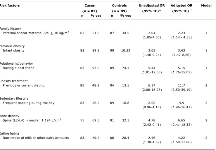
Table 1 shows the prevalence and odds ratios (unadjusted and adjusted) with the corresponding confidence intervals of the explanatory variables for cases and controls.
In Table 1, the variables concerned with family history, previous history of obesity and relationship/behavior have prevalence rates of 51.8; 29.2; 93.9% respectively, in cases, compared to their normal-weight counterparts, thus representing risks greater than 2 for obesity in the bivariate analyses that yielded unadjusted odds ratios. These variables corresponded to the first level (model 1) of the hierarchical model and, even after the control of the other variables, these three variables were regarded as important risks for overweight and obesity among the sampled adolescents.
The prevalence rates of the remaining variables for cases, such as obesity treatment (48.2%), sedentary
lifestyle (28.9%), bone density (69.3%), and eating habits (49.4%), were included at the second hierarchical level. The bivariate analyses also revealed risks greater than 2 with confidence intervals that did not include a single estimated value, thus assuring statistical significance. Such risks, also at the second level, showed a similar behavior in the multivariate analysis.
Specifically, in the hierarchical model, the bone density variable shows that adolescents with bone density above the median have 6.78 more chances of being overweight and obese. Notably, this risk persists after the control of the variables related to sedentary lifestyle and to the non-intake of milk and other dairy products on a daily basis.
Discussion
The adolescents assessed herein showed a 15.2% prevalence of overweight/obesity. This result is similar to
that obtained by Gama,23 16.2%, when evaluating 408
students from a private school of São Paulo; and to that found by Giuliano,24 14%, who studied 1,053 adolescents
(7-18 years) from a public school of Florianópolis.
This study recruited adolescents aged between 14 and 19 years who were in the final stage of maturation, with the aim of minimizing the influence of puberty over bone mass accumulation as a confounding factor in the interpretation of the results. In puberty, growth hormones and sex steroids actively participate in the bone structure development, as a result of normal growth.7
The bone density variable was categorized using the
median (1.194 g/cm2) of the sample as cutoff point.
Maynard et al.18 assessed spinal bone density in 186
normal-weight U.S. adolescents (15 to 18 years old) and
described average g/cm2 values ranging from 0.956
(SD = 0.13) to 1.111 (SD = 0.13). The cutoff point used in the present study is therefore similar to and slightly greater than the average values obtained for the group of normal-weight U.S. adolescents, which seems appropriate for the categorization of adolescents bone density into high and low as far as the analysis of associated factors is concerned.
The present study showed that an above-median prevalence of bone mineral density is twice as frequent among postpubertal adolescents who suffer from obesity and overweight (69.3%) than among their normal-weight counterparts (32.1%). In the bivariate analysis, such prevalence rates yielded an OR of 4.78. However, other confounding factors that could cause some interference were controlled, and the adjusted logistic regression model demonstrated that the association between obesity and spinal bone mineral density is even more intense when the OR increases to 6.65.
There is a paucity of studies in the literature about this association in obese adolescents at the end of puberty, without any associated pathology.
revealed that obese individuals have a higher bone mineral density than nonobese ones.25,26
The bone structure can easily adapt to stimuli. Excessive weight, represented both by body fat and skeletal muscle tissue, produces a mechanical force on
the bones, stimulating osteogenesis.27 However, it has
been shown that the positive effect of adipose tissue on the bones of adult ndividuals is far greater than that of the lean mass.28
On the other hand, weight loss among obese elderly men and women was an important risk factor for bone fractures.29
In adolescents, underweight is related to low bone density. A study carried out with 61 Polish girls (14 years
old) with diagnosis of anorexia nervosa for an average of 12.9 months demonstrated a 36.6% reduction in spinal bone mass. Girls with a body fat percentage above 15% have significantly larger total bone density than those with a smaller body fat percentage.30
Other research studies of obese adults have investigated hormonal factors, especially regarding serum levels of estrogen and insulin.28
During the menopause, with the reduction in estrogen levels, adipose tissue, which is an endocrinologically active organ, begins to synthesize this hormone through aromatization of androgens.31 Estrogen is important during
the bone formation process. The association of the mechanical load exerted by excessive weight and the
Risk factors Cases Controls Unadjusted OR Adjusted OR Model
(n = 83) (n = 89) (95% IC)* (95% IC)
n % yes n % yes
Family history
Paternal and/or maternal BMI > 30 kg/m2 83 51.8 87 34.5 2.04 2.23 1
(1.05-4.00) (1.14 - 4.35)
Previous obesity
Infant obesity 82 29.2 88 10.23 3.63 3.63 1
(1.46-9.24) (1.47-8.80)
Relationship/behavior
Having a best friend 83 93.9 89 74.1 5.44 5.15 1 (1.81-17.53) (1.76-15.07)
Obesity treatment
Previous or current dieting 83 48.2 84 13.1 6.17 11.7 2 (2.86-13.28) (3.50-39.19)
Sedentary lifestyle
Frequent napping during the day 83 28.9 89 16.8 2.00 4.9 2 (0.96-4.16) (1.46-16.41)
Bone density
Spine (L2-L4) > median 1.194 g/cm2 75 69.3 81 32.1 4.78 6.65 2
(2.42-9.41) (2.41-18.32)
Eating habits
Non-intake of milk or other dairy products 83 49.4 88 28.4 2.46 4.22 2 (1.30-4.62) (1.50-11.86)
Table 1 - Prevalence of risk factors among obese and overweight (cases) and normal-weight adolescents (controls), unadjusted and adjusted odds ratios (OR) with their respective 95% confidence intervals
BMI = bone mass index.
Model 1: Parents’ BMI, infant’s obesity, having a best friend, adjusted among themselves. Model 2: Adjusted for all variables in the table.
* Total n assessed 172.
9. Heaney RP, Abrams S, Dawson-Hughes B, Looker A, Marcus R, Matkovic V, et al. Peak bone mass. Osteoporos Int. 2000;11:985-1009.
10. Comisión europea. Informe sobre la osteoporosis en la comunidad europea: acción para la prevención. Luxemburgo: Oficina de Publicaciones Oficiales de las Comunidades Europeas; 1998.
11. National Institute of Health. Consensus development panel on osteoporosis prevention, diagnosis and therapy. JAMA. 2001;285:785-95.
12. Branca F, Valtuena S, Vatuena S. Calcium, physical activity and bone healthbuilding bones for a stronger future. Public Health Nutr. 2001;4:117-23.
13. Must A, Dallal GE, Dietz WH. Reference data for obesity: 85th and 95th percentiles of body mass index (wt/ht2) and triceps skinfold thickness. Am J Clin Nutr. 1991;53:839-46.
14. Szklo M, Nieto FJ. Epidemiology: beyond the basics. Gaithersburg (MD): Aspen Publishers; 2000.
15. Marshall WA Variations in pattern of pubertal changes in girls. Arch Dis Childhood. 1969;44:291-303.
16. Marshall WA, Tanner JM. Variations in pattern of pubertal changes in boys. Arch Dis Childhood. 1970;44:13-23.
17. Frisancho AR. Anthropometric standards for the assessment of growth and nutritional status. 4th ed. Ann Arbor (MI): University of Michigan Press; 1993.
18. Maynard LM, Guo SS, Chumlea WC, Roche AF, Wisemandle WA, Zeller CM, et al. Total-body and regional bone mineral content and area bone mineral density in children aged 8-18 y: the Fels Longitudinal Study. Am J Clin Nutr. 1998;68:1111-7.
19. Epi Info [computer program] version 6.02: a word processing, database and statistic program for epidemiology on microcomputers. Atlanta (GA): Center for Disease Control and Prevention; 1997.
20. Victora CG, Huttly SR, Fuchs SC, Olinto MT. The role of conceptual frameworks in epidemiological analysis: a hierarchical approach. Int J Epidemiol. 1997;26:224-7.
21. Mickey RM, Greenlands S. The impact of confounder selection criteria on effect estimation. Am J Epidemiol. 1989;129:125-37.
22. Stata statistical software [computer program] version 8.0. College Station (TX): Stata Corporation; 2003.
23. Gama CM. Consumo alimentar e estado nutricional de adolescentes matriculados em escolas da rede particular e estadual do bairro de Vila Mariana, São Paulo [tese]. São Paulo (SP): Universidade Federal de São Paulo; 1999.
24. Giuliano ICB. Perfil lipídico em crianças e adolescentes da rede escolar de Florianópolis [tese]. Florianópolis (SC): Universidade Federal de Santa Catarina; 2003.
25. Hasanoglu A, Bideci A, Cinaz P, Tumer L, Unal S. Bone mineral density in childhood obesity. J Pediatr Endocrinol Metab. 2000;13:307-11.
26. Ellis KJ, Shypailo RJ, Wong WW, Abrams SA. Bone mineral mass in overweight and obese children: diminished or enhanced? Acta Diabetol. 2003;40:S274-7.
27. Sugiyama T, Yamaguchi A, Kawai S. Effects of skeletal loading on bone mass and compensation mechanism in bone: a new insight into the mechanostat theory. J Bone Miner Metab. 2002;20:196-200.
28. Reid IR. Relationship among body mass, its components, and bone. Bone. 2002;31:547-55.
29. Knoke DJ, Connor-Barrett E. Weight loss: a determinant of hip bone loss in older men and women. The Rancho Bernardo Study. Am J Epidemiol. 2003;158:1132-8.
30. Jagielska G, Wolanczyk T, Komender J, Tomaszewicz-Libudzic C, Przedlacki J, Ostrowski K. Bone mineral density in adolescent girls with anorexia nervosaa cross-sectional study. Eur Child Adolesc Psychiatry. 2002;11:57-62.
31. Thomas T, Burguera B, Melton LJ III, Atkinson EJ, OFallon WM, Riggs BL, et al. Role of serum leptin, insulin, and estrogen levels as potential mediators of the relationship between fat mass and bone mineral density in men versus women. Bone. 2001;29: 114-20.
References
1. Hedley AA, Ogden CL, Johnson CL, Carroll MD, Curtin LR, Flegal KM. Prevalence of overweight and obesity among US children, adolescents, and adults, 1999-2002. JAMA. 2004;291:2847-50.
2. Abrantes MM, Lamounier JA, Colosimo EA. Prevalência de sobrepeso e obesidade nas regiões Nordeste e Sudeste do Brasil. Rev Assoc Med Bras. 2003;49:162-6.
3. Rossner S. Childhood obesity and adulthood consequences. Acta Paediatr. 1998;87:1-5.
4. Ribeiro IC, Taddei JA, Colugnati FA. Obesity among children attending elementary public schools in São Paulo, Brazil: a case-control study. Public Health Nutr. 2003;6:659-63.
5. Bowman SA, Gortmaker SL, Ebbeling CB, Pereira MA, Ludwig DS. Effects of fast-food consumption on energy intake and diet quality among children in a National Household Survey. Pediatrics. 2004;113:112-8.
6. Freedman DS, Dietz WH, Srinivasan SR, Berenson GS. The relationship of overweight to cardiovascular risk factor among children and adolescents: the Bogalusa Heart Study. Pediatrics. 1999;103:1175-82.
7. Bouillon R, Prodonova A. Growth hormone deficiency and peak bone mass. J Pediatr Endocrinol Metab. 2000;12:1327-36.
8. Silva CC, Goldberg TB, Teixeira AS, Dalmas JC. Mineralização óssea em adolescentes do sexo masculino: anos críticos para a aquisição de massa óssea. J Pediatr (Rio J). 2004;80:461-7.
synthesis of sex hormones in the adipose tissue have been implicated as a protective mechanism against
fractures in obese women.32
Another protective mechanism used to explain the lower frequency of fractures among obese adults is that insulin, steroid hormone found at high levels in obesity due to peripheral resistance, reduces the hepatic synthesis of sex hormone carriers. Thus, there is an increase in circulating sex hormones, which stimulates the activity of osteoblastic cells.33
In this study, we noted that lack of physical activity (sedentary lifestyle) and non-ingestion of milk and of other dairy products were risk factors for obesity. On the other hand, it is common knowledge that these risk factors are the same ones that hinder an age-appropriate
bone mass acquisition.9,12 Although the group of obese
adolescents had a higher bone density than normal-weight individuals, further investigation is necessary in order to identify the metabolic mechanisms associated with the stimulus to osteogenesis. Moreover, cohort studies will be necessary to assess the influence of this characteristic over bone resistance in adulthood and, consequently, over the incidence of osteopenia and osteoporosis at older ages.
Correspondence:
José Augusto de Aguiar Carrazedo Taddei Universidade Federal de São Paulo Rua Loefgreen, 1647, Vila Clementino CEP 04040-032 São Paulo, SP, Brazil Tel./Fax: +55 (11) 5573.1246 E-mail: taddei.dped@epm.br 32. van Coeverden SC, de Ridder CM, Roos JC, vant Hof MA,
Netelenbos JC, Delemarre-van de Waal HA. Pubertal maturation characteristics and the rate of bone mass development longitudinally toward menarche. J Bone Miner Res. 2001;16: 774-81.