High-Copy Overexpression Screening Reveals
PDR5
as the Main Doxorubicin Resistance
Gene in Yeast
Ayse Banu Demir1,2, Ahmet Koc1*
1Izmir Institute of Technology, Department of Molecular Biology and Genetics, Urla, Izmir, Turkey,2Dokuz Eylul University, Institute of Oncology, Department of Basic Oncology, Izmir, Turkey
*ahmetkoc@iyte.edu.tr
Abstract
Doxorubicin is one of the most potent anticancer drugs used in the treatment of various can-cer types. The efficacy of doxorubicin is influenced by the drug resistance mechanisms and its cytotoxicity. In this study, we performed a high-copy screening analysis to find genes that play a role in doxorubicin resistance and found several genes (CUE5,AKL1,CAN1,
YHR177WandPDR5) that provide resistance. Among these genes, overexpression of
PDR5provided a remarkable resistance, and deletion of it significantly rendered the toler-ance level for the drug. Q-PCR analyses suggested that transcriptional regulation of these genes was not dependent on doxorubicin treatment. Additionally, we profiled the global expression pattern of cells in response to doxorubicin treatment and highlighted the genes and pathways that are important in doxorubicin tolerance/toxicity. Our results suggest that many efflux pumps and DNA metabolism genes are upregulated by the drug and required for doxorubicin tolerance.
Introduction
Doxorubicin is an anthracycline with a strong anticancer activity. It exerts its effects through different mechanisms, such as intercalation into DNA and inhibition of DNA/RNA biosynthe-sis, formation of free radicals, inhibition of topoisomerase II, changing membrane properties and inhibition of RNA helicase [1,2]. Unfortunately, these different modes of actions for doxo-rubicin bring along serious side effects. The most notable one is its cardiotoxicity [3]. Doxoru-bicin leads to iron accumulation and ROS production, which eventually damages
mitochondria and leads to cardiac problems [4].
Multi-drug resistance (MDR) is believed to be an important cause of the treatment failure in metastatic cancer patients [5]. The efflux of the chemotherapeutics by membrane transporters is the main mechanism leading to MDR. Even though mechanisms of MDR have not been revealed exclusively, evading drug resistance and controlling MDR, have been a great issue in chemotherapy.
The mechanisms of doxorubicin resistance have been studied both in yeast and mammalian cells. In mammals, resistance mechanisms include primarily drug efflux from the cell via OPEN ACCESS
Citation:Demir AB, Koc A (2015) High-Copy Overexpression Screening RevealsPDR5as the Main Doxorubicin Resistance Gene in Yeast. PLoS ONE 10(12): e0145108. doi:10.1371/journal. pone.0145108
Editor:Michael Polymenis, Texas A&M University, UNITED STATES
Received:July 27, 2015
Accepted:November 27, 2015
Published:December 21, 2015
Copyright:© 2015 Demir, Koc. This is an open access article distributed under the terms of the
Creative Commons Attribution License, which permits unrestricted use, distribution, and reproduction in any medium, provided the original author and source are credited.
Data Availability Statement:All relevant data are within the paper and its Supporting Information files.
Funding:This work was supported by TUBITAK (Turkey) Grand No. 114Z701 to Ahmet Koc. The funders had no role in study design, data collection and analysis, decision to publish, or preparation of the manuscript.
[21], cytochrome oxidase subunit IV gene [22], and overexpression ofCLN1,CLN2andERG13
[23]. Additionally, checkpoint and recombination functions in G1 and early S phase [14], as well as several proteins involved in DNA repair, RNA metabolism, chromatin remodeling, amino acid metabolism, and heat shock response [15], play roles in doxorubicin resistance.
Identification of new genes that play role in cancer drug resistance may provide further prognostic information, which in turn may help to improve the development of new chemo-therapeutic agents and increase efficacy of chemochemo-therapeutics. In this study, we intended to identify doxorubicin resistance mechanisms by performing a high copy genomic DNA library screening in the presence of doxorubicin. Several new genes were found to cause resistance against high level of doxorubicin (500μM). Among these genes,PDR5had the most remarkable effect on doxorubicin resistance. We also profiled the expression pattern of yeast genome for doxorubicin treatment and highlighted the paths that played roles in resistance and detoxifica-tion for this drug.
Materials and Methods
Yeast strains, cell growth and plasmids
The BY4741(MATa,his3Δ1 leu2Δ0 met15Δ0 ura3Δ0)and BY4743 (MATa,his3Δ1/his3Δ1
leu2Δ0/leu2Δ0 LYS2/lys2Δ0 met15Δ0/MET15 ura3Δ0/ura3Δ0) strains of the budding yeast
Sac-charomyces cerevisiaewere used in this study. The high copy yeast genomic library (ATCC No. 37323) was used for genomic library screenings. Yeast transformations were performed by the standard LiAc method. Unless indicated otherwise, all experiments were performed on Yeast Nitrogen Base (YNB, 2% Glucose) media supplemented with appropriate amino acids and bases.
For yeast expression experiments, the genes that reside within the original YEp13 genomic clones that caused resistance against Doxorubicin, were each cloned separately into the pAG426-GPD plasmid (Addgene) and then expressed under control of the GPD promoter, except for PDR5 plasmid, which was obtained from Prof. Dr. Wenjun Guan (Zhejiang Univer-sity, China). For plasmid isolations, yeast cells were predigested by lyticase (5u/ml) for 30 min-utes in Tris-EDTA (TE) buffer before the isolation and plasmids were isolated from yeast cells by using GeneJET Plasmid Miniprep kit (Thermo-Molecular Biology) as described by the man-ufacturer. The isolated plasmids were amplified inE.coliDH5αcells and sequenced by using a pair of vector-specific primers at IzTech Biotechnology Center (Izmir). Doxorubicin was pur-chased from SABA pharmaceuticals (Cat No.: 8699511796063 /Turkey).
Gradient spot assays
contained normal medium and the plate was propped up slightly for agar to cover the entire bot-tom. When the agar was solidified, the dish was placed in a horizontal position and doxorubicin harbouring medium (50 ml) was added on top of the plate. Downward diffusion of doxorubicin resulted in its dilution proportional to the thickness of the agar layers and established a concen-tration gradient changing from approximately 0μM on one side to 500μM on the other [24].
The WT BY4741 and BY4743 strains carrying the plasmids were grown overnight diluted by growth media and incubated 3h to obtain exponentially growing cells. Cells were washed with dH2O, diluted to OD6000.02 and 5μl of cell solution was transferred to each spot. Plates were photographed after three days of incubation at 30°C.
RNA isolation and real-time PCR analysis
Total RNA samples from exponentially growing yeast cells were isolated using the RNeasy Mini Kit (Qiagen). Genomic DNA contaminations were removed by DNase treatment (Dnase RQ1, Promega). cDNA synthesis was performed using the First Strand cDNA Synthesis Kit (Fermentas) according to the manufacturer’s instructions. The cDNAs were used as templates to amplify internal parts of the selected genes.ACT1gene was used as an internal control. Real-time PCR assays were performed with IQ5 real-Real-time PCR system (BIO-RAD).
Microarray analysis
Concentrations and purity of RNA samples were determined by measuring their absorbances at 260/280nm, using a nanodrop spectrophotometer (Thermo Scientific), and the quality of RNAs were determined by Agilent RNA 6000 Nano Kit in Agilent Bioanalyzer.
Total RNA and spike-in mixes were prepared by mixing minimum 100ng of total RNA and spike-in kit for each qualified RNA sample. Total RNA sample was resuspended in nuclease-free water to obtain minimum of 50ng/μl RNA. The spike-in solution and T7 primer mix was added on diluted RNA samples and denaturation was performed by incubating the mix at 65°C for 10 minutes. cDNA master mix was prepared and added on each RNA sample mix. The mix was then incubated at 40°C for 2 hours; 70°C for 10 minutes and on ice for 5 minutes, respec-tively. The Cyanine-3-labeled and amplified RNA samples (cRNA) were purified and quantifi-cation was performed by using Nanodrop spectrophotometer (Thermo Scientific).
For hybridization, cRNA samples were incubated with a fragmentation buffer and gene expression blocking agent at 60°C for 30 minutes. The fragmented samples were loaded on arrays and the array slides were placed in a hybridization oven and the hybridization reaction was performed at 65°C for 18 hours. After the hybridization step, the samples were washed with a washing solution. The washing step was performed at room temperature for the first and the second washes. The third wash was performed at 37°C. After the washing step, the slides were scanned and signal intensities were obtained by a feature extraction program.
Statistical analysis
Student’s T-test was used for Real-time PCR analysis and fold-change analysis was used for microarray analysis data. P–value 0.05 was chosen as the significance level (p<0.05) for statis-tical analysis.
Results and Discussion
Screening for the genes that show resistance to doxorubicin
concentrations in liquid media for both haploid (BY4741) and diploid (BY4743) wild type cells. As seen inFig 1, the growth of both strains was completely inhibited in the presence of 200μM or higher amounts of doxorubicin. Next, we transformed wild type (BY4741) cells with a 2μ-based genomic expression library and isolated 6 transformants that could grow in the presence of 500μM of doxorubicin on solid media. By using a high concentration of doxorubi-cin for screening, we aimed to find genes that help cells tolerate very toxic levels of the drug. Plasmids from the transformants were isolated and amplified inE.coliand used for the
re-Fig 1. Growth curves.Wild type (A)haploid and (B)diploid yeast strains on 0μM, 100μM, 200μM and 300μM doxorubicin media. Error bars represent SD of the means for triplicate determinations.
transformation of fresh wild type cells for confirmation purposes. All the isolated plasmids conferred resistance to the new cells, thus, we confirmed that the resistance observed in the original transformants were provided by the plasmids.
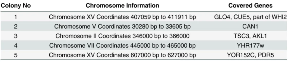
The nucleotide sequences of the expression cassettes in each plasmid were determined. Sequence analyses yielded nine intact genes (Table 1). In order to find out which one of these genes provided resistance, they were all cloned onto the pAG426-GDP plasmid individually and expressed in haploid (BY4741) and diploid (BY4743) wild type cells. As seen inFig 2A and 2B, only expression ofAKL1,CUE5,CAN1,YHR177wandPDR5genes provided resistance to doxorubicin. Particularly, cells overexpressingPDR5 gene were highly resistant to the drug and able to grow in the presence of 2 mM of doxorubicin (Fig 2C), which is the highest dose that could be tolerated by yeast cells as of our knowledge.
As the next step, we analyzed the deletion mutants ofAKL1,CUE5,CAN1,YHR177wand
PDR5genes in both haploid and diploid backgrounds to identify whether they were sensitive to doxorubicin (Fig 3A and 3B). In both backgrounds,pdr5Δmutants were the most sensitive cells and their growth was inhibited by 50μM of doxorubicin. In addition topdr5Δmutants,
akl1Δcells were also more sensitive to doxorubicin when compared to wild type cells, however the rest of the mutants (cue5Δ,can1Δandyhr177wΔ) showed a growth pattern similar to that of wild type cells.
Pdr5p is a member of the ATP-binding cassette family of transporters and mediates resis-tance to many xenobiotics such as mutagens, antifungals and steroids [25,26]. In addition to drug stress, it is also involved in cation resistance [27], and lipid transport in yeast cells [28]. Pdr5 resembles (orthologous) mammalian MDR1 which is a major factor in tumor resistance [29]. Regulation ofPDR5is controlled by transcription factorsPDR1andPDR3and deletion of these transcription factors leads to loss ofPDR5expression, while gain of function mutations in pdr1/3 leads to over expression of this protein [30–32].
Being an efflux pump,PDR5gene has previously shown to be associated with doxorubicin resistance. Golin et. al. showed thatpdr5Δmutants could not grow at 50μM doxorubicin [33] and Rogers et. al. showed thatpdr5Δmutants are sensitive to doxorubicin [34]. Kolaczkowski et al (1996) showed thatpdr1-3mutants, that overexpressPDR5, are doxorubicin resistant, while thepdr5Δmutant is sensitive to the drug [35]. A more recent study has shown that
pdr1-3mutation leads to upregulation of about twenty-five other genes in addition toPDR5[36], which may also affect the resistance level inpdr1-3mutants. Here, we showed that not only
pdr5Δmutants were doxorubucin sensitive but alsoPDR5overexpression from a plasmid increased doxorubicin resistance, which supports the findings from previous studies [33–35].
PDR5,SNQ2andYOR1are known to be major determinants of multidrug resistance in yeast and they all are controlled byPDR1[30,37,38]. In order to check ifPDR5overexpression may lead to resistance to doxorubucin in the absence of these genes, we overexpressedPDR5in
yor1Δ,snq2Δandpdr1Δmutants and analyzed them by a spotting assay (Fig 4). In addition to doxorubicin, transformants were also tested on clatrimazole, an inhibitor of ergosterol synthe-sis in fungi [39], and cerulenin, an inhibitor of fatty acid synthesis [40], since both are known
Table 1. Genomic sequences that were responsible for doxorubicin resistance.
Colony No Chromosome Information Covered Genes
1 Chromosome XV Coordinates 407059 bp to 411911 bp GLO4, CUE5, part of WHI2 2 Chromosome V Coordinates 30280 bp to 33605 bp CAN1
3 Chromosome II Coordinates 346000 bp to 366000 TSC3, AKL1 4 Chromosome VII Coordinates 445000 bp to 465000 bp YHR177w 5 Chromosome XV Coordinates 607000 bp to 627000 bp YOR152C, PDR5
to be substrates of Pdr5 (S1 Fig).PDR5overexpression made these cells resistant to all three drugs. Thepdr1Δstrain was more sensitive to the drugs compared to the wild type cells, whereas snq2Δandyor1Δmutants showed no sensitivity. Cerulenin was very toxic to cells and only cells that carriedPDR5plasmid were able to survive in the presence of this drug even at the minimal concentrations (S1 Fig). We also noticed thatPDR5overexpressingpdr1Δcells were less resistant to the drugs compared toPDR5overexpressing wild type cells. On the other hand, yor1Δmutants were previously shown to be sensitive to doxorubicin [34], however we did not observe this phenotype under our experimental conditions.
Another gene whose overexpression provided resistance and deletion rendered cells to doxorubicin wasAKL1. Akl1, which is a serine-threonine protein kinase involved in endocyto-sis and actin cytoskeleton organization [41], has previously been linked to doxorubicin resis-tance by others [14]. Sla1/Pan1/End3 complex is involved in endocytosis and affected by Akl1
Fig 2. Spotting assays for overexpression analyses.Candidate genes that play role in doxorubicin resistance were cloned into pAG426-GPD plasmid and expressed in(A)haploid and(B)diploid wild type strains in the presence of doxorubucin.(C)Overexpression of thePDR5gene could provide resistance to 2 mM of doxorubucin.
overexpression [20]. Akl1 phosphorylates Pan1p and leads to dissociation of the yeast Sla1/ Pan1/End3 complex, which regulates the internalization step of endocytosis [42]. Dissociation
Fig 3. Spotting assays for sensitivity analyses. (A)Haploid and(B)diploid deletion mutants of the candidate genes were analyzed by a spotting assay for their doxorubicin sensitivity. Serial dilutions (5μl) (OD6000.2, 0.02, 0.002 and 0.0002) for each strain were used for the sensitivity analyses.
doi:10.1371/journal.pone.0145108.g003
Fig 4.PDR5overexpression analyses inyor1Δ,snq2Δandpdr1Δmutants.PDR5gene was overexpressed in wild type,yor1Δ,snq2Δ,pdr1Δ, and pdr5Δcells and growth rates were determined by a spotting assay in the presence of doxorubicin gradient.
of the complex subsequently results in inhibition of endocytosis. Reduction in endocytosis did not affect doxorubicin accumulation significantly [20], and only very few drugs have been shown to enter cells through endocytosis [29]. Therefore, resistance to doxorubicin provided by Akl1p overexpression may be through mechanisms other than reduced endocytosis. However, reduc-tion in endocytosis may have indirect effects on doxorubucin resistance, becauseend4 endocyto-sis mutants accumulate Pdr5p at the plasma membrane [35,43]. To test if doxorubicin resistance inAKL1overexpressing strain is somehow affected by the presence of Pdr5, we overexpressed
AKL1inpdr5Δmutants and spotted cells in the presence of doxorubicin (Fig 5) and other drugs (S2 Fig). Our results indicated thatAKL1overexpression inpdr5Δmutants did not make cells resistant to doxorubicin or to other drugs, which suggested thatPDR5was required for Akl1 action. It is likely thatAKL1overexpression may act through Pdr5 stabilization in plasma mem-brane. Overexpression of AAK1, which is the human orthologue ofAKL1, also causes doxorubi-cin resistance in Hela cells [20], suggesting that the role ofAKL1in doxorubicin resistance is conserved in higher eukaryotes, but the mechanisms of resistance are not clear yet.
CAN1,YHR177WandCUE5are the other genes obtained from genomic library screening, of which their overexpression made cells resistance to doxorubicin, but their deletion mutants did not show any sensitivity. Can1p is a plasma membrane arginine permease, arginine-H+ symporter, which is exclusively associated with lipid rafts [44]. Possible doxorubicin resistance mechanism forCAN1may be upregulation of filamentous growth due to its location in ergos-terol-rich domains of the plasma membrane that harbors several proteins required for filamen-tous growth [45].
YHR177wis a putative transcription factor with a WOPR domain, of which its overexpres-sion causes either cell cycle delay or arrest [46]. Proteins with WOPR domains are important in pathogenesis [46] andYHR177woverexpression leads to invasive growth (pseudohyphae formation) in yeast [47]. Therefore,YHR177woverexpression may also cause doxorubicin resistance through activation of invasive growth.
Cue5p functions as ubiquitin-Atg8p adaptor in ubiquitin-dependent autophagy [48,49] and it is role in drug resistance is not clear.
Expression patterns of resistance genes in response to doxorubicin
treatment
Screening analyses showed that the overexpression ofPDR5,AKL1,CAN1,YHR177Wand
CUE5from a plasmid provided doxorubicin resistance, however it is not known whether these
Fig 5.AKL1overexpression analyses inpdr5Δmutants.AKL1was overexpressed in haploidpdr5Δmutants and doxorubicin resistance was tested by a spotting assay.
genes are upregulated or not in the presence of doxorubicin. In order to study expression pat-terns of these genes, we incubated cells with a sublethal dose of doxorubicin (80μM) for two hours and determined the transcript levels of each gene by a Real-Time PCR approach (Fig 6). Interestingly, expression of onlyAKL1increased slightly (1.5-fold) (p = 0,25) and the rest of the genes did not physiologically respond to the drug. Expression analyses also showed that
PDR1, a known activator ofPDR5, expression was not activated by doxorubicin treatment. Thus, these genes were not transcriptionally responsive to doxorubicin and provided resistance only if they were expressed ectopically. On the other hand, doxorubicin sensitivity ofpdr5Δ andakl1Δmutants suggested a possible role for these genes in doxorubicin resistance with their basal expression levels.
Global expression profiling for doxorubicin response
To further evaluate the mechanisms of doxorubicin resistance and toxicity, we analyzed the changes in the global gene expression profile of yeast cells after doxorubicin exposure.
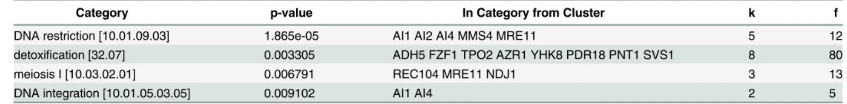
The genes showing more than 2-fold changes (p<0.05) are listed inS1 Table. Our microar-ray results also showed that doxorubicin resistance genes were not upregulated in response to the drug treatment and were consistent with the qPCR data. Out of approximately 6200 yeast genes, 211 of them were significantly upregulated, while 148 genes were downregulated. We categorized these genes by functional MIPS classification (Tables2and3) to highlight the path-ways that play role in doxorubicin tolerance and toxicity.
Funspec analysis [50] of the upregulated genes showed that paths related to transport dependent-detoxification systems and DNA metabolism such as DNA restriction, integration and recombination were upregulated in the presence of doxorubicin (Table 2).
Among the transporters (Table 2) polyamine transporterTPO2, azole resistance gene
AZR1, drug:H(+) antiporter YHK8, pleotropic drug resistance genePDR18and membrane
Fig 6. Real-Time PCR analyses of the genes that cause resistance to doxorubicin.The expression levels for genes were normalized by their untreated counterparts. Each sample was assayed at least 3 times and error bars show the SD of the means.ACT1gene was used as the internal control.
Methane Sulfonate Sensitivity) both function in DNA repair [52,53], and are related to doxoru-bicin sensitivity [15,54].
When we functionally categorized the down-regulated genes in response to doxorubicin treatment, only a small fraction of genes were clustered (Table 3).
Mainly, expression of iron transporters (FIT1,FIT2andFIT3), sugar transporters (HXT15,
HXT13,HXT16andHXT17) and proton driven antiporters (KHA1andATO2) were inhibited by doxorubicin.
Yeast responds to iron limitations by activating Aft1 transcription factor and expressing
FIT1,FIT2andFIT3genes. These genes encode for mannoproteins that are incorporated into the cell wall and play roles in retention of siderophore-iron in the cell wall [55]. Decreasing iron levels by inhibition ofFIT1,FIT2andFIT3genes might be a good defense system for doxorubicin toxicity, since iron plays role in doxorubicin cytotoxicity by producing ROS [4]. Sugar transporters that were downregulated by doxorubicin are all low affinity hexose trans-porters and their roles in drug response is not known.
Ion transportersKHA1andATO2play role as K(+)/H(+) antiporter and ammonia extruder, respectively [56,57]. Inhibition ofKHA1may lead to accumulation of K(+) and disruption of vacuole membrane potential, which might be important in doxorubicin transport/defense. However, resistance to doxorubicin decreases inkha1Δmutants [54]. Thus, the exact role of
KHA1in doxorubicin resistance is not clear. Similarly, possible benefits that could be gained by lowering the transcript levels ofATO2, ammonia transporter, are not clear.
Our microarray and real-time PCR results were consistent with each other. They both con-firmed that none of the genes obtained from genomic library screenings were upregulated by doxorubicin treatment. The genomic DNA library used in this work was a high copy number (2μ) library and supposedly genes on the plasmids were expressed at high levels. We observed thatPDR5mRNA level was 7-fold higher than that of empty vector carrying transformants (S3 Fig) (p = 0,039) and that was apparently enough for cells to tolerate 2mM doxorubicin.
Seemingly, doxorubicin did not activate the transcriptional machinery required for the expression ofPDR5,CAN1,YHR177WandCUE5genes since their mRNA levels did not change much upon the treatment. A specific support for the transcriptional inertness of these genes was the unaffected level ofPDR1, an activator ofPDR5gene (Fig 6). In addition to Pdr1, transcription factors Pdr3, Yap1, and Mig3 also play roles in expression ofPDR5in response to
Table 3. MIPS functional classification of>2-fold downregulated genes in response to Doxorubicin treatment (p value cutoff: 0.01).
Category p-value In Category from Cluster k f
ion transport [20.01.01] 0.0003473 FIT1 FIT2 FIT3 3 7
sugar transport [20.01.03.01] 0.004537 HXT15 HXT13 HXT16 HXT17 4 31
proton driven antiporter [20.03.02.03.01] 0.00948 KHA1 ATO2 2 7
drug/chemical stress exposure [58], however, their mRNA levels were not increased signifi-cantly by the doxorubicin treatment (S1 Table). Thus, even thoughPDR5played a major role in doxorubicin tolerance, the transcription factors that regulate it were not activated by doxo-rubicin treatment.
Conclusion
In this study, we screened a yeast genomic DNA library to identify genes that are responsible for doxorubicin resistance and found thatPDR5was the primary gene that played role in doxo-rubicin tolerance. Our screen also pointed out roles of other genes such as,AKL1,CAN1,
YHR177WandCUE5in protecting cells from doxorubicin toxicity. We also showed that tran-scriptional regulation of these genes was not dependent on doxorubicin treatment, however, overexpression of them makeS.cerevisiaecells resistant to high doses of doxorubicin.
Additionally, we analyzed the global expression profile of yeast cells after doxorubicin treat-ment and highlighted the genes and paths that might be important in doxorubicin tolerance and toxicity. Our results showed that membrane transporters
and DNA metabolism genes are upregulated in the presence of doxorubicin and these genes may function in doxorubicin detoxification/tolerance processes.
When we consider the genes whose overexpression caused doxorubicin resistance, exclud-ing Cue5, their common effect seemed to be the change in membrane asymmetry. Thus, chang-ing the composition of the cell membrane could be a common response of cells to high
doxorubicin levels.
Supporting Information
S1 Fig. Spotting assays for PDR5 overexpression.PDR5was cloned and expressed in wild type,yor1Δ,snq2Δ,pdr1Δ, andpdr5Δcells. Spotting assays were performed on (A) clatrimazole and (B) cerulenin.
(TIFF)
S2 Fig. Spotting assays forAKL1overexpression.AKL1was cloned and expressed inakl1Δ andpdr5Δ. Spotting assays were performed on (A) clatrimazole and (B) cerulenin.
(TIFF)
S3 Fig. Real-time PCR analyses forPDR5overexpression.PDR5transcript analyses in hap-loid and diphap-loid wild-type strains that overexpressPDR5.
(TIFF)
S1 Table. List of the genes that were up or down-regulated by 2-fold or more in response to doxorubicin treatment.
(XLSX)
Acknowledgments
We would like to thank Prof. Dr. Wenjun Guan, Zhejiang University China, for supplying the Yeplac195::PDR5 plasmid. We would like to thank Idil Uyan, for her experimental helps.
Author Contributions
PMC3904631.
5. Longley DB, Johnston PG. Molecular mechanisms of drug resistance. The Journal of pathology. 2005; 205(2):275–92. doi:10.1002/path.1706PMID:15641020.
6. Ueda K, Clark DP, Chen CJ, Roninson IB, Gottesman MM, Pastan I. The human multidrug resistance (mdr1) gene. cDNA cloning and transcription initiation. The Journal of biological chemistry. 1987; 262 (2):505–8. PMID:3027054.
7. Cole SP, Bhardwaj G, Gerlach JH, Mackie JE, Grant CE, Almquist KC, et al. Overexpression of a trans-porter gene in a multidrug-resistant human lung cancer cell line. Science. 1992; 258(5088):1650–4.
PMID:1360704.
8. Longhurst TJ, O'Neill GM, Harvie RM, Davey RA. The anthracycline resistance-associated (ara) gene, a novel gene associated with multidrug resistance in a human leukaemia cell line. British journal of can-cer. 1996; 74(9):1331–5. PMID:8912525; PubMed Central PMCID: PMC2074757.
9. Allen JD, Brinkhuis RF, Wijnholds J, Schinkel AH. The mouse Bcrp1/Mxr/Abcp gene: amplification and overexpression in cell lines selected for resistance to topotecan, mitoxantrone, or doxorubicin. Cancer research. 1999; 59(17):4237–41. PMID:10485464.
10. Slovak ML, Ho JP, Cole SP, Deeley RG, Greenberger L, de Vries EG, et al. The LRP gene encoding a major vault protein associated with drug resistance maps proximal to MRP on chromosome 16: evi-dence that chromosome breakage plays a key role in MRP or LRP gene amplification. Cancer research. 1995; 55(19):4214–9. PMID:7671223.
11. Withoff S, De Jong S, De Vries EG, Mulder NH. Human DNA topoisomerase II: biochemistry and role in chemotherapy resistance (review). Anticancer research. 1996; 16(4A):1867–80. PMID:8712715.
12. Singh SV, Nair S, Ahmad H, Awasthi YC, Krishan A. Glutathione S-transferases and glutathione peroxi-dases in doxorubicin-resistant murine leukemic P388 cells. Biochemical pharmacology. 1989; 38 (20):3505–10. PMID:2818642.
13. Shukla A, Hillegass JM, MacPherson MB, Beuschel SL, Vacek PM, Pass HI, et al. Blocking of ERK1 and ERK2 sensitizes human mesothelioma cells to doxorubicin. Molecular cancer. 2010; 9:314. doi: 10.1186/1476-4598-9-314PMID:21159167; PubMed Central PMCID: PMC3016286.
14. Westmoreland TJ, Wickramasekara SM, Guo AY, Selim AL, Winsor TS, Greenleaf AL, et al. Compara-tive genome-wide screening identifies a conserved doxorubicin repair network that is diploid specific in Saccharomyces cerevisiae. PloS one. 2009; 4(6):e5830. doi:10.1371/journal.pone.0005830PMID: 19503795; PubMed Central PMCID: PMC2688081.
15. Xia L, Jaafar L, Cashikar A, Flores-Rozas H. Identification of genes required for protection from doxoru-bicin by a genome-wide screen in Saccharomyces cerevisiae. Cancer research. 2007; 67(23):11411–
8. doi:10.1158/0008-5472.CAN-07-2399PMID:18056469; PubMed Central PMCID: PMC3635107. 16. Furuchi T, Takahashi T, Tanaka S, Nitta K, Naganuma A. Functions of yeast helicase Ssl2p that are
essential for viability are also involved in protection from the toxicity of adriamycin. Nucleic acids research. 2004; 32(8):2578–85. doi:10.1093/nar/gkh582PMID:15141027; PubMed Central PMCID:
PMC419470.
17. Takahashi T, Furuchi T, Naganuma A. A novel role for Bsd2 in the resistance of yeast to adriamycin. Journal of cellular physiology. 2005; 202(1):100–4. doi:10.1002/jcp.20082PMID:15389553.
18. Huang RY, Kowalski D, Minderman H, Gandhi N, Johnson ES. Small ubiquitin-related modifier pathway is a major determinant of doxorubicin cytotoxicity in Saccharomyces cerevisiae. Cancer research. 2007; 67(2):765–72. doi:10.1158/0008-5472.CAN-06-2839PMID:17234788.
20. Takahashi T, Furuchi T, Naganuma A. Endocytic Ark/Prk kinases play a critical role in adriamycin resis-tance in both yeast and mammalian cells. Cancer research. 2006; 66(24):11932–7. doi:
10.1158/0008-5472.CAN-06-3220PMID:17178891.
21. Schenk PW, Brok M, Boersma AW, Brandsma JA, Den Dulk H, Burger H, et al. Anticancer drug resis-tance induced by disruption of the Saccharomyces cerevisiae NPR2 gene: a novel component involved in cisplatin- and doxorubicin-provoked cell kill. Molecular pharmacology. 2003; 64(2):259–68. doi:10.
1124/mol.64.2.259PMID:12869630.
22. Kule C, Ondrejickova O, Verner K. Doxorubicin, daunorubicin, and mitoxantrone cytotoxicity in yeast. Molecular pharmacology. 1994; 46(6):1234–40. PMID:7808447.
23. Takahashi T, Nakashima S, Masuda T, Yoneda S, Hwang GW, Naganuma A. Overexpression of CLN1, CLN2, or ERG13 increases resistance to adriamycin in Saccharomyces cerevisiae. The Journal of toxicological sciences. 2011; 36(6):855–7. PMID:22129752.
24. Szybalski W, Bryson V. Genetic studies on microbial cross resistance to toxic agents. I. Cross resis-tance of Escherichia coli to fifteen antibiotics. Journal of bacteriology. 1952; 64(4):489–99. PMID:
12999676; PubMed Central PMCID: PMC169383.
25. Leppert G, McDevitt R, Falco SC, Van Dyk TK, Ficke MB, Golin J. Cloning by gene amplification of two loci conferring multiple drug resistance in Saccharomyces. Genetics. 1990; 125(1):13–20. PMID:
2160400; PubMed Central PMCID: PMC1203995.
26. Bauer BE, Wolfger H, Kuchler K. Inventory and function of yeast ABC proteins: about sex, stress, pleio-tropic drug and heavy metal resistance. Biochimica et biophysica acta. 1999; 1461(2):217–36. PMID:
10581358.
27. Miyahara K, Mizunuma M, Hirata D, Tsuchiya E, Miyakawa T. The involvement of the Saccharomyces cerevisiae multidrug resistance transporters Pdr5p and Snq2p in cation resistance. FEBS letters. 1996; 399(3):317–20. PMID:8985171.
28. Kihara A, Igarashi Y. Cross talk between sphingolipids and glycerophospholipids in the establishment of plasma membrane asymmetry. Molecular biology of the cell. 2004; 15(11):4949–59. doi:10.1091/
mbc.E04-06-0458PMID:15342785; PubMed Central PMCID: PMC524749.
29. Gottesman MM. Mechanisms of cancer drug resistance. Annual review of medicine. 2002; 53:615–27.
doi:10.1146/annurev.med.53.082901.103929PMID:11818492.
30. Balzi E, Wang M, Leterme S, Van Dyck L, Goffeau A. PDR5, a novel yeast multidrug resistance confer-ring transporter controlled by the transcription regulator PDR1. The Journal of biological chemistry. 1994; 269(3):2206–14. PMID:8294477.
31. Katzmann DJ, Hallstrom TC, Mahe Y, Moye-Rowley WS. Multiple Pdr1p/Pdr3p binding sites are essen-tial for normal expression of the ATP binding cassette transporter protein-encoding gene PDR5. The Journal of biological chemistry. 1996; 271(38):23049–54. PMID:8798494.
32. Gao C, Wang L, Milgrom E, Shen WC. On the mechanism of constitutive Pdr1 activator-mediated PDR5 transcription in Saccharomyces cerevisiae: evidence for enhanced recruitment of coactivators and altered nucleosome structures. The Journal of biological chemistry. 2004; 279(41):42677–86. doi:
10.1074/jbc.M406363200PMID:15294907.
33. Golin J, Ambudkar SV, Gottesman MM, Habib AD, Sczepanski J, Ziccardi W, et al. Studies with novel Pdr5p substrates demonstrate a strong size dependence for xenobiotic efflux. The Journal of biological chemistry. 2003; 278(8):5963–9. doi:10.1074/jbc.M210908200PMID:12496287.
34. Rogers B, Decottignies A, Kolaczkowski M, Carvajal E, Balzi E, Goffeau A. The pleitropic drug ABC transporters from Saccharomyces cerevisiae. Journal of molecular microbiology and biotechnology. 2001; 3(2):207–14. PMID:11321575.
35. Kolaczkowski M, van der Rest M, Cybularz-Kolaczkowska A, Soumillion JP, Konings WN, Goffeau A. Anticancer drugs, ionophoric peptides, and steroids as substrates of the yeast multidrug transporter Pdr5p. The Journal of biological chemistry. 1996; 271(49):31543–8. PMID:8940170.
36. DeRisi J, van den Hazel B, Marc P, Balzi E, Brown P, Jacq C, et al. Genome microarray analysis of tran-scriptional activation in multidrug resistance yeast mutants. FEBS letters. 2000; 470(2):156–60. PMID:
10734226.
37. Decottignies A, Lambert L, Catty P, Degand H, Epping EA, Moye-Rowley WS, et al. Identification and characterization of SNQ2, a new multidrug ATP binding cassette transporter of the yeast plasma mem-brane. The Journal of biological chemistry. 1995; 270(30):18150–7. PMID:7629127.
38. Katzmann DJ, Hallstrom TC, Voet M, Wysock W, Golin J, Volckaert G, et al. Expression of an ATP-binding cassette transporter-encoding gene (YOR1) is required for oligomycin resistance in Saccharo-myces cerevisiae. Molecular and cellular biology. 1995; 15(12):6875–83. PMID:8524254; PubMed
44. Malinska K, Malinsky J, Opekarova M, Tanner W. Visualization of protein compartmentation within the plasma membrane of living yeast cells. Molecular biology of the cell. 2003; 14(11):4427–36. doi:10.
1091/mbc.E03-04-0221PMID:14551254; PubMed Central PMCID: PMC266762.
45. Song Q, Kumar A. An Overview of Autophagy and Yeast Pseudohyphal Growth: Integration of Signal-ing Pathways durSignal-ing Nitrogen Stress. Cells. 2012; 1(3):263–83. doi:10.3390/cells1030263PMID:
24710476; PubMed Central PMCID: PMC3901118.
46. Lohse MB, Rosenberg OS, Cox JS, Stroud RM, Finer-Moore JS, Johnson AD. Structure of a new DNA-binding domain which regulates pathogenesis in a wide variety of fungi. Proceedings of the National Academy of Sciences of the United States of America. 2014; 111(29):10404–10. doi:10.1073/pnas.
1410110111PMID:24994900; PubMed Central PMCID: PMC4115540.
47. Shively CA, Eckwahl MJ, Dobry CJ, Mellacheruvu D, Nesvizhskii A, Kumar A. Genetic networks induc-ing invasive growth in Saccharomyces cerevisiae identified through systematic genome-wide overex-pression. Genetics. 2013; 193(4):1297–310. doi:10.1534/genetics.112.147876PMID:23410832;
PubMed Central PMCID: PMC3606104.
48. Shih SC, Prag G, Francis SA, Sutanto MA, Hurley JH, Hicke L. A ubiquitin-binding motif required for intramolecular monoubiquitylation, the CUE domain. The EMBO journal. 2003; 22(6):1273–81. doi:10.
1093/emboj/cdg140PMID:12628920; PubMed Central PMCID: PMC151082.
49. Lu K, Psakhye I, Jentsch S. Autophagic clearance of polyQ proteins mediated by ubiquitin-Atg8 adap-tors of the conserved CUET protein family. Cell. 2014; 158(3):549–63. doi:10.1016/j.cell.2014.05.048
PMID:25042851.
50. Robinson MD, Grigull J, Mohammad N, Hughes TR. FunSpec: a web-based cluster interpreter for yeast. BMC bioinformatics. 2002; 3:35. PMID:12431279; PubMed Central PMCID: PMC139976. 51. Cabrito TR, Teixeira MC, Singh A, Prasad R, Sa-Correia I. The yeast ABC transporter Pdr18 (ORF
YNR070w) controls plasma membrane sterol composition, playing a role in multidrug resistance. The Biochemical journal. 2011; 440(2):195–202. doi:10.1042/BJ20110876PMID:21831043; PubMed
Central PMCID: PMC3215286.
52. Johzuka K, Ogawa H. Interaction of Mre11 and Rad50: two proteins required for DNA repair and meio-sis-specific double-strand break formation in Saccharomyces cerevisiae. Genetics. 1995; 139 (4):1521–32. PMID:7789757; PubMed Central PMCID: PMC1206481.
53. Xiao W, Chow BL, Milo CN. Mms4, a putative transcriptional (co)activator, protects Saccharomyces cerevisiae cells from endogenous and environmental DNA damage. Molecular & general genetics: MGG. 1998; 257(6):614–23. PMID:9604884.
54. Brown JA, Sherlock G, Myers CL, Burrows NM, Deng C, Wu HI, et al. Global analysis of gene function in yeast by quantitative phenotypic profiling. Molecular systems biology. 2006; 2:2006 0001. doi:10. 1038/msb4100043PMID:16738548; PubMed Central PMCID: PMC1681475.
55. Philpott CC, Protchenko O, Kim YW, Boretsky Y, Shakoury-Elizeh M. The response to iron deprivation in Saccharomyces cerevisiae: expression of siderophore-based systems of iron uptake. Biochemical Society transactions. 2002; 30(4):698–702. doi: 10.1042/ PMID:12196168.
56. Maresova L, Sychrova H. Physiological characterization of Saccharomyces cerevisiae kha1 deletion mutants. Molecular microbiology. 2005; 55(2):588–600. doi:10.1111/j.1365-2958.2004.04410.xPMID:
15659172.
57. Vachova L, Palkova Z. Physiological regulation of yeast cell death in multicellular colonies is triggered by ammonia. The Journal of cell biology. 2005; 169(5):711–7. doi:10.1083/jcb.200410064PMID:
58. Teixeira MC, Monteiro PT, Guerreiro JF, Goncalves JP, Mira NP, dos Santos SC, et al. The YEAS-TRACT database: an upgraded information system for the analysis of gene and genomic transcription regulation in Saccharomyces cerevisiae. Nucleic acids research. 2014; 42(Database issue):D161–6.