Arq Neuropsiquiatr 2010;68(4):562-566
The surface electromyography
analysis of the non-plegic upper
limb of hemiplegic subjects
Heloyse U. Kuriki1,2, Raquel N. de Azevedo1, Augusto C. de Carvalho1, Fábio Mícolis de Azevedo1, Rúben F. Negrão-Filho1, Neri Alves1,2
ABSTRACT
Many authors have studied physical and functional changes in individuals post-stroke, but there are few studies that assess changes in the non-plegic side of hemiplegic subjects. This study aimed to compare the electromyographic activity in the forearm muscles of spastic patients and clinically healthy individuals, to determine if there is difference between the non-plegic side of hemiplegics and the dominant member of normal individuals. 22 hemiplegic subjects and 15 clinically healthy subjects were submitted to electromyography of the flexor and extensor carpi ulnaris muscles during wrist flexion and extension. The flexor muscles activation of stroke group (average 464.6 u.n) was significantly higher than the same muscles in control group (mean: 106.3 u.n.) during the wrist flexion, what shows that the non affected side does not present activation in the standart of normality found in the control group.
Key words: muscle spasticity, surface electromyography, hemiplegia.
Análise da eletromiografia de superfície do membro superior não plégico de hemiplégicos
RESUMO
Muitos autores estudaram as modificações funcionais e físicas em indivíduos pós-acidente vascular cerebral; porém, poucos estudos avaliam alterações no hemicorpo não plégico de indivíduos hemiplégicos. O objetivo deste estudo foi comparar a atividade eletromiográfica nos músculos do antebraço de pacientes espásticos e indivíduos clinicamente saudáveis, para averiguar se há diferença entre o lado não plégico de indivíduos hemiplégicos e o lado dominante de indivíduos clinicamente saudáveis. 22 indivíduos hemiplégicos e 15 clinicamente saudáveis foram submetidos à eletromiografia dos músculos flexor e extensor ulnar do carpo durante a flexão e extensão do punho. A ativação dos músculos flexores dos hemiplégicos (média: 464,6 u.n), foi significantemente maior que nos indivíduos do grupo controle (média: 106,3 u.n) durante o movimento de flexão do punho, o que demonstra que o hemicorpo não acometido dos pacientes estudados não apresenta o comportamento padrão de normalidade encontrado no grupo controle.
Palavras-chave: espasticidade muscular, eletromiografia de superfície, hemiplegia. Correspondence
Heloyse Uliam Kuriki
Laboratório de Fisioterapia Aplicada ao Movimento Humano
Departamento de Fisioterapia Rua Roberto Simonsen 305
19060-900 Presidente Prudente SP - Brasil E-mail: [email protected]
Support
Bolsa de Pesquisa FAPESP/ IC (proc. N. 06/51155-0)
Received 28 October 2009 Received in final form 4 January 2010 Accepted 14 January 2010
Laboratório de Fisioterapia Aplicada ao Movimento Humano - LAFAMH, FCT/ UNESP: 1University Estadual Paulista (UNESP),
Laboratory of Physical Therapy Applied to Human Movement, Presidente Prudente SP, Brazil; 2University of São Paulo (USP),
Post-Graduation Program Interunits Bioingeneering, São Carlos SP, Brazil.
After a stroke, a lot of hemiplegic pa-tients are able to walk during early rehabil-itation, but most of them are unable to use their upper extremities in their activities of daily living (ADL), after months of stan-dard occupational therapy and physiother-apy. It was estimated that 55% of stroke
recovery of upper limb of hemiplegics is known to occur mainly in the proximal upper limb (shoulder and elbow), but is always limited in the distal (wrist)1,2. hese facts in-dicate the need for more studies in order to understand changes found in the upper limbs of hemiplegics, as well as therapeutic approach aimed at them3.
It is known that the lesion in the motor cortex or cor-ticospinal tract, as occurs in a stroke, can result in loss of movement on the contralateral side of the body, rang-ing from a transient weakness, decreased accuracy and strength or a complete and lasting paralysis, depending on the kind and extent of the injury. However, there is a growing trend that these losses are not only contralater-al but contralater-also ipsilatercontralater-al4. Perhaps these losses are related to the mode of division and crossing of the cortico-spinal ibers that are responsible for motor control. Passing by pyramidal decussation, some of the ibers continues ven-trally, forming the corticospinal tract anterior or medial, and the remaining crosses to form the corticospinal tract side5. here is not a consensus in the literature about what percentage of ibers that does not cross, it is cited about 10 to 20%, and some authors suggest that part of that per-centage does not cross at the pyramidal decussation, but crosses to reach the segment end. hus, unlike the ma-jority of studies have reported the involvement of a neu-rological injury is not exclusively in the contralateral side of lesion; the hemiplegia is installed contralateral to the brain damage, but the ipsilateral side to the lesion is also, in less proportion, afected6-8.
Sunnerhagen et al.9 relates in his study that more sen-sitive tests for hemiplegic patients are needed to detect changes in muscle function in the half-body without mo-tor symptoms and the unafected side should not be con-sidered normal. he lower performance observed in the experiment on the side ipsilateral to the lesion could be the result of both lack of training on the unafected hemi-sphere, or the fact that approximately 10% of descending motor pathways do not cross to the other side9. his ex-planation is supported by the results of Sinkjaer and Mag-nusson who found that the stifness of the ankle relex on the unafected side was diferent from healthy subjects. In the clinical rehabilitation, the reduced performance of the unafected limb should be considered to train func-tional procedures involving both extremities10.
This study aimed to analyze the non-plegic side of hemiplegic individuals by surface electromyography, compared to the same movement in the dominant limb of normal subjects, in order to verify functional changes in the non-afected member.
Method
The focus of this study was the electromyographic analysis (EMG) of lexor and extensor carpi ulnaris for
determining the degree of muscle activation during ac-tive movements of wrist lexion and extension. In both movements were evaluated both the agonist and the an-tagonist, to allow the calculation of the agonist-antago-nist relationship.
Subjects
It was evaluated 37 individuals, divided into two groups: [i] hemiplegic group: 22 post-stroke hemiplegic patients - referred by physicians with a diagnosis of uni-lateral ischemic stroke with no other associated diseas-es - with 11.6±9 years of injury in mean, age of 64.2±11.7 years old, 16 males and 6 females individuals, 15 with right hemiplegia and 7 left; in this group the assessment was done in the non-plegic side and only individuals with spasticity degrees 0 to 2 in the Ashworth scale were eval-uated, because the individuals with 3 and 4 degrees do not present range of movement; [ii] control group: 15 clinically healthy individuals, with no history of neurolog-ical disease, mean age (60.1±9.5 years old) similar to that of hemiplegic and able to obey simple commands, 8 males and 7 females, all with right-side dominant, which was evaluated. he individuals assessed prior have signed an informed consent form after receiving information about their participation in the study and the ethical implica-tions involving the procedures proposed were approved by the Committee of Ethics in Research of FCT/UNE-SP (061/2005).
Instrumentation
For this experiment execution a support of PVC with wooden base was confectioned to locate the forearm in neutral position of prono-supination, in order to prevent interference with the instruments of measurement used and to allow that the wrist movements of lexion and ex-tension occurred freely. And an electrogoniometer con-stituted by a linear potentiometer of precision of 10 KW that registers the angular position of the wrist articula-tion was developed and confecarticula-tioned.
Signals were captured using 2 pairs of surface Ag/ AgCl electrodes (Meditrace model of 3M), 10 mm in di-ameter. he electrodes had been located in parallel, sep-arated between themselves for 20 mm. In the handle of the electrode is present a preampliier circuit with gain of 20 times, CMRR (Common Mode Rejection Ratio) big-ger than 80 dB and impedance of 1012 W.
canals present frequency of sampling of 2000 Hz. he ac-quisition and storage of the signals in archives of data had been made by software Bioinspector 1.8 (Lynx®).
As it doesn’t have in literature standardization of the positioning of the electrodes in forearm, the position was determined by the localization of motor points in the lexor and extensor carpi ulnaris muscles using an elec-tro stimulator and an elecelec-trode type “pen”. After the lo-calization the electrodes had been located approximate-ly 4 cm below of the point, prioritizing the region of the muscular womb.
Protocol
With the forearm in neutral position of prono-supina-tion, the individual executed the movement of wrist ex-tension and after that, the wrist lexion, with the non-ple-gic limb; and the individuals of the control group with the dominant side. Each movement was repeated ten times, in order to get an adequate amostral number.
Data processing and analysis
Extracted EMG signals of each cycle had been sub-mitted to a digital ilter band- pass type Butterworth, with order 4 and cut frequency of 20 and 500 Hz. After the iltering, it was gotten the linear wrap of the signal. he wraps gotten in the 10 cycles of extension and lexion had been normalized in the time and the amplitude. For the normalization in the amplitude the value of the average of the signal was used; and in the time, the interpolation of data by cubical splines. After the normalization it was gotten an average tracing of EMG signal of each muscle, for the 10 cycles of each movement. he IEMG (integral of electromyography signal) was tabulated and expressed in u.n. (normalized unit).
Initially, an exploratory analysis of the data through the application of descriptive statisticians was achieved to verify the proiles of the groups in study. After this, statistical tests were applied in order to verify diferences between the groups (p-value<0.05). For comparisons that the distributions of the groups were normal, the t-student
for unpaired samples was applied, however for those pop-ulations that the distribution was not Gaussian (deter-mined by the normality test of Kolmogorov-Smirnov), Mann-Whitney Rank Sum Test was used.
Results
Data were compared in two moments: lexion and ex-tension. For it movement it was calculated the diference between the muscles and the agonist-antagonist relation-ship. he Table shows all the results and the statistical dif-ferences. For the comparisons with signiicantly diference, the graphs were plotted and are illustrated in Fig 1 and 2.
Table. Values of mean and standard deviation (mean±SD, in u.n.) of evaluated muscles and agonist-antagonist relationships in the wrist lexion and extension movements separated into the groups (hemiplegic: non-plegic side evaluated; and control: dominant side evaluated).
Movement Muscle Hemiplegic Control p-value
Flexion Flexor
Extensor
Agonist-antagonist relationship
418.6±290.7* 71.6±22.1
5.8±3.5*
106.3±26.8* 64.2±14.9
1.6±0.4*
< 0.0001 0.26 < 0.0001
Extension Flexor Extensor
Agonist-antagonist relationship
77.3±21.7 106.1±19.1
1.4±0.4
85.8±22.7 110.6±19.4
1.3±0.2
0.25 0.45 0.45 *Refers to statistical signiicantly diferences
1000
500
0
Hemiplegic Control
IE
MG (u.n.)
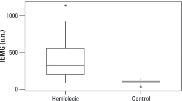
Fig 1. Comparison between lexor muscles (agonists) of hemiplegic and normal individuals during the wrist lexion.
15
10
5
0
Control Hemiplegic
Flexion
he comparisons between the muscles revealed that the agonists (lexor) are much more activated in the hemi-plegic patients (418.6±290.7 u.n.) when compared to clini-cally healthy individuals (106.3±26.8), the statistic test ap-plied in this case was Mann-Whitney that showed signif-icance between the groups (p<0.0001). his comparison is illustrated in the Fig 1. When the compared muscles were the antagonists (extensor), the diference were not signiicantly (p=0.26) by the t-student test; the hemiple-gic group achieved 71.6±22.1 u.n. and the control group 64.2±14.9. Consequently, when applied Mann-Whitney test, the agonist-antagonist relationship was signiicant-ly diferent too (p<0.0001), the relation was 5.8±3.5 for hemiplegics and 1.6±0.4 for control group (Fig 2).
Extension
In this movement, the diference was not signiicantly for any comparison. In relation to the agonists muscles, the extensor of hemiplegic groups demonstrated an ac-tivation of 106.1±19.1 u.n. and for the control group the activation was 110.6±19.4 (p=0.45). he antagonists (lex-or) showed an activation of 77.3±21.7 in the hemiplegic and 85.8±22.7 in the control (p=0.25). For these two com-parisons it was used t-student test. For agonist-antagonist relationship, it was used the Mann-Whitney test and the diference was not signiicantly too (p=0.45), the relation was 1.4±0.4 for hemiplegic and 1.3±0.2 for control.
discussion
During the wrist lexion, it was observed an important diference of the non-plegic side of hemiplegics in relation to the clinically healthy individuals; while the individuals of control group presented a mean activation of 106.3 u.n. in the lexor ulnaris, the hemiplegic individuals presented, in their normal side, a mean of 464.6 u.n. during the same movement, resulting in a p-value<0.0001. In the antago-nist muscles (extensor carpi ulnaris), it was not observed a signiicant diference. So, the agonist-antagonist rela-tionship during the wrist lexion have presented signii-cant diference between the groups; while in hemiplegic the mean relationship was 6.7, in control group it was 1.7 (p-value<0.0001). During the extension movement, there was no statistic diference between the evaluated groups. hese founds suggest that there is alteration in coor-dination and muscular compensation in the limb not af-fected by the stroke, showing that the non-plegic side of the hemiplegic individuals evaluated neither present the normal pattern found in the control group. It suggests that the central control of the motor units perhaps may be impaired, what contradicts a premise of some authors, that the motor units of the non-plegic limbs of subjects with hemiplegia were essentially normals11.
Mirbagueri et al.12 when studied the mechanical prop-erties of upper and lower extremities of hemiplegic have observed changes similar to these. he hemiplegic indi-viduals had intrinsic and relex stifness in the extremi-ties less afected by stroke, larger than the control sub-jects. One possible explanation comes from the hyperex-citability of stretch relexes in the non-paretic members and to the fact that the paths of the monoaminergic sys-tem are distributed bilaterally, and its activity may be in-creased due to stroke. Soon the corticospinal ibers that do not cross may have an increase in activity by altering the excitability of ipsilateral motoneurons. his physio-logical explanation may be the key for understand the al-tered patterns of activation found in our study.
Yarosh, Hofman and Stric13 in their study of surface electromyography of the extensor and lexor carpi during movement of the wrist found results similar to ours. Pa-tients with upper limb hemiparesis resultant of a unilater-al stroke had deicits in the ability to move the ipsilaterunilater-al wrist. he deicits were of dominant and non-dominant hemispheres injured: the ipsilateral wrist movements were less uncoordinated than the contralateral, but present-ed deicit in coordination to reach a target, were weak-er and slowweak-er than the healthy control subjects studied. In a previous study conducted by our group, when com-pared plegic and non-plegic sides of hemiplegic individu-als, it was showed that during wrist lexion there is a sig-niicantly lower activation of lexors in the plegic side. And for the extensor muscles there were no diference in rela-tion to the control group, both for lexion and the exten-sion14. It is known that in the stroke there is a preference of the spasticity for the lexor muscles in the upper limbs and extensor in the lower limbs15-17, what may explain why, ap-parently by the results, the stroke did not afect the pattern of neuro-motor activation of the extensor carpi ulnaris. Ponten et al.18 in a study about morphologyc properties of carpi lexor and extensor muscles in children with cerebral palsy, questioned why the lexor muscles are more strong as a group, suggested that may be possible that the hyper-activity of the nervous system simply activates both lex-ors and extenslex-ors. he lexlex-ors overlap the extenslex-ors, caus-ing the wrist lexion, because the moment arm of the lex-or is larger than the extenslex-or moment arm wrist in lexion, the lexor muscles have the appearance of being stronger.
Barela and Almeida19 compared the non-plegic side of individuals with hemiplegic spastic cerebral palsy with the dominant side of normal subjects in lexion of the shoulder and elbow. he results showed that the non-plegic side can-not be considered normal or intact, since the movements in the more distal were managed diferently from the prox-imal portions, which did not occur with normal subjects.
reported that the apparent weakness of the less afected limb by the stroke might be due to: lower percentage of the descending cortical tract ibers that are originated in the injured local and remains ipsilateral; or more general-ly, due to a sedentary lifestyle of hemiplegics, which may not be able to maintain the same force exerted by a non-dominant arm of a healthy person20.
Our results are added to a crescent group in literature that demonstrates the ipsilateral limb “non-afected” does not work normally after a unilateral stroke of brain motor areas21-23. Even with these studies pointing to the difer-ences inconsistencies are showed when we visualize the techniques used in the rehabilitation clinic of upper limbs of hemiplegics patients. One is the training of bilateral movements. his applies neurological postulates of motor coordination inter-members to activate motor synergies between members. Speciically, voluntary movements of the intact limb can facilitate voluntary movements in the paretic member. his activates the primary motor cortex and supplementary motor area for the member intact to increase the probability of voluntary muscle contraction (i.e. motor synergies) in the afected limb when symmet-rical movements are executed24.
he possible neural mechanisms underlying the bi-lateral movements are numerous. A basic assumption of the use of bilateral movement is that the therapy of bilat-eral symmetrical movements activate similar neural net-works in both hemispheres when homologous muscle groups are activated simultaneously. Bilateral symmet-rical movements, therefore, may allow the activation of the uninjured hemisphere to increase the activation of the injured hemisphere and facilitate control of plegic limb movements promoting neural plasticity.
When evaluated the reorganization of central nervous system with magnetic resonance functional during the therapy of bilateral symmetrical movements, the non-pa-retic hand’s movement have increased the activation of the uninjured hemisphere25. he bilateral training lead to an increased recruitment of sensory-motors areas of the contralateral hemisphere and the ipsilateral cerebelum. his recruitment is frequently explained due to the exis-tence of cortico-spinal ibers that do not cross in the py-ramidal decussation and are latent in healthy people. Its functional relevance is not yet clear. In patients with mo-tor deiciency after stroke, the rehabilitation with specif-ic bilateral repetitive therapy of upper extremity appears to induce the reorganization in the neural networks con-tralateral to the lesion, in brain hemisphere and cerebel-lum, and can operates by recruitment of these brain ar-eas in order to supply functional beneits24.
he literature in this area - studying motor control, es-pecially in upper extremities - is still scarce. here are a lit-tle amount of researches looking for diference in the
non-plegic side; in order to understand exactly what occurs in terms of motor control, more studies should be conducted. his study contributes to understand that the non-plegic side is not normal when compared with a control group, so it should be considered that although the patients have diagnostic of unilateral stroke, we can not ignore that there may be a microdamage contralateral to the plegia not ob-served on imaging studies, which may explain in part the observed changes in the electromyography spectrum.
RefeRences
Chae J, Hart R. Intramuscular hand neuroprosthesis for chronic stroke survi-1.
vors. Neurorehabil Neural Repair 2003;17:109-117.
Chae J, Yang G, Park BK, Labatia I. Muscle weakness and co-contraction in 2.
upper limb hemiparesis: relationship to motor impairment and physical dis-ability. Neurorehabil Neural Repair 2002;16:241-248.
Thrasher TA, Zivanovic V, McIlroy W, Popovic MR. Rehabilitation of reaching 3.
and grasping function in severe hemiplegic patients using functional electri-cal stimulation therapy. Neurorehabil Neural Repair 2008;22:706-714. Jankowska E, Edgley SA. How can corticospinal tract neurons contribute to 4.
ipsilateral movements? A question with implications for recovery of motor functions. Neuroscientist 2006;12:67-79.
Machado ABM. Neuroanatomia funcional. São Paulo: Atheneu, 1993. 5.
Lent R. Cem bilhões de neurônios: conceitos fundamentais de neurociências. 6.
São Paulo: Atheneu, 2004.
Brodal A. Anatomia neurológica com correlações clínicas. São Paulo: Rocca, 1984. 7.
Lundy-Ekman L. Neurociência: fundamentos para a reabilitação. Rio de Ja-8.
neiro: Guanabara Koogan, 2000.
Sunnerhagen KS, Svantesson U, Lonn L, Krotkiewski M, Grimby G. Upper mo-9.
tor neuron lesions: their efect on muscle performance and appearance in stroke patients with minor. Arch Phys Med Rehabil 1999;80:155-161. Sinkjaer T, Magnusson I. Passive, intrinsic and relex-mediated stifness in the 10.
ankle extensors of hemiparetic patients. Brain 1994;117:355-363. Tang A, Rymer WZ. Abnormal force: EMG relations in paretic limbs of hemipa-11.
retic humans subjects. J Neurol Neurosurg Psychiatry 1981;44:690-698. Mirbagheri MM, Alibiglou L, Thajchayapong M, Rymer,WZ. Muscle and relex 12.
changes with varying joint angle in hemiparetic stroke. J Neuroeng Rehab 2008;98:629-637.
Yarosh CA, Hofman DS, Strick PL .Deicits in movements of wrist ipsilateral to 13.
a stroke in hemiparetic subjects. J Neurophsiol 2004;92:3276-3285. Kuriki HU, Azevedo FM, Filho RFN, Alves N, Carvalho AC. Comparative anal-14.
ysis of electromyography pattern in the forearm muscles of hemiplegic pa-tients. Electromyogr Clin Neurophsysiol 2008;48:367-372.
Bobath B. Hemiplegia no adulto: avaliação e tratamento. São Paulo: Manole, 1978. 15.
Cook AS, Woollacott MH Controle motor: teoria e aplicações práticas. São 16.
Paulo: Manole, 2003.
Hu XL, Tong KY, Song R, et al. Quantitative evaluation of motor functional re-17.
covery process in chronic stroke patients during robot-assisted wrist train-ing. J Electromyogr Kinesiol 2008;19:639-650.
Ponten E, Frindén J, Thornell L, Lieber, R L.Spastic wrist lexors are more se-18.
verely afected than wrist extensors in children with cerebral palsy. Dev Med Child Neurol 2005;47:384-389.
Barela AMF, Almeida GL. Controle de movimentos voluntários no membro 19.
superior não plégico de portadores de paralisia cerebral hemiplégica espás-tica. Rev Bras Fisioterapia 2006;10:325-332.
Mc Crea PH, Eng JJ, Hodgson AJ. Time and magnitude of torque generations 20.
is impaired in both arms following stroke. Muscle Nerve 2003;28:46-53. Carey JR, Baxter TL, Di Fabio RP. Tracking control in the nonparetic hand of 21.
subjects with stroke. Arch Phsys Med Rehabil 1998;79:435-441.
Desrosiers J, Bourbonnais D, Bravo G, Roy P-M, Guay M. Performance of the “un-22.
afected” upper extremity of elderly stroke patients. Stroke 1996;27:1564-1570. Winstein CJ, Pohl PS. Efects of unilateral brain damage on the control of 23.
goal-directed hand movements. Exp Brain Res 1995;105:163-174. Stewart KC, Cauraugh JH, Summers JJ. Bilateral movement training and 24.
stroke rehabilitation: a systematic review and meta-analysis. J Neurol Sci 2006;244:89-95.
Luft AR, McCombe-Waller S, Whitall J, et al. Repetitive Bilateral arm training 25.