I
k
B Kinase in the
Drosophila
Toll Pathway
Jessica Daigneault, Liv Klemetsaune, Steven A. Wasserman*
Section of Cell and Developmental Biology, University of California San Diego, La Jolla, California, United States of America
Abstract
Toll receptors transduce signals that activate Rel-family transcription factors, such as NF-kB, by directing proteolytic degradation of inhibitor proteins. In mammals, the IkB Kinase (IKK) phosphorylates the inhibitor IkBa. AbTrCP protein binds to phosphorylated IkBa, triggering ubiquitination and proteasome mediated degradation. In Drosophila, Toll signaling directs Cactus degradation via a sequence motif that is highly similar to that in IkBa, but without involvement of IKK. Here we show that Pelle, the homolog of a mammalian regulator of IKK, acts as a Cactus kinase. We further find that the flybTrCP protein Slimb is required in cultured cells to mediate Cactus degradation. These findings enable us for the first time to trace an uninterrupted pathway from the cell surface to the nucleus forDrosophilaToll signaling.
Citation:Daigneault J, Klemetsaune L, Wasserman SA (2013) The IRAK Homolog Pelle Is the Functional Counterpart of IkB Kinase in theDrosophilaToll Pathway. PLoS ONE 8(9): e75150. doi:10.1371/journal.pone.0075150
Editor:Andreas Bergmann, University of Massachusetts Medical School, United States of America
ReceivedJuly 30, 2013;AcceptedAugust 9, 2013;PublishedSeptember 23, 2013
Copyright:ß2013 Daigneault et al. This is an open-access article distributed under the terms of the Creative Commons Attribution License, which permits unrestricted use, distribution, and reproduction in any medium, provided the original author and source are credited.
Funding:Funding provided by NIH RO1 GM05054516. The funders had no role in study design, data collection and analysis, decision to publish, or preparation of the manuscript.
Competing Interests:The authors have declared that no competing interests exist.
* E-mail: stevenw@ucsd.edu
Introduction
Toll and Toll-like receptors (TLRs) govern the nuclear localization and activity of NF-kB and other Rel-related transcription factors via an evolutionarily conserved signal transduction pathway [1,2,3]. Prior to Toll signaling, Rel proteins are held in the cytoplasm by a tightly bound inhibitor that belongs to the IkB family. Activated Toll receptors relay signals that trigger IkB protein degradation, freeing the Rel proteins to translocate into nuclei, bind DNA, and regulate gene expression.
In the fruit flyDrosophila melanogaster, Toll signaling functions in both development and immunity [4,5]. Toll first acts in the syncytial embryo, where spatially graded signaling establishes the dorsoventral axis. In larvae and adults, Toll mediates the humoral immune response to fungi and Gram (+) bacteria. Embryonic axis formation requires the Rel protein Dorsal, whereas innate immune responses involve either Dorsal or the Dorsal-related immune factor (Dif), another Rel protein [6]. Cactus, a fly IkB protein, is the inhibitor for both Dif and Dorsal. The adaptor proteins MyD88 and Tube, as well as the protein kinase Pelle, transduce signals from Toll to Cactus. These three signal-relay proteins each contain a death domain, a protein-protein interaction motif that mediates formation of a submembranous Toll signaling complex [7,8].
LikeDrosophila Toll, most mammalian TLRs signal via three
death domain proteins. Indeed, mammalian MyD88, IRAK4 and IRAK1 are the counterparts of fly MyD88, Tube, and Pelle, respectively [9,10]. However, additional factors link the death domain complex to IkB in mammalian innate immune signaling. In particular, signaling by the IRAK 1,2, and 4 proteins requires the adaptor TRAF6, the TAB proteins, the protein kinase TAK1, and the IkB Kinase (IKK) complex [1,11]. IKK-mediated phosphorylation of IkBa at two sites, Ser32 and Ser36, triggers
ubiquitination, leading to proteasome mediated IkBadegradation [12,13].
The Toll responsive sites in IkBaand Cactus share substantial sequence similarity [12,14,15]. In fact, the signal-responsive domain of IkBacan functionally substitute for the corresponding region of the Cactus protein [16]. Surprisingly, theDrosophilaIKK does not function in the fly Toll pathway [17,18,19].
The fact that Toll-directed Cactus phosphorylation is IKK independent leads to two important questions. First, why are the signal responsive sites conserved when the kinase is not? Second, what kinase phosphorylates these sites in response to Toll activation? Here we address these two questions.
Results
Drosophilab-TrCP can Mediate Cactus Degradation
To explain the sequence similarity of the signal responsive sites in IkBaand Cactus, it has been proposed that Cactus, like IkBa, is targeted for degradation by a b-TrCP protein acting as the substrate recognition subunit for an SCF E3 ubiquitin ligase [16,20]. In mammals, the F-box/b-TrCP protein of the SCF (Skp1/Cullin/F-Box) complex binds specifically to the phosphor-ylated form of the IkBa motif DS32GLDS36. This recognition
initiates ubiquitination, generating the signal for proteasomal recognition and proteolysis [1,21,22]. The recognition site forb -TrCP is thus a degron that initiates protein degradation in response to phosphorylation. Characterization of the degrons of a number ofb-TrCP targets has demonstrated that they are highly similar in sequence, with a consensus sequence of DSGxxS, but are regulated by a diverse set of protein kinases (Figure 1A).
recognition site. In flies, however, evidence for a role ofb-TrCP protein in Cactus degradation is somewhat contradictory. The slimb locus is the sole b-TrCP gene in the Drosophila genome. Whereas a loss-of-function mutation in slimb blocks the Toll-dependent transcription of the patterning genessnailandtwistin embryos, Toll-dependent transcription of the antifungal gene drosomycin is unaffected in adults [20,23]. Furthermore, ectopic expression of the viralb-TrCP inhibitor VPU in flies reduces but does not eliminate Pelle-mediated degradation of Cactus [20].
To investigate further the role of the Slimbb-TrCP in Cactus degradation, we used S2 cells transfected with a construct fusing the extracellular domain of EGFR to the transmembrane and intracellular domains of Toll. We have previously demonstrated that this chimeric receptor renders the Toll pathway responsive to EGF, directing Cactus degradation that is detectable within one minute of EGF addition [24]. To assay the requirement for Slimb
in Toll signaling, we used EGF activation of EGFR-Toll in combination with RNA interference (RNAi). The results were unequivocal. As shown in Figure 1C, RNAi against slimb abrogated Toll-directed degradation of Cactus. Indeed, eliminat-ingslimbfunction had the same effect as did RNAi againstMyD88, an essential component of the fly Toll pathway. In contrast, RNAi againsteaster, a gene that acts upstream of Toll, or againstImd, a gene essential to the IKK-dependent immune pathway, was without consequence. We conclude that Toll-directed Cactus degradation in these cells requires theb-TrCP protein Slimb.
Pelle Phosphorylates Signal-dependent Sites in Cactus
Cactus degradation [15,25,26,27]. Furthermore, Pelle undergoes autophosphorylation, autoactivation, and colocalization with Tube, events that occur concomitant with signaling [28,29,30]. There is also the fact that forward genetic screens and genome-wide RNAi screens have failed to identify any other protein kinase required for Cactus degradation [31,32,33,34]. We therefore set out to assay Pelle as a Cactus kinase.
We began by expressing recombinant forms of Cactus and Pelle inEscherichia coli. We expressed full-length Cactus (500 aa), isolated the protein from inclusion bodies, and carried out solubilization and renaturation by the method of Nu¨sslein-Volhard and colleagues [35]. Expressing Pelle in E. coli proved more problematic, due to the frequent appearance of mutations that blocked Pelle production or inactivated the kinase domain. To circumvent the toxicity of an active Pelle kinase in bacteria, we adopted the strategy of co-expressing an antagonizing protein phosphatase [36,37,38]. For this purpose we chose l protein phosphatase, which contains just 221 residues, is readily expressed in bacteria, and has very broad substrate specificity [39,40].
To ensure coordinated expression of Pelle and thel phospha-tase, we constructed a cistron consisting of a GST-Pelle fusion gene immediately 59 to the phosphatase gene. We induced expression inE. coliand used affinity chromatography to purify the Pelle fusion protein away from the l phosphatase and other proteins. Upon incubation at high concentration with Mg2+and
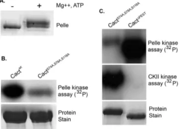
ATP, recombinant Pelle protein underwent autoactivation and autophosphorylation, as evident in a slight decrease in electro-phoretic mobility (Figure 2a). Pelle from Drosophila tissues similarly undergoes autoactivation and autophosphorylation fol-lowing phosphatase treatment [30].
With an active Pelle preparation in hand, we set out to investigate Cactus as a potential substrate. We prepared two His6
-Cactus isoforms, the wild-type Cactwt, and the triple mutant CactS74A,S78A,S116A. In the latter isoform, the serine-to-alanine substitutions at positions 74, 78, and 116 (see Figure 1B), are sufficient to block Toll-dependent degradation of Cactus in vivo [14,15,16]. We incubated Pelle with the Cactus proteins in the presence of [c-32P]-ATP and assayed phosphorylation by gel
electrophoresis and autoradiography. We observed robust phos-phorylation of wild-type Cactus, but dramatically reduced phosphorylation of the triple mutant (Figure 2B). Indeed, although the residues altered in this mutant represent only 6% of the serines in Cactus, mutating these three sites consistently reduced phosphorylation by 75–80%.
Next, we compared the site specificity of Pelle with that of Casein Kinase II (CKII). We have previously demonstrated that CKII phosphorylates target residues in the carboxyl-terminal PEST domain, mediating Toll-independent destabilization of Cactus [41]. To compare the activity of the two kinases toward Cactus, we used as substrate CactS74A,S78A,S116A, in which the signal-dependent sites are mutated, and CactDPEST, in which the signal-independent sites are deleted. As shown in Figure 2C, Pelle and CKII exhibited reciprocal specificity. Under conditions where Pelle had only minor activity toward substrate CactS74A,S78A,S116A,
CKII mediated extensive radiolabeling of this protein. Similarly, Pelle had robust activity toward CactDPEST, whereas CKII catalyzed phosphorylation of this substrate was virtually undetect-able. We conclude that Pelle acts as a Cactus kinase and preferentially phosphorylates Cactus at the serines required for signal responsiveness.
Pelle Phosphorylates Signal-responsive Sites in IkBa
Stein and colleagues have shown thatDrosophilaToll signaling can target the signal dependent sites of IkBa as well as Cactus [16]. We therefore predicted that Pelle should exhibit in vitro activity toward the two serines in the IkBadegron, GS32GLDS36.
To test this hypothesis we turned to a chimeric substrate, Ik B-CactD144, in which the signal-responsive region (residues 1–68) of IkBareplaces the corresponding region (residues 1–144) of Cactus (see teal colored regions in Figure 1B). This chimera not only functionally substitutes for Cactus in fly embryos, but also strictly requires residues Ser32 and Ser36 for this activity [16].
We expressed and purified IkB-CactD144 from bacteria. We then assayed Pelle activity using an anti-IkBaserum that is specific for the IkBa phosphorylated at both Ser32 and Ser36 in the degron. As shown in Figure 3, the phospho-specific antiserum detects a strong, Pelle-dependent signal, confirming that, as predicted, Pelle directly modifies the IkBadegron, GS32GLDS36.
Discussion
Phosphoregulation of Cactus Stability
Studies in fly embryos have identified two processes, one signal independent and one signal dependent, that regulate Cactus stability [42]. The kinase responsible for each activity is now known. We previously used an in-gel kinase assay to purify the kinase mediating signal independent degradation [41]. We identified this kinase as Casein Kinase II (CKII) and showed that
Figure 2. Pelle-mediated Cactus phosphorylation exhibits specificity for Toll-responsive sites. A. Autophosphorylation of recombinant GST-Pelle, stained with Coomassie brilliant blue. B. (Top) Autoradiogram of Cactus isoforms phosphorylated with GST-Pelle in the presence of [c-32P]-ATP. (Bottom) SDS-PAGE of unlabeled substrates. C.
Autoradiograms of Cactus isoforms radiolabelled with GST-Pelle or Casein Kinase II (CKII) in the presence of [c-32P]-ATP.
doi:10.1371/journal.pone.0075150.g002
Figure 3. Pelle phosphorylates the IkBadegron.Isoforms of the IkB-CactD144 chimera were incubated with or without recombinant Pelle and the reaction products immunoblotted with an antiserum specific for the phosphorylated forms of serines 32 and 36.
it destabilized Cactus by modifying consensus target residues in the carboxyl-terminal PEST domain. Mammalian CKII similarly modifies and destabilizes IkBa [43]. Here, we have shown that Pelle phosphorylates the signal-regulated sites in the amino-terminal domain of Cactus. Work from our lab and others has also shown that Pelle protein kinase activity is required downstream of Tube to direct Cactus degradation in response to Toll activation. Pelle thus has the in vivo and in vitro properties expected of the Cactus kinase.
Functionally, Pelle-catalyzed phosphorylation of Cactus is analogous to that of the mammalian IKK complex acting on IkBa. Different kinases mediate phosphorylation, but at highly similar sites. This site conservation has been suggested to reflect a need for SCF mediated recognition, a hypothesis supported by our finding that the Slimbb-TrCP is required in S2 cells for Cactus degradation. We note, however, that the Slimbb-TrCP may not mediate Cactus degradation in all cells and at all stages, since Lemaitre and colleagues have provided evidence that Slimb is not the sole mediator of Cactus degradation in adult flies [20]. Involvement of an additional F box protein could explain why the Cactus of flies and other invertebrates typically contains two degrons, with the more carboxyl-terminal motif deviating slightly but consistently from the b-TrCP consensus (Figure 1 and unpublished data).
Pelle as theDrosophilaToll Pathway Kinase
The kinase activity of Pelle appears to fulfill multiple roles in Toll signaling. We have shown here that Pelle modifies Cactus. In addition, we and others have shown previously that Pelle phosphorylates Tube in vitro [27,28]. This activity may play an in vivorole in terminating signal transduction, since Pelle activity as a protein kinase mediates feedback regulation on Tube localization [27]. In addition, it is known that Dorsal undergoes signal dependent phosphorylation and can respond to Toll signaling in the absence of Cactus [44,45,46,47]. We speculate that Pelle is also responsible for modification of Dorsalin vivo.
By themselves, Pelle and Cactus do not appear to stably interact [28,48]. However, Pelle binds to Tube; Tube and Pelle both bind to Dorsal; and Dorsal binds to Cactus [28,45,46,48,49,50,51]. Taking all of these interactions into consideration, we can now generate an illustration of the overall pathway in Drosophila embryos for signaling by activated Toll receptors (Figure 4).
The protein-protein interactions shown in Figure 4 likely increase the efficiency or specificity of Pelle activity when Dorsal is the Toll target. They may, for example, provide steric constraints on the interaction of Pelle and Cactus that enhance site preference. They might also explain why a Cactus isoform lacking the destabilizing effect of the PEST domain requires the presence of Dorsal for efficient response to Toll signaling [16]. However, the same interactions may not be important when Dif is the target, since Tube fails to bind Dif under conditions in which it stably binds to Dorsal [9]. Why this difference? One possibility is that the establishment of a nuclear concentration gradient of Dorsal across the embryo requires much more tightly regulated protein localization and diffusion than does the nuclear import of Dif in response to immune challenge.
Although recruitment of Toll components into a signaling complex likely influences Pelle activity, several lines of evidence reveal that such a complex is not strictly required for the action of Pelle on Cactus. First, deleting the region of Tube that mediates binding to Dorsal has a relatively mild effect on Toll signaling [48,50,52]. Second, expression of a fusion construct that targets Pelle to the plasma membrane can effect signaling in the absence of Tube [28,53]. Third, activation of Toll in embryos by injection
of the Toll ligand Spa¨tzle can trigger a detectable level of Cactus degradation in the absence of Dorsal [42]. Consistent with these results, we find that Pelle preferentially phosphorylates signal responsive sites in Cactusin vitroin the absence of any other Toll pathway proteins.
Given that mammals require eight proteins in the portion of the Toll pathway that in flies is occupied by Pelle, it is appealing to consider theDrosophila pathway a model of simplicity. We note, however, that the history of Toll studies in flies and mammals is full of unexpected parallels as well as minor differences of major significance. Having offered the model shown in Figure 4, we will leave judgments regarding simplicity or complexity to others.
Materials and Methods
Molecular Biology
The GST Pelle-l protein phosphatase (PPase) operon was generated by subcloning thel-PPase gene from a pT7 vector (gift of Jack Dixon) into the Not I site of a pGEX 4T-1 vector (Amersham), and then inserting the Pelle coding sequence as an in-frame fusion with GST using EcoRI and XhoI restriction sites. The result was a 27 bp separation between coding regions of GST-Pelle andl-PPase, with a Shine-Delgarno sequence 8 bases upstream of the phosphatase translational start site. For Cactus, PCR amplification was used to introduced an amino-terminal His6
tag, with the tagged construct being inserted into the BamHI and Figure 4. Model for Toll signaling inDrosophilaembryos.Upon binding activated (processed) Spa¨tzle, Toll dimerizes. The resulting conformational change allows the Toll TIR domain to bind to the TIR domain of the adaptor protein MyD88. (For simplicity, interactions and downstream events are shown for only one monomer of a dimeric Toll receptor). MyD88 brings along with it the adaptor protein Tube, an association mediated by a death domain in each protein. The bivalent Tube death domain then binds the death domain of Pelle, recruiting the inactive protein kinase into the signaling complex. Within the complex, Pelle undergoes autoactivation via autophosphorylation. Pelle then interacts with Dorsal-bound Cactus, an interaction enhanced by the binding of Dorsal to both Tube and Pelle. Pelle phosphorylates Cactus, triggering recognition by the SlimbbTrCP and subsequent ubiquitina-tion and proteasome mediated degradaubiquitina-tion. Pelle likely also phosphor-ylates Dorsal, enhancing its gene regulatory activity upon release from Cactus, and Tube, initiating negative feedback regulation on Toll signaling.
EcoRI restriction sites in the pRSET-A vector (Invitrogen). To generate the IkBachimera, IkBa(a gift from Michael Karin) was fused with cactusDPEST and ligated into the pRSET-B vector (Invitrogen) in frame with the polyhistidine tag. All point mutations were generated by PCR SOEing [54].
S2 Cell Studies
S2 cells stably transfected with EGFR-Toll were treated with dsRNA and with mouse EGF, as described [24]. After 20 min, cells were harvested and 15mg samples resolved on 8% SDS-PAGE and immunoblotted onto PVDF. Cactus was detected with a rabbit polyclonal antiserum (1:10,000), described previously [15].
Expression and Purification of Pelle
BL21 cells were transformed with Pelle-lPPase construct and protein expression was induced as described [39]. BL21 cells transformed with the Pelle-lPPase construct were grown for 5 hours at 28uC following IPTG induction. Cells from a 500 ml culture were pelleted and resuspended in 40 ml buffer containing 50 mM Tris HCl, pH 7.5; 2 mM EGTA; 0.5 M NaCl; and a protease inhibitor cocktail tablet (Roche). Cells were frozen and thawed; incubated with lysozyme, RNase, and DNase I in the presence of MgCl; and then lysed by three cycles of freezing (liquid nitrogen) and thawing (37uC). The lysate was spun at 25,000 g for 20 min at 4uC and the supernatant was decanted and incubated with 2 ml 50% glutathione Sepharose 4B resin slurry (GE Healthcare) in PBS for 2 hours at 4uC with gentle rocking. The beads were recovered by centrifugation at 500 g for 3 min at 4uC and the resin was washed three times in 10 ml PBS. GST-Pelle was eluted by adding 1 ml of elution buffer (10 mM glutathione, 50 mM Tris HCl, pH 8) and incubated for 1 hr at 4uC with gentle rocking.
Expression and Purification of His6-cactus and Chimera Wild-type and mutant forms of Cactus and the IkB-CactD144 chimera were each expressed with an amino-terminal His6tag in
the pRSET vector in BL21 cells. Cactus proteins were renatured according to the protocol of Nu¨sslein-Volhard and colleagues [35]. Wild-type and mutant forms of Cactus and the IkB-CactD144 chimera were each expressed with an amino-terminal His6tag in
the pRSET vector in BL21 cells. Expression was induced with 1 mM IPTG at 37uC for 3 hours. The lysate from a 500 ml culture was prepared by freeze/thaw cycles as described above and spun at 9000 g for 20 min. The pellet was resuspended in
20 ml buffer (20 mM Tris HCl, pH 8, 100 mM NaCl, 2 mM DTT, 2 mM EDTA, 1% Triton-X 100, K6complete protease inhibitor tablet (Roche), stirred at 4uC for 1 hour, spun at 9000 g for 20 min, and resuspended in the same buffer without Triton-X 100. After incubation with stirring at 4uC for 1 hr, the preparation was pelleted and then resuspended to 10 mg/ml protein in urea buffer (20 mM Tris HCl, pH 8, 10 mM DTT, 1 mM PMSF, 8M urea) prior to renaturation.
Radiolabeling Assay of Pelle
GST-pelle (10mg) was pre-incubated for 30 min at 30uC in 16kinase buffer (10mM ATP, 10 mM MgCl2, 50 mM b
-glycerophosphate, 25mM HEPES, pH 6.5) to allow activation by autophosphorylation. The activated Pelle (0.5mg) was then incubated with recombinant His6-Cactus (1.6mg) in a volume of
40ml in the presence of [c-32P]-ATP (13.3mCurie, Perkin Elmer) in 16kinase buffer. Following reaction for 5 min at room temperature, samples were mixed with 10ml 56SDS loading sample buffer, boiled, and loaded (30ml) on an 8% SDS protein gel. The gel was fixed twice for 30 min in fixing solution (50% methanol, 10% acetic acid) on a shaker, before being dried in a gel dryer for 1 hr. The gel was then exposed to a film and developed. Quantitation was performed with ImageJ software.
Immunoblot Assay of Pelle
Reactions were carried out as described above, except the kinase reactions were carried out with 11.2mg of the chimera for 5 min at 30uC without radiolabel. Immunoblotting of the proteins transferred to a nitrocellulose membrane used rabbit anti-IkBa [pSpS32/36] phosphospecific antibody (Invitrogen, 1:10,000) and a-rabbit secondary antibody (Sigma, 1:5,000), followed by detection using the Western Lightning-ECL chemiluminescence substrate (Perkin Elmer).
Acknowledgments
We thank David Stein, Richard Honkanen, Sailen Barik, Jack Dixon, Michael Karin, Allen Ninh, Huaiyu Sun and Par Towb for technical assistance and gifts of reagents. We also thank members of the Wasserman and McGinnis research groups for helpful comments on the manuscript.
Author Contributions
Conceived and designed the experiments: SW LK JD. Performed the experiments: LK JD. Analyzed the data: SW LK JD. Wrote the paper: SW LK.
References
1. Karin M, Ben-Neriah Y (2000) Phosphorylation meets ubiquitination: the control of NF-[kappa]B activity. Annu Rev Immunol 18: 621–663. 2. Silverman N, Maniatis T (2001) NF-kappaB signaling pathways in mammalian
and insect innate immunity. Genes Dev 15: 2321–2342.
3. Akira S (2006) TLR signaling. Curr Top Microbiol Immunol 311: 1–16. 4. Lemaitre B, Hoffmann J (2007) The host defense of Drosophila melanogaster.
Annual review of immunology 25: 697–743.
5. Lindsay SA, Wasserman SA (2013) Conventional and non-conventional Drosophila Toll signaling Developmental & Comparative Immunology: in press. 6. Ip YT, Reach M, Engstrom Y, Kadalayil L, Cai H, et al. (1993) Dif, a dorsal-related gene that mediates an immune response in Drosophila. Cell 75: 753–763. 7. Sun H, Bristow BN, Qu G, Wasserman SA (2002) A heterotrimeric death domain complex in Toll signaling. Proc Natl Acad Sci U S A 99: 12871–12876. 8. Moncrieffe MC, Grossmann JG, Gay NJ (2008) Assembly of oligomeric death domain complexes during Toll receptor signaling. The Journal of Biological Chemistry 283: 33447–33454.
9. Towb P, Sun H, Wasserman SA (2009) Tube Is an IRAK-4 homolog in a Toll pathway adapted for development and immunity. Journal of Innate Immunity 1: 309–321.
10. Lin SC, Lo YC, Wu H (2010) Helical assembly in the MyD88-IRAK4-IRAK2 complex in TLR/IL-1R signalling. Nature 465: 885–890.
11. Kawai T, Akira S (2006) TLR signaling. Cell Death Differ 13: 816–825. 12. Brown K, Gerstberger S, Carlson L, Franzoso G, Siebenlist U (1995) Control of
IkB-a proteolysis by site-specific, signal-induced phosphorylation. Science 267: 1485–1488.
13. Chen ZJ, Parent L, Maniatis T (1996) Site-specific phosphorylation of IkappaBalpha by a novel ubiquitination-dependent protein kinase activity. Cell
84: 853–862.
14. Bergmann A, Stein D, Geisler R, Hagenmaier S, Schmid B, et al. (1996) A gradient of cytoplasmic Cactus degradation establishes the nuclear localization gradient of thedorsalmorphogen inDrosophila. Mechanisms of Development 60: 109–123.
15. Reach M, Galindo RL, Towb P, Allen JL, Karin M, et al. (1996) A gradient of cactus protein degradation establishes dorsoventral polarity in the Drosophila embryo. Developmental Biology 180: 353–364.
16. Fernandez NQ, Grosshans J, Goltz JS, Stein D (2001) Separable and redundant regulatory determinants in Cactus mediate its dorsal group dependent degradation. Development 128: 2963–2974.
18. Rutschmann S, Jung AC, Zhou R, Silverman N, Hoffmann JA, et al. (2000) Role of Drosophila IKK gamma in a toll-independent antibacterial immune response. Nat Immunol 1: 342–347.
19. Lu Y, Wu LP, Anderson KV (2001) The antibacterial arm of the drosophila innate immune response requires an IkappaB kinase. Genes Dev 15: 104–110. 20. Leulier F, Marchal C, Miletich I, Limbourg-Bouchon B, Benarous R, et al. (2003) Directed expression of the HIV-1 accessory protein Vpu in Drosophila fat-body cells inhibits Toll-dependent immune responses. EMBO reports 4: 976– 981.
21. Skaar JR, Pagan JK, Pagano M (2013) Mechanisms and function of substrate recruitment by F-box proteins. Nature reviews Molecular cell biology 14: 369– 381.
22. Kanarek N, Ben-Neriah Y (2012) Regulation of NF-kappaB by ubiquitination and degradation of the IkappaBs. Immunological reviews 246: 77–94. 23. Spencer E, Jiang J, Chen ZJ (1999) Signal-induced ubiquitination of IkBa by the
F-box protein Slimb/b-TrCP. Genes and Development 13: 284–294. 24. Sun H, Towb P, Chiem DN, Foster BA, Wasserman SA (2004) Regulated
assembly of the Toll signaling complex drives Drosophila dorsoventral patterning. The EMBO journal 23: 100–110.
25. Shelton CA, Wasserman SA (1993) pelle encodes a protein kinase required to establish dorsoventral polarity in theDrosophilaembryo. Cell 72: 515–525. 26. Hecht PM, Anderson KV (1993) Genetic characterization of tube and pelle,
genes required for signaling between Toll and dorsal in the specification of the dorsal- ventral pattern of the Drosophila embryo. Genetics 135: 405–417. 27. Towb P, Bergmann A, Wasserman SA (2001) The protein kinase Pelle mediates
feedback regulation in the Drosophila Toll signaling pathway. Development 128: 4729–4736.
28. Grosshans J, Bergmann A, Haffter P, Nusslein-Volhard C (1994) Activation of the kinase Pelle by Tube in the dorsoventral signal transduction pathway of Drosophila embryo. Nature 372: 563–566.
29. Towb P, Galindo RL, Wasserman SA (1998) Recruitment of Tube and Pelle to signaling sites at the surface of the Drosophila embryo. Development 125: 2443– 2450.
30. Shen B, Manley JL (2002) Pelle kinase is activated by autophosphorylation during Toll signaling in Drosophila. Development 129: 1925–1933. 31. Brennan CA, Anderson KV (2004) Drosophila: the genetics of innate immune
recognition and response. Annu Rev Immunol 22: 457–483.
32. Huang HR, Chen ZJ, Kunes S, Chang GD, Maniatis T (2010) Endocytic pathway is required for Drosophila Toll innate immune signaling. Proc Natl Acad Sci U S A 107: 8322–8327.
33. Kuttenkeuler D, Pelte N, Ragab A, Gesellchen V, Schneider L, et al. (2010) A large-scale RNAi screen identifies Deaf1 as a regulator of innate immune responses in Drosophila. J Innate Immun 2: 181–194.
34. Valanne S, Myllymaki H, Kallio J, Schmid MR, Kleino A, et al. (2010) Genome-wide RNA interference in Drosophila cells identifies G protein-coupled receptor kinase 2 as a conserved regulator of NF-kappaB signaling. J Immunol 184: 6188–6198.
35. Geisler R, Bergmann A, Hiromi Y, Nu¨sslein-Volhard C (1992) Cactus, a gene involved in dorsoventral pattern formation of Drosophila, is related to the IkB gene family of vertebrates. Cell 71: 613–621.
36. Weijland A, Neubauer G, Courtneidge SA, Mann M, Wierenga RK, et al. (1996) The purification and characterization of the catalytic domain of Src expressed in Schizosaccharomyces pombe. Comparison of unphosphorylated
and tyrosine phosphorylated species. European journal of biochemistry/FEBS 240: 756–764.
37. Seeliger MA, Young M, Henderson MN, Pellicena P, King DS, et al. (2005) High yield bacterial expression of active c-Abl and c-Src tyrosine kinases. Protein science : a publication of the Protein Society 14: 3135–3139.
38. Wang YH, Ayrapetov MK, Lin X, Sun G (2006) A new strategy to produce active human Src from bacteria for biochemical study of its regulation. Biochemical and Biophysical Research Communications 346: 606–611. 39. Zhuo S, Clemens JC, Hakes DJ, Barford D, Dixon JE (1993) Expression,
purification, crystallization, and biochemical characterization of a recombinant protein phosphatase. The Journal of biological chemistry 268: 17754–17761. 40. Elling RA, Tangonan BT, Penny DM, Smith JT, Vincent DE, et al. (2007)
Mouse Aurora A: expression in Escherichia coli and purification. Protein expression and purification 54: 139–146.
41. Liu Z-P, Galindo RL, Wasserman SA (1997) Phosphorylation of Cactus by CKII is required for wild-type Cactus function in Drosophila embryos. Genes & Development 11: 3413–3422.
42. Belvin MP, Jin Y, Anderson KV (1995) Cactus protein degradation mediates Drosophila dorsal-ventral signaling. Genes & Development 9: 783–793. 43. Barroga CF, Stevenson JK, Schwarz EM, Verma IM (1995) Constitutive
phosphorylation of I kappa B alpha by casein kinase II. Proceedings of the National Academy of Sciences of the United States of America 92: 7637–7641. 44. Roth S, Hiromi Y, Godt D, Nu¨sslein-Volhard C (1991) Cactus, a maternal gene required for proper formation of the dorsoventral morphogen gradient in Drosophila embryos. Development 112: 371–388.
45. Whalen AM, Steward R (1993) Dissociation of the dorsal-cactus complex and phosphorylation of the dorsal protein correlate with the nuclear localization of dorsal. Journal of Cell Biology 123: 523–534.
46. Gillespie SK, Wasserman SA (1994) Dorsal, a Drosophila Rel-like protein, is phosphorylated upon activation of the transmembrane protein Toll. Molecular & Cellular Biology 14: 3559–3568.
47. Drier EA, Govind S, Steward R (2000) Cactus-independent regulation of Dorsal nuclear import by the ventral signal. Current Biology 10: 23–26.
48. Edwards DN, Towb P, Wasserman SA (1997) An activity-dependent network of interactions links the rel protein Dorsal with its cytoplasmic regulators. Development 124: 3855–3864.
49. Kidd S (1992) Characterization of the Drosophila cactus locus and analysis of interactions between cactus and dorsal proteins. Cell 71: 623–635.
50. Yang J, Steward R (1997) A multimeric complex and the nuclear targeting of the Drosophila Rel protein Dorsal. Proc Natl Acad Sci U S A 94: 14524–14529. 51. Xiao T, Towb P, Wasserman SA, Sprang SR (1999) Three-dimensional
structure of a complex between the death domains of Pelle and Tube. Cell 99: 545–555.
52. Letsou A, Alexander S, Wasserman SA (1993) Domain mapping of tube, a protein essential for dorsoventral patterning of the Drosophila embryo. EMBO Journal 12: 3449–3458.
53. Galindo RL, Edwards DN, Gillespie SK, Wasserman SA (1995) Interaction of the pelle kinase with the membrane-associated protein tube is required for transduction of the dorsoventral signal in Drosophila embryos. Development 121: 2209–2218.