Effect of Natural and Semisynthetic
Pseudoguianolides on the Stability of NF-
κ
B:
DNA Complex Studied by Agarose Gel
Electrophoresis
Rodrigo Villagomez1,2, Rajni Hatti-Kaul3, Olov Sterner1, Giovanna Almanza2, Javier A. Linares-Pastén3*
1Centre for Analysis and Synthesis, Lund University, P.O. Box 124, 221 00 Lund, Sweden,2Instituto de Investigaciones Químicas, Facultad de Ciencias Puras y Naturales, Universidad Mayor de San Andrés, P.O. Box 303 La Paz, Bolivia,3Biotechnology, Dept. of Chemistry, Lund University, P.O. Box 124, SE-22 100 Lund, Sweden
Abstract
The nuclear factorκB (NF-κB) is a promising target for drug discovery. NF-κB is a heterodi-meric complex of RelA and p50 subunits that interact with the DNA, regulating the expres-sion of several genes; its dysregulation can trigger diverse diseases including inflammation, immunodeficiency, and cancer. There is some experimental evidence, based on whole cells studies, that natural sesquiterpene lactones (Sls) can inhibit the interaction of NF-κB with DNA, by alkylating the RelA subunit via a Michael addition. In the present work, 28 nat-ural and semisynthetic pseudoguianolides were screened as potential inhibitors of NF-κB in a biochemical assay that was designed using pure NF-κB heterodimer, pseudoguianolides and a ~1000 bp palindromic DNA fragment harboring two NF-κB recognition sequences. By comparing the relative amount of free DNA fragment to the NF-κB–DNA complex, in a
routine agarose gel electrophoresis, the destabilizing effect of a compound on the complex is estimated. The results of the assay and the following structure-activity relationship study, allowed the identification of several relevant structural features in the pseudoguaianolide skeleton, which are necessary to enhance the dissociating capacity of NF-κB–DNA
com-plex. The most active compounds are substituted at C-3 (α-carbonyl), in addition to having theα-methylene-γ-lactone moiety which is essential for the alkylation of RelA.
Introduction
The nuclear factorkB (NF-kB) is considered to be a promising target for drug discovery. It regulates the transcription of pro-inflammatory and anti-apoptotic proteins, among others; dysregulation can lead to the development of chronic inflammation, immunodeficiency and cancer. Hence, NF-kB plays an important role in oncogenesis, proliferation and cancer a11111
OPEN ACCESS
Citation:Villagomez R, Hatti-Kaul R, Sterner O, Almanza G, Linares-Pastén JA (2015) Effect of Natural and Semisynthetic Pseudoguianolides on the Stability of NF-κB:DNA Complex Studied by Agarose Gel Electrophoresis. PLoS ONE 10(1): e0115819. doi:10.1371/journal.pone.0115819
Academic Editor:Heidar-Ali Tajmir-Riahi, University of Quebect at Trois-Rivieres, CANADA
Received:September 3, 2014
Accepted:November 27, 2014
Published:January 23, 2015
Copyright:© 2015 Villagomez et al. This is an open access article distributed under the terms of the Creative Commons Attribution License, which permits unrestricted use, distribution, and reproduction in any medium, provided the original author and source are credited.
Data Availability Statement:All relevant data are within the paper and its Supporting Information files.
Funding:This work was financially supported by the Research Department of the Swedish International Development Cooperation Agency (SIDA-SAREC). The funders had no role in study design, data collec-tion and analysis, decision to publish, or preparacollec-tion of the manuscript.
metastasis [1–6]. The NF-kB regulation pathway is complex but has been thoroughly investi-gated, and involves several steps [7,8] in addition to advanced“crosstalk”with other signaling pathways [9]. In practice, two major pharmaceutical approaches have received most attention: a) inhibition of the proteolytic activity of proteasome 26S, and b) inhibition ofkB protein kinase (IKK). Both enzyme activities are indispensable for the degradation and phosphoryla-tion of inhibitor-kB protein (IkB), and consequently for the activaphosphoryla-tion of NF-kB [10–12].
Several steps in the NF-kB activation pathway can be evaluated via biochemical and cell-based assays. The transcription capacity of the factor can be monitored by different methods and those mostly used are based on reporter gene assays. Examples are the firefly luciferase gene (LUC) [13,14], secreted embryonic alkaline phosphatase (SEAP) [15], chloramphenicol acetyltranferase (CAT) [16] andb-galactosidase [17]. The binding of NF-kB to DNA can be studied with Electrophoretic Mobility Shift Assays (EMSA) by polyacrylamide gel electropho-resis (PAGE) [18], and recently a highly sensitive biochemical method based on luminescent switch-on probe was reported [19]. Other methods are more specific for the early stages of the pathway, e.g. the degradation of IkB using a inhibitory protein labeled with a fragment of b-galactosidase (ProLabel) [20], and monitoring the nuclear translocation of the activated NF-kB by fluorescence cytometry [21]. Western blot is usually used to determine IKK activa-tion as well as the degree of phosphorylaactiva-tion and degradaactiva-tion of the IkB protein [22–24]. Nevertheless, majority of these techniques are either expensive or time demanding, and most of them are performed in whole cells. Biochemical assays allow a direct study of interactions between NF-kB and potential inhibitors providing insights of structure—activity relationships (SAR), which is the basis for the rational design of new drugs. In addition, the low cost and fast assays allow screening of large number of inhibitor candidates.
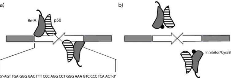
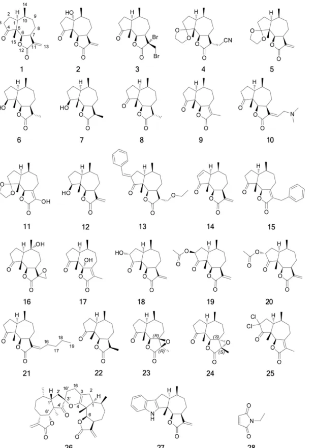
Several natural products are potential lead structures for the development of NF-kB inhibi-tors, and an important group is the sesquiterpene lactones (SLs) [25,26]. Almost all the experi-mental evidence demonstrates that the SLs act as alkylating agents by a Michael addition mechanism, and an especially interesting target is residue cys-38 in the NF-kB transcriptional subunit RelA (canonical pathway), as the formation of the complex of NF-kB with DNA is pre-vented if the thiol group of this cysteine is alkylated [27,28]. Michael acceptors as well as other alkylating agents have traditionally been associated with toxicity, but the possibility to increase their selectivity makes them interesting alternatives for drug discovery [29]. Based on this knowledge, we developed a biochemical assay to study the effects of SLs on the interaction between a designed DNA-recognition target (fragment ~1000 bp containing two kB-recogni-tion sites oriented in a palindrome way), and the canonical NF-kB heterodimer (RelA/p50) (Fig. 1). Both interactions, NF-kB:DNA and NF-kB:DNA:SLs, were observed by agarose gel electrophoresis. The evaluated SLs were derivatives of damsin (1) (Fig. 2), a natural product with a recognized NF-kB inhibitory capacity [30]. In total 27 compounds were tested, repre-senting a substantial structural diversity, facilitating an initial SAR analysis.
Materials and Methods
Chemicals
All chemicals of analytical grade were purchased from different commercial suppliers and were used without further purification unless otherwise stated.
Strains and Plasmids
Escherichia coliNovaBlue andE. coliBL21(DE3) strains for cloning and expression,
co-expression vector pCOLADuet was from Novagen. Ampicillin (100mg/mL) was added to the medium to select theE. coliNovaBlue harboring pUC19 derivative constructs, and 30mg/ mL kanamicyn was included for the selection ofE. coliBL21(DE3) harboring pCOLADuet de-rivative constructs.
Cloning of RelA and p50 Genes
The strategy used was focused on the co-expression of the RelA/p50 heterodimer, with a 6X histidine tag in the N-terminal end of the RelA subunit for an effective purification of soluble heterodimer, similar to the one reported previously [40,41]. Synthetic genes to overexpress the truncated human RelA (residues 20–291) and p50 (residues 42–353) inE. coliwere designed. The codon optimization was performed using the Optimizer Server (http://genomes.urv.es/ OPTIMIZER/) and the analysis of the optimal codons byE. coliCodon Usage Analyzer 2.1 (www.faculty.ucr.edu/~mmaduro/codonusage/usage.htm). Every optimized gene sequence was synthesized and inserted into pUC57 vectors (GeneScript USA Inc). The genes were amplified individually from pUC57 using the following primers: RelA-Forward (5’-GGA TCC GGC CTA CGT TGA AAT CAT CGA ACA GCC GAA ACA GC-3’), RelA-Reverse (5’- GCG GCC GCG TCC GGC AGG TAC TGG AAT TCC ATC GG-3’), p50-Forward (5’- CAT ATG GGT CCG TAC CTG CAG ATC CTG GAA CA-3’) and p50-Reverse (5’- TTA ATT AAG CTT CCG GGT AGT ACA GGA ACG GTT TCG G-3’). The PCR products were inserted individu-ally into pUC19/smaI vector for proliferation and finally subcloned into theBamHI/NotI (RelA) andPacI/NdeI (p50) sites of the co-expression vector pCOLADuet.
Expression and Purification of the Proteins
RelA/p50 heterodimer was co-expressed inE. coliBL21(DE3). The cells were grown in shake flasks at 30°C in LB medium supplemented with 25mg/ml kanamycin. The induction was done with 1 mM isopropylb-D-thiogalactopyranoside (IPTG) when the optical density reached 0.6 at 600 nm, followed by overnight incubation at room temperature with shaking at 100 rpm. The cells were harvested by centrifugation (9300 g for 20 min), re-suspended in binding buffer (20 mM HEPES and 500 mM NaCl at pH 7.4) and disrupted by sonication (10 x 2 min pulses with 2 min interval). The cell lysate was centrifuged for 20 minutes at 22000 rpm and the
Figure 1. Schematic view of interactions. a)Complexation between the palindromic DNA-recognition target and two RelA/p50 heterodimers.b)Effect of inhibitors on RelA (cys38) hindering the formation of the complex.
Figure 2. Compounds tested as inhibitors of the NF-κB:DNA-recognition target complex formation.
supernatant was used for protein purification. The recombinant proteins were purified by af-finity chromatography using 1 mL Histrap HP column with 20 mM HEPES, 500 mM NaCl pH 7.4 as the binding buffer, and the buffer with 500 mM imidazole as the elution buffer. The col-umn fractions were analyzed by SDS-PAGE, and imidazole was removed by dialysis against 2 x 1 L of binding buffer.
Construction of the DNA Recognition Target
A DNA fragment of ~1000 bp containing NF-kB recognition sequences was constructed as fol-lows: a palindromic synthetic oligonucleotide (NF-kB target) AGT TGA GGG GAC TTT CCC AGG CCT GGG AAA GTC CCC TCA ACT was inserted in pUC19/SmaI vector. Subsequent-ly, a fragment containing the palindromic sequence (NF-kB target) in the middle was generat-ed by PCR using DreamTaq PCR Master Mix (Thermo Scientific). The primers usgenerat-ed were kB-Forward GAA GGC AAA ATG CCG CAA AAA AGG andkB-Reverse GCG TCG ATT TTT GTG ATG CTC GTC. The mixture was incubated for 2 min at 98°C, 35 amplification cycles were performed (denaturation for 20 s at 98°C, annealing for 30 s at 60°C, and extension for 40 s at 72°C) and this was followed by a final extension for 7 min at 72°C. The quality of the product was confirmed by electrophoresis on 1% (w/v) agarose gel and visualization with GelRed Nucleic Acid Gel Stain (Invitrogen, USA).
Determination of NF-
κ
B:DNA Recognition Target Interaction and
Optimal Molar Ratio
The reaction was carried out in a volume of 10mL for 2 h at 37°C. The protein:DNA molar ratio was varied in three different ranges:1. From 2:1 to 13:1:using 1.5mL of DNA target (85 ng/mL) the amount of NF-kB (28 ng/mL) was varied from 1mL (ratio 2:1) to 6mL (13:1),
2. From 19:1 to 112:1:to 1.0mL of DNA target (85 ng/mL) was added varying volumes of NF-kB dilution (165 ng/mL) from 1mL (19:1) to 6mL (112:1),3. From 298:1 to 669:1:with 1.0mL of DNA target (85 ng/mL) the amount of NF-kB (660 ng/mL) was varied from 4mL (298:1) to 9 mL (669:1). The volumes of all the reaction mixtures were made up to 10mL with nuclease-free water. Two blanks were prepared, the first one with HEPES (B1) with 1mL of DNA target (100 ng/mL) in 9mL of HEPES buffer (20 mM HEPES and 500 mM NaCl at pH 7.4), and the second one without HEPES (B2): 1mL of DNA target (100 ng/mL) in 9mL of nuclease-free water. The range from 298:1 to 669:1 was performed in triple replicates including the blanks. The samples were analyzed by electrophoresis on 1% agarose gel (TAE 1X; 100V for 1.5 h) and visualized with GelRed Nucleic Acid Gel Stain. Quantification of the relative DNA concentration on the agarose gels was performed using Bio-Imaging Systems Mini Bis Pro (Dual UV configuration 254–365 nm), software GelQuant Pro v11.4.
Determination of the Optimal Inhibitor Concentration and Reaction Time
Determining the Effect of Different Inhibitors and Their Solubility in the
System
All the experiments were performed in triplicates in reactions, with 8mL of NF-kB and 1mL of inhibitor in a total volume of 10mL. The mixture was incubated for four hours at 37°C, after which 1mL of DNA target dissolution was added and incubated for two more hours at 37°C. Four different diluted reaction mixtures were prepared from the following solutions: 85 ng/mL DNA target, 660 ng/mL NF-kB and 90 mM inhibitors (compounds 1 and 4 dissolved in DMSO), giving different DNA:NF-kB molar ratios (Table A inS2 File). Thus, both the inhibi-tor and NF-kB were diluted in facinhibi-tors of 1, 3/4, 1/2 and 1/4, while the DNA target in facinhibi-tors 1, 1/2, 1/4 and 1/8.
Effect of DMSO
Reaction mixtures with 10%, 20% and 30% v/v DMSO were prepared. The addition of extra volume (2 and 3mL) of DMSO to reach 20% and 30% forced to decrease the volume of NF-kB (7 and 6mL) giving different final concentrations (528, 462 and 396 ng/mL respectively). Then, 1mL of inhibitor was added in each solution to give a final concentration of 9 mM. These pre-mixtures were incubated for 4 hours at 37°C, then 1mL of DNA target was added (giving a final concentration of 8.5 ng/mL) and the mixtures were incubated for two more hours at 37°C (Table B inS2 File).
Synthesis of Pseudoguianolides
Following pseudoguianolides were synthesized according to the protocols described inS1 File: (6S,9aR,9bR)-3-hydroxy-6,9a-dimethyl-4,5,6,6a,7,8,9a,9b-octahydro-2H-spiro[azuleno[4,5-b] furan-9,20-[1,3]dioxolan]-2-one(11); 3b-acetoxydamsin(19); (E)-13-n-Butildamsin(21); (7R, 11R)-Epoxy-13-hydrodamsin and (7S, 11S)-Epoxy-13-hydrodamsin(23 and 24); 3,3-Dichlorodamsin(25); (3aS,3a0S,6S,60S,8S,9aR,9bR,11b0R,11c0R)-6,60,9a,11b0-tetramethyl-3,30 -dimethylene- 3a,3a0,4,40,5,50,6,6a,60,6a0,7,70,80,90-tetradecahydro-2H-spiro[azuleno[4,5-b] furan-8,100-furo[30,20:7,8]azuleno[1,2-b]pyran]-2,20,9(3H,30H,9aH,9bH,11b0H,11c0H)-trione
(26)and (E)-13-Bromodamsin(29).
Structure Determination
HMRS (ESI) spectra of the pseudoguianolides were recorded with a Micromass Q-TOF Micro spectrometer. NMR spectra (in CDCl3) were recorded using a Bruker DRX 400 MHz at 400 Mhz (1H) and at 100 MHz (13C) and Bruker DRX 500 MHz at 500 MHz (1H) and 125 MHz (13C). Chemical shifts are given in ppm relative to TMS using the residual CHCl3in CDCl3solution as internal standard (7.25 ppm1H and 77.00 ppm13C). All flash chromatogra-phy was performed with 60Å30–75mm Silica gel. TLC analyses were made on Silica Gel 60 F254 (Merck) plates.
Results and Discussion
Determination of NF-
κ
B /DNA Complex Formation and Optimal Molar
Ratio
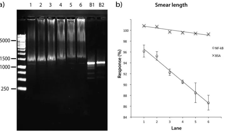
RelA/p50 heterodimers with one DNA molecule to obtain a higher molecular weight NF-kB-DNA complex (Fig. 1). The response is the quantified relative concentration (%) of the free DNA recognition target on the agarose gel.
In order to optimize the protein:DNA molar ratio, two responses were measured; the con-centration of free DNA-recognition target (that was not complexed by NF-kB) and the total length of smear. The response was measured for molar ratios ranging from 2:1 to 669:1. The experiment was performed in triplicates (Fig. 3a). Ratios higher than 298:1 affected significant-ly the normal mobility of the DNA band (the concentration was reduced to a minimum of 42%). In order to demonstrate that NF-kB is the only component in the mixture that affects the DNA mobility, two blanks were included, the first with HEPES buffer (used in the protein solutions) and the second with nuclease-free water (used for DNA dilutions); neither interfered with the DNA mobility. On the other hand, the smear length of the DNA recognition target was demonstrated to be linearly dependent on the NF-kB protein concentration as shown in
Fig. 3b. In addition, when the same experiment was repeated using bovine serum albumin pro-tein (BSA), no effect was observed on the DNA mobility, ruling out the high concentration of an unrelated protein as being responsible for the DNA retention (Fig. 3b). The results of these experiments suggested that best condition to observe a strong interaction between NF-kB and the DNA recognition target is NF-kB:DNA molar ratio of 595:1 (protein concentration 528 ng/mL and DNA concentration 8.5 ng/mL).
Figure 3. Optimization of Molar Ratio NF-κB-DNA. a)Agarose gel showing the effect of NF-κB on the palindromic DNA-recognition target. NF-κB-DNA molar ratio per Lane: 1 (253:1), 2 (316:1), 3 (379:1), 4 (443:1), 5 (506:1), 6 (569:1), B1 (Blank with HEPES Buffer) and B2 (Blank with nuclease-free water).
b)(0) Standard addition curve for each molar ratio of NF-κB:DNA target (as lane number), y = -0.0199x + 0.9854, R2; = 0.98911; (X) Standard addition curve
for each molar ratio of Bovine serum albumin:DNA target (as lane number).
Optimization of Inhibitor Concentration and Reaction Time
Damsin(1)(Fig. 2) is a known inhibitor of NF-kB [30] and was used for the semisynthesis of several compounds tested in this study. In order to determine the optimal inhibitor concentra-tion and reacconcentra-tion time, a combinaconcentra-tion of different damsin concentraconcentra-tions and different incuba-tion times with NF-kB were evaluated. The response measured was the percentage of DNA released with respect to the control (8.5 ng/mL DNA in nuclease free water) (Fig. 4). The pro-tein:DNA molar ratio was fixed at 595:1 (see previous section). At the lowest concentration of inhibitor (3 mM) the response did not change with time, giving a constant value of DNA re-leased (about 10%). On the other hand, at the highest inhibitor concentration (12 mM), the response has shown a polynomial trend as a function of time, reaching a maximum of 60% after 4 h. However, a relatively lower response is preferred for the detection of highly active compounds or to neglect compounds with poor activity. Thus responses between 20 to 25% were considered as the most adequate for testing a range of damsin(1)derivatives,
Figure 4. Optimization of inhibitor concentration and reaction time.Reaction of NF-κB with 3–12 mM inhibitor (1) at 1–4 h reaction time. The vertical axis represents the response as the percentage of released DNA target compared to the control (pure DNA target at 8.5 ng/μL). The coordinates show the concentration of damsin (1) (inhibitor) and the reaction time between NF-κB and the inhibitor.
corresponding to 9 mM damsin concentration and 4 h incubation time at 37°C. An additional benefit of the selected inhibitor concentration is the constant response between 3 and 4 h of reaction. The same concentration was used for the positive control in further experiments.
Determination of Activity and Solubility of the Compounds in the
Reaction System
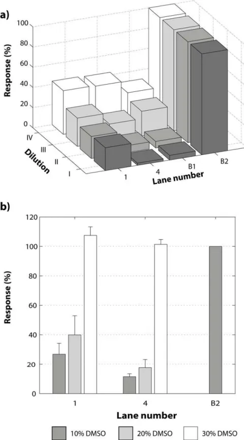
With the aim to evaluate the effect of active and non-active compounds on the NF-kB:DNA complex stability, two compounds were selected:1, that was used in the optimization process, which has aa-methylene-g-lactone moiety responsible for the alkylating capacity and com-pound4, lacking thea-methylene-g-lactone moiety and therefore presumed to be inactive. Furthermore, the sample solubility is a crucial factor in the development of biochemical and biological assays. Usually a co-solvent is necessary and the most frequently used is DMSO [32]. Hence, the effect of the selected compounds was tested at the same time as their solubility. In
Fig. 5a, different responses for1and4were observed and, as expected, compound1gave the same response as in the previous experiments while compound4proved to be inactive. In order to improve the system and the solubility of the components, the concentration of all compounds was decreased proportionally while the total reaction volume was fixed to 10mL for each dilution. For this purpose, three diluted reactions were prepared (Table A inS2 File), the inhibitor and the NF-kB protein were diluted in the same ratios for each dilution (3/4, 1/2 and 1/4), and the DNA recognition target was diluted in lower ratios (1/2, 1/4 and 1/8). Conse-quently, the NF-kB:DNA molar ratio was increased, while the concentration was reduced. This was done to favor the formation of the complex, nevertheless the complex demonstrated to be dissociated by the dilution (Fig. 5a). This effect becomes clear when the controls B1 (NF-kB: DNA complex without inhibitor) and B2 (free DNA recognition target in nuclease free water) are compared. The difference between B1 and B2 for the dilution I is 82% while for dilution IV it is 45%; the last dilution limited significantly the analytical range. Additionally, the resolution decreases with dilution, making it difficult to see a difference between active and inactive com-pounds. The second evaluated variable was the percentage of DMSO (10, 20 and 30% w/w) in the final mixture (Table B inS2 File). The results show that DMSO had a strongly negative ef-fect on the stability of the NF-kB:DNA complex and complete dissociation was observed at 30% DMSO concentration (Fig. 5b).
Preparation and Structure Elucidation of Damsin Derivatives
Isolation of natural products1and2, as well as the preparation of derivatives3–10,12–18,20,
22and27(Fig. 2), was done as reported previously [30,33,34]. Semisynthesis of compounds
11,19,21and23–26(Fig. 2) from1was also performed. Compound11was prepared by the
ozonolysis of5. This derivative was intended as a 11-keto intermediate to give access to 13-substituted pseudoguaianolides via Wittig olefinations, but unfortunately11only exists as the enolic tautomer as shown by NMR data, that is not useful for such reactions. A hydroxylic proton was observed at low field (d6.24 ppm), as well as two new unsaturated carbons corre-sponding to C-7 (d135.8 ppm) and C-11 (d136.8 ppm).
concentration.b)Effect of DMSO concentration on the response for two different inhibitors compared with one control: 1) compound1; 2) inactive compound4; B2) Free DNA target. The concentration of DMSO was increased at the same time as the protein concentration was decreased as follows: 10% DMSO (Protein:DNA molar ratio of 595:1); 20% DMSO (Protein:DNA molar ratio of 521:1) and 30% DMSO (Protein:DNA molar ratio of 446:1).
Compound19was synthesized by an inversion of the configuration of C-3 of18, by a Mit-sunobu reaction. The stereochemistry of the epimer19was confirmed by the correlation be-tween H-3aand H-1a(calculated distance 2.6Å) observed in the NOESY spectrum. The coupling constants of H-3awere also compared. The experimental coupling constant with H-2ais 8.2Hz and the calculated 7.3Hz, while the experimental coupling constant with H-2bis 11.4 Hz and the calculated 9.3Hz.
Compound21was synthesized via3and29(Fig. 6a) using Suzuki coupling conditions. However, rather than obtaining the coupled product (13-propyldamsin), (E)-13-butyldamsin (21) was formed. Presumably, the mechanism is a push-pull substitution of the bromine by the
n-butyl anion (BuLi). The configuration of the 11–13 double bond of21was determined by the NOESY correlations observed between H2–16 and H-7.
The epoxidation of the conjugated double bond in compound9to obtain products23and
24was carried out with hydrogen peroxide in NaOH solution under microwave irradiation, but the yield of the epoxidized products was low. The configuration of the epoxide23was de-termined by the NOESY correlations between H3–13 and H-6, H-9aand H-1. In the same way the epimer24was assigned by the NOESY correlation of H3–13 with H-8b, which also corre-lates with H3–14 and H3–15. An alternative reaction with sodium hypochlorite in pyridine was tested to obtain23and24. However, instead of the desired epoxides, the 3,3-dichloro deriva-tive25was obtained.
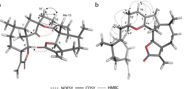
Finally, we wanted access to compound31(Fig. 6b) in order to have a second exocyclic Mi-chael acceptor, however, unexpectedly it dimerized through a regioselective hetero-Diels-Alder (HDA) reaction to form26. The structure of dimer26was elucidated comparing the COSY, HMBC and NOESY spectra with computational models (Fig. 7). The regioselectivity of the re-action corresponds to an inverse-demand HDA mechanism [36], only yielding theendo addi-tion adduct26. The COSY correlations confirmed the inverse-demand HDA, in particular the correlations between the protons on positions 16 and 16’, as well as their HMBC correlations. Theendoaddition was finally confirmed by the NOESY correlations between H-6 and H-6’
Figure 6. Reaction conditions. a)Suzuki coupling conditions: a. Et3N/DCM; b. PrI, Buli, B-methoxy-9-BBN/
Et2O, THF; c. K3PO4(aq),29, PdCl2(dppf)CH2Cl2/DMF.b)Mannich condensation: d. H2C = N+(CH3)2,
CF3CO2-/DCM; e. MeI/MeOH.
(calculated distance 2.8Å) and between H-1’and H3–15 (calculated distance 3.8Å). The dis-tances for the same proton pairs in the exo adduct are both greater than 6Å.
Compound31was prepared via a Mannich condensation (Fig. 6b) with dimethyl(methy-lene)ammonium trifluoroacetate followed by a Hoffmann elimination as reported previously [35], but it could not be isolated.
Screening of the Derivatives
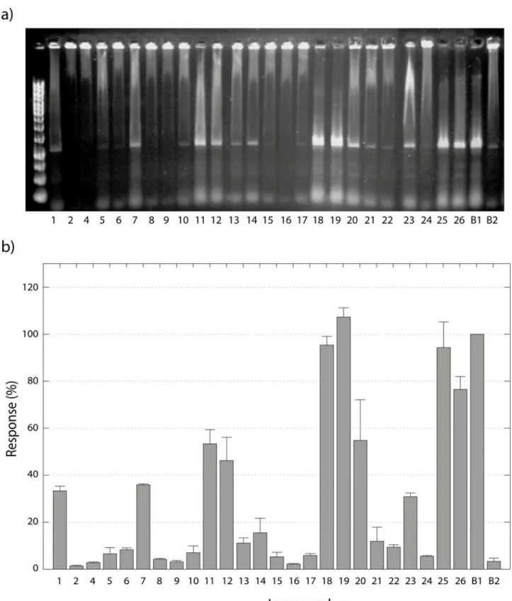
The compounds tested as inhibitors of NF-kB:DNA complex formation are shown inFig. 2. They were tested using the optimized screening conditions discussed above.N-Ethylmaleimide (NEM)(28)was used as a positive control as it is known to be a cysteine alkylating agent react-ing via a Michael addition mechanism [37]. The assays were performed in quadruple replicates, and outlying data was rejected using Grubbs test [38]. All the compounds except compounds3
and27showed acceptable replicability. Compound3is very active but is also highly electro-philic, a property that may cause it to react indistinctly with many components in the protein (e.g. cysteine and histidine residues) as well as guanine in the free DNA [39]. On the other hand, compound27was active but had low solubility in the HEPES buffer even with 10% DMSO. The other compounds showed satisfactory replicability with a specific response of each, facilitating a discussion of structure-activity relationships. The compounds were divided into four categories according to their responses (seeFig. 8):
A. Providing at least 90% of the response of the positive control. These compounds are con-sidered to be very active, and include compounds18,19and25. The common characteristics of this group are a substituent at C-3, in addition to thea-methylene-g-lactone, although how the C-3 substituent increases the activity remains unclear. A very important exception in this group is25, which is very active despite having the less reactive endocyclica,b-unsaturated lac-tone. This indicates that the two geminal chlorines at C-3 are at least partly responsible for the high activity.
B.Providing between 50 and 90% of the response of the positive control. These compounds are considered to be active, and include compounds11,20and26. The structural diversity in
Figure 7. Computational model of the less energy conformer of dimer 26.The experimental 2D-NMR NOESY, COSY and HMBC correlations are indicated with arrows.
Figure 8. Screening of derivatives and their effect on NF-κB/DNA complex stability. a)Agarose gel showing the effect of the tested compounds on NF-κB:DNA complex formation. Each lane indicates the number of the compound inFig. 2. B1 is the positive control28, while B2 is the negative control.
b)Response for each compound (Fig. 2) compared to the positive control28. The error bars indicate the standard deviation.
this group does not allow for the identification of common or similar structural features re-sponsible of the activity, and consequently these compounds will be discussed independently. Compound11is an exception among the active compounds, having features that diminish the activity (as will be described later), as a dioxalane ring in position 4 and lacks the
a-methylene-g-lactone moiety. However, the enol in the lactonic ring might be responsible for its activity. Compound20has two characteristics that also are present in groupA, a substitu-ent at C-3 and a Michael acceptor, but the lower activity suggests that the configuration of po-sition 3 may play an important role (compared with19). Compound26has two Michael acceptors giving the possibility of cross-linking in the protein, which could explain the higher activity compared to1.
C.Providing 10 to 50% of the response of the positive control. These compounds are considered to be poorly active, and include1,7,12,13,14,21and23. Some compounds in this group resemble damsin(1), e.g.12with the keto function reduced to a hydroxyl group. The fact that12is more active than1suggests that the presence of a hydroxyl group (hydrogen bond donor) in position 4, enables the molecule to interact differently with the target. Compound14with two Michael acceptors is nevertheless less potent than1. This might depend on a higher reactivity of the second Michael acceptor (the cyclopentenone) and, as a consequence, a low selectivity for the target residue (cys-38). Compound21is less active compared to1, which is logical when the steric hindrance from the butyl substitution in position 13 is considered. Three of the derivatives do not fit the previously described characteristics. Unexpectedly, compound7was demonstrated to be approximately as active as1, even if thea-methylene-g-lactone moiety is not present. Comparing its activity with1
and12, the notion that theb-hydroxyl group in position 4 is beneficial for the activity is reinforced. In the same way, compound13demonstrated to be slightly active even without the Michael acceptor, although it cannot be excluded that13spontaneously eliminates ethanol during the assay reverting to thea-methylene-g-lactone moiety. Finally, the activity of compound23could be explained by the presence of an alkylating epoxide.
D. Less than 10% of the response of the positive control. These compounds are considered to be inactive, and include2, 4, 5, 6, 8, 9, 10, 15, 16, 17, 22and24. The main characteristics that render them inactie are: a dioxalane group in position 4 (4and5), the lack of a Michael acceptor(4, 6, 8and22), and the presence of a less reactive endocyclica,b-unsaturated lactone9, 15and17). Other derivatives that are electrophilic but inactive are the epoxides (16and24). Compound2has a hydroxyl group in position 1, which somehow affects the activity negatively. Finally,10was designed to provide intramolecular basic catalysis for the Michael addition, is probably not chemically stable enough to survive the assay conditions.
Conclusions
A biochemical assay based on a DNA fragment constructed with two recognition sequences for the NF-kB binding, enabling the detection of free DNA from NF-kB–DNA complex on a 1% agarose gel electrophoresis, was developed. The assay is fast, low cost and applicable for screening of potential inhibitors of the NF-kB—DNA complex formation. The assay and the following SAR study facilitated the identification of relevant structural features in the pseudoguaianolide skeleton, which are necessary for, or enhance the capacity of dissociating the complex NF-kB—DNA. The most active compounds have a substitution at C-3
Supporting Information
S1 File. Synthesis.
NMR spectra of compounds 11, 19, 21, 23, 24, 25 and 26.
• Compound 11.
• 1H-NMR
• 13C-NMR
• Compound 19.
• 1H-NMR
• 13C-NMR
• NOESY
• Compound 21.
• 1H-NMR
• 13C-NMR
• NOESY
• Compound 23.
• 1H-NMR
• 13C-NMR
• NOESY
• Compound 24.
• 1H-NMR
• 13C-NMR
• NOESY
• Compound 25.
• 1H-NMR
• 13C-NMR
• Compound 26.
• 1H-NMR
• 13C-NMR
• NOESY (PDF)
S2 File. Supporting Information Tables.Table A. Dilutions prepared to test the effect of the concentration of the reaction components. Table B. Dilutions prepared to test the effect of DMSO concentration.
Author Contributions
Conceived and designed the experiments: JALP. Performed the experiments: RV. Analyzed the data: RV JALP OS RHK. Contributed reagents/materials/analysis tools: RHK OS GA. Wrote the paper: RV JALP GA OS RHK.
References
1. Baldwin AS (2001) Control of oncogenesis and cancer therapy resistance by the transcription factor NF-kB. J Clin Invest 107: 241–246. doi:10.1172/JCI11991PMID:11160144
2. Miller SC, Huang R, Sakamuru S, Shukla SJ, Attene-Ramos MS, et al. (2010) Identification of known drugs that act as inhibitors of NF-κB signaling and their mechanism of action. Biochem Pharmacol 79: 1272–1280. doi:10.1016/j.bcp.2009.12.021PMID:20067776
3. Kim HJ, Hawke N, Baldwin AS (2006) NF-kappaB and IKK as therapeutic targets in cancer. Cell death and differentiation, 13, 738–747. doi:10.1038/sj.cdd.4401877PMID:16485028
4. Orlowski RZ, Baldwin Jr AS (2002) NF-κB as a therapeutic target in cancer. Trends in Molecular Medicine, 8, 385–389. doi:10.1016/S1471-4914(02)02375-4PMID:12127724
5. Lin A, Karin M (2003) NF-κB in cancer: a marked target. Seminars in Cancer Biology, 13, 107–114. doi: 10.1016/S1044-579X(02)00128-1PMID:12654254
6. Verma IM (2004) Nuclear factor (NF)-κB proteins: therapeutic targets. Annals of the Rheumatic Dis-eases, 63, ii57–ii61. doi:10.1136/ard.2004.028266PMID:15479873
7. Gilmore TD (2006) Introduction to NF-[kappa]B: players, pathways, perspectives. Oncogene, 25, 6680–6684. doi:10.1038/sj.onc.1209954PMID:17072321
8. Hayden MS, Ghosh S (2008) Shared Principles in NF-κB Signaling. Cell, 132, 344–362. doi:10.1016/j. cell.2008.01.020PMID:18267068
9. Oeckinghaus A, Hayden MS, Ghosh S (2011) Crosstalk in NF-[kappa]B signaling pathways. Nat Immu-nol, 12, 695–708. doi:10.1038/ni.2065PMID:21772278
10. Nakanishi C, Toi M (2005) Nuclear factor-[kappa]B inhibitors as sensitizers to anticancer drugs. Nat Rev Cancer, 5, 297–309. doi:10.1038/nrc1588PMID:15803156
11. Gasparian AV, Guryanova OA, Chebotaev DV, Shishkin AA, Yemelyanov AY, et al. (2009) Targeting transcription factor NFκB: comparative analysis of proteasome and IKK inhibitors. Cell Cycle, 8, 1559–1566. doi:10.4161/cc.8.10.8415PMID:19372735
12. Olivier S, Robe P, Bours V (2006) Can NF-κB be a target for novel and efficient anti-cancer agents? Biochemical Pharmacology 72: 1054–1068. doi:10.1016/j.bcp.2006.07.023PMID:16973133 13. Lai C, Jiang X, Li X (2006) Development of luciferase reporter-based cell assays. Assay and drug
de-velopment technologies, 4, 307–315. doi:10.1089/adt.2006.4.307PMID:16834536
14. Israël N, Gougerot-Pocidalo MA, Aillet F, Virelizier JL (1992) Redox status of cells influences constitu-tive or induced NF-kappa B translocation and HIV long terminal repeat activity in human T and mono-cytic cell lines. The Journal of Immunology, 149, 3386–3393. PMID:1431113
15. Plummer SM, Holloway KA, Manson MM, Munks RJ, Kaptein A, et al. (1999) Inhibition of
cyclo-oxygenase 2 expression in colon cells by the chemopreventive agent curcumin involves inhibition of NF-kappaB activation via the NIK/IKK signalling complex. Oncogene, 18, 6013–6020. doi:10.1038/ sj.onc.1202980PMID:10557090
16. Sun SC, Ganchi PA, Béraud C, Ballard DW, Greene WC (1994) Autoregulation of the NF-kappa B transactivator RelA (p65) by multiple cytoplasmic inhibitors containing ankyrin motifs. Proceedings of the National Academy of Sciences, 91, 1346–1350. doi:10.1073/pnas.91.4.1346PMID:8108414 17. Lindenmeyer MT, Garci´a-Piñeres AJ, Castro V, Merfort I (2004) Sesquiterpene lactones inhibit
lucifer-ase but notβ-galactosidase activity in vitro and ex vivo. Analytical Biochemistry, 328, 147–154. doi:10. 1016/j.ab.2004.01.021PMID:15113690
18. Matsusaka T, Fujikawa K, Nishio Y, Mukaida N, Matsushima K, et al. (1993) Transcription factors NF-IL6 and NF-kappa B synergistically activate transcription of the inflammatory cytokines, interleukin 6 and interleukin 8. Proceedings of the National Academy of Sciences, 90, 10193–10197. doi:10. 1073/pnas.90.21.10193
19. Ma DL, Xu T, Chan DSH, Man BYW, Fong WF, et al. (2011) A highly selective, label-free, homogenous luminescent switch-on probe for the detection of nanomolar transcription factor NF-kappaB. Nucleic Acids Research, 39, e67. doi:10.1093/nar/gkr106PMID:21398636
signaling. Assay and drug development technologies, 1, 823–833. doi:10.1089/ 154065803772613453PMID:15090228
21. Ding GJF, Fischer PA, Boltz RC, Schmidt JA, Colaianne JJ, et al. (1998) Characterization and Quanti-tation of NF-κB Nuclear Translocation Induced by Interleukin-1 and Tumor Necrosis Factor-α: develop-ment and use of a high capacity fluorescence cytometric system. Journal of Biological Chemistry, 273, 28897–28905. doi:10.1074/jbc.273.44.28897PMID:9786892
22. Finco TS, Beg AA, Baldwin AS (1994) Inducible phosphorylation of I kappa B alpha is not sufficient for its dissociation from NF-kappa B and is inhibited by protease inhibitors. Proceedings of the National Academy of Sciences, 91, 11884–11888. doi:10.1073/pnas.91.25.11884
23. Riganti C, Doublier S, Costamagna C, Aldieri E, Pescarmona G, et al. (2008) Activation of Nuclear Factor-κB Pathway by Simvastatin and RhoA Silencing Increases Doxorubicin Cytotoxicity in Human Colon Cancer HT29 Cells. Molecular Pharmacology, 74, 476–484. doi:10.1124/mol.108.045286 PMID:18463201
24. Castro AC, Dang LC, Soucy F, Grenier L, Mazdiyasni H, et al. (2003) Novel IKK inhibitors:β-carbolines. Bioorganic & Medicinal Chemistry Letters, 13, 2419–2422. doi:10.1016/S0960-894X(03)00408-6 PMID:12824047
25. Bremner P, Heinrich M (2002) Natural products as targeted modulators of the nuclear factor-KB path-way. Journal of Pharmacy and Pharmacology, 54, 453–472. doi:10.1211/0022357021778637PMID: 11999122
26. Salminen A, Lehtonen M, Suuronen T, Kaarniranta K, Huuskonen J (2008) Terpenoids: natural inhibi-tors of NF-κB signaling with anti-inflammatory and anticancer potential. Cell. Mol. Life Sci., 65, 2979–2999. doi:10.1007/s00018-008-8103-5PMID:18516495
27. Rüngeler P, Castro V, Mora G, Gören N, Vichnewski W, et al. (1999) Inhibition of transcription factor NF-κB by sesquiterpene lactones: a proposed molecular mechanism of action. Bioorganic & Medicinal Chemistry, 7, 2343–2352. doi:10.1016/S0968-0896(99)00195-9PMID:10632044
28. Garcia-Pineres AJ, Castro V, Mora G, Schmidt TJ, Strunck E, et al. (2001) Cysteine 38 in p65/NF-κB plays a crucial role in DNA binding inhibition by sesquiterpene lactones. J. Biol. Chem., 276, 39713–39720. doi:10.1074/jbc.M101985200PMID:11500489
29. Johansson MH (2012) Reversible Michael additions: covalent inhibitors and prodrugs. Mini Rev Med Chem, 12, 1330–1344. doi:10.2174/13895575112091330PMID:22625413
30. Villagomez R, Rodrigo GC, Collado IG, Calzado MA, Muñoz E, et al. (2013) Multiple Anticancer Effects of Damsin and Coronopilin Isolated from Ambrosia arborescens on Cell Cultures. Anticancer Research, 33, 3799–3805. PMID:24023312
31. Chen FE, Huang DB, Chen YQ, Ghosh G (1998) Crystal structure of p50/p65 heterodimer of transcrip-tion factor NF-[kappa]B bound to DNA. Nature, 391, 410–413. doi:10.1038/34956PMID:9450761 32. Di L, Kerns EH (2006) Biological assay challenges from compound solubility: strategies for bioassay
optimization. Drug Discovery Today, 11, 446–451. doi:10.1016/j.drudis.2006.03.004PMID:16635808 33. Villagomez R, Collado JA, Muñoz E, Almanza G, Sterner O (2014) Natural and semi-synthetic
pseudo-guaianolides as inhibitors of NF-κB. Journal of Biomedical Science and Engineering. 7, 833–847. doi: 10.4236/jbise.2014.710083
34. Villagomez R (2014) Biological activities of natural and semi-synthetic pseudo-guaianolides: Inhibition of transcription factors, Doctoral Thesis, Lund University, Lund Sweden.
35. Ahond A, Cave A, Kan-Fan C, Husson HP, De Rostolan J, et al. (1968) Facile N-O bond cleavages of amine oxides. Journal of the American Chemical Society, 90, 5622–5623. doi:10.1021/ ja01022a063
36. Ding C, Wang L, Chen H, Wild C, Ye N, et al. (2014) ent-Kaurane-based regio- and stereoselective in-verse electron demand hetero-Diels–Alder reactions: synthesis of dihydropyran-fused diterpenoids. Organic & Biomolecular Chemistry, 12, 8442–8452. doi:10.1039/C4OB01040JPMID:25225052 37. Garcia-Pineres AJ, Lindenmeyer MT, Merfort I (2004) Role of cysteine residues of p65/NF-κB on the
in-hibition by the sesquiterpene lactone parthenolide and N-ethyl maleimide, and on its transactivating po-tential. Life Sci., 75, 841–856. doi:10.1016/j.lfs.2004.01.024PMID:15183076
38. Grubbs FE (1969) Procedures for detecting outlying observations in samples. Technometrics, 11, 1–21. doi:10.1080/00401706.1969.10490657
40. Chen FE, Kempiak S, Huang DB, Phelps C, Ghosh G (1999) Construction, expression, purification and functional analysis of recombinant NFκB p50/p65 heterodimer. Protein Engineering, 12, 423–428. doi: 10.1093/protein/12.5.423PMID:10360983