Wild-Type Phosphoribosylpyrophosphate Synthase (PRS)
from
Mycobacterium tuberculosis
: A Bacterial Class II
PRS?
Ardala Breda1,2, Leonardo K. B. Martinelli1,2, Cristiano V. Bizarro1, Leonardo A. Rosado1,2, Caroline B. Borges1,2, Dio´genes S. Santos1,2*, Luiz A. Basso1,2*
1Instituto Nacional de Cieˆncia e Tecnologia em Tuberculose (INCT-TB), Centro de Pesquisas em Biologia Molecular e Funcional (CPBMF), Programa de Po´s-Graduac¸a˜o em Biologia Celular e Molecular, Pontifı´cia Universidade Cato´lica do Rio Grande do Sul (PUCRS), Porto Alegre, Rio Grande do Sul, Brazil,2Programa de Po´s-Graduac¸a˜o em Biologia Celular e Molecular, Pontifı´cia Universidade Cato´lica do Rio Grande do Sul (PUCRS), Porto Alegre, Rio Grande do Sul, Brazil
Abstract
The 5-phospho-a-D-ribose 1-diphosphate (PRPP) metabolite plays essential roles in several biosynthetic pathways, including histidine, tryptophan, nucleotides, and, in mycobacteria, cell wall precursors. PRPP is synthesized from a-D-ribose 5-phosphate (R5P) and ATP by theMycobacterium tuberculosis prsAgene product, phosphoribosylpyrophosphate synthase (MtPRS). Here, we report amplification, cloning, expression and purification of wild-typeMtPRS. Glutaraldehyde cross-linking results suggest thatMtPRS predominates as a hexamer, presenting varied oligomeric states due to distinct ligand binding.
MtPRS activity measurements were carried out by a novel coupled continuous spectrophotometric assay.MtPRS enzyme activity could be detected in the absence of Pi. ADP, GDP and UMP inhibitMtPRS activity. Steady-state kinetics results indicate thatMtPRS has broad substrate specificity, being able to accept ATP, GTP, CTP, and UTP as diphosphoryl group donors. Fluorescence spectroscopy data suggest that the enzyme mechanism for purine diphosphoryl donors follows a random order of substrate addition, and for pyrimidine diphosphoryl donors follows an ordered mechanism of substrate addition in which R5P binds first to free enzyme. An ordered mechanism for product dissociation is followed byMtPRS, in which PRPP is the first product to be released followed by the nucleoside monophosphate products to yield free enzyme for the next round of catalysis. The broad specificity for diphosphoryl group donors and detection of enzyme activity in the absence of Pi would suggest thatMtPRS belongs to Class II PRS proteins. On the other hand, the hexameric quaternary structure and allosteric ADP inhibition would placeMtPRS in Class I PRSs. Further data are needed to classifyMtPRS as belonging to a particular family of PRS proteins. The data here presented should help augment our understanding ofMtPRS mode of action. Current efforts are toward experimental structure determination ofMtPRS to provide a solid foundation for the rational design of specific inhibitors of this enzyme.
Citation:Breda A, Martinelli LKB, Bizarro CV, Rosado LA, Borges CB, et al. (2012) Wild-Type Phosphoribosylpyrophosphate Synthase (PRS) fromMycobacterium tuberculosis: A Bacterial Class II PRS? PLoS ONE 7(6): e39245. doi:10.1371/journal.pone.0039245
Editor:Anil Kumar Tyagi, University of Delhi, India
ReceivedMarch 15, 2012;AcceptedMay 22, 2012;PublishedJune 20, 2012
Copyright:ß2012 Breda et al. This is an open-access article distributed under the terms of the Creative Commons Attribution License, which permits unrestricted use, distribution, and reproduction in any medium, provided the original author and source are credited.
Funding:This work was supported by funds of Decit/SCTIE/MS-MCT-CNPq-FNDCT-CAPES to National Institute of Science and Technology on Tuberculosis (INCT-TB) to Diogenes S. Santos and Luiz Basso. Luiz Basso and Diogenes S. Santos also acknowledge financial support awarded by FAPERGS-PRONEXCNPq–2009. (Fundac¸a˜o de Amparo A` Pesquisa do Estado do Rio Grande do Sul - The National Council for Scientific and Technological Development.) Luiz Basso (CNPq, 520182/99-5) and Diogenes S. Santos (CNPq, 304051/1975-06) are Research Career Awardees of The National Council for Scientific and Technological Development (CNPq). Ardala Breda, Leonardo K. B. Martinelli, Cristiano V. Bizarro, Leonardo A. Rosado and Caroline B. Borges acknowledge scholarships awarded by The National Council for Scientific and Technological Development (CNPq) and The Higher Education Co-ordination Agency (CAPES). The funders had no role in study design, data collection and analysis, decision to publish, or preparation of the manuscript.
Competing Interests:The authors have declared that no competing interests exist.
* E-mail: [email protected] (LB); [email protected] (DSS)
Introduction
Tuberculosis (TB) is a chronic infectious disease caused mainly by Mycobacterium tuberculosis, being the second leading cause of mortality by infectious diseases in human populations, killing about 1.7 million people worldwide in 2009 [1]. One third of the world population is estimated to be infected with latent TB. The latter is worsened by the spread of HIV-TB co-infection, which can lead to increased rates of TB reactivation, being up to 30% of deaths among HIV positive subjects caused by the TB bacilli [2]. TB infection is treated by a combination of four drugs that act upon different molecular targets [3]. The treatment regimen includes six month therapy with rifampicin and isoniazid, supplemented with pyrazinamide and ethambutol in the first two
antimycobacterial agent development include elucidation of the role played by proteins from biochemical pathways that are essential for mycobacterial growth [9].
Phosphoribosylpyrophosphate synthase (PRS; EC 2.7.6.1) plays central roles in a number of cellular processes, catalyzing the synthesis of 5-phospho-a-D-ribose 1-diphosphate (PRPP; a -D-5-phosphoribosylpyrophosphate; a-D-ribosyl diphosphate 5-phos-phate). PRS enzymes catalyze, in the presence of Mg2+, the
transfer ofb,c-diphosphoryl moiety of adenosine 59-triphosphate (ATP) to C1-hydroxyl group of a-D-ribose 5-phosphate (R5P), yielding PRPP [10,11] (Figure 1). PRPP is an essential metabolite for a number of distinct biochemical pathways includingde novo
and salvage pathways of purine and pyrimidine nucleotide synthesis, and biosynthesis of NAD, histidine and tryptophan [12–14]. PRPP is also associated with cell integrity inSaccharomyces cerevisiae [15]. The yeast genome encodes five distinct prs genes, whose products are combined to form hetero-oligomeric catalytic active PRS, with possible role in plasma membrane stability [16]. In Corynebacteriaceae, such as mycobacteria, PRPP is a co-substrate for the synthesis of polyprenylphosphate-pentoses, which are the source of arabinosyl residues of arabinogalactan, compo-nent of the mycobacterial cell wall, and lipoarabinomannan, a
highly immunogenic lipoglycan that is involved in modulating the host immune response [17,18].
PRS enzymes usually require Mg2+
NATP as diphosphoryl group
donor. The PRS proteins from Escherichia coli [19], Salmonella typhimurium [20] and mammals [21] have been shown to also require a second free Mg2+ion for increased catalytic rates. PRS
enzymes from these organisms, as well as fromBacillus subtilis[22], are representative of Class I (also known as ‘‘Classical’’) PRS proteins, with hexameric quaternary structure, allosteric inhibition by purines ribonucleoside diphosphate (adenosine 59-diphosphate, ADP; and guanosine 59-diphosphate, GDP), specificity for ATP (or dATP) as diphosphoryl group donor, and requirement of inorganic phosphate (Pi) for enzyme activity [23]. The three-dimensional structures of PRS enzymes fromB. subtilis(PDB ID: 1IBS) [21] andHomo sapiens(PDB ID: 2H06) [11] demonstrate that the functional enzyme is a hexamer of identical subunits, associated two by two, where each monomer is composed by two domains, both with high topological similarity to the type I family of phosphoribosyltransferases [24]. In addition, there is conservation of amino acid residues in the PRPP substrate binding site [22]. Class II PRS proteins share several structural charac-teristics with Class I enzymes. However, Class II PRSs are characterized by not being dependent on Pi for activity, have
Figure 1. Chemical reaction catalyzed byMtPRS (Rv1017c).This figure also shows the metabolic source of R5P and the biosynthetic pathways in which the reaction product PRPP plays central roles.
broad specificity for diphosphate donors (including guanosine 59 -triphosphate, GTP; cytosine 59-triphosphate, CTP; and uridine 59 -triphosphate, UTP), and are not allosteric inhibited by purines ribonucleosides diphosphate [23,25]. Class II PRS proteins appear to be specific for plants as they have been identified in spinach [26] and Arabidopsis thalianaisozymes 3 and 4 [27]. Nevertheless the PRS enzyme from pathogenic Gram negative enterobacteria S. typhimurium was reported as using GTP, ITP, CTP and UTP in addition to ATP as substrate [28]. More recently, a PRS enzyme from the archeonMethanocaldococcus jannaschiihas been shown to be tetrameric (PDB ID: 1U9Y), activated by Pi, non-allosteric inhibited by ADP, and that employs ATP as diphosphate donor [25]. These findings prompted the proposal thatM. jannaschiiPRS belongs to a new Class III of PRPP synthases [25].
Here we describe cloning ofprsA(Rv1017c) fromM. tuberculosis; and expression, purification, molecular and kinetic characteriza-tion of the non-tagged recombinant PRS (MtPRS). Glutaralde-hyde cross-linking results indicate that the oligomeric state of
MtPRS is predominantly hexameric in solution. However, the presence of ligands appears to stabilize alternative oligomeric states.MtPRS activity was assessed by a novel coupled continuous spectrophotometric assay that measures the decrease in orotate concentration catalyzed byM. tuberculosisorotate phosphoribosyl-transferase (MtOPRT) due to PRPP formation by recombinant
MtPRS enzyme activity. Steady-state data indicate that MtPRS can use both pyrimidine and purine nucleosides triphosphate as diphosphoryl group donors (broad specificity). In addition, enzyme activity measurements show thatMtPRS is catalytically competent in the absence of Pi. These data suggest that MtPRS belongs to Class II PRS family, as plant homologues, even though the primary amino acid structure is indicative of structural resem-blance to Class I PRS. Equilibrium binding data are also presented suggesting that MtPRS mechanism is likely random order of substrate addition for purine diphosphoryl donors and ordered addition of pyrimidine diphosphoryl donors, with ordered release of products in which PRPP dissociation is followed by the purine or pyrimidine nucleoside monophosphate products. The prsA -encoded protein has been predicted to be essential for in vitro
growth of M. tuberculosis based on transposon-site hybridization studies [29]. More recently, PRS fromCorynebacterium glutamicum, a model organism used to study M. tuberculosiscell physiology, has been shown to be essential for the maintenance of cellular integrity [30]. The results presented here are discussed in light of previous reports onMtPRS [30,31], and should thus contribute to a better understanding ofMtPRS mode of action.
Methods
Gene Amplification
The prsA gene (Rv1017c) was PCR amplified from total genomic DNA of M. tuberculosis H37Rv strain using specific primers designed to contain NdeI (primer sense 59 GCCATAT-GAGCCACGACTGGACCGATAATCG39) andBamHI (primer
antisense 59
GCGGATCCTCATGCGTCCCCGTC-GAAAAGT39) restriction sites (underlined). An internal restriction site forNdeI was removed from the gene sequence by site-directed mutagenesis at codon position 170, in which a thymine was replaced with a cytosine at codon’s third position (CAT to CAC), resulting in a sense mutation that maintained a histidine amino acid at this position. Dimethyl sulfoxide (DMSO) was added to the PCR reaction at final concentration of 10%. AmplifiedprsAgene was cloned into pET-23a(+) expression vector (Novagen). The integrity of constructs was confirmed in all cases by appropriate selections and digests with restriction enzymes (New England
Biolabs). Inserted sequences were confirmed by automated DNA sequencing.
Expression and Purification of RecombinantMtPRS CompetentE. coliBL21(DE3) (Novagen) cells were electropo-rated with pET-23a(+)::prsA recombinant vector and selected on Luria-Bertani (LB) agar plates containing 50mg mL21ampicillin. Aliquots of a 5 mL cell culture grown from a single colony were used to inoculate 500 mL of Terrific Broth (TB) medium supplemented with 50mg mL21 ampicillin, grown at 37uC and
180 rpm to an optical density (OD600 nm) of 0.4–0.6. At this growth stage, culture temperature was lowered to 30uC and protein expression was carried out without isopropyl-b -D-thiogalactopyranoside (IPTG) induction, for 24 hours. Cells were harvested by centrifugation (11,800g) for 30 min at 4uC and stored at 220uC. Protein purification was performed by Fast-Performance Liquid Chromatography (FPLC) on A¨ kta Purifier System (GE HealthCare) at 4uC. Cell pellet (4 g) was suspended in 40 mL of buffer A (Tris HCl 50 mM pH 7.5) and stirred for 30 min. Cells were disrupted by sonication (12 pulses of 10 sec, with intervals of 1 min off) in presence of 0.2 mg mL21lysozyme (Sigma Aldrich), and the clarified supernatant (48,000gfor 30 min in all cases) was further treated with 1% (wt/vol) streptomycin sulfate (Sigma-Aldrich). The latter supernatant was treated with 2.5 M ammonium sulfate and the resulting precipitate was suspended in 40 mL of buffer A (crude extract). The crude extract was loaded on a Q-Sepharose Fast Flow anion exchange column (GE Healthcare) equilibrated with buffer A. Adsorbed material was eluted with 0% to 50% linear gradient of Tris HCl 50 mM NaCl 1 M pH 7.5 (buffer B) at 1 mL min21flow rate. Fractions containingMtPRS, as inferred by 12% SDS-PAGE polyacrilamide gel electrophoresis stained with Coomassie Brilliant Blue [32], were pooled, concentrated and loaded on a Superdex 200 size exclusion column (GE Healthcare) previously equilibrated with buffer A. Proteins were eluted in isocratic conditions and fractions containingMtPRS were loaded on a Mono Q HR 16/10 anion exchange column (GE Healthcare) equilibrated with buffer A. Proteins were eluted with 0% to 100% linear gradient of buffer B, at 1 mL min21flow rate (Table 1). HomogeneousMtPRS eluted at approximately 430 mM NaCl (Figure 2A). Fractions contain-ing homogeneousMtPRS were pooled, dialyzed against buffer A, concentrated, and stored at280uC up to 7 months without any loss of enzyme activity.
MtPRS Identification by Mass Spectrometry
LC-MS/MS peptide mapping experiments were performed to confirm the identity of MtPRS samples. Briefly, the purified samples were digested with trypsin using a protocol adapted from [33], and the digested peptides were chromatographically sepa-rated (Kinetex 2.6mm C18 core-shell particles - Phenomenex,
MS spectra obtained from in-silico tryptic digests of the M. tuberculosisH37Rv proteome (ftp://ftp.ncbi.nih.gov/genomes).
Determination ofMtPRS Subunit Molecular Mass Purified MtPRS samples were desalted, reconstituted in methanol 50%/formic acid 1% and directly injected into an IonMax electrospray ion source. The electrospray source param-eters were as follows: positive ion mode, 5 kV of applied voltage to the electrospray source, 37.6 V of capillary voltage, 310uC of capillary temperature, and 109 V of tube lens voltage. Full spectra (770–2000 m/z range) were collected during 20 min on a Thermo Orbitrap Discovery XL in profile mode at a nominal resolution r = 30,000 at m/z 400 using FT automatic gain control target value of 1,000,000 charges. The average spectrum was processed with the software MagTran [34] for charge state deconvolution.
MtPRS Quaternary Structure Assessment by Cross-linking Studies
Cross-linking studies of the protein’s oligomeric states were performed as described by Fadouloglou et al. [35], using crystallization supports with 120mL of 25% (v/v) glutaraldehyde
acidified with HCl in the reservoir. A cover slip was used to seal the reservoir, containing a 10mL drop of protein suspension
(0.3 mg mL21homogeneous recombinantMtPRS in buffer A) in its apo form and incubated with Pi50 mM, both in presence and absence of ATP 5 mM, R5P 5 mM, and ADP 5 mM. The plates were incubated at 30uC for different time intervals and protein drops were subsequently analyzed by 12% SDS-PAGE.
MtPRS Activity Assays
All chemicals were purchased from Sigma Aldrich. All enzyme activity assays were performed in triplicate. MtPRS activity was measured by a coupled continuous spectrophotometric assay in quartz cuvettes using a UV-visible Shimadzu spectrophotometer UV2550 equipped with a temperature-controlled cuvette holder.
MtPRS reaction (ATP+R5PRPRPP+AMP) was coupled toM. tuberculosis orotate phosphoribosyltransferase (MtOPRT, EC 2.4.2.10) forward reaction (OA+PRPPROMP+PPi), in which PRPP synthesis can be monitored by the decrease in orotate (OA) concentration at 295 nm, for 60 sec at 25uC, using an extinction coefficient value of 3950 M21cm21 [36], when ATP was the diphosphoril group donor of the reaction catalyzed byMtPRS. When GTP, CTP and UTP were used as substrates forMtPRS enzyme activity measurements, the decrease in OA concentration was monitored at 303 nm, for 60 sec at 25uC, using an extinction coefficient of 2200 M21cm21 [37] due to strong absorption of these nucleosides triphosphate at 295 nm. Homogeneous recom-binant MtOPRT was obtained as described elsewhere [38]. Coupled assay conditions so as the indicator enzyme (MtOPRT) did not limit the primary reaction (MtPRS) were employed according to [39] and [40]. The reaction mixture (500mL) contained 1.8 UMtOPRT, 300mM OA, 20 mM MgCl2, in Tris HCl 50 mM pH 8.0. Reaction was started by addition ofMtPRS (0.24–1.2mM). One unit ofMtPRS is defined as the amount of enzyme necessary to convert 1mmol of R5P to PRPP per min. Effect of Pi over MtPRS activity was assessed by varying Pi concentration (10–50 mM) in the reaction conditions described above.
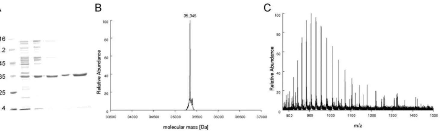
Figure 2. A)MtPRS purification steps. Lane 1: Protein Marker – Fermentas; lane 2: crude extract; lane 3: sample loaded on Q-Sepharose; lane 4: sample eluted from Q-Sepharose; lane 5: sample eluted from Superdex 200; lane 6: protein fraction eluted from Mono Q HR anion exchange step showing homogeneous recombinantMtPRS (approximately 35 kDa).B) Determination ofMtPRS molecular mass by mass spectrometry analysis. Deconvoluted spectra ofMtPRS resulted in a peak corresponding to the molecular mass of 35,345 Da.C) ESI-FTMS spectra showing the charge distribution obtained forMtPRS, spanning from charge state 25+to 45+.
doi:10.1371/journal.pone.0039245.g002
Table 1.Purification ofMtPRS from 4 g of wet cell paste ofE. coliBL21(DE3) host cells.
Purification step Total protein (mg)
Specific activity
(U mg21) Total enzyme activity (U) Yield % Purification fold
crude extract 524 0.028 145 100 1
Q-Sepharose FF 19.6 0.518 10.16 7 18.5
Superdex 200 19 0.220 4.17 2.8 7.9
MonoQ 16/10 3.5 1.16 4.06 2.8 41.4
Apparent steady-state kinetic constants,KMappandVmaxapp, were
determined by fitting the data for each substrate pairs to Henri-Michaelis-Menten equation,Eq. (1)[41], in which v,Vmax, [S],
and KM represent, respectively, steady-state reaction rate,
maxi-mum reaction rate, substrate concentration, and Henri-Michaelis-Menten constant for substrate S. The kcat values and substrate
inhibition constant (Ki) were calculated, respectively, fromEq. (2)
[42] and Eq. (3) [41]; in which kcat and [E]t correspond to,
respectively, catalytic constant, or turnover number, and total enzyme concentration, forEq. (2). ForEq. (3),Kirepresents the
dissociation constant for the inhibitory complex, and the remaining variables are as for Eq. (1). Data analysis was performed using SigmaPlot 10 Software.
v~Vmax½S=KMz½S ð1Þ
Vmax~kcat½Et ð2Þ
v~Vmax½S=KMz½S(1z½S=Ki) ð3Þ
Inhibition Assays
Inhibition assays were performed at fixed-saturating concentra-tions of R5P (60mM) and ATP (300mM), in either absence or presence of varied concentrations of ADP (20mM to 1.5 mM),
GDP (500mM to 5 mM) or UMP (1 mM to 12 mM). Reaction
was started by addition of 0.24mM MtPRS, under assay
conditions described above for substrate pair ATP/R5P. All measurements were performed in triplicate. The concentration of inhibitor required to reduce the fractional enzyme activity to half of its initial value in the absence of inhibitor (IC50) was obtained from fitting the data toEq. (4)for partial inhibition [41], in which
yis the fractional activity of the enzyme in the presence of inhibitor at concentration [I];y(max)is the maximum value ofyobserved at [I] = 0; and ymin is the minimum limiting value of y at high inhibitor concentrations.
y~ymax {ymin
1zIC½I
50
zymin ð4Þ
Intrinsic Tryptophan Fluorescence (ITF) Spectroscopy Intrinsic tryptophan fluorescence titration was carried out to assess binary complex formation at equilibrium betweenMtPRS and either substrate(s) or product(s) at 25uC [43]. All substrates (R5P, ATP, GTP, UTP and CTP), products (AMP and PRPP) and the enzyme were dissolved in buffer A containing MgCl2 20 mM. Fluorescence titrations were performed by making microliter additions of substrates and products at varying stock concentrations to 1 mL of MtPRS 3mM, with a maximum dilution of 6%. Ligand concentration ranges were as follow: R5P 0.99–126.83mM; ATP 0.9–169.65mM; GTP 0.9–309.24mM; UTP 0.9–389.25mM; CTP 0.9–389.25mM; AMP 0.99–
389.25mM; and PRPP 0.99–389.25mM. After each ligand
titration, the mixture was stirred for 3 minutes to ensure equilibrium binding prior to ITF measurements. Measurements of ITF of MtPRS employed excitation wavelength values of 292 nm (R5P) and 295 nm (PRPP, AMP, ATP, GTP, UTP and
CTP), and the emission wavelength ranged from 300 nm to 400 nm (maximum MtPRS lEM= 336 nm). In the binding experiments, different slits for, respectively, the excitation and emission wavelengths were employed: 1.5 nm and 5 nm for R5P, 1.5 nm and 10 nm for binding of ATP, GTP, UTP and CTP, and 1.5 nm and 10 nm for the products AMP and PRPP. Control experiments were performed in the same conditions in the absence ofMtPRS to verify any inner filter effect, and the values found in the control experiments were subtracted from those obtained in the presence of the enzyme. No corrections for effects of protein dilution on ITF upon addition of buffer A containing MgCl2 20 mM to MtPRS were necessary. Data from equilibrium fluorescence spectroscopy were fitted toEq. (5) for hyperbolic binding isotherms, in whichFis the observed fluorescence signal;
Fmaxis the maximal fluorescence intensity; andKDrepresents the
dissociation constant for binding of substrate and/or product to
MtPRS. Sigmoidal binding data were fittedEq. (6)[44], in which
F is the observed fluorescence signal, Fmax is the maximal fluorescence intensity,nis the Hill coefficient, andK’is a constant comprising interaction factors and the intrinsic dissociation constant [42].
F Fmax
~ FmaxS
KDzS
ð5Þ
F Fmax
~ A
n
K0zAn ð6Þ
Results
Cloning, Expression and Purification of Recombinant
MtPRS
Automated DNA sequencing confirmed the identity and integrity of the pET-23a(+)::prsAconstruct. RecombinantMtPRS protein was purified to homogeneity (Figure 2A) by a three-step chromatographic protocol, with 2.8% yield and approximately 41 fold purification (Table 1). Desorption of recombinant MtPRS from Q-Sepharose Fast Flow anion exchange column occurred at approximately 390 mM salt concentration, with removal of substantial amount of contaminants from the total protein sample. Salt removal after size exclusion step led to an activity loss that was reverted after homogeneousMtPRS elution from Mono Q HR at 430 mM salt concentration. Identity of recombinantMtPRS was assigned by LC-MS/MS peptide mapping experiments, with coverage of 61% of its primary sequence.
Mass Spectrometry Analyses
LC-MS/MS peptide mapping experiments. Apparently homogeneous MtPRS samples were desalted, digested with trypsin, and the peptide mixtures subjected to LC-MS/MS analysis as described in the Methods section. 188 spectra were obtained and identified with 27 different peptides derived from the trypsin digestion of theMtPRS protein. These peptides covered 61% of theMtPRS sequence.
MtPRS, consistent with the post-translational removal of the N-terminal methionine (theoretical subunit molecular mass of 35,477.47 Da with methionine and 35,346.28 without methionine) (Figure 2B). As the value for subunit molecular mass of E. coli
PRS is 34,218.2, the mass spectrometry analysis also demonstrates that the homogeneous protein is indeed recombinantMtPRS.
MtPRS Quaternary Structure Assignment
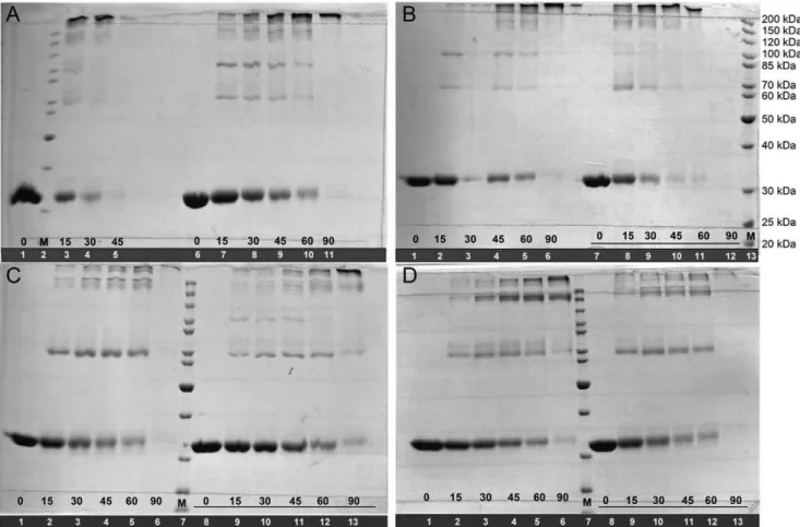
MtPRS quaternary structure could not be assigned by analytical HPLC gel filtration chromatography due to formation of protein aggregates under the experimental conditions described elsewhere [45]. Cross-linking experiments of apoMtPRS were thus pursued and indicates that there is a shift from monomeric, intermediate multi oligomeric states, to predominantly hexameric forms (approximately 220 kDa) after 45 min incubation time in the absence of Pi(Figure 3A, lanes 1, 3–5). Intermediate oligomers, mostly dimers (,70 kDa), trimers (,100 kDa) and tetramers (,150 kDa) could also be visualized on Coomassie Brilliant Blue stained gels. Although the presence of Pi50 mM did not change this oligomerization profile, it appears to have delayed the shift of
MtPRS to hexameric forms (Figure 3A, lanes 6–11). Pre-incubation with R5P 5 mM in either absence or presence of Pi 50 mM appears to have no noticeable effects on shifting the oligomeric states ofMtPRS (Figure 3B) as the profiles are similar to the apo form ofMtPRS (Figure 3A). Pre-incubation with ATP 5 mM seems to stabilize MtPRS dimeric state (Figure 3C), whereas pre-incubation with ADP 5 mM suggests that there is an increase in the tetrameric state ofMtPRS over time (Figure 3D), both in the absence and presence of Pi50 mM.
Enzyme Activity, Substrate Specificity and Inhibition Assays
MtPRS enzyme activity could be detected in the absence of Pi, and in the presence of varying concentrations of ATP dipho-sphoryl group donor at fixed 60mM of R5P (Figure 4A). When substrate ATP was fixed at saturating concentration (300mM) and
enzyme activity measurements at varying R5P concentrations were carried out, substrate inhibition was observed at R5P concentration values larger than 60mM (Figure 4B). Addition of
10–50 mM of Pito the assay mixtures abrogatedMtPRS enzyme activity detection due to inhibition of coupled enzymeMtOPRT (data not shown), likely due to chelating effect of PO4
32anions on
Mg2+
cations [46]. As Mg2+
NPRPP is the true substrate of MtOPRT [38], addition of Pi into the reaction mixture would result in no formation of the true substrate and ensuing lack of activity of MtOPRT coupled enzyme. Accordingly, all MtPRS enzyme activity assays henceforth described were carried out in the absence of Pi.
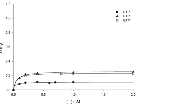
MtPRS enzyme activity could be detected when, under the same experimental conditions, the ATP diphosphoryl group donor was replaced with either purine (GTP) or pyrimidine (CTP and UTP) nucleoside 59-triphosphates (Figure 5). Although the values for the catalytic rate constants (kcat) of GTP, UTP and CTP are lower than the value for ATP, the apparent overall dissociation constant (KM) values are somewhat similar (Table 2). The lower kcat values and similar KM values result in lower values for the
specificity constant (kcat/KM) of GTP, UTP and CTP in
comparison to ATP (Table 2). These results indicate thatMtPRS has broad substrate specificity being able to use ATP, GTP, CTP and UTP as diphosphoryl group donors.
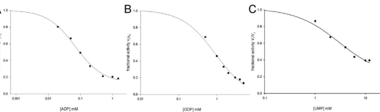
Addition of both ADP (Figure 6A) and GDP (Figure 6B) to
MtPRS reaction mixture (ATP and R5P fixed at, respectively, 300mM and 60mM, under assay conditions described in the
Methods section) resulted in partial inhibition of enzyme activity.
The data on partial enzyme inhibition were fitted to Eq. (4), yielding IC50 values of, respectively, 0.07 (60.01) mM and 0.9 (60.1) mM for ADP and GDP. Addition UMP to the reaction mixture also resulted in partial inhibition of MtPRS, and data fitting to Eq. (4) yielded an IC50 value of 3.0 (60.8) mM (Figure 6C).
To ascertain whether or not these experimental data were due to effects on MtPRS activity and not on MtOPRT coupled enzyme, measurements of activity of the latter enzyme were performed in the presence of the diphosphoryl group donors (ATP, GTP, CTP, and UTP), and nucleoside diphosphate or monophosphate inhibitors (ADP, GDP and UMP). The presence of these compounds in the assay mixtures employed in the coupled assays did not have any effect onMtOPRT enzyme activity to any extent (data not shown). Accordingly, the effects of the alternative diphosphoryl group donors, or nucleoside 59-diphosphate or monophosphate inhibitors, were solely due to changes inMtPRS enzyme activity.
ITF Spectroscopy
Binary complex formation between substrates (R5P, ATP, GTP) or products (AMP, PRPP) and MtPRS was assessed by equilibrium fluorescence spectroscopy to ascertain the order (if any) of addition of these chemical compounds. Titration ofMtPRS with R5P, ATP and GTP were hyperbolic (Figure 7A). These data were thus fitted toEq. (5), yieldingKDvalues of 61 (63)mM for R5P, 18 (62) mM for ATP, and 21 (62) mM for GTP. Titration ofMtPRS with AMP product was sigmoidal (Figure 7B), and data fitting toEq. (6)yielded a value of 109 (63)mM forK’.
There was no intrinsic protein fluorescence change upon PRPP binding to MtPRS, suggesting that PRPP cannot bind to free enzyme. Binding experiments were also carried out in an attempt to determine whether or not there is binary complex formation betweenMtPRS and the alternative pyrimidine substrates UTP and CTP, which can also substitute for ATP as diphosphoryl group donors. No change in protein fluorescence could be detected upon titration ofMtPRS enzyme with UTP and CTP.
Discussion
Recently, Alderwick and workers [30] and Lucarelli and co-workers [31] have also reported biochemical characterization of
MtPRS. Both reported protocols for cloning and purification of recombinant MtPRS are significantly different from the one described herein, sinceMtPRS reported here was produced as a non-His-tagged protein. Although many protocols use histidine tags to facilitate protein purification by the nickel-affinity chromatography strategy, adding histidine tags may alter the protein structure and the biological activity [47,48]. We have thus deemed appropriate to make efforts to produce recombinant
MtPRS without any fusion partner to avoid any possible effect that the latter may have on the former. Notwithstanding, it should be pointed out that steady-state kinetics results were shown by Lucarelli and co-workers [31] to be quite similar for His-tagged
C-Figure 3.MtPRS quaternary structure assignment by glutaraldehyde cross-linking experiments.Incubation times (numbers in black) are shown at the bottom of each lane. Underlined incubation times indicate the presence of 50 mM Piin the reaction mixtures. Lane numbers are in
white in a solid black background. M: Page Ruler Marker (Fermentas).A)ApoMtPRS.B)MtPRS incubated with R5P 5 mM.C)MtPRS incubated with ATP 5 mM.D)MtPRS incubated with ADP 5 mM.
doi:10.1371/journal.pone.0039245.g003
Figure 4. Apparent steady-state kinetic constants forMtPRS, measured under standard assay conditions (Methods), for substrate pair ATP/R5P.A) Varied ATP concentrations in presence of 60mM R5P. B) Varied R5P concentrations in presence of saturating ATP (300mM).
terminal His-tagged MtPRS was stable in solution up to 2 mg mL21 in KH2PO4 buffer at pH 7.9 containing 150 mM NaCl, 1 mM DTT, 10% glycerol. It has been reported that addition of ammonium sulfate or Mg2+
NATP was needed to preserve 20% of MtPRS activity in 50 mM Tris-HCl pH 8.0 and 50 mM Hepes-NaOH pH 8.0 buffers [31]. Lucarelli et al. [31] also reported that full activity of MtPRS could be maintained with addition of 50 mM Pi. No loss of activity could be observed forMtPRS in Tris HCl 50 mM pH 7.5 buffer for the protein preparation here described. The possible explanations for these conflicting exper-imental observations are rather elusive at the moment.
MtPRS quaternary structure could not be unequivocally determined by size exclusion liquid chromatography, in agreement with previous reports on PRS enzymes showing a tendency of these proteins to exist in multiple aggregated states in solution, ranging from dimeric to octameric quaternary structures [49,50]. Accordingly, the glutaraldehyde cross-linking method followed by SDS-PAGE analysis [35] method was employed to determine the
MtPRS protein oligomerization state in solution. These data suggest that recombinantMtPRS may adopt multiple oligomeric states over time, in which the homo hexameric form is the
predominant quaternary structure for apo MtPRS after 45 min incubation time in absence of Pi. Addition of 50 mM Pito apo
MtPRS seems to delay this quaternary structure organization shift from monomer to hexamer (Figure 3A). This apparent delay might be related to Pi mediated stabilization of recombinant
MtPRS at alternative organization of quaternary states (in dimeric or trimeric structures), as inorganic phosphate concentration of at least 25 mM has been proposed as essential for PRS complete stability [28]. Alternatively, binding of PitoMtPRS may lead to protection of lysine, tyrosine, histidine and arginine residues, slowing the reaction of glutaraldehyde cross-linking over time. However, whether the presence of Piresults in oligomeric state stabilization or in reduction in cross-linking remains to be established. Addition of R5P 5 mM appears to have no effect on time-dependent shifting of MtPRS oligomeric states when compared to its apo form (Figure 3B). Nevertheless, MtPRS incubation with ATP 5 mM (Figure 3C) leads to a shift towards dimeric quaternary structure with concomitant reduction in the hexameric form, whereas incubation with ADP 5 mM (Figure 3D) enhanced theMtPRS tetrameric organization. These results are in agreement with previous reports onM. tuberculosis
Figure 5. Apparent steady-state kinetic constants forMtPRS, measured under standard assay conditions (Methods), for substrates GTP, CTP and UTP, varied in the presence of fixed-saturating concentration of R5P (60mM).
doi:10.1371/journal.pone.0039245.g005
Table 2.Apparent kinetic parameters forMtPRS reaction.
Substrate pair Kinetic parameters
KM(mM) Vmax(mmol21min21mg21) kcat(s21) kcat/KM(M21s21) Ki(mM)
ATP/R5P 25 (64) 1.12 (60.03) 0.66 (60.02) 26 (64)6103 –
R5P/ATP 14 (62) 1.41 (60.07) 0.83 (60.04) 59 (68)6103 211 (628)
GTP/R5P* 37 (69) 0.237 (60.004) 0.140 (60.002) 3.8 (60.9)6103 –
UTP/R5P* 52 (67) 0.264 (60.005) 0.155 (60.003) 3.0 (60.4)6103 –
CTP/R5P* 26 (61) 0.111 (60.003) 0.065 (60.001) 2.5 (60.1)6103 –
recombinant PRS quaternary structure (Table 3). Sedimentation velocity experiments in 50 mM KH2PO4pH 7.9 buffer contain-ing either R5P, ATP and ADP at the same concentrations described here led to somewhat similar changes in oligomerization states of MtPRS [30]. Namely, R5P has no effect and ATP increased the hexameric species with concomitant decrease in homodimeric state ofMtPRS [30]. On the other hand, ADP has been reported to affect the molar mass distribution increasing the hexameric state with a concomitant reduction in trimeric species [30]. This shift could be related to human PRS isoform 1 [11] and
B. subtilis[22] ADP binding site identification on the interface of three subunits in the hexamer, a quaternary structure that might be stabilized by the presence of ADP in solution. Interestingly, analytical gel filtration results suggested that apo M. tuberculosis
PRS eluted as a single symmetrical peak consistent with the hexameric state in phosphate buffer [31]. The data here presented on glutaraldehyde cross-linking (Figure 3A) and elution of a single peak from Superdex 200 size exclusion column (protein purifica-tion protocol) suggest thatMtPRS exists as a hexamer in Tris HCl buffer and absence of ligands. Quaternary structure assignment of PRS enzymes in solution remains ambiguous with varying results in presence and absence of ligands [50]. It has been proposed that
ADP binding to an allosteric site ofMtPRS induces stabilization of an inactive, hexameric oligomeric species [30]. Further efforts appear thus to be warranted to ascertain whether or not the dynamic equilibrium of MtPRS has any bearing on enzyme activity.
PRS enzyme activity is often assessed by radiochemical assays with either [14C]-R5P [30] or [c-32P]-ATP detection [11,25,49,51], by enzyme coupling with myokinase, pyruvate kinase and lactate dehydrogenase [52], or by a recently developed HPLC-based method that follows AMP formation [31]. Here we present, to the best of our knowledge, a novel coupled continuous spectrophotometric assay that measures the decrease in orotate concentration catalyzed by MtOPRT in the presence of PRPP formed in solution by MtPRS enzyme activity, a assay first proposed as a suitable alternative to follow PRS activity in the late 709s, by Switzer and co-workers [28].
MtPRS-catalyzed PRPP formation could be measured in the presence of R5P and ATP in absence of Pi (Figure 4A). Interestingly, it has been reported that Piis required for MtPRS enzyme activity [30,31]. The reason for this discrepancy is not apparent at the moment. Measurements of MtPRS enzyme activity here presented were carried out in the complete absence Figure 6. Inhibition ofMtPRS enzyme activity by A) ADP; B) GDP; and C) UMP.MtPRS expressed as its fractional activity; and ADP, GDP and UMP concentrations were plotted on log scale.
doi:10.1371/journal.pone.0039245.g006
Figure 7. A)Hyperbolic equilibrium binding of R5P, ATP and GTP to MtPRS assessed by ITF. B) Sigmoidal equilibrium binding of AMP toMtPRS assessed by ITF.
of Pisince the enzyme was stored in Tris HCl 50 mM pH 7.5 and activity measurements assessed in Tris HCl 50 mM MgCl2 20 mM pH 8.0, OA 300mM, MtOPRT 1.8 U, and varied
concentrations of ATP and R5P. It is possible that an explanation for this discrepancy may be attributed to coupled assay sensitivity, allowing MtPRS activity detection even in absence of Pi. In addition, the steady-state kinetic constants for MtPRS enzyme reported in this work (Table 2) are considerably distinct from previous reports (Table 4) [30,31]. It could be argued that the rather low values for the kinetic constants reported here are not representative ofMtPRS full activity, as it has being described that lower Pi concentrations led to partial enzyme activity [30]. However, it appears more plausible that the rather low value for theMtPRS catalytic constant is due to the limiting value for the maximum velocity of the coupled enzyme (0.6 s21) as reported elsewhere [38]. Notwithstanding, the results presented here demonstrate that Piis not an obligatory requirement forMtPRS catalytic activity. An interesting feature was identification of substrate inhibition by R5P when it is varied in the presence of saturating ATP concentration (Figure 4B), with Ki value of 211mM. Substrate inhibition by R5P has been reported for rat
liver PRS [53], as well as forE. coli[54] andM. tuberculosis[30], both in the presence of non-saturating Piconcentrations.
The dependence of MtPRS activity upon varying Mg2+
concentrations could not be assessed as this cation is essential for
MtOPRT coupled enzyme activity [38]. We have thus fixed the Mg2+ concentration at 20 mM based on both the optimum
concentration for activity ofMtOPRT (larger concentration values are inhibitory) [38] and previously reported saturation curve for the dependence ofMtPRS activity on increasing Mg2+
concen-tration [31]. It has been shown that the enzyme requires free Mg2+
as an activator and as Mg2+
NATP co-substrate, and that free Mg2+
is likely to be an allosteric effector of the K-type enzyme model for cooperativity [31].
Substrate specificity measurements showed that MtPRS can accept GTP, CTP, and UTP, in addition to ATP, as diphosphoryl group donors (Figure 5, Table 2), thereby showing broad substrate specificity. Although theKMvalues are similar, the values
for the catalytic rate constants of GTP, CTP and UTP are lower than ATP, MtPRS main substrate. S. typhimurium PRS, another bacterial PRS, has been described as being specific for ATP, although capable of using GTP, ITP, CTP and UTP as alternative substrates to a lesser extent (3% of maximum reported activity using ATP as substrate) [28].
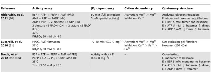
The purine nucleoside diphosphates ADP and GDP were reported as PRS allosteric inhibitors [23,25]. Under assay conditions here described, both behave partial inhibitors with IC50 values of, respectively, 0.0760.01 mM (Figure 6A) and 0.960.1 mM (Figure 6B). ADP has been shown to be a non-competitive inhibitor ofMtPRS with overall inhibition constant Table 3.Comparison of biochemical data onM. tuberculosisPRS.
Reference Activity assay [Pi] dependency Cation dependency Quaternary structure
Alderwick, et al. 2011[30]
R5P+ATPRPRPP+AMP (PRS) AMP+ATPR2ADP (MK)
ADP+PEPR2 piruvate+2 ATP (PK) 2 piruvate+2 NADH+2HR2 lactate+2 NAD+ (LDH)
37uC
KH2PO450 mM pH 8.0
50 mM (full activation) 5 mM (partial activity)
Activation: Mn2+.Mg2+ Inhibition: Ca2+
Analytical ultracentrifugation: E: trimer and hexamer (equilibrium); E+R5P 5 mM: trimer and hexamer; E+ATP 5 mM:Qhexamerqdimer; E+ADP 5 mM:Qtrimerqhexamer.
Lucarelli, et al. 2010[31]
HPLC, AMP formation 37uC
KH2PO450 mM pH 8.0
10–40 mM (59.7 U mg21) Activation: Mn2+.Mg2+ Inhibition: Cu2+.Fe2+. Ca2+
Size exclusion gel filtration: Hexamer (220 KDa).
Breda, et al. 2012(this work)
R5P+ATPRPRPP+AMP (MtPRS) PRPP+OARPPi+OMP (MtOPRT) 25uC
Tris HCl 50 mM pH 8.0
Activity without Pi (1.16 U mg21)
– Cross-linking:
E: monomer to hexamer;
E+R5P 5 mM: monomer to hexamer; E+ATP 5 mM:Qhexamerqdimer; E+ADP 5 mM:qtetramer.
doi:10.1371/journal.pone.0039245.t003
Table 4.Comparative of apparent kinetic parameters forMtPRS substrate pair ATP/R5P.
Reference Kinetic parameters
KM(mM) Vmax(mmol21min21mg21) k
cat(s21) kcat/KM(M21s21) Ki(mM)
R5P
Alderwick, et al. 2011[30] 8.2 530 60.68 74306103 –
Lucarelli, et al. 2010[31] 71 – 37.1 5216103 –
Breda, et al. 2012(this work) 14 (62) 1.41 (60.07) 0.83 (60.04) 59 (68)6103 211 (628)
ATP
Alderwick, et al. 2011[30] S0.5= 62.65/n = 1.68 601 – – –
Lucarelli, et al. 2010[31] S0.5= 260 (650)/n = 1 – 34.6 (63) 133.16103 –
Breda, et al. 2012(this work) 25 (64) 1.12 (60.03) 0.66 (60.02) 26 (64)6103 –
values ranging from 320mM to 522mM [30]. On the other hand, it has been reported an IC50 value ranging from 0.26 mM (at saturating ATP and non-saturating Piconcentrations) to 0.4 mM (saturating ATP and saturating Piconcentrations) for ADP and an IC50larger than 5 mM for GDP inhibition ofMtPRS activity in the presence of Pi[31]. These results prompted the proposal of a regulatory site to which both ADP inhibitor and Piactivator can bind [31]. Accordingly, ADP binding to the regulatory site hinders Pibinding resulting in inhibition ofMtPRS enzyme activity [31]. In addition, the sigmoidal curve for ADP inhibition ofMtPRS has been shown to affect the maximum velocity only, without affecting the value ofK9and the degree of cooperativity [31]. To evaluate if
MtPRS activity was responsive to pyrimidine regulation, the effect of UMP titration upon standard activity assay (Methods) was assessed (Figure 6C), which yielded an IC50value of 3.0 (60.8) mM. These findings seem to indicate that MtPRS activity is regulated in response to M. tuberculosis energetic demand, being more responsive to purine inhibition (ADP and GDP), although variation in pyrimidine intermediates (UMP) could also regulate its activity. The regulation ofMtPRS enzyme activity by variations in both purine and pyrimidine intermediates is in accordance with the role of PRPP as a key intermediate in the metabolic pathways for the synthesis and recycling of both purine and pyrimidine nucleotides.
Hyperbolic binding isotherms determined from ITF measure-ments indicated that substrates R5P (KD = 61mM), ATP (KD = 18mM), and GTP (KD = 21mM) can bind to free apoMtPRS (Figure 7A). Dissociation constant values for ATP and GTP are somewhat similar, an indicative that there might be no substrate preference between these purine nucleosides 59-triphosphates, which is in agreement with their similar KM values (Table 2).
Although the results of steady-state kinetic experiments have shown that CTP and UTP can act as diphosphoryl group donors, no change in IFT could be detected in the absence of R5P substrate. These findings might suggest that binding of pyrimidine nucleosides 59-triphosphate results in no change in tryptophan fluorescence or that there is an alternative order of substrate addition for pyrimidine nucleotides. No change in ITF could be detected upon PRPP titration into free MtPRS. On the other hand, AMP product showed hyperbolic variation of ITF upon titration to free MtPRS, with K9 value of 109mM and a Hill coefficient value of 3.2 (Figure 7B), an indicative of positive homotropic cooperativity. Further experiments of isothermal titration calorimetry could be pursued to address the issue of whether or not pyrimidine nucleotides are capable of binding to freeMtPRS.
Data on steady-state kinetics and equilibrium binary complex formation suggest that the enzyme mechanism of MtPRS for purine (ATP and GTP) diphosphoryl donors follows a random-ordered substrate addition with random-ordered product dissociation, in which PRPP is the first product to be released followed by purine nucleoside monophosphate products (AMP or GMP) to yield free enzyme for the next round of catalysis (Figure 8A). Although the order of substrate addition can be proposed as it is based on solid experimental evidence, the order of product release has to be considered with caution. For instance, it is possible that PRPP binding to freeMtPRS enzyme results in no change in ITF, which would imply in a random-order mechanism of product release. Isothermal titration calorimetry can thus be used to address the issue of whether or not PRPP is capable of binding to freeMtPRS. The enzyme mechanism for pyrimidine (UTP or CTP) diphos-phate donors might obey an ordered mechanism of substrate addition and product release; in which R5P binds to free enzyme followed by the diphosphate donors, and PRPP release is followed by pyrimidine nucleoside monophosphate products (UMP or CMP) to yield freeMtPRS (Figure 8B).
Considering their molecular and kinetic characterization, three different classes of PRS enzymes have been described. Classifica-tions of PRS proteins as belonging to Class I (also known as ‘‘Classical’’). Class II or Class III are based on specificity for diphosphoryl donors, requirement of Pi for activity, allosteric inhibition by purine ribonucleoside diphosphates, and oligomeric states [23,25,31]. It has been proposed that there is also a proportional relationship amongKM,Vmaxand PRS classes [25], in which Class III enzymes have largerKM values for R5P and ATP substrates, Class I with the lowest values, and Class II with intermediate values [25]. The extent to which these criteria could be used for classifying PRS enzymes are still not clear due to limited number of representatives of Classes II and III PRSs [25].
MtPRS has approximately 41% identity to the three human PRS isoforms, as well as toA. thalianaand spinach Class I enzymes (isoforms 1 and 2). The degree of primary sequence conservation drops to 18–23% when theM. tuberculosissequence is compared to Class II PRS enzymes from the latter two organisms (isoforms 3 and 4). As previously demonstrated [11,25,31], the amino acids involved in substrate binding are the most conserved regions:
MtPRS Tyr88-Ser104 and Asp166-Arg169 for ATP binding, and
Figure 6A and with previous reports showing that ADP is an allosteric inhibitor of MtPRS [30,31]. Despite low amino acid conservation, secondary structure prediction showed that homo-trimeric spinach PRS isozyme 4 (a Class II enzyme) and hexamericB. subtilisPRS (a Class I enzyme) have a similar folding pattern [23]. No Class II PRS structure has been solved so far, thus any inferences about amino acids substitution that might account for this class broader substrate specificity are, based on available structural data, somewhat speculative. PRS nucleotide binding pocket is located in a wide cleft, and the secondary structure elements might undergo conformational rearrangements upon ligand binding to accommodate both purine and pyrimidine bases, as well as properly positioning of amino acids side chains to specifically hydrogen bond each diphosphoryl group donor.
The broad specificity for diphosphoryl group donors and detection of enzyme activity in the absence of Piwould suggest that
MtPRS belongs to Class II PRS proteins. On the other hand, the hexameric quaternary structure assembly, as suggested by cross-linking experiments (Figure 3) would indicate that it belongs to Class I PRS enzymes. In addition, allosteric inhibition by ADP [30,31] (Figure 6A) would place MtPRS in Class I PRSs. Accordingly, it has previously been suggested thatMtPRS belongs
to Class I [30]. Further data are thus needed to classifyMtPRS as belonging to a particular family of PRS proteins.
It should be pointed out that the results here presented extend previous studies onMtPRS [30,31]. To the best of our knowledge, the results here presented are, along withS. typhimuriumPRS data, the first experimental evidence for a bacterial PRS enzyme that can use both pyrimidine and purine nucleosides triphosphate as diphosphoryl group donors since broad substrate specificity has been described for plants only. In addition, this is the first report on MtPRS enzyme mechanism for purine and pyrimidine diphosphate donors. Current efforts are towards experimental structure determination ofMtPRS to provide a solid foundation for the rational design of, hopefully, specific inhibitors of this enzyme without affecting to a great extent the host PRS.
Author Contributions
Conceived and designed the experiments: AB LAB DSS. Performed the experiments: AB CBB LKBM CVB LAR. Analyzed the data: AB CVB LKBM LAR. Contributed reagents/materials/analysis tools: LAB DSS. Wrote the paper: AB LAB.
References
1. World Health Organization (2010) Global Tuberculosis Control 2010. Geneva: WHO Press.
2. World Health Organization (2010) The Global Plan to Stop TB 2011–2015: Transforming the fight towards elimination of tuberculosis. Available: http:// www.stoptb.org. Accessed 2011 April 04.
3. Ma Z, Lienhardt C, McIlleron H, Nunn AJ, Wang X (2010) Global tuberculosis drug development pipeline: the need and the reality. Lancet 375: 2100–2109. 4. World Health Organization (2009) A ministerial meeting of high M/XDR-tb
burden countries. Available: http://www.who.int/tb/challenges. Accessed 2011 April 16.
5. Aziz MA, Wright A, Laszlo A, Muynck AD, Portaels F, et al. (2006) Epidemiology of antituberculosis drug resistance (the global project on anti-tuberculosis drug resistance surveillance): an updated analysis. Lancet 368: 2142–2154.
6. Svenson S, Ka¨llenius G, Pawlowski A, Hamasur B (2010) Towards new tuberculosis vaccines. Human Vaccines 6: 309–317.
7. Velayati AA, Masjedi MR, Farnia P, Tabarsi P, Ghanavi J, et al. (2009) Emergence of new forms of totally drug-resistant tuberculosis bacilli super extensively drug-resistant tuberculosis or totally drug-resistant strains in Iran. Chest 136: 420–425.
8. Udwadia ZF, Amale RA, Ajbani KK, Rodrigues C (2011) Totally drug-resistant tuberculosis in India. Clin Infect Dis Advance Access DOI: 10.1093/cid/cir889. 9. Ducati RG, Basso LA, Santos DS (2007) Mycobacterial shikimate pathway
enzymes as targets for drug design. Curr Drug Targets 8: 423–435. 10. Khorana HG, Fernandes JF, Kornberg A (1958) Pyrophosphorylation of ribose
5-phosphate in the enzymatic synthesis of 5-phosphorylribose 1-pyrophosphate. J Biol Chem 230: 941–948.
11. Li S, Lu Y, Peng B, Ding J (2007) Crystal structure of human phosphoribo-sylpyrophosphate synthetase 1 reveals a novel allosteric site. Biochem J 401: 39– 47.
12. Hove-Jensen B (1988) Mutation in the phosphoribosylpyrophosphate synthetase gene (prs) that results in simultaneous requirements for purine and pyrimidine nucleosides, nicotinamide nucleotide, histidine, and tryptophan inEscherichia coli. J Bacteriol 170: 1148–1152.
13. Ames BN, Martin RG, Garry BJ (1961) The first step of histidine biosynthesis. J Biol Chem 236: 2019–2026.
14. Zoref E, De Vries A, Sperling O (1975) Mutant feedback-resistant phosphori-bosylpyrophosphate synthetase associated with purine overproduction and gout. J Clin Invest 56: 1093–1099.
15. Schneiter R, Carter AT, Hernando Y, Zellnig G, Schweizer LM, Schweizer M (2000) The importance of the five phosphoribosyl-pyrophosphate synthetase (Prs) gene products ofSaccharomyces cerevisiaein the maintenance of cell integrity and the subcellular localization of Prs 1p. Microbiology 146: 3269–3278. 16. Hove-Jensen B (2004) Heterooligomeric phosphoribosyl diphosphate synthase of
Saccharomyces cerevisiae. J Biol Chem 279: 40345–40350.
17. Scherman MS, Kalbe-Bournonville L, Bush D, Xin Y, Deng L, et al. (1996) Polyprenylphosphate-pentoses in mycobacteria are synthesized from 5-phos-phoribose pyrophosphate. J Biol Chem 271: 29652–29658.
18. Wolucka BA (2008) Biosynthesis of D-arabinose in mycobateria - A novel bacterial pathway with implication for antimycobaterial therapy. FEBS J 275: 2691–2711.
19. Hove-Jensen B, Harlow KW, King CJ, Switzer RL (1986) Phosphoribosylpy-rophosphate synthetase ofEscherichia coli. Properties of the purified enzyme and primary structure of the prs gene. J Biol Chem 261: 6765–6771.
20. Switzer L (1969) Regulation and mechanism of phosphoribosylpyrophosphate synthetase I: Purification and properties of the enzyme from Salmonella typhimurium.J Biol Chem 244: 2854–2863.
21. Tatibana M, Kita K, Taira M, Ishijima S, Sonoda T, et al. (1995) Mammalian phosphoribosylpyrophosphate synthetase. Adv Enzyme Regul 35: 229–249. 22. Eriksen TA, Kadziola A, Bentsen AK, Harlow KW, Larsen S (2000) Structural
basis for the function ofBacillus subtilisphosphoribosyl-pyrophosphate synthetase. Nat Struct Biol 7: 303–308.
23. Krath BN, Hove-Jensen B (2001) Implications of secondary structure prediction and amino acid sequence comparison of class I and class II phosphoribosyl diphosphate synthases on catalysis, regulation, and quaternary structure. Protein Sci 10: 2317–2324.
24. Sinha SC, Smith JL (2001) The PRT protein family. Curr Opin Struct Biol 11: 733–739.
25. Kadziola A, Jepsen CH, Johansson E, McGuire J, Larsen S, et al. (2005) Novel class III phosphoribosyl diphosphate synthase: structure and properties of the tetrameric, phosphate-activated, non-allosterically inhibited enzyme from Methanocaldococcus jannaschii. J Mol Biol 354: 815–828.
26. Krath BN, Hove-Jensen B (2001) Class II recombinant phosphoribosyl diphosphate synthase from spinach. J Biol Chem 276: 17851–17856. 27. Krath BN, Eriksen TA, Poulsen TS, Hove-Jensen B (1999) Cloning and
sequencing of cDNAs specifying a novel class of phosphoribosyl diphosphate synthase inArabdopsis thaliana. Biochim Biophys Acta 1430: 403–408. 28. Switzer RL, Gibson KJ, (1978) Phosphoribosylpyrophosphate synthase
(ribose-5-phosphate pyrophosphokinase) fromSalmonella typhimurium. Methods Enzymol 51: 3–11.
29. Sassetti CM, Boyd DH, Rubin EJ (2003) Genes required for mycobacterial growth defined by high density mutagenesis. Mol Microbiol 48: 77–84. 30. Alderwick LJ, Lloyd GS, Lloyd AJ, Lovering AL, Eggeling L, et al. (2011)
Biochemical characterization of theMycobacterium tuberculosis phosphoribosyl-1-pyrophosphate synthetase. Glycobiology 21: 410–425.
31. Lucarelli AP, Buroni S, Pasca MR, Rizzi M, et al. (2010)Mycobacterium tuberculosis phosphoribosylpyrophosphate synthase: Biochemical features of a crucial enzyme for mycobacterial cell wall biosynthesis. PloS ONE 5(11): e315494. 32. Laemmli UK (1970) Cleavage of structural proteins during the assembly of the
head of bacteriophage T4. Nature 227: 680–685.
33. Klammer AA, MacCoss MJ (2006) Effects of modified digestion schemes on the identification of proteins from complex mixtures. J Proteome Res 5: 695–700. 34. Zhang Z, Marshall AG (1998) A universal algorithm for fast and automated
charge state deconvolution of electrospray mass-to-charge ratio spectra. J Am Chem Soc Mass Spectrom 9: 225–233.
35. Fadouloglou VE, Kokkinidis M, Glykos NM (2008) Determination of protein oligomerization state: two approaches based on glutaraldehyde cross linking. Anal Biochem 373: 404–406.
37. Ozturk DH, Dorfman RH, Scapin G, Sacchettini JC, Grubmeyer C (1995) Locations and functional roles of conserved lysine residues in Salmonella typhimuriumorotate phosphoribosyltransferase. Biochemistry 34: 10755–10763. 38. Breda A, Rosado LA, Lorenzini DM, Basso LA, Santos DS (2012) Molecular,
kinetic and thermodynamic characterization ofMycobacterium tuberculosisorotate phosphoribosyltransferase. Mol BioSyst 5: 572–586.
39. Cook PF, Cleland WW (2007) Enzyme assays. In: Enzyme kinetics and mechanism. Garland Science. 19–34.
40. Harris DA (1987) Spectrophotometric assays. In: Harris DA, Bashford CL, editors. Spectrophotometry and spectrofluorimetry – A practical approach. IRL Press. 49–90.
41. Copeland RA (2000) Enzymes – A practical introduction to structure, mechanism and data analysis. Wiley-VCH, New York, USA.
42. Segel IH (1993) Enzyme kinetics - Behavior analysis of rapid equilibrium and steady-state enzyme systems. Wyley-Interscience Publication, John Wiley & Sons, Inc, New York, USA.
43. Harris DA, Bashford CL, editors. Spectrophotometry & spectrofluorimetry – A practical approach. IRL Press, Oxford, UK.
44. Hill AV (1913) The combinations of haemoglobin with oxygen and with carbon monoxide. J Biochem 7: 471–480.
45. Martinelli LKB, Ducati RG, Rosado LA, Breda A, Selbach BP, et al. (2011) RecombinantEscherichia coliGMP reductase: kinetic, catalytic and chemical mechanisms, and thermodynamics of enzyme–ligand binary complex formation. Mol Biosyst 7: 1289–1305.
46. Dawson RMC, Elliot DC, Elliot WH and Jones KM (1989) Data for biochemical research. Oxford Science Publications. 3rd
Ed. p.408.
47. Chant A, Kraemer-Pecore CM, Watkin R, Kneale GG (2005) Attachment of a histidine tag to the minimal zinc finger protein of theAspergillus nidulansgene regulatory protein AreA causes a conformational change at the DNA-binding site. Protein Expr Purif 39: 152–159.
48. Fonda I, Kenig M, Gaberck-Porekar V, Prostovaek P, Menart V (2002) Attachment of histidine tags to recombinant tumor necrosis factor-alpha drastically changes its properties. Sci World J 2: 1312–1325.
49. Arnvig K, Hove-Jensen B, Switzer RL (1990) Purification and properties of phosphoribosyl-diphosphate synthetase fromBacillus subtilis. Eur J Biochem 192: 195–200.
50. Schubert KR, Switzer RL (1975) Studies of the quaternary structure and the chemical properties of phosphoribosylpyrophosphate synthetase fromSalmonella typhimurium. J Biol Chem 250: 7492–7500.
51. Nosal JM, Switzer RL, Becker MA (1993) Overexpression, purification, and characterization of recombinant human 5-phosphoribosyl-1-pyrophosphate synthetase isozymes I and II. J Biol Chem 268: 10168–10175.
52. Braven J, Hardwell TR, Seddon R, Whittaker M (1984) A spectrophotometric assay of phosphoribosyl pyrophosphate synthetase. Ann Clin Biochem 21: 366– 371.
53. Roth DG, White C, Deuel TF (1978) Ribosephosphate pyrophosphokinase (rat liver). Methods Enzymol 51: 12–17.