Possible Role of Autoimmunity in Patients with
Premature Ovarian Insuf f iciency
Renata Košir Pogačnik, M.D.1*, Helena Meden Vrtovec, Ph.D.1, Alenka Vizjak, Ph.D.2, Alenka Uršula
Levičnik, M.D.1, Nina Slabe, M.D.1, Alojz Ihan, Ph.D.3
1. Department of Obstetrics and Gynecology, University Medical Centre Ljubljana, Ljubljana, Slovenia 2. Institute of Pathology, Faculty of Medicine, University of Ljubljana, Ljubljana, Slovenia 3. Institute of Microbiology and Immunology, Faculty of Medicine, University of Ljubljana,
Ljubljana, Slovenia
Abstract
Background:To evaluate the involvement of immune abnormality in patients with
idi-opathic premature ovarian insuficiency (POI). In addition to the known etiology, autoim
-mune disorders may be a pathologic mechanism for POI.
Materials and Methods:Our study was a prospective controlled trial. Twenty women
with POI, reasons other than autoimmune excluded, were enrolled in this study. The control group consisted of 17 healthy women. In both groups, family and per
-sonal history were taken and the levels of follicle stimulating hormone, luteinizing hormone, thyroid-stimulating hormone, prolactin, anti-Müllerian hormone, inhibin B, antithyroglobulin and antithyroid peroxidase antibodies were determined.
Anti-ovarian antibodies and subpopulations of peripheral blood T-lymhocytes were also
determined.
Results: Participants in the study group exhibited hypergonadotropichypogonadism,
while high levels of follicle stimulating hormone and low levels of inhibin B and anti-Müllerian hormone were observed. In 16 (80%) patients, POI was associated in their personal and familial history with another autoimmune disease. Fifty percent of patients presented highly elevated antithyroid antibodies. The lymphocyte subset, especially B cells, was signiicantly higher (p=0.014), and peripheral regulatory lymphocytes CD25+ high were signiicantly lower (p=0.015) in the study group than in the control group.
Anti-ovarian antibodies were detected in 20% of patients with POI.
Conclusion:We presume that the presence of anti-ovarian antibodies together with ab
-normalities of cellular immunity may in some cases potentially represent the
involve-ment of an autoimmune mechanism in idiopathic POI.
Keywords: Autoantibody, Thyroid Stimulating Antibody, Cell Immunity, Premature
Ovarian Insuficiency, T-Lymphocyte
Citation: Košir Pogačnik R, Meden Vrtovec H, Vizjak A, Uršula Levičnik A, Slabe N, Ihan A. Possible role of autoim
-munity in patients with premature ovarian insuficiency. Int J Fertil Steril. 2014; 7(4): 281-290.
Received: 17 Nov 2012, Accepted: 07 Apr 2013
* Corresponding Address: Department of Obstetrics and Gynecol
-ogy, University Medical Centre Ljubljana, Šlajmerjeva 3, SI-1000 Ljubljana, Slovenia
Email: renata.kosir@inbox.com Royan Institute
International Journal of Fertility and Sterility
Introduction
Premature ovarian insuficiency (POI) is charac
-terized by hypergonadotropic amenorrhea due to cessation of ovarian function before the age of 40 years. The diagnosis is based on amenorrhea be
-fore the age of 40 associated with follicle stimulat
-ing hormone (FSH) levels >40 IU/l, detected on two occasions at least one month apart (1). POI causes female infertility, while is a signiicant psy
Although there are multiple etiologies of POI (genetic, chromosomal, infectious, and iatrogenic causes), the etiology cannot be identiied in most patients and this is referred to as idiopathic POI; up to 30% of idiopathic cases may have an auto
-immune cause (4). The most convincing evidence coming from the commonly observed association of POI with other autoimmune disorders (5, 6) are demonstration of anti-ovarian antibodies (AOA, 7, 8) and histological indings of ovarian tissue from affected women. Roughly, one third of POI patients have AOA and/or antithyroid antibodies in their serum (1, 9). Various organ-speciic and
systemic autoimmune diseases cause autoimmune
ovarian insuficiency in up to 30% of women with POI (4). According to the literature, 2-10% of POI cases are known to be associated with adrenal au
-toimmunity (10). One of the irst signs that auto -immunity may be responsible for ovarian function failure came from the observation that ovarian
failure may precede the onset of Addison’s disease by 8-14 years (11). Autoimmune Addison’s dis
-ease seldom develops in isolation, whereas several other endocrine glands and organs are generally affected, leading to an autoimmune polyglandular syndrome (APGS, 12). Two main forms of APGS can be clinically discerned, APGS types 1 and 2. APGS type 1 is characterized by an association of mucocutaneous candidiasis, hypoparathyroidism and Addison’s disease. In about 60% of cases, there is also an association with ovarian insufi
-ciency. Blizzard et al. (13) and Irvine et al. (14) found that POI commonly presents with adrenal cytoplasmic antibodies, called steroid cell antibod
-ies (SCA); they react with cytoplasmic antigens of other steroid-producing cells present in the ovary, testis and placenta. Alteration of lymphocytes and their speciic subsets, as well as T-cell mediated injury are likely to play an important role in the pathogenesis of autoimmune oophoritis. Surface markers of peripheral blood mononuclear cells (PBMC) have been shown to be deranged in early autoimmune phases and to be persisted through the disease, even after targeted disruption (15).
The presence of pathogenic factors might accel -erate the process of apoptosis and atresia of
ovar-ian follicles during the fetal and post-natal period (16). This interpretation is based on the dogma that the number of ovarian follicles at birth is inal and that there is no possibility of regeneration or re
-newal of reserve follicles in adulthood (17). Ex
-perimental work on animals suggests a possibility
of renewal of the follicle reserve from proliferative
germinal ovarian cells even after birth; veriication of which is being sought in studies on human ova
-ries (18, 19). It has been shown that undifferenti -ated ovarian stem cells differentiate into structures
similar to egg cells under certain laboratory condi
-tions (20). We faced two problems: whether to ac
-cept the standard understanding of a inal number
of ovarian follicles or to focus on the hypothetic
possibility of renewal of the follicular reserve. The
aim of this study was to evaluate the involvement of immune abnormality in patients with idiopathic
POI.
Materials and Methods
Subjects
Our study was a prospective randomized con
-trolled trial. The study group consisted of 20 wom
-en with POI (mean age 31.8 years, range 20-39 years) and no use of medications or oral contracep
-tives for at least 4 months prior to the study. The di
-agnosis was based on the presence of amenorrhea before the age of 40, associated with two serum FSH levels above 40 IU/l at least one month apart. All women with POI underwent karyotyping and genetic testing of the fragile X mental retardation 1 (FMR1). Women with infectious or iatrogenic causes were excluded from the study. The control group consisted of 17 healthy women volunteers. Inclusion criteria were a regular menstrual cycle, age between 18 and 39 years (mean 30.8 years),
and no use of medications or oral contraceptives
for at least 4 months prior to the study. They also had to be exempt from autoimmune disease or in
-fertility problems. All women provided a complete personal and family history, with a stress on pos
-sible immune-mediated, particularly autoimmune processes (allergy, asthma, diabetes, thyroiditis, rheumatoid arthritis, andatopiceczema), and all underwent physical and vaginal ultrasound exami
-nations.
Hormone and serological analyses
Serum levels of luteinizing hormone (LH), FSH, estradiol (E2), prolactin (PRL), inhibin B, thy
measured, and immunological investigations at cellular and humoral levels were performed. The adrenocorticotropic hormone (ACTH) stimulation test was performed in the study group, only. The standard Synacthen stimulation test is clinically widely used as a sensitive screening method for symptomatic adrenal insuficiency. Each ampoule of Synacthen contains 250 μg of the active ingre
-dient, tetracosactrin (Novartis Pharmaceuticals, North Ryde NSW, Australia). Thirty minutes after 250 μg Synacthen I.M. (Alliance Pharmaceutical Wiltshire, UK), blood cortisol was measured by electro-chemiluminescence immunoassay. A nor
-mal cortisol response to Synacthen was deined as a post-stimulation peak cortisol value of >500 nmol/l at 30 minutes.
Serum AMH in peripheral blood was deter
-mined by a Personal Lab analyser using the enzyme linked immunosorbent assay (ELISA) method with Beckman Coulter reagent. The normal range of AMH levels is 0.7-3.5 mg/l.
Values below 0.3 mg/l indicate a reduced ovar
-ian reserve. Hormones were determined on a LI
-AISON analyser by quantitative direct competi
-tive chemiluminescence immunoassays (CLIA). Each test is a modified two-step process, in
which the specific antibodies of a certain
hor-mone bind to magnetic cells. Normal ranges for the follicular phase of the cycle are as follows: FSH: 3.5-9.2 IU/l; LH: 1.1-11.6 IU/l; LH around ovulation: 17-77 IU/l; PRL: 6.2-23.5 μg/l; E2: on day 3 up to 310 pmol/L (0.31 nmol/L); E2 postmenopause: 0-110 pmol/L (0-0.11 nmol/l); and TSH: 0.3-3.6 mE/l. Serum Inhibin-B cut-off level on day 3 was 45 pg/mL. Serum aTG and
aTPO concentrations measured by immunoas
-say using direct chemiluminometric technology on an ADVIA CENTAUR analyser (Siemens Medical Solutions Diagnostics, Tarrytown, USA). The normal range for serum aTG and aTPO is <60 KE/l. Detection of AOA was by
indirect immunofluorescence (IIF) on cryosec
-tions of normal human ovarian tissue.
Non-human primate ovaries for the detection of AOA are not commercially available, while
normal human ovarian tissue is available at
the Institute of Pathology, Ljubljana, Slovenia,
when ovaries are removed due to various
patho-logical processes, particularly tumors, and are sent for pathohistological examination. Ovarian
tissue was incubated with patient serum diluted
1:10. The second incubation was with luorescein isothiocyanate-labelled anti-human IgG antibody (Dakopatts, Copenhagen, Denmark). Positive re
-actions were semi-quantitatively evaluated (on a scale of 1– 4+). Negative control omitting the pa
-tient’s serum was regularly included (21). At the cellular level fresh PBMC were studied by low cytometry (Becton Dickinson FACS, NJ, USA), and percentages of the following blood lympho
-cyte populations were determined: T cells (CD3+),
helper T cells (CD4+), cytotoxic T cells (CD8+),
natural killer cells (CD56+CD16+), regulatory T
cells (CD25+high) and B cells (CD19+) (22). Im
-munoluorescence labelling was performed by in
-cubating PBMCs with monoclonal antibodies to CD3, CD4, CD8, CD25, CD56/16 and CD45. A
differential blood count with a standard laboratory
procedure was taken to obtain concentrations of individual lymphocyte subtypes.
Statistical analysis
Normal data distribution was tested with the Kolmogorov-Smirnov test. Where variables were normally distributed, we used the Pearson’s chi-square test. If variables were not normally dis
-tributed, we used a nonparametric Mann-Whitney test. Statistical analysis was done using Statistical Package for the Social Sciences, version 18 (SPSS Inc., Chicago, IL, USA). The results were consid
-ered statistically signiicant at p<0.05.
Ethical considerations
The study protocol was approved by the Nation
-al Ethics Committee, and -all patients gave written informed consent.
Results
General clinical and biological parameters
The subjects were comparable with controls by
age. One patient presenting a mosaic 45X0/46XX was excluded from the inal analysis. Other pa
-tients had a 46XX karyotype. Anamnestic data of patients showed that four had had mumps during their childhood; they were thus excluded from the inal analysis. Table 1 shows the clinical and en -docrine characteristics of patients and of healthy
women. All endocrine parameters in controls were
Table 1: Hormone and ovarian peptide levels in patients and in healthy controls
Control group (n=17) Study group (n=20)
Mean value ± SD
6.25 ± 2.77 88.04 ± 47.93
FSH (IU/L)
11.64 ± 31.1 37.85 ± 18.71
LH (IU/L)
210 ± 130 (0.21 ± 0.13 nmol/L) 180 ± 200 (0.18 ± 0.2 nmol/L)
E2 (pmol/L)
8.74 ± 5.51 11.14 ± 6.42
PRL (μg/L)
32.63 ± 24.05 13.36 ± 10.66
Inhibin B (pg/ml)
3.54 ± 1.58 0.36 ± 0.37
AMH (mg/L)
POI; Premature ovarian insuficiency, FSH; Follicle stimulating hormone, LH; Luteinizing hormone, E2; Estradiol, PRL;
Prolactine and AMH; Anti-müllerian hormone.
Prevalence of autoimmune diseases and associ-ated autoimmune abnormalities
We collected targeted history information on personal and familial autoimmune disorders (Ta
-ble 2). Sixteen patients (80%) had an associated autoimmune disease in their personal and/or fa
-milial history. Four women (20%) with POI had irst grade relatives with ovarian insuficiency before the age of 40. Thyroid disorders were the most common (15%) of the autoimmune diseases associated with POI in personal histories. Before
entering the study, three patients had been treated for autoimmune thyroid dysfunction (Hashimoto thyroiditis). In the study group, 55% of women had autoimmune disease in the family history; the most frequent autoimmune disorder was diabetes type I. The overall personal and familial incidence
of autoimmune diseases was lower in the control
group. In the study group, 50% of women had
aTG. One healthy woman had evidence of auto
-immune thyroid dysfunction, manifested by an el
-evated aTG serum level, and was excluded from the inal analysis (Tables 2, 3).
Table 2: Personal and family history on autoimmune diseases in patients and healthy controls Control group (n=17) Study group (n=20)
Personal history Family history
Personal history Family history
1 7
Diabetes type I
1 1
2 2
Psoriasis
2
Rheumatism
1 2
3
Thyroid disease
1 1
Vitiligo
4
POI
1 1
Atopic dermatitis
Table 3: Associated autoimmune abnormalities (some participants showed more than one condition)
Control group Study group
Associated autoimmune indings
0 10 (50%)
Anti-thyroglobulin antibody (aTG)
0 5 (25%)
Positive ACTH test
0 4 (20%)
Anti-ovarian antibodies (AOA)
2 (11.1%) 6 (30%)
Personal history of autoimmune disease
4 (22.2%) 11 (55%)
Family history of autoimmune disease
0 4 (20%)
POI in family members
POI; premature ovarian insuficiency.
Anti-ovarian antibodies
Analysis of AOA in serum was performed for all patients (Fig 1). AOA were detected in 4 (20%) patients, and in 3 patients, we found an associated autoimmune disease; Hashimoto thyroiditis. There was a clear positive immune luorescent reaction of AOA in the serum of one woman who had mumps during her childhood, and she was excluded from the analysis. AOA were not detected in any of the controls (Table 3).
AOA negative AOA positive
80%
20%
Fig 1: Prevalence of serum anti-ovarian antibodies (AOA) in patients.
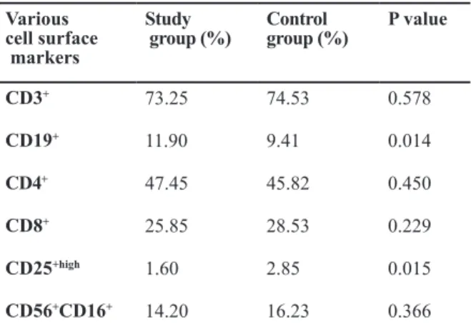
Lymphocyte subtypes
Cellular autoimmune reaction occurring due to a changed T cell function was analysed as a po
-tential cause of POI. Cell abnormalities were more frequent in women with POI than in healthy wom
-en (Table 4).
Peripheral T cell count is expressed as a percent
-age of various cell surface markers. In patients with POI, peripheral regulatory T lymphocytes (CD25+high, p=0.015) were low and peripheral
blood B cells (CD19+) were high (p=0.014); T lymphocyte parameters were normal in the control
group.
Table 4: Prevalence (%) of analysed peripheral blood lymphocyte samples for various cell surface markers in
patients and in controls
P value Control
group (%) Study
group (%) Various
cell surface markers
0.578 74.53
73.25
CD3+
0.014 9.41
11.90
CD19+
0.450 45.82
47.45
CD4+
0.229 28.53
25.85
CD8+
0.015 2.85
1.60
CD25+high
0.366 16.23
14.20
CD56+CD16+
Markers for peripheral blood lymphocytes: T lymphocytes (CD3+), helper T lymphocytes (CD4+), cytotoxic T lympho
-cytes, (CD8+), natural killer cells (CD56+CD16+), regulatory
Discussion
To present POI as a possible autoimmune ab
-normality, we focused on three potentially inter
-connected factors: personal and familial history of autoimmune disorders, peripheral blood T-lym
-phocytes levels and presence of AOA.
We performed a targeted history of personal and
familial autoimmune disorders and found
associat-ed autoimmune disorders in our patients. Thyroid
disorders were common in personal histories and
diabetes type 1 in familial histories, conirming the indings of previous studies (5, 6, 15). One of the reasons for suspecting an autoimmune etiology of POI is its frequent association with nearly all organ-speciic autoimmune diseases (1, 3). Auto
-immune diseases are signiicantly more frequent in young women than in men. This phenomenon may be explained by the effect of sex steroids on the components of the cellular immune system, which might contribute to the development and progres
-sion of autoimmune POI (23).
Hoek et al. (24) found that 60% of patients with secondary amenorrhea and Addison’s disease have a detectable SCA serum titre. These antibodies are shown by 60-80% of patients with APGS type I. In 25% of our patients, the Synacthen test revealed an abnormal cortisol response. We interpreted this
as the possibility that patients could be positive for
SCA, but had a normal cortisol response. Patients with this disorder have an insidious onset; to con
-irm subclinical autoimmune adrenal insuficien
-cy, measurement of adrenal antibodies might be a more effective screening method (25, 26).
Thyroid disorders and the presence of antithy-roid antibodies are often mentioned in association
with POI. Thyroid disorders are the most com -mon of the autoimmune diseases associated with
POI, found in 12-39% of women with POI (27-29). Thyroid disorders are often associated with endometriosis and polycystic ovary syndrome, two conditions often resulting in infertility (2). We found a greater involvement of aTG and aTPO in the study than in the control group, and the expla
-nation being that ovarian failure may have been present in the latent period of thyroid disease. We found a strong correlation between autoimmune thyroid disease and autoimmune POI; 50% of pa
-tients tested positive for aTG. We concluded that not all patients with positive aTG necessarily have
a clinically expressed thyroid disorder and the disease can have an insidious onset. Furthermore, greater involvement of other immune-mediated diseases, particularly auto immune disorders, such as allergy, psoriasis, atopic dermatitis and vitiligo in the study group suggests an autoimmune cause of POI, consistent also with the indings in the lit
-erature (9, 15, 30, 31).
Genetic predisposition is known to be one of the primary causes of autoimmunity and it is generally
observed that patients with autoimmune diseases
have several types of antibodies, as also conirmed in our study. We found AOA in 20% of patients; these results are consistent with other studies (9, 21, 32-35). The prevalence of AOA in women with POI, studied with IF on ovary tissue, varies great
-ly from 2 to 70%, which supports their role as a marker of an immune dysfunction process against ovaries. There could be several reasons for the dif
-ferences among study results, including the diverse origin of tissue sections, which include human and non-human primate ovaries, as well as different study inclusion criteria. Many studies have shown that the presence of serum AOA does not corre
-late with the clinical manifestation of POI. Despite these antibodies being present, their pathogenetic role is highly questionable (36, 37). AOA may oc -cur several years before the oc-currence of
clini-cal symptoms, as detected in 33-61% of women with unexplained infertility; a situation that may indicate early stages of autoimmune ovarian insuf
-iciency (38, 39). For most autoimmune diseases, screening for speciic antibodies is probably the best way of evaluating immunological involve
-ment. Many ovarian structures are potential tar
-gets for autoimmune events with POI. In addition
to AOA, SCA, aTG and aTPO, antibodies to sev
-eral potential ovarian antigens have been proposed as markers of ovarian autoimmunity, which could potentially mediate autoimmune damage in POI. Betterle et al. (40) found antibodies against steroid genetic enzymes in some cases with anti-adrenal autoimmunity. Antibodies against gonadotropin receptors and gonadotropins have also been found in patients with POI, but they are still the subject of research. The general opinion is that more re
AOA were determined semi-quantitatively by the IIF method. IIF is a basic method and widely used for determining auto antibodies, while more spe
-ciic tests enabling detection of AOA for spe-ciic antigen targets are not available in our laboratory.
Infertile women with AOA also have a decreased response to gonadotropin stimulation and reduced pregnancy rate after treatment (46, 47). In 2002, it was found that low-responders to gonadotropins with AOA are younger than low-responders with
-out AOA (48). Detection of autoimmune processes
that affect the ovarian response should thus be
in-cluded in the diagnostic workup before any infer
-tility treatment, particularly in women with low or no response to gonadotropins (34). Determination of AOA, as a speciic test, is important in the diag
-nosis of diseases with an autoimmune etiology, but it should not be the only reliable diagnostic tool for optimal selection of patients who may beneit from immune modulatory therapy that could, at least temporarily, re-establish their ovarian func
-tion and fertility.
Abnormalities of cellular immunity of T lym
-phocytes, macrophages and dendritic cells play an important role in autoimmune events. Some of
these abnormalities have been seen in women with
POI, conirming the potential existence of an auto
-immune mechanism of the disease. Mignot et al. (49) found that the absolute number and percent
-age of peripheral blood T lymphocytes, especially CD4+ T cells, are increased in patients with POI.
Moreover, Miyake et al. (50) found that patients with POI have low levels of CD8+/CD57+ Tcells
(cytotoxic T lymphocytes) and an increased ratio of CD4+ to CD8+ cells, which may relect cell cyto
-toxic cell migration from blood to inlamed tissue. Multiple animal studies have suggested that the ba
-sis of POI is a cell-mediated autoimmune reaction caused by an alteration in T cell regulation (18). CD4+ T cells with constantly expressed α chain
(CD25) receptor for IL-2 of were the irst detected
mediators of inhibition of autoimmune diseases in mice alteration of suppressor T cell subsets and T
cell abnormalities are likely to play an important role in the pathogenesis of autoimmune diseases (51). Regulatory CD4+25+ T cells show a potent immunosuppressive function in vitro and in vivo,
and contribute to immunologic self-tolerance by
suppressing potentially auto-reactive CD4+ T cells.
There is known to be a number of immunological
mechanisms associated with the failure of immune
tolerance and the development of autoimmunity. Although studying regulatory T cells in human autoimmune diseases is dificult, and at times, indings have been contradictory, the data sug
-gest that defects in CD4+ CD25+ regulatory T cells
mediated suppression (52) are a major subset of
immune cells responsible for peripheral immune
self-tolerance.
In agreement with studies in patients with sys
-temic lupus erythematosus, multiple sclerosis, rheumatoid arthritis and autoimmune vasculitis, we conirmed a reduced number of CD4+CD25+high
T cells in the peripheral blood of our patients. High expressions of CD25 and CD4 surface mark
-ers have classically been used for identiication of regulatory T cells. This may be problematic since CD25 is also expressed on antigen-responding activated non-regulatory T cells. The additional measurement of cellular expression of Foxp3 pro
-tein allowed a more speciic analysis of Treg cells (CD4+CD25+Foxp3+ cells). However, Foxp3 is also
transiently expressed in activated human effector T cells, thus complicating a correct Treg analysis. The large majority of Foxp3-expressing regulatory T cells express high levels of the interleukin-2 re
-ceptor alpha chain (CD25). Since there are no cell surface markers that are uniquely and speciically expressed on all Foxp3-expressing regulatory T cells, the measurement of CD4+CD25+high T cells is still in use in clinical studies of peripheral blood
lymphocytes.
We interpreted the reduced number of
CD4+CD25+high T cells as a possible mechanism
contributing to the formation of an autoimmune response in association with the presence of AOA and aTG. We also found an increased number of B cells in peripheral blood. A similar picture has
been observed with other autoimmune
endocri-nopathies; therefore, we interpreted the elevated
B cell count as activation of the humoral immune
system, crucial for autoantibody production. Some authors, though, have tried estrogen substitution in women with POI without any effect on peripheral B cell count (53, 54).
The hormones inhibin B, FSH and AMH have been proposed as potential markers for determin
level decreases signiicantly over time, whereas other markers associated with ovarian aging, such as FSH, inhibin B and antral follicle count (AFC), do not change during this time period. Since it de
-creases at a time when concentrations of FSH and inhibin B are still normal, AMH has been proposed
as the best indicator of ovarian reserve and as a
marker of ovarian aging (55-59). In contrast, AMH is almost undetectable in women with POI (60), which our study also conirmed. In our group of patients, there was a small AFC or these structures were no longer seen on ultrasound, which agrees with the data in the literature (61).
Conclusion
Clinical and biological characteristics of women without known causes of disease suggest a pos
-sibility of autoimmune pathogenesis. In some pa
-tients, a combination of various autoimmune pro
-cesses has been found. The presence of AOA and anti-thyroid antibodies, together with abnormali
-ties of cellular immunity, potentially represent an autoimmune mechanism of POI. There is thus an increasing need to ind a reliable and simple diag -nostic procedure to determine the true prevalence
of autoimmune ovarian disease. In women with POI, more attention should be paid to evaluation of associated autoimmune disorders.
Acknowledgements
We declare that we had no inancial support from any pharmaceutical or other commercial company. The study was inanced from research funds from the University Medical Centre Ljubljana, Slovenia, project number: 20100434. There is no conlict of interest in this study.
References
1. Conway GS. Premature ovarian failure. Br Med Bull.
2000; 56(3): 643-649.
2. Goswami D, Conway GS. Premature ovarian failure.
Horm Res. 2007; 68(4): 196-202.
3. Anasti JN. Premature ovarian failure: an update. Fertil
Steril. 1998; 70(1): 1-15.
4. Conway GS, Kaltsas G, Patel A, Davies MC, Jacobs
HS. Characterisation of idiopathic premature ovarian failure. Fertil Steril. 1996; 65(2): 337-341.
5. Husebye ES, Løvås K. Immunology of Addison’s
dis-ease and premature ovarian failure. Endocrinol Metab Clin North Am. 2009; 38(2): 389-405.
6. Poppe K, Velkeniers B. Female infertility and the
thy-roid. Best Pract Res Clin Endocrinol Metab. 2004; 18(2): 153-165.
7. Edassery SL, Shatavi SV, Kunkel JP, Hauer C,
Bruck-er C, Penumatsa K, et al. Autoantigens in ovarian au-toimmunity associated with unexplained infertility and premature ovarian failure. Fertil Steril. 2010; 94(7): 2636-2641.
8. Welt CK. Autoimmune oophoritis in the adolescent.
Ann NY Acad Sci. 2008; 1135: 118-122.
9. Ashrai M, Fallahian M, Eshrati B, Salman Yazdi R. The
presence of anti thyroid and anti ovarian auto-antibodies in familial POF. Int J Fertil Steril. 2008; 1(4): 171-174. 10. Irvine WJ, Barnes EW. Addison’s disease, ovarian
failure and hypoparathyroidism. Clin Endocrinol Me-tab.1975; 4(2): 379-434.
11. Turkington RW, Lebovitz HE. Extra-adrenal endocrine
deficiencies in Addison’s disease. Am J Med. 1967; 43(4): 499-507.
12. Muir A, Schatz DA, MacLaren NK. Autoimmune pol
-yglandular syndrome. In: de Groot LS, editor.
Endo-crinology. 3rd ed. Philadelphia: WB Saunders; 1995;
3013-3024.
13. Blizzard RM, Chee D, Davies W. The incidence of
adrenal and other antibodies in sera of patients with idiopathic adrenal insufficiency (Addison’s disease). Clin Exp Immunol. 1967; 2(1): 19-30.
14. Irvine WJ, Chan MM, Scarth L, Kolb FO, Hartog M, Bayliss RI, et al. Immunological aspects of premature ovarian failure associated with idiopathic Addison’s disease. Lancet. 1968; 2(7574): 883-887.
15. Forges T, Monnier-Barbarino P, Faure GC, Bene MC.
Autoimmunity and antigenic targets in ovarian pathol-ogy. Hum Reprod Update. 2004; 10(2): 163-175.
16. Morita Y, Tilly JL. Oocyte apoptosis: like sand through
an hourglass. Dev Biol. 1999; 213(1): 1-17.
17. Zuckerman S. The number of oocytes in the mature
ovary. Recent Prog Horm Res. 1951; 6(1): 63-108. 18. Byskof AG, Faddy MJ, Lemmen JG, Andersen CY.
Eggs forever?. Differentiation. 2005; 73(9-10): 438-446.
19. Liu Y, Wu C, Lyu Q, Yang D, Albertini DF, Keefe DL, et al. Germline stem cells and neo-oogenesis in the adult human ovary. Dev Biol. 2007; 306(1): 112-120. 20. Bukovsky A, Virant-Klun I, Svetlikova M, Willson I.
Ovarian germ cells. Methods Enyzmol. 2006; 419:
208-258.
21. Damewood MD, Zacur HA, Hoffman GJ, Rock JA. Cir-culating antiovarian antibodies in premature ovarian failure. Obstet Gynecol. 1986; 68(6): 850-854. 22. Groselj-Grenc M, Ihan A, Derganc M. Neutrophil and
monocyte CD64 and CD163 expression in critically ill neonates and children with sepsis: comparison of fluorescence intensities and calculated indexes. Me-diators Inflamm. 2008; 2008: 202646.
23. Lahita RG. Predisposing factors to autoimmune dis-ease. Int J Fertil Womens Med. 1997; 42(2): 115-119. 24. Hoek A, Schoemaker J, Drexhage HA. Premature
ovarian failure and ovarian autoimmunity. Endocr Rev. 1997; 18(1): 107-134.
25. Bakalov VK, Vanderhoof VH, Bondy CA, Nelson LM. Adrenal autoantibodies detected asymptomatic auto-immune adrenal insufficiency in young women with spontaneous premature ovarian failure. Hum Reprod. 2002; 17(8): 2096-2100.
26. Brozzetti A, Marzotti S, La Torre D, Bacosi ML, Morelli
autoim-mune ovarian insufficiency and in Addison’s disease are IgG1 dominated and suggest a predominant, but
not exclusive, Th1 type of response. Eur J Endocrinol.
2010; 163(2): 309-317.
27. Dragojevic-Dikic S, Marisavljevic D, Mitrovic A, Dikic
S, Jovanovic T, Jankovic-Raznatovic S. An immuno
-logical insight into premature ovarian failure (POF). Autoimmun Rev. 2010; 9(11): 771-774.
28. Betterle C, Rossi A, Dalla Pria S, Artifoni A, Pedini B, Gavasso S, et al. Premature ovarian failure: auto-immunity and natural history. Clin Endocrinol. 1993; 39(1): 35-43.
29. Goswami R, Marwaha RK, Goswami D, Gupta N, Ray
D, Tomar N, et al. Prevalence of thyroid autoimmunity
in sporadic idiopathic hypoparathyroidism in compari-son to type 1 diabetes and premature ovarian failure. J Clin Endocrinol Metab. 2006; 91(11): 4256-4259. 30. Irvine WJ. Premature menopause in autoimmune
dis-ease. Lancet. 1969; 1(7588): 264.
31. Vallotton MB, Forbes AP. Antibodies to cytoplasm of ova. Lancet. 1966; 2(7457): 264-265.
32. Gleicher N,Weghofer A, Oktay K, Barad D. Do etiolo-gies of premature ovarian aging (POA) mimic those of premature ovarian failure (POF)?. Hum Reprod. 2009; 24(10): 2395-2400.
33. Goswami D, Conway GS. Premature ovarian failure. Hum Reprod Update. 2005; 11(4): 391-410.
34. Luborsky J. Ovarian autoimmune disease and ovarian autoantibodies. J Womens Health Gend Based Med. 2002; 11(7): 585-599.
35. Yan G, Schoenfeld D, Penney C, Hurxthal K, Taylor A,
Faustman D. Identification of premature ovarian fail-ure patients with underlying autoimmunity. J Womens Health Gend Based Med. 2000; 9(3): 275-287.
36. Monnier-Barbarino P, Forges T, Faure GC, Béné MC.
Gonadal antibodies interfering with female reproduc-tion. Best Pract Res Clin Endocrinol Metab. 2005; 19(1): 135-148.
37. Coulam CB, Ryan RJ. Prevalence of circulating anti-bodies directed towards ovaries among women with premature ovarian failure. Am J Reprod Immunol Mi-crobiol. 1985; 9(1): 23-24.
38. Gleicher N, Weghofer A, Barad D. Female infertility due to abnormal autoimmunity: frequently overlooked and greatly underappreciated. Part II. Expert Rev Ob-stet Gynecol. 2007; 2(4): 465-475.
39. Gleicher N, Weghofer A,Barad D. Female infertility due to abnormal autoimmunity: frequently overlooked and greatly underappreciated. Part I. Expert Rev Ob-stet Gynecol. 2007; 2(4): 453-464.
40. Betterle C, Rossi A, Dalla Pria S, Artifoni A, Pedini B, Gavasso S, et al. Premature ovarian failure: auto-immunity and natural history. Clin Endocrinol. 1993; 39(1): 35-43.
41. Wheatcroft NJ, Toogood AA, Li TC, Cooke ID, Weet
-man AP. Detection of antibodies to ovarian antigens in women with premature ovarian failure. Clin Exp Im-munol. 1994; 96(1): 122-128.
42. Luborsky JL, Visintin I, Boyers S, Asari T, Caldwell
B, DeCherney A. Ovarian antibodies detected by
im-mobilized antigen immunoassay in patients with pre
-mature ovarian failure. J Clin Endocrinol Metab. 1990; 70(1): 69-75.
43. Moncayo H, Moncayo R, Benz R, Wolf A, Lauritzen
C. Ovarian failure and autoimmunity: detection of au-toantibodies directed against both the unoccupied
lu-teinizing hormone/human chorionic gonadotropin re
-ceptor and the hormone-re-ceptor complex of bovine corpus luteum. J Clin Invest. 1989; 84(6): 1857-1865.
44. Sotsion F, Bottazzo GF, Doniach D. Immunoofluo
-rescence studies on antibodies to steroid-producing cells, and to germline cells in endocrine disease and infertility. Clin Exp Immunol. 1980; 39(1): 97-111. 45. Horejsi J, Martinek J, Novakova D, Madar J,
Brande-jska M. Autoimmune antiovarian antibodies and their
impact on the success of an IVF/ET program. Ann N
Y Acad Sci. 2000; 900: 351-356.
46. Meyer WR, Lavy G, DeCherney AH, Visintin I, Econo-my K, Luborsky JL. Evidence of gonadal and gonado-tropin antibodies in woman with suboptimal ovarian response to exogenous gonadotropin. Obstet Gy-necol. 1990; 75(5): 795-799.
47. Luborsky J, Pong R. Pregnancy outcome and ovarian antibodies in infertility patients undergoing controlled ovarian hyperstimulation. Am J Reprod Immunol. 2000; 44(5): 261-265.
48. Luborsky JL, Thiruppathi P, Rivany B, Roussev R,
Coulam C, Radwanska E. Evidence for different eti-ologies of low estradiol response to FSH: age-related
accelerated luteinization of follicles of presence of
ovarian autoantibodies. Hum Reprod. 2002; 17(10): 2641-2649.
49. Mignot MH, Drexhage HA, Kleingeld M, Van de Plass-che-Boers EM, Rao BR, Schoemaker J. Premature ovarian failure. II: Considerations of cellular immunity defects. Eur J Obstet Gynecol Reprod Biol. 1989; 30(1): 67-72.
50. Miyake T, Sato Y, Takeuchi S. Implications of circulat
-ing autoantibodies and peripheral blood lymphocyte subset for the genesis of premature ovarian failure. J Reprod Immunol. 1987; 12(3): 163-171.
51. Sakaguchi S, Sakaguchi N, Asano M, Itoh M, Toda M.
Immunologic self-tolerance maintained by activated T
cells expressing IL-2 receptor alpha-chains (CD25). Breakdown of a single mechanism of self-tolerance causes various autoimmune diseases. J Immunol. 1995; 155(3): 1151-1164.
52. Long SA, Buckner JH. CD4+FOXP3+ T regulatory
cells in human autoimmunity: more than a numbers game. J Immunol. 2011; 187(5): 2061-2066.
53. Chernysmov VP, Radysh TV, Gura IV, Tatarchuk TP,
Khominskaya ZB. Immune disorders in women with premature ovarian failure in initial period. Am J Re-prod Immunol. 2001; 46(3): 220-225.
54. Ho PC, Tang GW, Lawton JW. Lymphocyte subsets
and serum immunoglobulins in patients with prema-ture ovarian failure before and after estrogen replace-ment. Hum Reprod. 1993; 8(5): 714-716.
55. Van Houten EL, Themmen AP, Visser JA. Anti-Müllerian
hormone (AMH): regulator and marker of ovarian func-tion. Ann Endocrinol (Paris). 2010; 71(3): 191-197.
56. Baird DD, Steiner AZ. Anti-Müllerian hormone: a po
-tential new tool in epidemiologic studies of female fe-cundability. Am J Epidemiol. 2012; 175(4): 245-249. 57. Van Rooij IA, Broekmans FJ, Scheffer GJ, Looman
CW, Habbema JD, de Jong FH, et al. Serum anti
müllerian hormone levels best reflect the reproduc
fertility: a longitudinal study. Fertil Steril. 2005; 83(4): 979-987.
58. Domingues TS, Rocha AM, Serafini PC. Tests for
ovarian reserve: reliability and utility. Curr Opin Ob-stet Gynecol. 2010; 22(4): 271-276.
59. Meden-Vrtovec H, Osredkar J. Anti-Müllerian hor
-mone-a predictor of ovarian reserve. Zdrav Vestn. 2010; 79(6): 507-511.
60. Nelson SM, Anderson RA, Broekmans FJ,
Raine-Fen-ning N, Fleming R, La Marca A. Anti-Müllerian hor
-mone: clairvoyance or crystal clear?. Hum Reprod. 2012; 27(3): 631-636.
61. Knauff EA, Eijkemans MJ, Lambalk CB, ten
Kate-Booij MJ, Hoek A, Beerendonk CC, et al. Anti-Mül