Addition of Arsenic Trioxide into Induction
Regimens Could Not Accelerate Recovery of
Abnormality of Coagulation and Fibrinolysis
in Patients with Acute Promyelocytic
Leukemia
Ye Zhang, SiJing Wu, Dan Luo, JianFeng Zhou, DengJu Li*
Department of Hematology, Tongji Hospital, Tongji Medical College, Huazhong University of Science and Technology, Wuhan, Hubei, China
*lidengju@163.com
Abstract
Aim
All-trans retinoic acid combined to anthracycline-based chemotherapy is the standard regi-men of acute promyelocytic leukemia. The advent of arsenic trioxide has contributed to improve the anti-leukemic efficacy in acute promyelocytic leukemia. The objectives of the current study were to evaluate if dual induction by all-trans retinoic acid and arsenic trioxide could accelerate the recovery of abnormality of coagulation and fibrinolysis in patients with acute promyelocytic leukemia.
Methods
Retrospective analysis was performed in 103 newly-diagnosed patients with acute promye-locytic leukemia. Hemostatic variables and the consumption of component blood were com-parably analyzed among patients treated by different induction regimen with or without arsenic trioxide.
Results
Compared to patients with other subtypes of de novo acute myeloid leukemia, patients with acute promyelocytic leukemia had lower platelet counts and fibrinogen levels, significantly prolonged prothrombin time and elevated D-dimers (P<0.001). Acute promyelocytic leuke-mia patients with high or intermediate risk prognostic stratification presented lower initial fibrinogen level than that of low-risk group (P<0.05). After induction treatment, abnormal coagulation and fibrinolysis of patients with acute promyelocytic leukemia was significantly improved before day 10. The recovery of abnormal hemostatic variables (platelet, prothrom-bin time, fibrinogen and D-dimer) was not significantly accelerated after adding arsenic tri-oxide in induction regimens; and the consumption of transfused component blood (platelet
OPEN ACCESS
Citation:Zhang Y, Wu S, Luo D, Zhou J, Li D (2016) Addition of Arsenic Trioxide into Induction Regimens Could Not Accelerate Recovery of Abnormality of Coagulation and Fibrinolysis in Patients with Acute Promyelocytic Leukemia. PLoS ONE 11(1): e0147545. doi:10.1371/journal.pone.0147545
Editor:Ken Mills, Queen's University Belfast, UNITED KINGDOM
Received:August 27, 2015
Accepted:January 4, 2016
Published:January 26, 2016
Copyright:© 2016 Zhang et al. This is an open access article distributed under the terms of the Creative Commons Attribution License, which permits unrestricted use, distribution, and reproduction in any medium, provided the original author and source are credited.
Data Availability Statement:All relevant data are within the paper.
Funding:This work was supported by the Natural Science Foundation of Hubei Province of China (No. 2013CFB075) (http://www.hbstd.gov.cn/) and National Natural Science Foundation of China (No.
81200353). DJL received the funding.
and plasma) did not dramatically change either. Acute promyelocytic leukemia patients with high or intermediate risk prognostic stratification had higher platelet transfusion demands than that of low-risk group (P<0.05).
Conclusions
Unexpectedly, adding arsenic trioxide could not accelerate the recovery of abnormality of coagulation and fibrinolysis in acute promyelocytic leukemia patients who received all-trans retinoic acid combining chemotherapy.
Introduction
Acute promyelocytic leukemia (APL) is a distinct subtype of acute myeloid leukemia (AML), and accounts for approximately 5–10% of cases of AML [1]. It is characterized by an excess of abnormal hypergranular promyelocytes in the bone marrow and other hematopoietic organs, and chromosomal translocation t(15;17)(q22;q21) leading to fusion of the genes encoding pro-myelocytic leukemia protein(PML) and retinoic acid receptor alpha (RARα) to generate the PML-RARαoncoprotein [2]. The abnormality of coagulation and fibrinolysis in APL is unique, and it account for early death in 10–30% of patients with APL [3].
The prompt combination of all-trans retinoic acid (ATRA) with chemotherapy has become a consensus regimen for treating newly diagnosed APL patients currently. The therapeutic effi-cacy of the regimen with ATRA and chemotherapy has been confirmed by a series prospective randomized clinical trials. The clinical complete remission rate was observed in 90%-95% of patients. 6-year disease-free survival rate is 68%, and 6-year overall survival rate is up to 83.9% [4–7]. Apparently, ATRA/chemotherapy combination regimen is superior to ATRA or chemo-therapy alone. However, the death rate in early 28 days from diagnosis is still high, with hemor-rhagic death at 5–11% being the major cause [8–10]. Since 1990s, the use of arsenic trioxide (ATO) has improved the clinical benefit of refractory or relapsed as well as newly diagnosed APL [11]. The regimens containing ATO was later approved by the US FDA for these refrac-tory or relapsed APL. A randomized European Phase III trial compared a synergistic targeted therapy of ATRA plus ATO with ATRA plus standard chemotherapy. The results showed that the non-chemotherapy dual-differentiation agents for induction and consolidation therapy were superior to chemotherapy regimen in both two-year event-free and overall survival rates in patients with low-to-intermediate-risk APL [12]. Recently, another study by the Austral-asian Leukemia and Lymphoma Group using ATO superimposed on ATRA plus chemother-apy standard regimen for induction, while ATRA and ATO without chemotherchemother-apy for 2 cycle consolidation, also reported improved outcome with increased freedom from relapse and fail-ure-free survival when compared to their previously reported ATRA/chemotherapy-based protocol. However, the rate of early death rate and overall survival were of no significant differ-ence between the two groups [13].
Methods
Patients
A total of 103 hospitalized patients with newly diagnosed de-novo APL were treated at Tongji hospital (Wuhan, China) during March 2008 to January 2015. These cases consisted of 60 males and 43 females, with age ranging from 14 to 74 years and a median of 37 years. 263 de novo AML (other than APL) were retrospectively analyzed at the same period as control group for comparisons of laboratory parameters at initial diagnosis. The diagnostic criteria of AML were based on the of World Health Organization Classification of Tumors- Pathology and Genetic of Tumors of Haematopoietic and Lymphoid Tissue (2008) and FAB (1976)[14]. Other inclusion criteria were: no serious liver disease or other hemorrhagic diseases, and no usage of anticoagulants during initial induction therapy. We collected the data from December 2014 to January 2015 and identify the information during and after data collection. This study has been approved by the ethics committee of Tongji Hospital Affiliated of Huazhong Univer-sity of Science and Technology. Written informed consent was obtained from all enrolled sub-jects, including the next of kin on behalf of the minors recruited in our study.
ATRA 20 mg/m2treatment was begun immediately at the time after APL was suspected. Chemotherapy and/or ATO (0.16 mg/kg/d, maximum10 mg/d) was prescribed according to prognostic risk stratification and individual physicians’decisions. Therapeutic platelet or fresh frozen plasma (FFP) or cryoprecipitate transfusions were done only when clinically relevant bleeding occurred. Prophylactic platelet transfusion strategy was done when the platelet count was 30×109/L or lower [15]. For platelet transfusions, patients only accepted random ABO-identical (non-HLA-typed) apheresis platelets when available. In China, one apheresis units are standardized to contain 2.5×10¹¹platelets or more with less than 5×10⁸leucocytes. Prophy-lactic transfusion of FFP or cryoprecipitate mainly based on the fibrinogen level1g/L. One unit cryoprecipitate was converted to 200ml plasma in favor of subsequent statistical analysis.
Laboratory studies and clinical outcomes
The obtained information included case mix (age, gender,), clinical (initial bleeding events, early hemorrhagic death events, ATRA differentiation syndrome and consumption of trans-fused component blood), and laboratory variables [white blood cell (WBC) counts, platelet (PLT) counts, prothrombin time (PT), activated partial thromboplastin time (APTT), fibrino-gen (Fbg), D-dimer, creatinine, uric acid, lactate dehydrofibrino-genase (LDH), bcr3 transcript type and blasts and promyelocytic percentage]. The rating criteria of bleeding were based on World Health Organization bleeding scale [16,17]. Routine blood tests were carried out using a Sys-mex XE-5000 Hematology Analyzer (SysSys-mex, Kobe, Japan) on EDTA-anticoagulated blood samples. The STA Compact Automated Hemostasis Analyzer (Diagnostica Stago, Gennevil-liers, France) was used for detecting coagulation and fibrinolysis parameters, such as APTT, PT, Fbg level (Clauss method), D-dimer(Immuno-turbidimetric method). Blood biochemical test were done on COBAS INTEGRA 800 biochemical analyzer (Roche, Switzerland) on hepa-rin-anticoagulated blood samples. Fusion gene transcript from chromosome aberrations was analyzed by reverse transcription polymerase chain reaction. Blasts and promyelocytic percent-age was determined by microscopic examination of the bone marrow by two experienced phy-sicians separately.
Statistical analysis
Hemostatic variables and the consumption of transfused component blood were comparably analyzed between two groups using Mann-Whitney test for two-sample analysis.
Comparisons also were conducted between different prognostic risk groups of APL patients using Kruskal-Wallis test for multi-sample analysis. The prognostic risk stratification of APL is based on widely recognized risk evaluation standard which originate from the Italian
GIMEMA and the Spanish PETHEMA trials: WBC10×109/L and PLT>40×109/L as low risk, WBC10×109/L and PLT40×109/L as intermediate-risk, and WBC10×109/L as high-risk groups [18].
Hemostatic variables and the consumption amount of transfused component blood were expressed in median (range) format. All P-values were two-sided and less than 0.05 were con-sidered as statistically significant. Statistical analysis was accomplished by SPSS software 20.0 (SPSS Inc., Chicago, IL, USA).
Results
Analysis of abnormality of coagulation and fibrinolysis
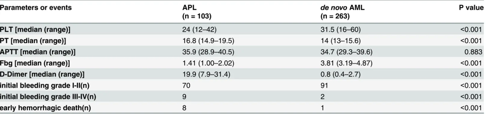
Compared to patients with other subtypes of de novo acute myeloid leukemia(AML), patients with APL had lower platelet counts and Fbg levels, significantly prolonged prothrombin time (PT) and elevated D-dimers(P<0.001). The APTT median values of both groups were all in normal range (P>0.05). Besides, APL patients had more bleeding events and early hemor-rhagic death than AML patients (P<0.001) (Table 1). Among the patients with APL, 48 patients bled at skin or soft tissue, 43 at oral or nasal, 11 at genitourinary system, 9 at central nervous system, 8 at pulmonary, 1 at gastrointestinal system and 1 at invasive sites.
Next, we analyzed the change in the trend of hemostatic variables in patients with APL. In order to minimize interference as much as possible, 14 cases were excluded due to early death (8 cases), pregnancy (2 cases), and withdrawing of treatment in 7 days after diagnosis (4 cases). The remaining cases (60 males and 43 females) were incorporated into the following research. The recording time points were respectively set at the first visit (day 0), and after treatment with ATRA/ATO/chemotherapy (day 1, day 4, day 7, day 10, day 13, day19 and day 25).
The median of PT returned to normal range after only 7 days of therapy. The median of Fbg kept rising step by step and had fallen in normal range since day 10. The elevated D-dimers showed a relatively slow downtrend and still maintained at a slightly high level in the fourth week of induction therapy (Table 2).
vs. 10/25/18, P = 1.000) and incidence of ATRA differentiation syndrome (4/36 vs. 5/53, P = 1.000).
The fibrinolytic or hemostatic variables of APL patients with different prognostic stratifica-tion were comparatively analyzed, too. The statistical differences existed in fibrinogen level at the time of initial diagnosis (day 0), PT at day 4, D-dimer at day 7. High risk group had a lower Fbg level at day 0 (P = 0.012) and longer PT at day 7 than low risk and intermediate risk groups. The value of PT, APTT and D-dimers at day 0 had no obvious difference among differ-ent risks groups (P>0.05). The whole change in the trend of fibrinolytic or hemostatic variables is approximately consistent during the induction treatment (Table 4). The result showed that the pace of coagulation recovery was not affected by prognostic stratification of APL.
Analysis of consumption of transfused component blood
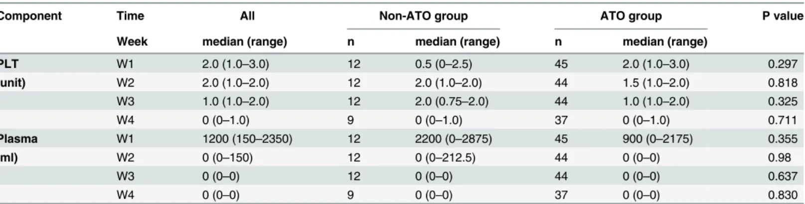
The median transfusion volume of plasma were 1200 ml/week (interquartile range 150–2350 ml/week) in the first week, but decreased sharply from the second week. In contrast, the median transfusion amount of platelet maintained at 1.5 unit/week during the first three weeks of induction therapy, and finally declined in the fourth week (Table 5).
There were slightly differences between non-ATO group and ATO group in the consump-tion of PLT and plasma transfusion. But no statistical significance was found in two groups (Table 5). The result indirectly shows that the most serious abnormality of coagulation appears Table 1. Evaluation of hemostatic parameters and Incidence of bleeding events in patients with APL or de novo AML (other than APL).
Parameters or events APL de novoAML P value
(n = 103) (n = 263)
PLT [median (range)] 24 (12–42) 31.5 (16–60) <0.001
PT [median (range)] 16.8 (14.9–19.5) 14 (13–15.6) <0.001
APTT [median (range)] 35.9 (28.9–40.5) 34.7 (29.3–39.6) 0.883 Fbg [median (range)] 1.41 (1.00–2.02) 3.81 (3.19–4.87) <0.001 D-Dimer [median (range)] 19.9 (7.9–31.4) 0.8 (0.4–2.7) <0.001
initial bleeding grade I-II(n) 70 91 <0.001
initial bleeding grade III-IV(n) 9 2 <0.001
early hemorrhagic death(n) 8 1 <0.001
APL, acute promyelocytic leukemia; AML, acute myeloid leukemia; PLT, platelet count, (40–100)*109/L; PT, prothrombin time, 11.5
–14.5 s; APTT, activated partial thromboplastin time, 28.5–41.5 s; Fbg,fibrinogen, 2.00–4.00 g/L; D-dimer,<0.5mg/ml; n, number of events.
doi:10.1371/journal.pone.0147545.t001
Table 2. Change in the trend of hemostatic parameters in patients with APL.
Time PLT PT Fbg APTT D-dimer
median (range) median (range) median (range) median (range) median (range)
d0 25 (13–40) 16.7 (14.8–19.8) 1.41 (1.01–2.06) 34.4 (28.7–40.2) 19.9 (8–33.1) d4 42 (29–51)* 14.7 (13–15.9)* 1.88 (1.55–2.59)* 32 (27.6–36.2)* 6.5 (2.9–13.7)*
d7 37 (26–52)* 14.4 (12.6–15.1)* 1.89 (1.55–2.96)* 31.8 (27.2–36.7) 3.4 (1.4–8.3)*
d10 31 (20–48) 13.9 (12.6–14.8)* 2.41 (1.76–3.11)* 33.3 (27.5–38.3) 3.3 (1.7–7.2)*
d13 26 (18–44) 14.1 (12.6–14.8)* 2.9 (2.13–4.06)* 33.4 (29.5–37.9) 2.3 (0.8–4.8)*
W3 29 (17–45) 13.5 (11.9–14.4)* 3.21 (2–4.32)* 33.6 (28.941.4) 1.9 (0.6–2.9)*
W4 45 (31–104)* 13.5 (12.4–14.6)* 2.83 (2.11–4.42)* 34.7 (27.7–44.1) 1 (0.6–1.9)*
*, compare to the initial level, Wilcoxon signed rank test’s P value<0.01.
in the early stages of the treatment. The pace of improvement was not related with the applica-tion of ATO in inducapplica-tion regiments.
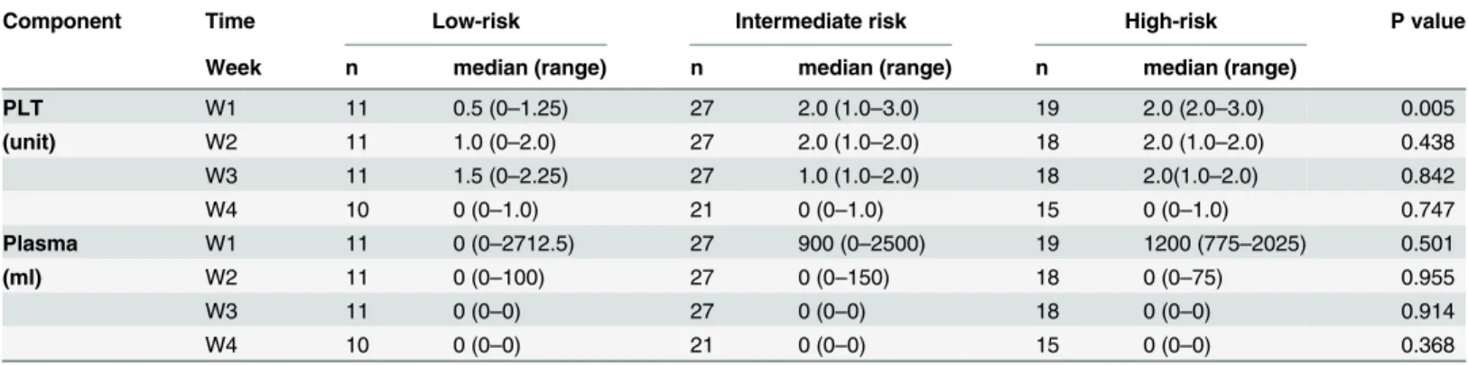
The consumption of PLT and plasma transfusion was higher in high-risk group and inter-mediate-risk group than low-risk group in the first week. But statistical differences only were found in the platelet transfusion (P<0.05) (Table 6).
Discussion
APL-associated coagulopathy is more complex than simple disseminated intravascular coagu-lation (DIC). Activation of the clotting system, increased fibrinolytic activity and non-specific Table 3. Compared analysis of hemostatic parameters between non-ATO group and ATO group.
Non-ATO group ATO group P value
n median (range) n median (range)
PLT d0 36 27 (15.3–43) 52 23 (10.7–34.5) 0.243
d4 36 37.5 (29–51.5) 52 44 (29.4–51) 0.662
d7 36 38 (21.3–52.5) 52 35.5 (29.5–53.3) 0.389
d10 29 29 (24.5–46.5) 44 31.5 (18.7–49.2) 0.857
d13 29 22 (15.5–38.5) 44 31.2 (19.5–51) 0.159
W3 20 26 (15.5–44) 43 31 (17–46) 0.701
W4 11 37 (28–52) 30 51.5 (32–126.3) 0.215
PT d0 36 16.8 (4.4–20.6) 52 16.6 (15–19.2) 0.769
d4 36 13.8 (11.7–14.9) 52 15.1 (14.1–16) 0.003
d7 36 12.9 (11.2–14.6) 52 14.7 (14–15.4) <0.001
d10 29 13.2 (12.1–14.6) 44 14.1 (12.8–14.9) 0.082
d13 29 14 (12.3–14.7) 44 14.3 (13.4–15) 0.144
W3 20 13.4 (11.4–14.3) 43 13.6 (12.4–14.4) 0.434
W4 11 13.5 (11.5–14.4) 30 13.6 (12.6–14.8) 0.717
Fbg d0 36 1.55 (1.06–2.23) 52 1.37 (0.79–1.94) 0.116
d4 36 1.88 (1.56–2.76) 52 1.88 (1.5–2.55) 0.799
d7 36 1.89 (1.43–3.02) 52 1.93 (1.63–2.95) 0.725
d10 29 2.56 (1.75–3.31) 44 2.39 (1.76–3.12) 0.774
d13 29 3.95 (2.76–4.81) 44 2.52 (2.03–3.38) 0.002
W3 20 3.99 (3.02–4.69) 43 2.88 (1.87–3.72) 0.009
W4 11 3.9 (2.81–5.32) 30 2.71 (1.88–3.4) 0.023
APTT d0 36 31.7 (27.7–39.9) 52 35.9 (29.7–40.2) 0.281
d4 36 29.1 (25.3–33.5) 52 34.1 (29.9–37.1) 0.001
d7 36 27.8 (25.7–33) 52 34.9 (29.3–41) <0.001
d10 29 31.5 (25.2–35.1) 44 34.5 (29.7–39.7) 0.010
d13 29 32.8 (26.2–36.2) 44 33.6 (30.5–39.1) 0.083
W3 20 31.3 (27.6–40.3) 43 34.6 (29–41.4) 0.447
W4 11 30.8 (24.5–41.9) 30 34.9 (28.1–46.4) 0.632
D-dimer d0 36 22.1 (7.2–39.6) 52 19.9 (8.4–30.7) 0.767
d4 21 6.4 (2.8–14.5) 22 8.2 (2.7–12.8) 0.950
d7 21 3.1 (2.3–7.7) 22 3.7 (1.1–9.1) 0.875
d10 16 3.5 (1.8–6.8.) 18 3.2 (1.6–8.4) 0.959
d13 16 2.4 (1.2–4.7) 18 1.9 (0.6–4.9) 0.567
W3 7 2.3 (0.6–2.9) 16 1.8 (0.6–3.5) 0.922
W4 4 0.8 (0.6–1.7)) 12 1.2 (0.6–2.7) 0.684
protease activity, with hyperfibrinolysis predominating are all included [19]. Recent studies have revealed that the unique abnormalities of coagulation and fibrinolytic function are associ-ated with increased amounts of tissue factor(TF), cancer procoagulant (CP) as well as elements of the fibrinolytic system, including tissue plasminogen activator, annexin A2, and plasmino-gen activator inhibitor type 1 expressed by leukemic promyelocytes in APL [20].
In our study, abnormalities of the 103 newly diagnosed patients with APL in routine hemo-static variables include low platelet counts, prolonged PT, low Fbg levels, elevated D-dimers, more bleeding events and higher early hemorrhagic death rate, which are consistent with previ-ous reports [19,21,22]. Low platelets are usually due to a result of impaired platelet production Table 4. Change in the trend of hemostatic parameters in APL patients with different prognostic stratification.
Low-risk Intermediate risk High-risk P value
Day n median (range) n median (range) n median (range)
PT d0 19 16 (13.3–19.9) 42 17.1 (14.4–19.7) 27 16.7 (14.9–20.8) 0.651 d4 19 13.6 (11.7–15) 42 14.7 (13–16) 27 15 (14.2–16) 0.042 d7 19 13.1 (11.2–15) 42 14.4 (12.9–15.2) 27 14.6 (13.2–15.1) 0.294 d10 16 13.1 (12.3–14.6) 33 14 (12.7–14.9) 24 14.1 (12.6–14.8) 0.500 d13 16 13.7 (12.4–14.6) 33 14.1 (13.1–14.9) 24 14.3 (13.4–14.9) 0.335 Fbg d0 19 2.10 (1.35–2.97) 42 1.38 (1.02–1.94) 27 1.13 (I0.75–1.62) 0.012 d4 19 2.06 (1.7–2.62) 42 1.82 (1.33–2.49) 27 2.01 (1.56–2.68) 0.426 d7 19 2.25 (1.75–3.04) 42 1.89 (1.41–2.85) 27 1.85 (1.59–3.03) 0.379 d10 16 3.19 (1.79–4.72) 33 2.31 (1.52–2.89) 24 2.42 (1.85–3.01) 0.144 d13 16 3.44 (2.24–5.56) 33 2.45 (1.68–3.88) 24 2.91 (2.36–4.32) 0.102 APTT d0 19 35.9 (29.7–40.7) 42 32.2 (27.5–40.7) 27 35.2 (30.1–39.4) 0.592 d4 19 30.7 (28.8–34.2) 42 33.2 (27.7–38.5) 27 31.1 (26.8–36.3) 0.449 d7 19 30.2 (26.9–35.8) 42 23.9 (27.5–38.8) 27 31.1 (26.6–36.6) 0.664 d10 16 32.2 (27.9–38) 33 33.8 (26.6–37.8) 24 31.9 (27.7–38.4) 0.890 d13 16 33.7 (28.4–38.1) 33 33.7 (30–38.6) 24 33.1 (29.5–37.1) 0.892 D-dimer d0 15 18.9 (9.7–39.6) 38 23 (7.8–35.6) 20 17.3 (7.9–28.1) 0.623 d4 7 1.6 (0.5–5.1) 21 9.2 (4.5–15.7)) 15 7 (2.9–13.9) 0.067 d7 7 2.2 (0.5–7.3) 21 6.5 (2.9–12.8) 15 2.5 (1–6.2) 0.047 d10 7 1.6 (1.2–3.2) 17 5.8 (2.1–16.7) 10 3.5 (2.4–4.7) 0.063 d13 7 1.5 (0.6–2.8) 17 2.4 (0.9–8.4) 10 2.4 (0.6–4.2) 0.454
doi:10.1371/journal.pone.0147545.t004
Table 5. Compared analysis of consumption of component blood between non-ATO group and ATO group.
Component Time All Non-ATO group ATO group P value
Week median (range) n median (range) n median (range)
PLT W1 2.0 (1.0–3.0) 12 0.5 (0–2.5) 45 2.0 (1.0–3.0) 0.297
(unit) W2 2.0 (1.0–2.0) 12 2.0 (1.0–2.0) 44 1.5 (1.0–2.0) 0.818
W3 1.0 (1.0–2.0) 12 2.0 (0.75–2.0) 44 1.0 (1.0–2.0) 0.325
W4 0 (0–1.0) 9 0 (0–1.0) 37 0 (0–1.0) 0.711
Plasma W1 1200 (150–2350) 12 2200 (0–2875) 45 900 (0–2175) 0.355
(ml) W2 0 (0–150) 12 0 (0–212.5) 44 0 (0–0) 0.98
W3 0 (0–0) 12 0 (0–0) 44 0 (0–0) 0.637
W4 0 (0–0) 9 0 (0–0) 37 0 (0–0) 0.830
and consumption. Increase in D-dimer and decrease in the fibrinogen level are the evidence of hyperfibrinolysis [20,23]. Clearly, bleeding events and high early hemorrhagic death rate are resulted from the coagulopathy described above.
Several studies have already confirmed the change in the trend of hemostatic variables dur-ing the first or second weeks of treatment [24,25]. Our study provides other convincing evi-dences, such as significant recovery of abnormal hemostatic markers and phasedown of consumption of transfused component blood, which supported the restoration of coagulation and fibryolysis by induction therapy.
Recent studies revealed that ATRA and ATO could specially bind to RARαand PML moie-ties of PML-RARαoncoprotein respectively and leading to their degradation [26,27]. Further-more, double induction of ATRA and ATO could cause APL cells differentiation, apoptosis [28–30]. Several studies have confirmed benefits of ATRA-ATO combination for newly diag-nosed APL in long-term follow-up [12,13,29,31]. Compared to those treated with either sin-gle agent, more encouraging outcomes were achieved in pilot studies with patients receiving dual induction of ATRA and ATO, including shorter time needed to achieve CR and higher rate of CR, enhanced 5-year disease-free survival, event-free survival rates and overall survival rate, less hematologic toxicity and fewer infections but more hepatic toxicity [12,32–34]. Another study also showed improved clearance of PML-RARA transcripts in patients receiving the combination therapy of ATRA and ATO [29]. Most studies focused on the therapeutic effect comparison of patients with APL by different induction regiments including ATRA and/ or ATO. Our study paid more attention to the change in the trend of coagulation and fibrinoly-sis during the initial treatment. We found that adding ATO into induction regiments neither accelerate the recovery of abnormality of coagulation and fibrinolysis nor decrease the con-sumption of transfused component blood in patients with APL.
Previous reports suggested high WBC count (>10×109/L) as an adverse prognostic factors for bleeding complications in APL [35,36]. However, our results did not find that the pace of coagulation recovery was affected by prognostic stratification of APL. But we found that the demand amounts of PLT and plasma transfusion increased in high-risk group and intermedi-ate risk group than low-risk group in the first week.
The limitations of this study were the relatively small sample size, missing data on some patients, and biases of judgment on bleeding diathesis which will directly affect the transfu-sion demand of PLT and plasma. More APL specific and sensitive laboratory tests such as, lev-els of thrombin antithrombin complex, prothrombin fragment, amount of tissue factor and cancer procoagulant and plasminogen activator and annexin A2 levels were not included this Table 6. Change in the trend of consumption of component blood in patients with APL with different prognostic stratification.
Component Time Low-risk Intermediate risk High-risk P value
Week n median (range) n median (range) n median (range)
PLT W1 11 0.5 (0–1.25) 27 2.0 (1.0–3.0) 19 2.0 (2.0–3.0) 0.005
(unit) W2 11 1.0 (0–2.0) 27 2.0 (1.0–2.0) 18 2.0 (1.0–2.0) 0.438
W3 11 1.5 (0–2.25) 27 1.0 (1.0–2.0) 18 2.0(1.0–2.0) 0.842
W4 10 0 (0–1.0) 21 0 (0–1.0) 15 0 (0–1.0) 0.747
Plasma W1 11 0 (0–2712.5) 27 900 (0–2500) 19 1200 (775–2025) 0.501
(ml) W2 11 0 (0–100) 27 0 (0–150) 18 0 (0–75) 0.955
W3 11 0 (0–0) 27 0 (0–0) 18 0 (0–0) 0.914
W4 10 0 (0–0) 21 0 (0–0) 15 0 (0–0) 0.368
time due to the limitation of retrospective study, and we will make up in the follow up future studies.
In conclusion, ATO/ATRA plus chemotherapy regimen relieves the coagulopathy burden in the induction period. Our study found unexpectedly that adding ATO could not accelerate the recovery of abnormality of coagulation and fibrinolysis in APL patients. Moreover, it was necessary to pay more attention to satisfy the high demand of component blood transfusion in initial treatment, which will substantially decrease the serious bleeding episodes related to abnormality of coagulation and fibrinolysis.
Acknowledgments
We are grateful to Liang Huang for his advice on the sample collecting and analysis.
Author Contributions
Conceived and designed the experiments: YZ DJL. Performed the experiments: YZ DL SJW JFZ. Analyzed the data: YZ. Contributed reagents/materials/analysis tools: YZ JFZ DJL. Wrote the paper: YZ DJL.
References
1. Lengfelder E, Hofmann WK and Nowak D. Impact of arsenic trioxide in the treatment of acute promyelo-cytic leukemia. Leukemia. 2012; 26(3):433–42. doi:10.1038/leu.2011.245PMID:21904379
2. Breen KA, Grimwade D and Hunt BJ. The pathogenesis and management of the coagulopathy of acute promyelocytic leukaemia. Br J Haematol. 2012; 156(1):24–36. doi:10.1111/j.1365-2141.2011.08922.x
PMID:22050876
3. Biondi A, Luciano A, Bassan R, Mininni D, Specchia G, Lanzi E, et al. CD2 expression in acute promye-locytic leukemia is associated with microgranular morphology (FAB M3v) but not with any PML gene breakpoint. Leukemia. 1995; 9(9):1461–6. PMID:7658712
4. Wang ZY and Chen Z. Acute promyelocytic leukemia: from highly fatal to highly curable. Blood. 2008; 111(5):2505–15. doi:10.1182/blood-2007-07-102798PMID:18299451
5. Tallman MS, Andersen JW, Schiffer CA, Appelbaum FR, Feusner JH, Woods WG, et al. All-trans reti-noic acid in acute promyelocytic leukemia: long-term outcome and prognostic factor analysis from the North American Intergroup protocol. Blood. 2002; 100(13):4298–302. PMID:12393590
6. Sanz MA, Martin G, Gonzalez M, Leon A, Rayon C, Rivas C, et al. Risk-adapted treatment of acute pro-myelocytic leukemia with all-trans-retinoic acid and anthracycline monochemotherapy: a multicenter study by the PETHEMA group. Blood. 2004; 103(4):1237–43. PMID:14576047
7. Asou N, Kishimoto Y, Kiyoi H, Okada M, Kawai Y, Tsuzuki M, et al. A randomized study with or without intensified maintenance chemotherapy in patients with acute promyelocytic leukemia who have become negative for PML-RARalpha transcript after consolidation therapy: the Japan Adult Leukemia Study Group (JALSG) APL97 study. Blood. 2007; 110(1):59–66. PMID:17374742
8. Sanz MA, Iacoboni G and Montesinos P. Conventional induction and post-remission therapy in APL: have we arrived? Best Pract Res Clin Haematol. 2014; 27(1):33–8. doi:10.1016/j.beha.2014.04.004
PMID:24907015
9. Lehmann S, Ravn A, Carlsson L, Antunovic P, Deneberg S, Mollgard L, et al. Continuing high early death rate in acute promyelocytic leukemia: a population-based report from the Swedish Adult Acute Leukemia Registry. Leukemia. 2011; 25(7):1128–34. doi:10.1038/leu.2011.78PMID:21502956
10. de la Serna J, Montesinos P, Vellenga E, Rayon C, Parody R, Leon A, et al. Causes and prognostic fac-tors of remission induction failure in patients with acute promyelocytic leukemia treated with all-trans retinoic acid and idarubicin. Blood. 2008; 111(7):3395–402. doi:10.1182/blood-2007-07-100669PMID:
18195095
11. Niu C, Yan H, Yu T, Sun HP, Liu JX, Li XS, et al. Studies on treatment of acute promyelocytic leukemia with arsenic trioxide: remission induction, follow-up, and molecular monitoring in 11 newly diagnosed and 47 relapsed acute promyelocytic leukemia patients. Blood. 1999; 94(10):3315–24. PMID:
10552940
12. Lo-Coco F, Avvisati G, Vignetti M, Thiede C, Orlando SM, Iacobelli S, et al. Retinoic acid and arsenic tri-oxide for acute promyelocytic leukemia. N Engl J Med. 2013; 369(2):111–21. doi:10.1056/
13. Iland HJ, Bradstock K, Supple SG, Catalano A, Collins M, Hertzberg M, et al. All-trans-retinoic acid, idarubicin, and IV arsenic trioxide as initial therapy in acute promyelocytic leukemia (APML4). Blood. 2012; 120(8):1570–80; quiz 1752. PMID:22715121
14. Vardiman JW, Thiele J, Arber DA, Brunning RD, Borowitz MJ, Porwit A, et al. The 2008 revision of the World Health Organization (WHO) classification of myeloid neoplasms and acute leukemia: rationale and important changes. Blood. 2009; 114(5):937–51. doi:10.1182/blood-2009-03-209262PMID:
19357394
15. Wandt H, Schaefer-Eckart K, Wendelin K, Pilz B, Wilhelm M, Thalheimer M, et al. Therapeutic platelet transfusion versus routine prophylactic transfusion in patients with haematological malignancies: an open-label, multicentre, randomised study. The Lancet. 2012; 380(9850):1309–1316.
16. Miller AB, Hoogstraten B, Staquet M and Winkler A. Reporting results of cancer treatment. Cancer. 1981; 47(1):207–14. PMID:7459811
17. Bercovitz RS and O'Brien SH. Measuring bleeding as an outcome in clinical trials of prophylactic plate-let transfusions. Hematology Am Soc Hematol Educ Program. 2012; 2012(157–60. PMID:23233575
18. Candoni A, Damiani D, Michelutti A, Masolini P, Michieli M, Michelutti T, et al. Clinical characteristics, prognostic factors and multidrug-resistance related protein expression in 36 adult patients with acute promyelocytic leukemia. Eur J Haematol. 2003; 71(1):1–8. PMID:12801292
19. Falanga A and Rickles FR. Pathogenesis and management of the bleeding diathesis in acute promye-locytic leukaemia. Best Pract Res Clin Haematol. 2003; 16(3):463–82. PMID:12935963
20. Kwaan HC. The unique hemostatic dysfunction in acute promyelocytic leukemia. Semin Thromb Hemost. 2014; 40(3):332–6. doi:10.1055/s-0034-1370792PMID:24590422
21. Rovelli A, Biondi A, Cantu Rajnoldi A, Conter V, Giudici G, Jankovic M, et al. Microgranular variant of acute promyelocytic leukemia in children. J Clin Oncol. 1992; 10(9):1413–8. PMID:1517784
22. Tallman MS and Kwaan HC. Reassessing the hemostatic disorder associated with acute promyelocytic leukemia. Blood. 1992; 79(3):543–53. PMID:1732003
23. Stein E, McMahon B, Kwaan H, Altman JK, Frankfurt O and Tallman MS. The coagulopathy of acute promyelocytic leukaemia revisited. Best Pract Res Clin Haematol. 2009; 22(1):153–63. doi:10.1016/j. beha.2008.12.007PMID:19285282
24. Dombret H, Scrobohaci ML, Ghorra P, Zini JM, Daniel MT, Castaigne S, et al. Coagulation disorders associated with acute promyelocytic leukemia: corrective effect of all-trans retinoic acid treatment. Leu-kemia. 1993; 7(1):2–9. PMID:8418375
25. Federici AB, Falanga A, Lattuada A, Di Rocco N, Barbui T and Mannucci PM. Proteolysis of von Willeb-rand factor is decreased in acute promyelocytic leukaemia by treatment with all-trans-retinoic acid. Br J Haematol. 1996; 92(3):733–9. PMID:8616045
26. Raelson JV, Nervi C, Rosenauer A, Benedetti L, Monczak Y, Pearson M, et al. The PML/RAR alpha oncoprotein is a direct molecular target of retinoic acid in acute promyelocytic leukemia cells. Blood. 1996; 88(8):2826–32. PMID:8874178
27. Lallemand-Breitenbach V, Jeanne M, Benhenda S, Nasr R, Lei M, Peres L, et al. Arsenic degrades PML or PML-RARalpha through a SUMO-triggered RNF4/ubiquitin-mediated pathway. Nat Cell Biol. 2008; 10(5):547–55. doi:10.1038/ncb1717PMID:18408733
28. Jing Y, Wang L, Xia L, Chen GQ, Chen Z, Miller WH, et al. Combined effect of all-trans retinoic acid and arsenic trioxide in acute promyelocytic leukemia cells in vitro and in vivo. Blood. 2001; 97(1):264–9. PMID:11133770
29. Hu J, Liu YF, Wu CF, Xu F, Shen ZX, Zhu YM, et al. Long-term efficacy and safety of all-trans retinoic acid/arsenic trioxide-based therapy in newly diagnosed acute promyelocytic leukemia. Proc Natl Acad Sci U S A. 2009; 106(9):3342–7. doi:10.1073/pnas.0813280106PMID:19225113
30. Zheng PZ, Wang KK, Zhang QY, Huang QH, Du YZ, Zhang QH, et al. Systems analysis of transcrip-tome and proteome in retinoic acid/arsenic trioxide-induced cell differentiation/apoptosis of promyelo-cytic leukemia. Proc Natl Acad Sci U S A. 2005; 102(21):7653–8. PMID:15894607
31. Powell BL, Moser B, Stock W, Gallagher RE, Willman CL, Stone RM, et al. Arsenic trioxide improves event-free and overall survival for adults with acute promyelocytic leukemia: North American Leukemia Intergroup Study C9710. Blood. 2010; 116(19):3751–7. doi:10.1182/blood-2010-02-269621PMID:
20705755
32. Aribi A, Kantarjian HM, Estey EH, Koller CA, Thomas DA, Kornblau SM, et al. Combination therapy with arsenic trioxide, all-trans retinoic acid, and gemtuzumab ozogamicin in recurrent acute promyelocytic leukemia. Cancer. 2007; 109(7):1355–9. PMID:17326049
34. Estey E, Garcia-Manero G, Ferrajoli A, Faderl S, Verstovsek S, Jones D, et al. Use of all-trans retinoic acid plus arsenic trioxide as an alternative to chemotherapy in untreated acute promyelocytic leukemia. Blood. 2006; 107(9):3469–73. PMID:16373661
35. Dally N, Hoffman R, Haddad N, Sarig G, Rowe JM and Brenner B. Predictive factors of bleeding and thrombosis during induction therapy in acute promyelocytic leukemia-a single center experience in 34 patients. Thromb Res. 2005; 116(2):109–14. PMID:15907524