897
IN VITRO ANTIFUNGAL AND DEMELANIZING ACTIVITY OF NEPETA RTANJENSIS
ESSENTIAL OIL AGAINST THE HUMAN PATHOGEN BIPOLARIS SPICIFERA MILICA LJALJEVIĆ-GRBIĆ*, M. STUPAR, JELENA VUKOJEVIĆ and D. GRUBIŠIĆ
University of Belgrade, Faculty of Biology, Institute of Botany and Botanical Garden “Jevremovac”, 11000 Belgrade, Serbia
Abstract – he antifungal activity of Nepeta rtanjensis Diklić & Milojevićessential oil was tested against the human patho-genic fungus Bipolaris spicifera (Bainier) Subramanianvia mycelial growth assay and conidia germination assay. he mini-mally inhibitory concentration (MIC) of the oil was determined at 1.0 μg ml-1, while the MIC for the antifungal drug Bifonazole in a positive control was determined at 10.0 μg ml-1. he maximum of conidia germination inhibition was ac-complished at 0.6 μg ml-1. In addition, at 0.6 μg ml-1 and 0.8 μg ml-1 the oil was able to cause morphophysiological changes in B. spicifera. he most signiicant result is the bleaching efect of the melanized conidial apparatus of the test fungi, since the melanin is the virulence factor in human pathogenic fungi. hese results showed the strong antifungal properties of N. rtanjensis essential oil, supporting its possible rational use as an alternative source of new antifungal compounds.
Key words:Bipolaris spicifera (Bainier) Subram., essential oil, antifungal activity, melanin, human pathogen, depigmenta-tion
UDC 615.282:58
INTRODUCTION
Bipolaris spicifera (Bainier) Subram. (telemorph:
Cochliobolus spicifer R.R. Nelson) is dematiaceous hyphomycete very oten isolated from plant mate-rial, which in regions with a hot and dry climate are among the most frequent air-borne fungi encoun-tered (Alcorn, 1988). B. spicifera appears frequently in medical literature as a cause of human and ani-mal diseases: cutaneous and subcutaneous phaeohy-phomycosis (Mc Ginnis et al., 1992), fungal sinusitis (Buzina et al., 2003), cutaneous disease (Straka et al., 1989), mycotic keratitis (Hemashettar et al., 1992; Saha and Das 2005), allergic fungal sinusitis (Taguchi et al., 2004), systemic dual mycosis with Torulopsis glabrata in a dog (Waurzyniak et al., 1992), fatal en-darteritis ater aortic valve replacement (Ogden et al., 1992), fungal peritonitis (Bava et al., 2003), men-ingitis (Latham, 2000) and disseminated disease in a neonate (Moore et al., 2001). Dematiaceous fungi,
fungi contain melanin within their cell wall struc-ture (e.g. Aspergillus fumigatus, A. nidulans, A. niger,
Alternaria alternata, Cladosporium carionii, Crypto-coccus neoformans, Exophiala jeanselmei, Fonsecaea compacta, Fonsecaea pedrosoi, Hendersonula toruloi-dii, Histoplasma capsulatum, Paracoccidioides bra-siliensis, Penicillium marnefei, Phaeoannellomyces wernickii, Phialophora richardsiae, P. verrucosu, Spo-rothrix schenckii, Wangiella dermatitidis). For several of these fungi, melanin has been described as a viru-lence factor due to its ability to reduce a pathogen’s susceptibility for killing by host antimicrobial mech-anisms and by inluencing the host immune response (Youngchimet al., 2004; Nosanchuk and Casadevall, 2006). Due to protective role of fungal melanin, de-maticeous fungi are extremely diicult to treat with antifungal drugs (Nosanchuk and Casadevall, 2006). Plant secondary metabolites could be a good alterna-tive for the treatment of fungalinfections in light of increasing fungal resistance to commercial antifun-gal agents (Vivek et al., 2009). It has been shown that the essential oil isolated from Nepeta rtanjensis Diklić & Milojević (Lamiaceae), an endemic and critically endangered aromatic plant from south-east Serbia, has a strong antifungal activity and can inhibit the mycelial growth of some fungi in vitro (Stojanović et al., 2005; Ljaljević Grbić et al., 2008). he main components of the N. rtanjensis essential oil include
α-pinene (3.3%), β-pinene (0.37%), 2-metoxy-p-cresol (1.14%), 4aβ,7α,7aβ nepetalactone (6.30%),
α-copaene (1.33%), 4aα,7α,7aβ nepetalactone (79.89%), germacrene D (1.80), δ-cadinene (2.12%) (Ljaljević Grbić et al., 2008).
he present research emphasizes the antifungal activity of N. rtanjensis essential oil against B.s pic-ifera and its potential to cause the demelanizing (bleaching) of B. spicifera reproductive structures.
MATERIAL AND METHODS
Essential oil
he essential oil was isolated from air-dried aeri-al parts of Nepeta rtanjensis, collected during the pre-lowering stage, by hydrodistilation for 2 h in a
Clavenger-type apparatus. he extracted essential oil was kept in sealed glass vials at + 4˚C until further analysis.
Fungal strain used
Bipolaris spicifera (Bainier) Subram. was originally isolated from the wall of a storage room of the Ser-bian National Museum. Due to the high concentra-tion of indoor air fungal spores, the room sufered from “sick building syndrome”. he fungus was de-posited to the Mycotheca of the Department of Algo-logy, Mycology and LychenoAlgo-logy, Faculty of BioAlgo-logy, University of Belgrade. he fungus was maintained on a malt extract agar (MEA), and potato dextrose agar (PDA), stored at + 4˚C and subcultured once in a month.
Test for antifungal activity
Mycelial growth assay
Diferent concentrations of essential oil (0.2 – 1.4 μg ml-1) were diluted in Petri dishes with 10 ml of
MEA. For each treatment and each dose tested, three replicate Petri dishes were used. he culture medium was inoculated with 5 mm agar discs from an ac-tively growing culture of B. spicifera. Ater 21 days of incubation in the dark at + 25˚C, the diameter of the colonies was recorded. Antifungal activity was expressed in terms of percentage of mycelia growth inhibition and calculated using the formula of Pan-dey et al., 1982):
growth inhibition %= 100 (dc – dt)/dc
dc = average diameter of fungal colony in control
dt = average diameter of fungal colony in treatment.
Conidia germination assay
Conidia germination assays were carried out on Petri dishes containing MEA amended with diferent N.
rtanjensis essential oil concentrations (0.2 – 1.4 μg ml-1). A MEA without essential oil was used as a
neg-ative control. For each treatment and each dose test-ed, three replicate Petri dishes were used. Petri dishes were inoculated by covering the entire surface with a suspension of 200 μl of B. spicifera conidia (104 ml−1),
obtained from the sporulated mycelia of 10-day-old cultures, and incubated in the dark at +25˚ C. Ater 24 h, germinated and non-germinated conidia were counted under a microscope (Zeiss Axio Imager M.1, with AxioVision Release 4.6 sotware). At least 200 conidia were counted for each observation and scored by hemocytometer. Conidia were considered germinated when the germ tube length was at least half the length of the diameter of the conidia or long-er. he experiments were repeated twice.
Screening of morphophysiological changes
Light microscopy
A sample of mycelium was taken from the periphery of a colony grown on MEA enriched with diferent concentrations of N. rtanjensis essential oil. he sam-ples were dyed and ixed with lactophenol – cotton blue and observed under a light microscope (Zeiss Axio Imager M.1, with AxioVision Release 4.6 sot-ware) to examine structural abnormalities. Samples
from the control plate without oil were also stained and observed.
Scanning electron microscopy (SEM)
Treated and control B. spicifera colonies were used for SEM observations. 5 x 10 mm segments were cut from the culture growing on the MEA and placed in vials containing 3% glutaraldehyde in 0.05 M phos-phate bufer (pH 6.8) at 4˚C. Samples were kept in this solution for 48 h and then washed with distilled water and dehydrated in an ethanol. hen the sam-ples were dried in liquid carbon dioxide and placed in desiccators until further use. he fungal materials were deposited on adhesive tape ixed to specimen tabs and then ion sputter coated with gold. Micro-structure characterization of the samples was carried out with a JEOL JSM 6460 LV instrument equipped with an OXFORD INSTRUMENTS EDS analyzer.
Statistical analysis
One way ANOVA was performed for mycelial growth assay and conidia germination assay. A P value less than 0.05 was considered statistically signiicant.
RESULTS
Nepeta rtanjensis essential oil showed a strong anti-fungal and bleaching activity against mycelial growth and conidia germination of Bipolaris spicifera (Table 1). he radial growth of the fungal colony was
sig-Table 1. Inhibition of mycelial growth and conidia germination of Bipolaris spicifera in MA amended with diferent Nepeta rtanjensis
essential oil doses:
Concentration of oil (μg ml-1)
Radial growth inhibition (%)
Conidia germination inhibition (%)
Depigmentation
0.2 1.93 ± 0.05 69.78 ± 0.37
-0.4 7.07 ± 0.45 91.40 ± 1.92
-0.6 38.98 ± 0.57 100 ±
0.8 59.99 ±1.30 100 +
1.0 100 100 /
1.2 100 100 /
1.4 100 100 /
niicantly reduced in response to diferent concen-trations of essential oil. At 1.0 μg ml-1 the inhibition
of fungal development reached its maximum (100% of radial growth inhibition) and this concentration was regarded as the MIC for the test fungus (P<0.05). he MIC for the commercial fungicide Bifonazole was determined at 10 μg ml-1. In the conidia
germi-nation assay the maximum of conidia germigermi-nation inhibition (100%) was accomplished at 0.6.μg ml-1
(P<0.05). In addition to inhibited mycelial growth and inhibition of conidia germination, the presence of diferent concentrations of essential oil seemed to exhibit distinct morphophysiological changes. hese
variations included lack of sporulation, visible loss of pigmentation and aberrant development of co-nidiophores. Colonies formed on the MEA enriched with 0.8 μg ml-1 essential oil were hyaline (Fig 1Ad).
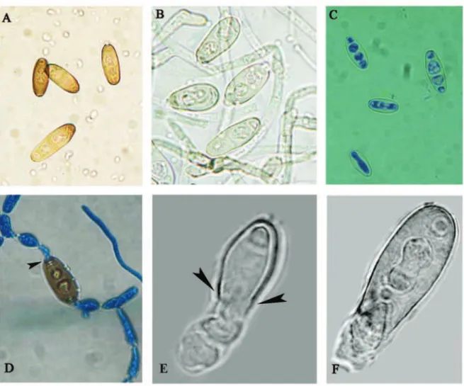
he intensity of sporulation was slightly reduced in the positive control (Fig 1D) and very poor on the MEA with essential oil added (Fig 1E). Light micro-photographs showed changes in conidiogenous ap-paratus of B. spicifera. Typical micromorphology of
B. spicifera, dark and pheoid septate hyphae, zig-zag conidiophores with thick walled cylindrical poro-conidia appeared in the negative control (Fig. 2A). Low aberrant conidiophore, with only terminal
zig-Fig. 1. he inluence of Nepeta rtanjensis essential oil on Bipolaris spicifera mycelia growth. A. a) Negative control; b) Positive control; c) 0.6 μg ml-1; d) 0.8 μg ml-1; B. the detail of Ad; C. Normal conidia production in control culture; D. he lowest conidia production in
zagging and slightly melanized, occurred on 0.6 μg ml-1 essential oil added (Fig. 2B). Aberrant
conidi-ophores, non-zig-zagging, hyaline, non-melanized and hyaline poroconidia were recorded on 0.8 μg ml-1 essential oil added (Fig. 2C, D). he essential
oil concentration at 0.8 μg ml-1 induced bleaching of
the conidia (Fig.3B, C) and cell wall rupture (Fig.3D) with cell content lowing outside (Fig.3E). SEM mi-crophotographs showed typical, roughed, zig-zag co-nidiophore with several poroconidia in the control (Fig.4D), in comparison to smooth sterile conidio-phores or conidioconidio-phores with only terminal
poroco-nidia (Fig. 4A, B). Cell wall rupture was also visible on the SEM microphotographs (Fig. 4C).
DISCUSSION
Nepeta rtanjensis essential oil showed the ability to interfere with all stages in the reproduction cycle of the human pathogenic fungus Bipolaris spicifera: conidia germination, mycelial growth and intensity of sporulation which is demonstrated with radial mycelial growth inhibition, inhibition of conidia germination and low conidia production in the
Fig. 3. Morphological changes of Bipolaris spicifera conidia inluenced by Nepeta rtanjensis essential oil: A. Negative control-typical
melanized poroconidia; B,C. Hyaline, bleached conidia-0.8 μg ml-1 essential oil added; D. Negative control-typical bipolar germination
of poroconidia (arrow pointed terminal pore with germinating hyphae). E,F. Ungerminated, lysed conidia with cell wall rupture (arrow)
treated samples. he high fungicidal activity of the tested oil was proven with a MIC value (1.0 μg ml-1)
and the MIC value for the referent antifungal agent Bifonazole was much higher (10.0 μg ml-1).
Espinel-Ingrof et al. (2002) tested the optimal conditions for determining the MICs for uncommon molds, including B. spicifera, and reported that the MICs for amphotericin B ranged from 0.06 to 8 μg ml-1,
for itraconazole from 0.03 to 8 μg ml-1, for
posa-conazole from 0.007 to 8 μg ml-1, for ravuconazole
from 0.2 to 8 μg ml-1 and for voriconazole from 0.12
to 8 μg ml-1. Essential oils are known to cause
mor-phophysiological changes in fungi through a lack of sporulation, depigmentation and aberrant develop-ment of conidiophores (eg. Sharma and Tripathi,
2008; Moreira et al., 2010). he most signiicant documented morphophysiological changes in our investigation included demelanization (bleaching) and an aberrant conidial apparatus of B. spicifera. Further investigations are required to determinate whether depigmentation is a result of an inhibition of a melanin biosynthesis or melanin bleaching phe-nomenon. Reinoculation of hyaline B. spicifera on sterile Petri dishes with MEA suggested that depig-mentation was reversible. Essential oils are known to interfere with the cell metabolism of fungi. Many components of essential oils can act as regulators of intermediary metabolism; they can change the membrane structure and interfere with the nutrient uptake from the medium; they can afect enzyme
Fig. 4. Morphological changes of Bipolaris spicifera conidiogenous apparatus inluenced by Nepeta rtanjensis essential oil (SEM): A,B.
Smooth, sterile or conidiophores with only terminal conidia-0.8 μg ml-1 essential oil added. C. Type of conidia without pore and wall
synthesis at nuclear or ribosomal level, or they can substitute the limiting factor in intermediary me-tabolism (Fries, 1973). Previous investigations of
N. rtanjensis essential oil’s chemical composition showed the presence of phenolic and terpenoid compounds with 4aα, 7α, 7aβ-nepetalactone as the major component (Ljaljević-Grbić et al., 2008). Ac-cording to Mossier et al. (1993) phenolic derivates are well-known causes of depigmentation. Experi-ments with other human pathogenic fungi suggest-ed that melanins are not essential for fungal growth but appear to be important as a virulence factor. he mechanisms by which melanins enhance viru-lence in fungi are yet to be determined, but it has been reported that pigmented cells of Aspergillus fumigatus (Tsai et al., 1997), Exophiala dermatidis
(Dixon et al., 1992) and Cryptococcus neoformans
(Kwon Chung et al., 1982) are more virulent than hyaline cells. According to Polak (1999), the loss of the production of melanin signiicantly reduces the pathogenic power of Dematiaceae (24). he present study showed the in vitro antifungal activity of N. rtanjensis essential oil against B. spicifera. In vitro
data may be helpful in determining the potential usefulness of the plants for treatment of Bipolaris
infections.
Acknowledgments - his work is a part of the research realized within project No. 143041, inancially supported by the Min-istry of Science and Technological Development of Republic of Serbia.
REFFERENCES
Alcorn, J.L. (1988). he taxonomy of “Helminthosporium” spe-cies. Annu. Rev. Phytopathol. 26, 37-56.
Babitskaya, V.G., Shcherba, V.V. and N.V. Lkonnikova (2000). Melanin complex of the fungus. Inonotus obliquus. Appl. Biochem. Microbiol. 36, 377-381.
Bava, A.J., Fayad A., Cespedes, C. and M. Sandoval (2003). Fun-gal peritonitis caused by Bipolaris spicifera. Med. Mycol.
41, 529-531.
Butler, J.M. and A.W.Day (1998). Fungal melanins: a review.
Can. J. Microbiol.44, 1115–1136.
Butler, J.M., Day, A.W., Henson, J.M. and N.P. Money (2001). Pathogenic properties of fungal melanins. Mycologia93, 1-8.
Buzina, W., Braun, H., Schimpl, K.and H.Stammberger (2003).
Bipolaris spicifera causes fungus balls of the sinuses and triggers polypoid chronic rhinosinusitis in an immuno-competent patient. J. Clin. Microbiol.41, 4885-4887.
Castelnuovo, P., De Bernardi, F., Cavanna, C., Pagella, F., Bossole-si, P., Marone, P. and C. Farina (2003). Invasive fungal sinusitis due to Bipolaris hawaiiensis. Mycoses47, 76-81.
Dixon, D.M., Migliozzi, J., Cooper, C.R., Solis, O., Breslin, B. and
P.J. Szaniszlo (1992). Melanized and non-melanized mul-ticellular form mutants of Wangiella dermatidis in mice:
mortality and histopathological studies. Mycoses 35,
7-21.
Espinel-Ingrof, A., Chaturvedi, V., Fothergill, A. and M.G.Rinaldi
(2002). Optimal testing conditions for determining MICs and minimum fungicidal concentrations of new and es-tablished antifungal agents for uncommon molds: NCCLS collaborative study. J. Clin. Microbiol.40, 3776-3781.
Fries, N. (1973). Efects of volatile organic compounds on the growth and development of fungi. Trans. Br. Mycol.Soc.
60, 1-21.
Hemashettar, B.M., Veerappa, T.S., Verma, P.V., Hanchinamani, S., Patil, C.S. and A. hammayya (1992). Mycotic keratitis caused by Bipolaris spicifera. Indian. J. Pathol. Microbiol.
35, 274-227.
Ishii, H. (1995). Monitoring of fungicide resistance in fungi:
bio-logical to biochemical approaches. In: Molecular Methods
in Plant Pathology (Eds. S.U. Singh and P.R. Singh), 483-495. Lewis Publisher Boca Ratton.
Kawamura, C., Moriwaki, J., Kimura, N., Fujita, Y., Fuji, S., Hi-rano, K., Koizumi, S. and T.Tsuge (1997). he melanin bio-synthesis genes of Alternaria alternata can restore
patho-genicity of the melanin deicient mutant of Magnaporthe
grisea. Mol. Plant-Microbe Interact.10, 445 – 453.
Kwon Chung, K.J., Polacheck, K.J.I. and T.J Popkin (1982). Mela-nin-lacking mutants of Cryptococcus neoformans and their virulence for mice. J Bacteriol. 150, 1414-1421.
Latham, R.H. (2000). Bipolaris spicifera meningitis complicating a neurosurgical procedure. Scand. J. Infect. Dis.32, 102-103.
Ljaljević Grbić, M., Stupar, M., Vukojević, J., Soković, M., Mišić, D., Grubišić, D and M.Ristić (2008). Antifungal activity of Nepeta rtanjensis essential oil. J. Serb. Chem. Soc. 73, 961–965.
Mc Ginnis, M.R., Campbell, G., Gourley, W.K. and H.L. Lucia (1992). Phaeohyphomycosis caused by Bipolaris spicifera: an informative case. Eur J. Epidemiol.383-386.
Moore, M.L., Collins, G.R., Hawk, B.J. and T.S. Russell (2001). Disseminated Bipolaris spicifera in a neonate. J. Perinatol.
Moreira, A.C.P., Lima, E.O., Wanderley, P,A., Carmo, E.S. and
E.L.de Souza (2010). Chemical composition and antifun-gal activity of Hyptis suaveolens (L.) Poit leaves essential oil against Aspergillus species. Braz. J. Microbiol.41, 28-33.
Mossier., D.B., Fitapatrick, T.B., Hori, Y. (1993). Disorder of pig-mentation. Dermatology in general medicine. 4th ed. (Eds. T.B. Fitapatrick, A.Z. Eisen and K. Wolf) 949. McGraw-Hill, New York.
Nosanchuk, J.D. and A. Casadevall (2006). Impact of melanin on microbial virulence and clinical resistance to antimi-crobial compounds. Antimicrob. Agents. Chemother. 50, 3519-3528.
Ogden, P.E., Hurley, D.L. and Cain, P.T. (1992). Fatal fungal en-darteritis caused by Bipolaris spicifera following replace-ment of the aortic valve. Clin. Infect. Dis.14, 596-598.
Pandey, D.K., Tripathi N.N., Tripathi, R.D. and S.N.Dixit (1982). Fungitoxic and phytotoxic properties of the essential oil of H. suaveolens. Zeit. Plazenkran. Planzensch. 89, 344– 349.
Polak, A. (1999). he past, present and future of antimycotic
combination therapy. Mycoses42, 355–370.
Romero-Martinez, R., Wheeler, M., Guerrero-Plata, A., Rico, G.
and H. Torres-Guerrero (2000). Biosynthesis and func-tion of melanin in Sporothrix schenckii. Infect. Immun. 67, 2845-2853.
Saha, R. and Das, S. (2005). Bipolaris keratomycosis. Mycoses
48, 453-455.
Sharma, N. and A. Tripathi, (2008). Efect of Citrus sinensis (L.) Osbeck epicarp essential oil on growth and
morphogen-esis of Aspergillus niger (L.) Van Tieghem. Microbiol. Res.
163, 337-344.
Stojanović, G., Radulović, N., Lazarević, J., Miladinović, D. and D. Đoković, (2005). Antimicrobial Activity of Nepeta rtanjen-sis essential oil. J. Essent. Oil. Res.17, 587- 589.
Straka, B.F., Cooper, P.H. and B.A.Body (1989). Cutaneous Bipo-laris spicifera infection. Arch Dermatol.125, 1383-1386.
Taguchi, K., Kawabata, T., Wakayama, M., Oharaseki, T., Yok-ouchi, Y., Takahashi, K., Naoe, S., Ogoshi, T., Iwabuchi, S., Shibuya, K. and K.Nishimura (2004). A case of aller-gic fungal sinusitis caused by Bipolaris spicifera. Nippon. Ishinkin .Gakkai. Zasshi.45, 239-245.
Tsai, H.F., Chang, Y.C., Washburn, G.R. and K.J.Kwon Chung
(1997). Aspergillus fumigatusarp1 modulates conidial
pig-mentation and complement deposition. Mol Microbial26,
175-183.
Vivek, K.B., Jung, I.Y. and C.K.Sun (2009). Antifungal potential of essential oil and various organic extracts of Nandina do-mestica hunb. against skin infectious fungal pathogens.
Appl. Microbiol. Biotechnol.83, 1127–1133.
Waurzyniak, B.J., Hoover, J.P., Clinkenbeard, K.D. and R.D.Welsh
(1992). Dual systemic mycosis caused by Bipolaris spicifera
and Torulopsis glabrata in a dog. Vet. Pathol. 29, 566-569.
Youngchim, S., Morris-Jones, R., Hay, R.J. and A.J. Hamilton