Pediatric acute hematogenous osteomyelitis: analysis of patients
assisted in a university hospital
Osteomielite hematogênica aguda em Pediatria: análise de casos atendidos em hospital universitário Osteomielitis hematogénica aguda en Pediatría: análisis de casos atendidos en hospital universitario
Pedro Fiorini Puccini1, Maria Aparecida G. Ferrarini2, Antônio Vladir Iazzetti3
Instituição:Escola Paulista de Medicina da Universidade Federal de São Paulo (Unifesp), São Paulo, SP, Brasil
1Graduação em Medicina pela Escola Paulista de Medicina da Unifesp,
São Paulo, SP, Brasil
2Mestre em Pediatria pela Escola Paulista de Medicina da Unifesp, São
Paulo, SP, Brasil
3Doutor em Pediatria; Professor Adjunto na Escola Paulista de Medicina da
Unifesp, São Paulo, SP, Brasil
ABSTRACT
Objective: To describe occurrence, evolution, and out-come of acute hematogenous osteomyelitis in children and adolescents.
Methods:A descriptive study of 21 cases with patients aged zero to 14 years-old, diagnosed with acute hematog-enous osteomyelitis assisted at the Pediatric Infectious Disease Follow-Up Clinic of Escola Paulista de Medicina, between 2005 and 2009. The medical records were reviewed. Descriptive analysis and Spearman’s rank correlation were performed, with a 95% confidence interval.
Results: The incidence in males was higher than in females, and children over five years of age were the most affected ones. Fever and pain were the main symptoms, and the long bones were the most often affected. The most commonly recovered pathogen was Staphylococcus aureus. The time interval between the onset of symptoms and the diagnosis was 9.7 days, the length of hospital stay was 24.7 days, and the duration of treatment was 71.7 days. Complete resolution occurred in 71.4% of the cases and complications appeared in 28.6% of them, being chronic osteomyelitis the main one.
Conclusions:Data regarding gender, age, etiology, and evolution were in accordance with literature reports. The duration of the treatment was ten weeks, which is longer than usual reports. There were no significant correlations between duration of symptoms before the diagnosis,
dura-tion of hospital stay, and duradura-tion of treatment, considering the small sample size.
Key-words: osteomyelitis; children; /etiology; clinical evolution; treatment.
RESUMO
Objetivo:Descrever a ocorrência, a evolução e o desfecho de pacientes com osteomielite hematogênica aguda na faixa etária pediátrica.
Métodos:Estudo descritivo de 21 casos de pacientes de zero a 14 anos com diagnóstico de osteomielite hematogênica aguda, em acompanhamento no Ambulatório de Infectologia Pediátrica da Escola Paulista de Medicina entre 2005 e 2009. A coleta de dados ocorreu pelo levantamento de prontuários. Realizaram-se a análise descritiva e o teste de correlação de Spearman, com intervalo de confiança de 95%.
Resultados: Foi identificada maior incidência no sexo masculino e em crianças maiores de cinco anos. Febre e dor foram os sintomas mais frequentes. Os ossos longos foram os mais acometidos. O principal agente etiológico identificado foi o Staphylococcus aureus. Em média, o tempo de sintoma-tologia até o diagnóstico foi de 9,7 dias, o de internação, 24,7 dias, e o total de antibioticoterapia foi de 71,7 dias. A resolução completa do quadro ocorreu em 71,4% dos casos, com permanência de sequelas em 28,6% deles, sendo a evo-lução para osteomielite crônica a principal delas.
Endereço para correspondência: Pedro Fiorini Puccini
Avenida Piassanguaba, 1.923 – Planalto Paulista CEP 04060-003 – São Paulo/SP
E-mail: puccinipedro@gmail.com Conflito de interesse:nada a declarar Recebido em: 23/11/2011
Conclusões:As características dos pacientes e da doença referentes a sexo, idade, etiologia e evolução mostram-se concordantes com o descrito na literatura. O tempo de tra-tamento foi de aproximadamente dez semanas, valor acima do habitualmente encontrado nos diferentes estudos. Não foram encontradas correlações significantes entre o tempo de sintomatologia até o diagnóstico, o tempo de internação e o tempo total de antibioticoterapia, havendo a limitação do tamanho da amostra.
Palavras-chave: osteomielite; crianças; /etiologia; evo-lução clínica; tratamento.
RESUMEN
Objetivo: Describir la ocurrencia, la evolución y el des-enlace de pacientes con osteomielitis hematogénica aguda en la franja de edad pediátrica.
Métodos: Estudio descriptivo de 21 casos de pacientes de cero a 14 años con diagnóstico de osteomielitis hematogénica aguda, en seguimiento en el Ambulatorio de Infectología Pediátrica de la Escola Paulista de Medicina, entre 2005 y 2009. La recolección de datos ocurrió por el inventario de prontuarios. Se realizaron análisis descriptivo y la prueba de correlación de Spearman, con intervalo de confianza de 95%.
Resultados: Se identificó mayor incidencia en el sexo mas-culino y en niños con más de cinco años. Fiebre y dolor fueron los síntomas más frecuentes, y los huesos largos fueron los más atingidos. El principal agente etiológico identificado fue el Staphylococcus aureus. En promedio, el tiempo de sintoma-tología hasta el diagnóstico fue de 9,7 días; el de internación, 24,7 días y el total de antibioticoterapia fue de 71,7 días. La resolución completa del cuadro ocurrió en 71,4% de los casos, habiendo permanencia de secuelas en 28,6% de ellos, siendo la evolución para osteomielitis crónica la principal de ellas.
Conclusiones: Las características de los pacientes y de la enfermedad referentes a sexo, edad, etiología y evolución se mostraron concordantes con lo descrito por la literatura. El tiempo de tratamiento fue de aproximadamente diez sema-nas, valor superior al habitualmente encontrado en los dis-tintos estudios. No se encontraron correlaciones significantes entre el tiempo de sintomatología hasta el diagnóstico, el tiempo de internación y el tiempo total de antibioticoterapia, habiendo la limitación del tamaño de la muestra.
Palabras clave: osteomielitis; niños; etiología; evolución clínica; tratamiento.
Introduction
Hematogenous osteomyelitis is an invasive bacterial dis-ease currently classiied as an important cause of morbidity in childhood. Its annual incidence, unchanged in the last decades, ranges from 1:5,000 to 1:10,000 children and is two times higher among boys(1,2).
Signs and symptoms of osteomyelitis, although well known, are not speciic and vary according to age due to the dissemination of local infection, which may reach even the adjacent joint in newborns. Its diagnosis depends on clinical suspicions primarily and, when made at an appropriate time and followed by the initiation of adequate treatment, leads to good outcomes for most patients, with complication rates of about 5% and mortality close to zero(3,4).
Surgery is not always necessary in cases of hematogenous osteomyelitis. Treatment is basically deined as general patient care, such as hydration, analgesia and treatment of anemia, when necessary, as well as the administration of antibiotics to ight the probable pathogen and to avoid progressive tissue necrosis and complications. Such compli-cations may include the progression to chronic osteomyelitis, pathological fracture, joint instability and changes in the epiphyseal plate(5).
In all age groups, empirical treatment with antibiotics is initiated according to the most probable pathogen and should include Staphylococcus aureus. For newborns, addi-tional coverage should be provided against Gram-negative bacteria(6). The duration of IV antibiotic administration
should be deined by the fever curve and clinical progres-sion, and it is usually about two weeks. In cases there are signs of uncontrolled infection one week after the initiation of adequate antibiotics, the chance of complications should be evaluated(6,7). Total antibiotic treatment time is usually
long and ranges from four to eight weeks for acute osteo-myelitis in different studies(1-4). Its discontinuation should
be decided based on the improvement of the lesion and the general condition of the patient, as well as on inlammation markers, particularly erythrocyte sedimentation rate (ESR). Studies in the literature suggest that good progression is primarily associated with use of adequate antibiotics, absence of comorbidities and early diagnosis(5).
Recent studies compared different diagnostic methods, fo-cusing on whether an early diagnosis may be established(8-12).
worse prognoses, such as etiology and the patterns of patho-gen resistance to antimicrobial drugs(13-17).
Therefore, this study discusses the characteristics of a group of patients to provide a better understanding of clini-cal presentations, treatment strategies and disease sion. For that purpose, it describes the occurrence, progres-sion and outcome of acute hematogenous osteomyelitis in patients seen in the Pediatric Infectious Disease Outpatient Clinic of Escola Paulista de Medicina, Universidade Federal de São Paulo (Unifesp), São Paulo, Brazil.
Methods
This descriptive case series included patients aged zero to 14 years with a diagnosis of hematogenous osteomyelitis followed up in the Pediatric Infectious Disease Outpatient Service of Escola Paulista de Medicina, Unifesp, from February 2005 to October 2009.
The variables under study were those that describe the disease, such as sex, age, site affected, isolated pathogen and type of treatment, as well as time to diagnosis, hospital stay and duration of antibiotic treatment.
Medical charts were reviewed after identifying the patients with osteomyelitis according to outpatient visit records. Sixty-six cases of osteomyelitis were found, and their medical charts in the Medical and Statistical Filing Service of Hospital São Paulo were reviewed. Sixty charts were retrieved and analyzed, and 21 were cases of acute he-matogenous osteomyelitis. Of the cases excluded, 17 were not hematogenous, 12 were chronic osteomyelitis, seven had an unspeciic diagnosis.
Excel 2000 for Windows 7 Home Premium was used to build the database and analyze data descriptively. The Statistical Package for the Social Sciences (SPSS) 10.0 was used for statistical analysis (Spearman rank correlation) with 95% conidence intervals (α=0.05).
This study was approved by the Ethics in Research Committee of Unifesp and Hospital São Paulo.
Results
The 21 cases of acute hematogenous osteomyelitis were analyzed according to two age groups: 0 to less than 5, and 5 to 14 years. The irst group had 38.1% of the cases, and the second, 61.9%. In both groups, there were more boys, at a rate of 76.2% of all patients in the two age groups.
The analysis of trauma in clinical history and use of anti-biotics before the diagnosis of osteomyelitis were analyzed revealed that trauma was denied in 76.2% of the cases, but prior use of antibiotics was recorded for 47.6%.
The most frequent symptoms were fever and pain, found in over 70% of the cases. The analysis of site of infection re-vealed that osteomyelitis of the femur was the most frequent and accounted for one third of the cases (Table 1).
The analysis of underlying or previous pathologies re-vealed that three patients had a history of septic arthritis and
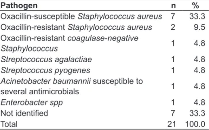
Table 2 - Isolated pathogen* in culture and susceptibility tests of acute hematogenous osteomyelitis cases seen in the Pedia-tric Infectious Disease Outpatient Clinic of Escola Paulista de Medicina from 2005 to 2009
Pathogen n %
Oxacillin-susceptible Staphylococcus aureus 7 33.3
Oxacillin-resistant Staphylococcus aureus 2 9.5
Oxacillin-resistant coagulase-negative
Staphylococcus 1 4.8
Streptococcus agalactiae 1 4.8
Streptococcus pyogenes 1 4.8
Acinetobacter baumannii susceptible to
several antimicrobials 1 4.8
Enterobacter spp 1 4.8
Not identiied 7 33.3
Total 21 100.0
*Isolated in blood, bone fragment or secretion culture.
Table 1 - Characteristics of acute hematogenous osteomyelitis cases seen in the Pediatric Infectious Disease Outpatient Clinic of Escola Paulista de Medicina from 2005 to 2009
Characteristic Yes % No % Total
Symptoms – clinical conditions
Fever 15 71.4 6 28.6 21
Pain 15 71.4 6 28.6 21
Edema 11 52.4 10 47.6 21
Functional disability 11 52.4 10 47.6 21
Hyperemia 5 23.8 16 76.2 21
High temperature 5 23.8 16 76.2 21
Affected site
Femur 7 33.3 14 66.7 21
Tibia 6 28.6 15 71.4 21
Humerus 3 14.3 18 85.7 21
Iliac 2 9.5 19 90.5 21
Fibula 1 4.8 20 95.2 21
Foot 1 4.8 20 95.2 21
two had hemoglobinopathies. The following comorbidities were found in one case each: juvenile idiopathic arthritis, systemic hypertension, cellulitis and genetic syndrome.
Pathogens were identiied in 14 of the 21 cases (66.7%) in blood culture (six cases), bone fragment culture (four cases), or secretion culture (eight cases). Table 2 shows that Staphylococcus was responsible for 71.4% of the cases for which the pathogen was isolated. Infection by coagulase-negative Staphylococcus was recorded for one case in which the same pathogen was isolated in bone fragment and secretion cul-tures. Although not usual, that patient was at school age and had no immunodeiciency.
The initial empirical antibiotic regimens were based on the most frequent pathogens of osteomyelitis. In eight cases (38.1%), initial treatment used oxacillin and ceftriaxone; in six (28.6%), oxacillin; and in two (9.5%), cephalo-thin. Vancomycin was introduced as the initial empirical treatment in two cases (9.5%) with a history of previous hospitalizations due to septic arthritis. The associations of oxacillin and amikacin, cephalothin and amikacin, and ampicillin and gentamicin were administered in one case each (4.8%) for newborns.
In addition to treatment with antibiotics, 14 patients (66.7%) also underwent at least one surgical debridement procedure. Treatment in hyperbaric chamber, however, was not used in any case in this study.
Treatment had to be changed during the progression of the cases due to different reasons. Hospital discharge, when IV antibiotics have to be replaced with oral equivalents, was the main reason and accounted for 35% of the changes. The results of antimicrobial susceptibility tests and the fact that oxacillin was not available in the hospital accounted for 20% of the changes each. Clinical factors were responsible for 15% of the changes, and laboratory indings, for 10%.
During outpatient follow-up, six patients (28.6%) had sequelae, such as progression to chronic osteomyelitis, seen in three cases. Deformities and limitation of movement range were found in one case each and were associated in another case. The two patients that had movement range limitations had osteomyelitis in the neonatal period.
ESR was the main test for inlammation during the clini-cal and laboratory follow-up of these cases of osteomyelitis. Graph 1 compares the dispersion of ESR values at admission, discharge and discontinuation of antibiotic treatment. ESR values at admission varied greatly and lied within the normal range in some cases. In contrast, at the time of antibiotic discontinuation, they were clearly more homogeneous.
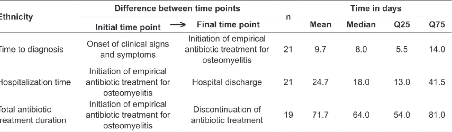
Time to diagnosis, duration of hospital stay and duration of antibiotic treatment are shown in Table 3. Mean values for each time were 9.7, 24.7 and 71.7 days.
The correlation between progression time for the cases under study was analyzed to investigate whether a shorter time to diagnosis would result in shorter of hospitalization and antibiotic treatment duration. However, there were no statistically signiicant correlations between these variables (Spearman rank correlation, p>0.05).
Discussion
In this descriptive study of 21 cases of acute hematogenous osteomyelitis in a pediatric population, the male-to-female ratio was 3.2:1. Although Riise et al(16) did not ind any
difference between sexes in the incidence of osteomyelitis, most studies(1,13-15) effectively demonstrated a predominance
of male sex. Zaoutis et al(17), for example, found that boys
were affected in 62% of the cases.
The analysis of distribution according to age group in our study showed that the incidence in children younger than ive years and in those ive years or older was 38.1% and 61.9%. This result is in agreement with that reported by Zaoutis et al(17), who studied children aged 2 months to
17 years and found an incidence of 42% and 58% for the same age groups.
Graph 1 - Comparison of ESR values at three time points in the progression of acute hematogenous osteomyelitis cases
ESR at hospital admission
ESR at discharge
ESR in mm/h
120
100
80
60
40
20
0
Q3=97.5
Q3=66.0
Q3=16.0 Q2=10.0 Q1=10.0 Q2=45.0
Q1=24.3 Q2=76.0
Q1=40.0
*
ESR at antibiotic discontinuation
In agreement with other studies in the literature, the most frequently affected sites were the long bones, with the femur in 34% of the cases, the tibia in 28% and the humerus in 14%. According to Weichert et al(1), the femur is affected in
36% of the cases, the tibia in 33%, and the humerus in 10%. In our study, pathogens were detected in 67% of the 21 cases, most often in secretion cultures. Arnold et al(14)
identiied pathogens in 48% of their cases of osteomyelitis, and Goergens et al(18), in 45%, whereas Moumile et al(13)
collected data for 186 cases of bone infection and identiied pathogens in 19%.
Staphylococcus aureus was isolated in 64% of the cases for which the pathogen was identiied. Streptococcus pyogenes and Streptococcus agalactiae were isolated in one case each (0.7%). Moumile et al(13) found Staphylococcus aureus in 60%
of the cases with isolates, whereas Streptococcus pyogenes and Streptococcus agalactiae were responsible for 0.8% of the cases each. Streptococcus pneumoniae was isolated in 13% of the cases for which the pathogen was known in the study conducted by Moumile et al(13), but was not found in our study.
Of the nine bacteria of the Staphylococcus aureus iso-lated in our study, two (28%) were resistant to oxacillin. Arnold et al(14) identiied Staphylococcus aureus in 34 cases
of osteomyelitis, 23 (67%) of which were caused by oxacillin-resistant pathogens.
In the service where our study was conducted, there are no routine methods to identify Kingella kingae, a pathogen that has been shown to play an important role in bone and joint infections in children, particularly those younger than 2 years(14,19).
The median ESR value at admission was 76.0mm/h (Q1=40.0 and Q3=97.5). For the same time point, Riise et al(16) found lower and more homogeneous values, at 41mm/h
(Q1=27 and Q3=52). The same authors also found that median duration of symptoms until admission was 8 days, the same value found in our study.
Mean duration of antibiotic treatment in our study was about 10 weeks (71.7 days). As mentioned before, treatment duration is long and variable in different studies. Weichert et al(1) reviewed publications about the duration of acute
osteomyelitis treatment in children and found that mean antibiotic treatment lasted up to 12 weeks according to Blockey and Watson(20); from six to nine weeks in the study
by Kolyvas et al(21); six weeks, according to Kulhanjian et
al(22); two weeks according to Jacobs et al(23); and in the study
by Peltola et al(24), three weeks.
Although an early diagnosis is extremely important in he-matogenous osteomyelitis, there are no data in the literature to estimate statistically signiicant values for time of diagno-sis after irst symptom and case progression. Because of the size of our study sample, no correlation was established be-tween time to diagnosis and duration of hospitalization and antibiotic treatment (Spearman rank correlation, p>0.05).
This study showed that acute hematogenous osteomyeli-tis is an important infection among children, particularly those in pre-school or school. The disease should be classi-ied as a severe bacterial infection that requires hospitaliza-tion and antibiotic treatment for a long time, and whose progression results in permanent sequelae for a substantial number of patients.
→
Table 3 - Time in days between events in the progression o acute hematogenous osteomyelitis cases seen in the Pediatric Infec-tious Disease Outpatient Clinic of Escola Paulista de Medicina from 2005 to 2009
Ethnicity
Difference between time points
n
Time in days
Initial time point Final time point Mean Median Q25 Q75
Time to diagnosis Onset of clinical signs
and symptoms
Initiation of empirical antibiotic treatment for
osteomyelitis
21 9.7 8.0 5.5 14.0
Hospitalization time
Initiation of empirical antibiotic treatment for
osteomyelitis
Hospital discharge 21 24.7 18.0 13.0 41.5
Total antibiotic treatment duration
Initiation of empirical antibiotic treatment for
osteomyelitis
Discontinuation of
antibiotic treatment 19 71.7 64.0 54.0 81.0
References
1. Weichert S, Sharland M, Clarke NM, Faust SN. Acute haematogenous osteomyelitis in children: is there any evidence for how long we should treat? Curr Opin Infect Dis 2008;21:258-62.
2. Kaplan SL. Osteomyelitis in children. Infect Dis Clin North Am 2005;19:787-97. 3. Darville T, Jacobs R, Richard F. Management of acute hematogenous
osteomyelitis in children. Pediatr Infect Dis J 2004;23:255-7.
4. Gutierrez K. Bone and joint infections in children. Pediatr Clin North Am 2005;52:779-94.
5. Staniski CL. Changes in pediatric acute hematogenous osteomyelitis management. J Pediatr Orthop 2004;24:444-5.
6. Iazzetti AV. Osteomielite e artrite séptica. In: Farhat CK, Carvalho LH, Succi RC, editors. Infectologia pediátrica. 3ª edição. São Paulo: Atheneu; 2007. p. 151-4.
7. Lew DP, Waldvogel FA. Osteomyelitis. Lancet 2004;364:369-79.
8. Abuamara S, Louis JS, Guyard MF, Barbier-Frebourg N, Lechavallier J. Osteoarticular infection in children: evaluation of a diagnostic and management protocol. Rev Chir Orthop Reparatrice Appar Mot 2004;90:703-13. 9. Aloui N, Nessib N, Jalel C, Ellouze S, Ben Chehida F, Sayed M et al. Acute
osteomyelitis in children: early MRI diagnosis. J Radiol 2004;85:403-8. 10. Kocher MS, Lee B, Dolan M, Weinberg J, Shulman ST. Pediatric orthopedic
infections: early detection and treatment. Pediatr Ann 2006;35:112-22. 11. Bayam L, Bruce CE, Sampath J, Bayam FB, Abernethy L. Importance
of communication between medical specialities: a case series. Injury 2008;39:623-6.
12. Collado P, Naredo E, Calvo C, Crespo M. Role of power Doppler sonography in early diagnosis of osteomyelitis in children. J Clin Ultrasound 2008;36:251-3. 13. Moumile K, Merckx J, Glorion C, Pouliquen JC, Berche P, Ferroni A.
Bacterial aetiology of acute osteoarticular infections in children. Acta Paediatr 2005;94:419-22.
14. Arnold SR, Elias D, Buckingham SC, Thomas ED, Novais E, Arkader A et al. Changing patterns of acute hematogenous osteomyelitis and septic arthritis:
emergence of community- -associated methicillin-resistant staphylococcus aureus. J Pediatr Orthop 2006;26:703-8.
15. Malcius D, Barauskas V, Uzkuraite R. Some aspects of long-term results of treatment of acute hematogenous osteomyelitis. Medicina (Kaunas) 2007;43:472-7.
16. Riise OR, Kirkhus E, Handeland KS, Flato B, Reiseter T, Cvancarova M
et al. Childhood osteomyelitis-incidence and differentiation from other acute onset musculoskeletal features in a population-based study. BMC Pediatr 2008;8:45.
17. Zaoutis T, Localio AR, Leckerman K, Saddlemire S, Bertoch D, Keren R. Prolonged intravenous therapy versus early transition to oral antimicrobial therapy for acute osteomyelitis in children. Pediatrics 2009;123:636-42. 18. Georgens ED, McEvoy A, Watson M, Barrett IR. Acute osteomyelitis and septic
arthritis in children. J Paediatr Child Health 2005;41:59-62.
19. Kiang KM, Ogunmodede F, Juni BA, Boxrud DJ, Glennen A, Bartkus JM et al. Outbreaks of osteomyelitis/septic arthritis caused by Kingella kingae among child care center attendees. Pediatrics 2005;116:e206-13.
20. Blockey NJ, Watson JT. Acute osteomyelitis in children. J Bone Joint Surg Br 1970;52:77-87.
21. Kolyvas E, Ahronheim G, Marks MI, Gledhill R, Owen H, Rosenthall L. Oral antibiotic therapy of skeletal infections in children. Pediatrics 1980;65:867-71. 22. Kulhanjian J, Dunphy MG, Hamstra S, Levernier K, Rankin M, Petru A et al.
Randomized comparative study of ampicillin/sulbactam vs. ceftriaxone for treatment of soft tissue and skeletal infections in children. Pediatr Infect Dis J 1989;8:605-10.
23. Jacobs RF, Darville T, Parks JA, Enderlin G. Safety proile and eficacy of cefotaxime for the treatment of hospitalized children. Clin Infect Dis 1992;14:56-65.