Impact of Estrogen Therapy on Lymphocyte
Homeostasis and the Response to Seasonal
Influenza Vaccine in Post-Menopausal
Women
Flora Engelmann1, Andrea Rivera1, Byung Park2, Marci Messerle-Forbes3, Jeffrey T. Jensen3,4, Ilhem Messaoudi1,4,5*
1Division of Biomedical Sciences, School of Medicine, University of California Riverside, Riverside, California, United States of America,2Division of Biostatistics, Department of Public Health and Preventive Medicine, Oregon Health and Science University, Portland, Oregon, United States of America,3Department of Obstetrics and Gynecology, Oregon Health and Science University, Portland, Oregon, United States of America,4Division of Reproductive Sciences, Oregon National Primate Research Center, Beaverton, Oregon, United States of America,5Division of Pathobiology and Immunology, Oregon National Primate Research Center, Beaverton, Oregon, United States of America
*messaoud@ucr.edu
Abstract
It is widely recognized that changes in levels of ovarian steroids modulate severity of auto-immune disease and auto-immune function in young adult women. These observations suggest that the loss of ovarian steroids associated with menopause could affect the age-related decline in immune function, known as immune senescence. Therefore, in this study, we determined the impact of menopause and estrogen therapy (ET) on lymphocyte subset fre-quency as well as the immune response to seasonal influenza vaccine in three different groups: 1) young adult women (regular menstrual cycles, not on hormonal contraception); 2) post-menopausal (at least 2 years) women who are not receiving any form of hormone therapy (HT) and 3) post-menopausal hysterectomized women receiving ET. Although the numbers of circulating CD4 and CD20 B cells were reduced in the post-menopausal group receiving ET, we also detected a better preservation of naïve B cells, decreased CD4 T cell inflammatory cytokine production, and slightly lower circulating levels of the pro-inflamma-tory cytokine IL-6. Following vaccination, young adult women generated more robust anti-body and T cell responses than both post-menopausal groups. Despite similar vaccine responses between the two post-menopausal groups, we observed a direct correlation between plasma 17βestradiol (E2) levels and fold increase in IgG titers within the ET group. These findings suggest that ET affects immune homeostasis and that higher plasma E2 levels may enhance humoral responses in post-menopausal women.
OPEN ACCESS
Citation:Engelmann F, Rivera A, Park B, Messerle-Forbes M, Jensen JT, Messaoudi I (2016) Impact of Estrogen Therapy on Lymphocyte Homeostasis and the Response to Seasonal Influenza Vaccine in Post-Menopausal Women. PLoS ONE 11(2): e0149045. doi:10.1371/journal.pone.0149045
Editor:Stephen Todryk, Northumbria University, UNITED KINGDOM
Received:October 2, 2015
Accepted:January 25, 2016
Published:February 9, 2016
Copyright:© 2016 Engelmann et al. This is an open access article distributed under the terms of the
Creative Commons Attribution License, which permits unrestricted use, distribution, and reproduction in any medium, provided the original author and source are credited.
Data Availability Statement:The authors have attached original data as a Supporting Information file, which is sufficient to replicate all the findings presented in the study.
Funding:This work was supported by a grant from the Center for Gender Differences at OHSU, and a Leadership in Aging Research Fellowship from the Brookdale Foundation.
Introduction
In addition to their role in sexual differentiation and reproduction, female sex hormones mod-ulate immune function. For instance,β-estradiol (E2) treatment exacerbates the severity of sys-temic lupus erythematosus and myasthenia gravis [1,2]. On the other hand, the severity and incidence of rheumatoid arthritis and multiple sclerosis are decreased during pregnancy [3] when circulating levels of progesterone are high. Moreover, cytokine production by peripheral blood T cells varies throughout the menstrual cycle. Specifically, the number of PBMC able to secrete IL-4 in response to stimulation correlated with estrogen levels [4] and serum levels of the cytokines IL-6, IL-1β, IL-10, and IL-8 peak during the follicular phase when estrogen levels are highest [5–7]. In addition, vaccination studies in humans indicate that vaginal immuniza-tions are more effective for induction of genital tract antibodies when performed during the mid-follicular phase of the menstrual cycle [8].
The mechanisms by which ovarian steroids affect immune function are beginning to emerge. T and B cells express ERαand ERβreceptors [9] indicating that E2 can directly modu-late lymphocyte function. E2 treatment of B cells increases: 1) the expression of the anti-apo-ptotic molecule Bcl-2 [10–12]; 2) B cell activation [13]; 3) IgG production [14]; and 4) the expression of activation-induced deaminase (AID) [15] leading to increased frequency of somatic hypermutation and class-switch recombination. Similarly, E2 was shown to inhibit activation-induced apoptosis of T cells from lupus patients by down-regulating the expression of Fas ligand [16]. In vitro studies also suggest a potential bias towards Th2, Th17, and Treg polarization in E2 treated T cell cultures [17,18]. Estrogen and progesterone can also indirectly influence T and B cells by affecting the function of innate immune cells such as dendritic cells and macrophages that influence T and B cell differentiation [19]. For example, progesterone treatment reduces the ability of dendritic cells to take up antigenic peptides, stimulate T cell responses [20], and secrete the potent antiviral cytokine IFNα[21]. In contrast, estradiol treat-ment increases the ability of macrophages to secrete inflammatory cytokines [22].
Aging is associated with a decline in immune function; a phenomenon commonly referred to as immune senescence and believed to result in greater infectious disease related morbidity and mortality in the elderly [23]. Given the influence of ovarian steroids on immune function, their loss during menopause could exacerbate immune senescence [24,25]. This hypothesis is supported by the observation that rhinovirus infection induces a higher IFNγand IL-13 response in young women than men, however this sex difference is no longer detected after the age of 50 coincident with typical onset of menopause and the associated loss of ovarian steroids [26]. Similarly, hepatitis vaccines induce higher antibody titers and seroconversion rates in young women, but this sexual dimorphism is no longer evident in vaccinees over the age of 60 [27]. A recent study looking at sex differences in gene expression in human peripheral blood found differences between men and women become smaller when women reach menopause and larger when women use hormonal contraceptives [28]. The biological process gene ontol-ogy category in female-biased genes with the greatest enrichment was immune system process [28].
Animal studies also support this hypothesis. Ovariectomy of young female rats resulted in decreased leukocyte chemotaxis and LPS-induced proliferation, reduced NK cell lysis, and increased oxidative damage and inflammatory cytokine production by peritoneal macrophages suggestive of premature immunosenescence [29,30]. Studies from our laboratory have shown that ovariectomized female rhesus macaques generate reduced T and B cell responses to vacci-nation compared to age-matched control animals [31]. Similarly, in a mouse model of HSV-2 challenge, ovariectomy resulted in a reduced response to an experimental vaccine and abro-gated protection from vaginal challenge [32].
Additional studies have shown that post-menopausal women have higher plasma levels of the inflammatory cytokines TNFαand IFNγcompared to pre-menopausal subjects [33–35]. In contrast, HT reduces the levels of both of these inflammatory mediators [33,35], in addition to IL-6 [36,37]. Moreover, surgical menopause results in several changes associated with immune senescence such as a decreased CD4/CD8 ratio, an increase in the percentage of NK cells and a decrease in circulating B cells [38] and HT also reverses the age-related decrease in CD4 and B cell numbers, T cell proliferative capacity and the increase in NK cells numbers [38–40].
However, the impact of HT on the response to infection or vaccination remains poorly char-acterized. A mouse model of menopause showed a significant reduction of influenza specific antibodies in ovariectomized mice; these levels were rescued to that of sham operated mice by the administration of estrogen [41]. This indicates that HT may boost vaccine responses in post-menopausal women. Since vaccination represents an important preventative health mea-sure in older adults, and the role of HT is controversial, studies in this area have significant public health impact. In this study, we investigated the impact of menopause and estrogen ther-apy (ET) on lymphocyte frequency and the immune response to seasonal influenza vaccine in healthy hysterectomized women. We compared values of these parameters to those observed in young women of reproductive age as well as post-menopausal women not receiving any form of HT.
Materials and Methods
Subjects
The human subjects Institutional Review Board of Oregon Health and Science University approved all study procedures and documents. Every participant gave written consent before participation and research information was kept confidential. The study consisted of 3 arms: A) 15 young adults 19–38 years of age with normal menstrual cycles not currently taking con-traceptives (Table 1); B) 15 women aged 55–63, at least two years post-menopausal and had not received hormone replacement therapy for at least 4 years (Table 2); C) 15 post-meno-pausal hysterectomized women aged 47 to 64 who were receiving estrogen therapy (Table 3). Women who were smokers, pregnant or breast-feeding, using hormonal birth control, on com-bined hormone replacement therapy, or using corticosteroids were excluded from the study. A 10ml heparinized blood sample was obtained on the day subjects received their seasonal flu shot (Day 0), one week (window 7–10 days) and one month (window 30–35 days) after immu-nization. Cycling women were asked to arrange their first visit to fall on one of the first 12 days of their cycle. Subjects received the seasonal trivalent inactivated influenza vaccine Fluzone (Sanofi Pasteur).
Blood sample analysis
Complete blood cell counts (CBCs) were obtained with a Hemavet complete blood count machine (Drew Scientific, Waterbury, CT). Blood samples were centrifuged over a ficoll den-sity gradient in order to isolate peripheral blood mononuclear cells (PBMC) and plasma.
Determination of plasma hormone levels
ng/ml, respectively. The E2 antibody used by the Immulite 2000 platform does not cross-react with Equilin, the main estrogen component within Premarin, alpha Equilenin, Ethiny-Estra-diol, and 3- or 17-substrates of sulfate, valerate or propionate.
Table 1. Young Adult Women.
Subject ID Age Estrogen Levels BMI (KG/m2) History of smoking
Visit 1 Visit 2 Visit 3
1 23 117.0 40.8 47.3 25.2 N
2 24 31.9 118.0 59.2 19.5 N
4 35 98.9 172.0 57.5 20.5 Y
7 38 74.4 32.4 217.0 23.5 Y
9 19 37.4 <20.0 72.1 25.0 N
15 36 39.0 <20.0 30.1 24.8 N
16 29 108.0 101.0 69.9 27.0 N
19 26 124.0 23.9 74.0 21.0 N
20 35 115.0 21.0 135.0 21.4 N
21 24 109.0 56.0 81.7 25.5 N
22 22 21.2 45 22.5 N
23 28 <20.0 <20.0 97.3 21.5 Y
24 24 <20.0 61.9 26.5 22.5 Y
27 25 50.1 39.7 64.0 20.0 N
29 33 228.0 50.9 57.2 21.6 Y
Average 28.1±5.9
Ethnicity:Subject 9-Black, Subject 20-Asian, All other subjects in this group were White
doi:10.1371/journal.pone.0149045.t001
Table 2. Post-menopausal (P.M.) Women no HT.
Subject ID Age Years P.M. Previous hormone use and time
since last dose (years)
BMI (KG/m2) History of smoking
3 57 4 Never NA 24.0 N
6 55 4 Never NA 28.0 N
10 63 34 Combined 7 19.8 Y
11 56 8 Prempro 7 21.7 N
12 56 4 Never NA 26.0 N
13 59 3 Never NA 26.4 N
14 55 2 Never NA 26.6 N
18 60 20 Combined 16 22.8 N
25 63 13 Combined 5 22.0 N
26 61 7 Never NA 28.5 Y
30 56 14 Premarin 10 21.0 N
31 59 5 Climara 5 23.6 N
33 60 4 Estrodiol/Prometrium 4 24.2 Y
34 59 14 Prempro 5 23.4 N
37 61 12 Never NA 21.6 Y
Average 58.7±2.7
Ethnicity:All subjects in this group were White. NA indicates hormonal therapy was never received.
Determination of T and B cell numbers and subsets
To measure T and B cell numbers, PBMCs were stained with anti- CD20 (eBioscience, San Diego, CA), CD8b (Beckman Coulter, Miami, FL), CD4, CD28, CD95, and CD27 (BioLegend, San Diego, CA) antibodies. Naïve and memory CD4 and CD8 T cells subsets were determined as previously described [42]. Naïve T cells were identified as CD28+CD95-, central memory as CD28+CD95+ and effector memory as CD28-CD95+. Memory B cells were identified based on the expression of CD27 [43]. The samples were acquired using an LSRII flow cytometer (Beckton Dickenson, San Jose, CA) and the data were analyzed using FlowJo software (TreeS-tar, Ashland, OR). The percentages of T cell subsets were converted to absolute numbers perμL using lymphocyte numbers from corresponding whole blood CBCs.
Human IL-6 enzyme linked immuno-sorbant assay
IL-6 ELISA were performed using an‘UltraSensitive’Human IL-6 ELISA kit (Invitrogen, Cam-arillo, CA) following manufacturers instructions. Briefly, 100ul of sample serum was added in duplicate to precoated plates and incubated for 3 hours at 37°C. Plates were washed six times before the addition of 100ul of biotin conjugate per well, which was then incubated for 45 min-utes at room temperature. Plates were again washed six times before a second 45 min incuba-tion at room temperature with 100ul of streptavidin-HRP per well. After another six washes, plates were incubated at room temperature with 100ul of stabilized chromogen and the reac-tion stopped after 30 minutes with 100ul of stop solureac-tion per well. Color development was then read on a Spectramax 190 (Molecular Devices, Sunnyvale, CA) microplate reader at 450nm. Each plate contained manufacturer provided standards against which sample concentrations were determined, as well as an in house control.
Table 3. Post-menopausal (P.M.) Women receiving ET.
Subject ID Age Years P.M. ET regimen and length of time on regimen (years)
Estrogen Levels (pg/ml) BMI (KG/m2) History of smoking Visit 1 Visit 2 Visit 3
8 61 25 Premarin (0.625mg) 25 <20.0 <20.0 <20.0 22.5 N
17 57 34 Estrace (2mg) 34 158 200 185 23.8 N
28 58 3 Vagifem (25mg) 2 <20.0 <20.0 <20.0 26.0 N
35 61 30 Ogen (0.6mg) 20 29.3 39.5 30.0 26.3 N
36 57 9 Estrogel (1.25mg) 5 47.5 34.9 41.2 23.1 N
38 59 5 Estradiol (0.5mg) 5 57.2 60.6 46.0 28.1 Y
39 47 11 Estrace (0.5mg) 5 60.9 64.0 54.9 29.1 N
40 64 19 Estratest HS 17 or 7* 29.7 <20.0 32.9 26.4 N
42 61 11 Vivelle Dot (0.05mg) 11 37.3 46.1 36.4 25.8 N
43 64 12 Vivelle Dot (0.05mg) 12 44.9 <20.0 32.0 26.0 N
44 57 8 Estrace (0.5mg) 4 29.3 28.5 30.1 23.8 N
45 56 19 Vivelle Dot (0.05mg) 2 32.3 23.6 43.0 29.2 N
46 64 7 Premarin (0.3mg) 7 36.6 38.6 56.1 28.8 N
47 55 6 Femring (0.05mg) 5 26.8 27.2 24.8 25.1 N
48 55 23 Premarin (0.45mg) 5 <20.0 24.1 22.5 25.6 N
Average 58.4±4.5
Ethnicity:All subjects in this group were White.
*Participant 40 also participated in the WHI and it is unknown whether she received placebo or Estrogen.
Measuring antibody responses
IgG end point titers were determined by standard ELISA using plates coated with the same Influenza Virus Vaccine (Fluzone, Sanofi Pasteur) that subjects received. After blocking and washing, threefold dilutions of plasma were added in triplicates. Bound IgG were visualized by the addition of HRP conjugated goat anti-human IgG (Open Biosystems, Huntsville, AL) and OPD substrate. The optical density was measured at 490 nm using an ELISA plate reader (SpectraMax 190, Molecular Devices, Sunnyvale, CA). Endpoint titers were determined by lin-ear regression following log-log transformation and using 0.1 as the cut off and then normal-ized to a control sample included on each plate.
Measuring frequency of influenza specific T cells
PBMCs from each subject were either untreated (negative control), stimulated with influenza virus PR8 overnight at 37°C in the presence of brefeldin A (Sigma, St Louis, MO), or stimulated for 6hr with anti-CD3 in the presence of brefeldin A (positive control). At the end of the incubation, PBMCs were stained with anti-CD8β(Beckman Coulter) and CD4 (BioLegend) antibodies. The cells were then fixed and permeabilized to allow for intracellular staining with anti-IFNγand TNFαantibodies (Biolegend). Fluorescence was measured on an LSRII flow cytometer and the data analyzed using FlowJo software.
Statistical analysis
Statistical analysis was performed using the SAS Software version 9.2 (SAS Institute Inc., USA) and Statistica (StatSoft, Tulsa, OK). Repeated measures ANOVA followed by Tukey post-hoc comparisons of means was used. Bayesian information comparison was used to determine the optimal correlation within subject. P values were adjusted by BMI and past smoking history.
Results
Study Population
A total of 45 subjects were enrolled (15 in each group), and all subjects completed the study. All young adult women were White except for 2 individuals (Black and Asian), had an average age of 28.1± 5.9, and BMI of 22.8± 2.3 (Table 1). All subjects within group B (post menopausal women not currently receiving HT) were White with an average age of 58.7±2.7 and BMI of 24 ±2.6. Approximately 50% of the women in Group B previously used HT (Table 2) but all women discontinued it at least 4 years prior to enrolling in this study. Subjects in group C (hys-terectomized women receiving ET) were White with an average age of 58.4±4.5 and BMI of 26 ±2.1, and used different ET products summarized inTable 3. Plasma E2 levels were measured at each visit for all subjects. As expected, the levels of variation in plasma E2 levels between sub-ject and visit were highest in young adult women (30pg/ml—100 pg/ml E2, Group A,Table 1). Plasma E2 levels also showed considerable variation in women receiving ET (Table 2). As expected, post-menopausal women not currently receiving HT had no detectable E2 levels. None of the subjects were current smokers, although 4 women, 5 women and 1 women in groups A, B and C respectively had a previous history of smoking.
Menopause and ET impact on lymphocyte subset distribution
line with this observation, the number of CD4 and CD20 B cells was also reduced in hysterecto-mized women receiving ET compared to post-menopausal women not receiving HT (Fig 1B). Interestingly, the number of CD8 T cells was significantly lower in both post-menopausal women groups compared to young adult women (Fig 1B).
We next investigated the impact of menopause and ET on naïve and memory lymphocyte subset distribution. The number of naïve CD4 T cells was reduced only in hysterectomized women receiving ET compared to young adult women (Fig 1C). This difference in frequency of naïve CD4 T cells accounted for the decreased frequency of total CD4 T cells in
post-Fig 1. Impact of menopause and ET on lymphocyte frequencies.(A) The number of lymphocytes on visit 1 /μl blood based on complete blood cell counts (CBC) values. (B) Frequency of CD4 T cells, CD8 T cells and CD20 B cells was determined in PBMC using flow cytometry (FCM). The percentages were then converted to absolute numbers of cells/μl blood using the lymphocyte counts obtained from the CBCs. (C) Frequency of naïve (Na), central memory (CM) and effector memory (EM) CD4 T cells was determined using FCM and were then converted to number/μl of blood using the CD4 numbers obtained earlier. (D) Numbers of Na, CM and EM CD8 T cells was determined as described for CD4 T cells. (E) Frequencies of naïve (CD27-) and
memory (CD27+) B cells were determined by FCM and then converted to absolute numbers of cells/μl blood using the CD20 numbers obtained earlier.
menopausal women receiving ET. On the other hand and as described for total CD8 T cell numbers, we detected a significant decrease in the frequency of naïve CD8 T cells in both groups of post-menopausal women compared to adult women (Fig 1D). In contrast to these observations, we detected a decreased frequency of naïve B cells (CD27-) and a concomitant increase in the frequency of CD27+ memory B cells in post-menopausal women not receiving HT compared to young adult women (Fig 1E), whereas the numbers of naïve and memory B cells in post-menopausal women receiving ET were comparable to those in young adult women (Fig 1E).
Menopause and ET modulate inflammatory cytokine production
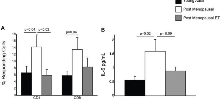
Aging is associated with increased production of pro-inflammatory cytokines by T cells [47,48]. Therefore, we measured inflammatory cytokine production by T cells in response to polyclonal CD3 stimulation using PBMC collected during visit 1 (Fig 2A). Specifically, we mea-sured the frequency of CD4 and CD8 T cells that produced TNFα, IFNγ, or both using intracel-lular cytokine staining. We detected increased frequency of CD4 and CD8 T cells producing TNFα, IFNγor both in post-menopausal women not receiving HT compared to young adult women consistent with previous studies comparing cytokine production by T cells collected from young and old subjects [47,49,50]. In contrast, frequencies of CD4 and CD8 T cells pro-ducing TNFα, IFNγor both were comparable in post-menopausal women receiving ET (group C) and young adult women (group A) (Fig 2A).
Aging is also associated with increased circulating levels of inflammatory cytokines, notably IL-6 and CRP, often referred to as inflammaging [44,48,51,52]. Thus, we next measured plasma levels of the key inflammatory cytokine IL-6 during the first visit (prior to vaccination). Our analysis revealed that post-menopausal women not receiving HT had a significantly higher level of plasma IL-6 than young women (Fig 2B). Plasma levels of IL-6 were previously shown to be lower in post-menopausal women receiving unopposed estrogen compared to women
Fig 2. Impact of menopause and ET on IL-6 levels and TNFαand IFNγproduction.(A) Frequency of CD4 and CD8 T cells that secrete TNFα, IFNγor both in response to CD3 stimulation was determined by intracellular cytokine staining and FCM using PBMC collected during visit 1 before the administration of the influenza vaccine. (B) Plasma IL-6 levels using samples collected during visit 1 were determined by ELISA.
not receiving HT [36,53]. Although not statistically significant, we also detected a clear trend towards lower plasma IL-6 levels in the ET group compared to non-HT group (p = 0.09).
The immune response to seasonal influenza vaccine
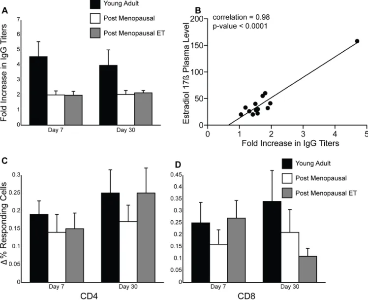
Previous studies have shown that hemagglutination inhibition (HI) assays are not a good corre-late of vaccine efficacy in the elderly [54]. Therefore, we measured the fold increase in IgG titers in response to influenza vaccination by standard ELISA (Fig 3A). IgG titers increased in all three groups at day 7 post-vaccination and remained stable at day 30 as previously reported for adults receiving the trivalent inactivated influenza vaccine [55] As expected, young adult women generated a robust antibody response compared to older post-menopausal women. There was no difference in the fold increase in IgG titers between the two post-menopausal
Fig 3. Impact of menopause and ET on immune response to seasonal influenza vaccine.(A) IgG end point titers were determined using standard ELISA and then the fold increase over baseline was calculated for visits 2 and 3. (B) Correlation between plasma E2 levels on visit 1 (day of vaccination) and fold increase in IgG titer during visit 2 was calculated for women in group C. (C) Frequency of flu-specific CD4 T cells was measured using intracellular cytokine staining and flow cytometry. The increased frequency of responding T cells was determined by subtracting the values obtained on visits 2 and 3 from those obtained on visit 1. (D) Same analysis was carried out for CD8 T cells.
groups, but given the differences in E2 plasma levels, we refined the analysis by measuring the correlation coefficient between plasma E2 levels on the day of vaccination and IgG fold increase on day 7 post-vaccination in post-menopausal women receiving ET (Fig 3B). Our analysis revealed a positive correlation of 0.98 with a p value<0.0001 within group C. This correlation remained significant after removing the subject with the highest E2 plasma levels tion = 0.59; p-value = 0.0439). This correlation remained significant on day 30 (correla-tion = 0.77; p-value = 0.0008) but once the subject with highest plasma E2 levels was omitted, the correlation was no longer significant (correlation = 0.32; p-value = 0.2634). Additionally, we measured the correlation coefficient between plasma E2 levels on the day of vaccination and IgG fold increase on days 7 and 30 post-vaccination in young adult women and saw no correlation (slope<0.01 and p>0.9 in both cases). These data suggest that plasma E2 levels may only play a role in post-menopausal women.
We next evaluated the impact of ET on the T cell response to the seasonal influenza vaccine (Fig 3). Following vaccination all subjects showed an increase in the frequency of influenza-specific CD4 and CD8 T cells on both day 7 and 30 post vaccination. There were no statistically significant differences in the increase in T cell responses between all three groups. On day 30 post vaccination, we detected a trend indicating a further increase in CD4 T cell response in young adult women and post-menopausal women receiving ET but not in post-menopausal women not receiving HT (Fig 3C). In contrast, CD8 T cell response in post-menopausal women receiving ET on day 30 post vaccination are slightly reduced compared to day 7 (Fig 3D).
Discussion
Aging is associated with a general dysregulation of the immune system referred to as immune senescence that leads to diminished capacity to respond to infection and vaccination [23]. Since several studies have shown that ovarian steroids modulate immune response in women
[56–58], menopause-associated loss of ovarian steroids is likely to have a significant impact on
immune senescence. Indeed, both clinical and experimental observations suggest that the loss of ovarian steroids correlates with several immunosenescent changes that are in turn modu-lated by hormone therapy (HT) [35,40]. However, the impact of HT on lymphocyte homeosta-sis and the immune response to vaccination and/or infection remains poorly understood. In this study we specifically investigated the impact of estrogen therapy (ET) on T and B cell sub-set distribution and function.
Kumuru study received transdermal E2. This current study is underpowered to examine the impact of age at hysterectomy and onset of ET and the type of ET on lymphocyte homeostasis but this will be the focus of future studies.
An alternative explanation for the decreased lymphocyte numbers is that ET interferes with lymphopoiesis given that puberty correlates with the onset of thymic involution [59]. The decreased frequency of naïve CD4 T cells observed in post-menopausal women receiving ET would support this hypothesis. In contrast to CD4 T cells, frequency of naïve CD8 T cells decreased in both groups of post-menopausal women compared to young adult women which suggests that aging has a more significant impact on loss of naïve CD8 T cells than menopause. However, the decreased frequency of CD4 T cells in post-menopausal women receiving ET did not impact the T cell response to influenza vaccination since we reported a comparable increase in the frequency of influenza T cells in all three groups.
Although post-menopausal women receiving ET had fewer B cells, we detected improved preservation of naïve B cells compared to post-menopausal women not receiving HT. Naïve lymphocytes are the body’s reserve to respond to novel pathogens that have not been previ-ously encountered. Thus, increased frequency of these cells could confer advantage to the host in the face of infection. Interestingly, fold increase in IgG titers on day 7 post vaccination corre-lated significantly with plasma E2 levels measured on the day of vaccination in post-meno-pausal women receiving ET. These findings are in line with previous studies in rodents, which showed that E2 treatment of control or ovariectomized mice enhanced protection against HSV-2 challenge [32]. In those studies, although antibody titers in E2 treated mice were not significantly higher than those observed in untreated mice, the neutralization potential was sig-nificantly improved [32]. Our data are also in agreement with previous in vitro studies that showed increased IgG and IgM production by human PBMC after E2 stimulation [14]. It should be noted that the correlation between plasma E2 levels and vaccine-specific IgG titers was only significant for day 7 and only in post-menopausal women, suggesting that plasma E2 levels might be particularly important for older women. We measured no additional increase in IgG titers after day 7 post vaccination, which is in line with previous reports from adults vac-cinated with live attenuated influenza viruses or a trivalent inactivated vaccine where serum IgG levels increased at 14 and remained stable at 28 days post vaccination [55]. On the other, ET had no effect on the T cell response to vaccination. However, the seasonal influenza vaccine is a fully inactivated vaccine that elicits primarily an antibody response, thus the induction of T cell responses is not usually substantial following vaccination.
women receiving ET could potentially result in better outcomes after influenza infection com-pared to post-menopausal women not receiving HT.
In summary, although hysterecotmized post-menopausal women receiving ET had a decreased number of circulating lymphocytes, they exhibited better preservation of naïve B cells, lower levels of inflammatory cytokine production by T cells, and slightly reduced levels of circulating IL-6 levels. We further show that the decreased number of T cells did not negatively impact their ability to generate a T cell response following seasonal influenza vaccination and that higher plasma E2 levels correlate with increased antibody IgG titers to influenza vaccine early after vaccination. This is a small cohort and our observations need to be validated in a larger study. Nevertheless, given the benefits conferred by ET on bone density, cognitive func-tion and cardiovascular disease [68], the studies presented here emphasize the need for addi-tional studies to elucidate the optimum route of E2 delivery and in particular the effect of oral (subject to hepatic first pass effects) versus transdermal (including vaginal) administration. They also highlight the need to identify a target blood level of E2 needed to enhance immune response to vaccination in post-menopausal women and achieve a better understanding of the impact of acute and chronic exposure to estrogen on immunity.
Supporting Information
S1 File. All Original Data. (XLSX)Acknowledgments
We would like to thank the staff of the Center for Women’s Health at Oregon Health and Sci-ence University for their tremendous help in recruiting subjects for this study.
Author Contributions
Conceived and designed the experiments: IM JTJ. Performed the experiments: FE MMF. Ana-lyzed the data: FE AR BP JTJ IM. Contributed reagents/materials/analysis tools: IM JTJ. Wrote the paper: FE AR BP MMF JTJ IM.
References
1. Carlsten H, Nilsson N, Jonsson R, Backman K, Holmdahl R, Tarkowski A (1992) Estrogen accelerates immune complex glomerulonephritis but ameliorates T cell-mediated vasculitis and sialadenitis in auto-immune MRL lpr/lpr mice. Cell Immunol 144: 190–202. PMID:1394437
2. Delpy L, Douin-Echinard V, Garidou L, Bruand C, Saoudi A, Guery JC (2005) Estrogen enhances sus-ceptibility to experimental autoimmune myasthenia gravis by promoting type 1-polarized immune responses. J Immunol 175: 5050–5057. PMID:16210608
3. Confavreux C, Hutchinson M, Hours MM, Cortinovis-Tourniaire P, Moreau T (1998) Rate of pregnancy-related relapse in multiple sclerosis. Pregnancy in Multiple Sclerosis Group. N Engl J Med 339: 285– 291. PMID:9682040
4. Verthelyi D, Klinman DM (2000) Sex hormone levels correlate with the activity of cytokine-secreting cells in vivo. Immunology 100: 384–390. PMID:10929062
5. Angstwurm MW, Gartner R, Ziegler-Heitbrock HW (1997) Cyclic plasma IL-6 levels during normal men-strual cycle. Cytokine 9: 370–374. PMID:9195137
6. Al-Harthi L, Wright DJ, Anderson D, Cohen M, Matity Ahu D, Cohn J, et al. (2000) The impact of the ovu-latory cycle on cytokine production: evaluation of systemic, cervicovaginal, and salivary compartments. J Interferon Cytokine Res 20: 719–724. PMID:10954915
8. Kozlowski PA, Williams SB, Lynch RM, Flanigan TP, Patterson RR, Cu-Uvin S, et al. (2002) Differential induction of mucosal and systemic antibody responses in women after nasal, rectal, or vaginal immuni-zation: influence of the menstrual cycle. J Immunol 169: 566–574. PMID:12077289
9. Suenaga R, Evans MJ, Mitamura K, Rider V, Abdou NI (1998) Peripheral blood T cells and monocytes and B cell lines derived from patients with lupus express estrogen receptor transcripts similar to those of normal cells. J Rheumatol 25: 1305–1312. PMID:9676761
10. Verthelyi DI, Ahmed SA (1998) Estrogen increases the number of plasma cells and enhances their autoantibody production in nonautoimmune C57BL/6 mice. Cell Immunol 189: 125–134. PMID: 9790726
11. Rider V, Jones S, Evans M, Bassiri H, Afsar Z, Abdou NI (2001) Estrogen increases CD40 ligand expression in T cells from women with systemic lupus erythematosus. J Rheumatol 28: 2644–2649. PMID:11764210
12. Evans MJ, MacLaughlin S, Marvin RD, Abdou NI (1997) Estrogen decreases in vitro apoptosis of peripheral blood mononuclear cells from women with normal menstrual cycles and decreases TNF-alpha production in SLE but not in normal cultures. Clin Immunol Immunopathol 82: 258–262. PMID: 9073549
13. Paavonen T, Andersson LC, Adlercreutz H (1981) Sex hormone regulation of in vitro immune response. Estradiol enhances human B cell maturation via inhibition of suppressor T cells in pokeweed mitogen-stimulated cultures. J Exp Med 154: 1935–1945. PMID:6459399
14. Kanda N, Tamaki K (1999) Estrogen enhances immunoglobulin production by human PBMCs. J Allergy Clin Immunol 103: 282–288. PMID:9949320
15. Karpuzoglu E, Zouali M (2009) The multi-faceted influences of estrogen on lymphocytes: toward novel immuno-interventions strategies for autoimmunity management. Clin Rev Allergy Immunol 40: 16–26. 16. Kim WU, Min SY, Hwang SH, Yoo SA, Kim KJ, Cho CS (2010) Effect of oestrogen on T cell apoptosis
in patients with systemic lupus erythematosus. Clin Exp Immunol 161: 453–458. doi: 10.1111/j.1365-2249.2010.04194.xPMID:20529085
17. Polanczyk MJ, Hopke C, Vandenbark AA, Offner H (2006) Estrogen-mediated immunomodulation involves reduced activation of effector T cells, potentiation of Treg cells, and enhanced expression of the PD-1 costimulatory pathway. J Neurosci Res 84: 370–378. PMID:16676326
18. Khan D, Dai R, Karpuzoglu E, Ahmed SA (2010) Estrogen increases, whereas IL-27 and IFN-gamma decrease, splenocyte IL-17 production in WT mice. Eur J Immunol 40: 2549–2556. doi:10.1002/eji. 201040303PMID:20623549
19. Kovats S, Carrera E, Agarwal H (2010) Sex Steroid receptors in Immune Cells. In: Klein SL, Roberts CW, editors. Sex Hormones and Immunity to Infection. Berlin: Springer. pp. 53–91.
20. Butts CL, Shukair SA, Duncan KM, Bowers E, Horn C, Belyavskaya E, et al. (2007) Progesterone inhib-its mature rat dendritic cells in a receptor-mediated fashion. Int Immunol 19: 287–296. PMID:
17289656
21. Hughes GC, Thomas S, Li C, Kaja M- K, Clark EA (2008) Cutting Edge: Progesterone Regulates IFN-{alpha} Production by Plasmacytoid Dendritic Cells. J Immunol 180: 2029–2033. PMID:18250406 22. Calippe B, Douin-Echinard V, Delpy L, Laffargue M, Lelu K, Krust A, et al. (2010) 17Beta-estradiol
pro-motes TLR4-triggered proinflammatory mediator production through direct estrogen receptor alpha sig-naling in macrophages in vivo. J Immunol 185: 1169–1176. doi:10.4049/jimmunol.0902383PMID: 20554954
23. McElhaney JE, Effros RB (2009) Immunosenescence: what does it mean to health outcomes in older adults? Current Opinion in Immunology 21: 418–424. doi:10.1016/j.coi.2009.05.023PMID:19570667 24. Gameiro CM, Romao F, Castelo-Branco C (2010) Menopause and aging: changes in the immune
sys-tem—a review. Maturitas 67: 316–320. doi:10.1016/j.maturitas.2010.08.003PMID:20813470 25. Gameiro C, Romao F (2010) Changes in the immune system during menopause and aging. Front
Biosci (Elite Ed) 2: 1299–1303.
26. Carroll ML, Yerkovich ST, Pritchard AL, Davies JM, Upham JW (2011) Adaptive immunity to rhinovi-ruses: sex and age matter. Respir Res 11: 184.
27. Klein SL, Jedlicka A, Pekosz A (2010) The Xs and Y of immune responses to viral vaccines. Lancet Infect Dis 10: 338–349. doi:10.1016/S1473-3099(10)70049-9PMID:20417416
28. Jansen R, Batista S, Brooks AI, Tischfield JA, Willemsen G, van Grootheest G, et al. (2014) Sex differ-ences in the human peripheral blood transcriptome. BMC Genomics 15: 33. doi: 10.1186/1471-2164-15-33PMID:24438232
30. Baeza I, De Castro NM, Gimenez-Llort L, De la Fuente M (2010) Ovariectomy, a model of menopause in rodents, causes a premature aging of the nervous and immune systems. J Neuroimmunol 219: 90– 99. doi:10.1016/j.jneuroim.2009.12.008PMID:20096467
31. Engelmann F, Barron A, Urbanski H, Neuringer M, Kohama SG, Park B, et al. (2010) Accelerated immune senescence and reduced response to vaccination in ovariectomized female rhesus macaques. Age (Dordr).
32. Pennock JW, Stegall R, Bell B, Vargas G, Motamedi M, Milligan G, et al. (2009) Estradiol improves gen-ital herpes vaccine efficacy in mice. Vaccine 27: 5830–5836. doi:10.1016/j.vaccine.2009.07.052 PMID:19660586
33. Deguchi K, Kamada M, Irahara M, Maegawa M, Yamamoto S, Ohmoto Y, et al. (2001) Postmenopausal changes in production of type 1 and type 2 cytokines and the effects of hormone replacement therapy. Menopause 8: 266–273. PMID:11449084
34. Vural P, Canbaz M, Akgul C (2006) Effects of menopause and postmenopausal tibolone treatment on plasma TNFalpha, IL-4, IL-10, IL-12 cytokine pattern and some bone turnover markers. Pharmacol Res 53: 367–371. PMID:16503406
35. Vural P, Akgul C, Canbaz M (2006) Effects of hormone replacement therapy on plasma pro-inflamma-tory and anti-inflammapro-inflamma-tory cytokines and some bone turnover markers in postmenopausal women. Pharmacol Res 54: 298–302. PMID:16879975
36. Saucedo R, Rico G, Basurto L, Ochoa R, Zarate A (2002) Transdermal estradiol in menopausal women depresses interleukin-6 without affecting other markers of immune response. Gynecol Obstet Invest 53: 114–117. PMID:11961386
37. De Martinis M, Franceschi C, Monti D, Ginaldi L (2005) Inflamm-ageing and lifelong antigenic load as major determinants of ageing rate and longevity. FEBS Lett 579: 2035–2039. PMID:15811314 38. Kumru S, Godekmerdan A, Yilmaz B (2004) Immune effects of surgical menopause and estrogen
replacement therapy in peri-menopausal women. J Reprod Immunol 63: 31–38. PMID:15284002 39. Giglio T, Imro MA, Filaci G, Scudeletti M, Puppo F, De Cecco L, et al. (1994) Immune cell circulating
subsets are affected by gonadal function. Life Sci 54: 1305–1312. PMID:8190002
40. Porter VR, Greendale GA, Schocken M, Zhu X, Effros RB (2001) Immune effects of hormone replace-ment therapy in post-menopausal women. Exp Gerontol 36: 311–326. PMID:11226745
41. Nguyen DC, Masseoud F, Lu X, Scinicariello F, Sambhara S, Attanasio R (2011) 17beta-Estradiol restores antibody responses to an influenza vaccine in a postmenopausal mouse model. Vaccine 29: 2515–2518. doi:10.1016/j.vaccine.2011.01.080PMID:21310192
42. Hsu HC, Scott DK, Zhang P, Zhou J, Yang P, Wu Q, et al. (2006) CD8 T-cell immune phenotype of suc-cessful aging. Mech Ageing Dev 127: 231–239. PMID:16313945
43. Agematsu K (2000) Memory B cells and CD27. Histol Histopathol 15: 573–576. PMID:10809378 44. Franceschi C, Bonafe M, Valensin S, Olivieri F, De Luca M, Ottaviani E, et al. (2000) Inflamm-aging. An
evolutionary perspective on immunosenescence. Ann N Y Acad Sci 908: 244–254. PMID:10911963 45. Weiskopf D, Weinberger B, Grubeck-Loebenstein B (2009) The aging of the immune system.
Trans-plant International 22: 1041–1050. doi:10.1111/j.1432-2277.2009.00927.xPMID:19624493 46. Wagner WM, Ouyang Q, Sekeri-Pataryas K, Sourlingas TG, Pawelec G (2004) Basic biology and
clini-cal impact of immunosenescence. Biogerontology 5: 63–66. PMID:15152619
47. Zanni F, Vescovini R, Biasini C, Fagnoni F, Zanlari L, Telera A, et al. (2003) Marked increase with age of type 1 cytokines within memory and effector/cytotoxic CD8+ T cells in humans: a contribution to understand the relationship between inflammation and immunosenescence. Exp Gerontol 38: 981– 987. PMID:12954485
48. Effros RB (2005) The role of CD8 T cell replicative senescence in human aging. Discov Med 5: 293– 297. PMID:20704891
49. McNerlan SE, Rea IM, Alexander HD (2002) A whole blood method for measurement of intracellular TNF-alpha, IFN-gamma and IL-2 expression in stimulated CD3+ lymphocytes: differences between young and elderly subjects. Exp Gerontol 37: 227–234. PMID:11772508
50. Sandmand M, Bruunsgaard H, Kemp K, Andersen-Ranberg K, Schroll M, Jeune B (2003) High circulat-ing levels of tumor necrosis factor-alpha in centenarians are not associated with increased production in T lymphocytes. Gerontology 49: 155–160. PMID:12679605
51. Krabbe KS, Pedersen M, Bruunsgaard H (2004) Inflammatory mediators in the elderly. Exp Gerontol 39: 687–699. PMID:15130663
53. Brooks-Asplund EM, Tupper CE, Daun JM, Kenney WL, Cannon JG (2002) Hormonal modulation of interleukin-6, tumor necrosis factor and associated receptor secretion in postmenopausal women. Cytokine 19: 193–200. PMID:12297113
54. McElhaney JE, Xie D, Hager WD, Barry MB, Wang Y, Kleppinger A, et al. (2006) T cell responses are better correlates of vaccine protection in the elderly. J Immunol 176: 6333–6339. PMID:16670345 55. Clements ML, Murphy BR (1986) Development and persistence of local and systemic antibody
responses in adults given live attenuated or inactivated influenza A virus vaccine. J Clin Microbiol 23: 66–72. PMID:3700610
56. Al-Harthi L, Kovacs A, Coombs RW, Reichelderfer PS, Wright DJ, Cohen MH, et al. (2001) A menstrual cycle pattern for cytokine levels exists in HIV-positive women: implication for HIV vaginal and plasma shedding. Aids 15: 1535–1543. PMID:11504986
57. Baeten JM, Nyange PM, Richardson BA, Lavreys L, Chohan B, Martin HL Jr., et al. (2001) Hormonal contraception and risk of sexually transmitted disease acquisition: results from a prospective study. Am J Obstet Gynecol 185: 380–385. PMID:11518896
58. Kaushic C, Ashkar AA, Reid LA, Rosenthal KL (2003) Progesterone increases susceptibility and decreases immune responses to genital herpes infection. J Virol 77: 4558–4565. PMID:12663762 59. Calder AE, Hince MN, Dudakov JA, Chidgey AP, Boyd RL (2011) Thymic involution: where
endocrinol-ogy meets immunolendocrinol-ogy. Neuroimmunomodulation 18: 281–289. doi:10.1159/000329496PMID: 21952680
60. Stopinska-Gluszak U, Waligora J, Grzela T, Gluszak M, Jozwiak J, Radomski D, et al. (2006) Effect of estrogen/progesterone hormone replacement therapy on natural killer cell cytotoxicity and immunoreg-ulatory cytokine release by peripheral blood mononuclear cells of postmenopausal women. J Reprod Immunol 69: 65–75. PMID:16236362
61. Salem ML (2004) Estrogen, a double-edged sword: modulation of TH1- and TH2-mediated inflamma-tions by differential regulation of TH1/TH2 cytokine production. Curr Drug Targets Inflamm Allergy 3: 97–104. PMID:15032646
62. Giuliani N, Sansoni P, Girasole G, Vescovini R, Passeri G, Passeri M, et al. (2001) Serum interleukin-6, soluble interleukin-6 receptor and soluble gp130 exhibit different patterns of age- and menopause-related changes. Exp Gerontol 36: 547–557. PMID:11250125
63. Yasui T, Maegawa M, Tomita J, Miyatani Y, Yamada M, Uemura H, et al. (2007) Changes in serum cytokine concentrations during the menopausal transition. Maturitas 56: 396–403. PMID:17164077 64. Weiss NS, Ure CL, Ballard JH, Williams AR, Daling JR (1980) Decreased risk of fractures of the hip and
lower forearm with postmenopausal use of estrogen. N Engl J Med 303: 1195–1198. PMID:7421945 65. Jentes ES, Poumerol G, Gershman MD, Hill DR, Lemarchand J, Lewis RF, et al. (2011) The revised
global yellow fever risk map and recommendations for vaccination, 2010: consensus of the Informal WHO Working Group on Geographic Risk for Yellow Fever. Lancet Infect Dis 11: 622–632. doi:10. 1016/S1473-3099(11)70147-5PMID:21798462
66. Kaiser L, Fritz RS, Straus SE, Gubareva L, Hayden FG (2001) Symptom pathogenesis during acute influenza: interleukin-6 and other cytokine responses. J Med Virol 64: 262–268. PMID:11424113 67. La Gruta NL, Kedzierska K, Stambas J, Doherty PC (2007) A question of self-preservation:
immunopa-thology in influenza virus infection. Immunol Cell Biol 85: 85–92. PMID:17213831