Sandfly-Borne Phlebovirus Isolations from
Turkey: New Insight into the
Sandfly fever
Sicilian
and
Sandfly fever Naples
Species
Cigdem Alkan1,2¤*, Ozge Erisoz Kasap3, Bulent Alten3, Xavier de Lamballerie1,2, Rémi N. Charrel1,2
*
1UMR "Emergence des Pathologies Virales" (EPV: Aix-Marseille University—IRD 190—Inserm 1207— EHESP), Marseille, France,2Fondation IHU Méditerranée Infection, APHM Public Hospitals of Marseille 13385, Marseille, France,3Faculty of Science, Department of Biology, Ecology Section, ESR Laboratories, Hacettepe University, Ankara, Turkey
¤ Current address: Institute de Biologie de l’Ecole Normale Supérieure de Paris, Paris, France *cgdmalkan@gmail.com(CA);remi.charrel@univ-amu.fr(RNC)
Abstract
Southern Anatolia in Turkey at the border with Syria, where many refugee camps are set-tled, is endemic for sandfly-borne leishmaniasis. Sandfly-borne phleboviruses are also known to circulate in this region, although their relevance in terms of medical implications is virtually unknown. Therefore, the specific objectives of our study were firstly to identify iso-late and characterise potentially pathogenic phleboviruses in sandflies; secondly to deter-mine the complete genomic sequence of any viruses that we were able to isolate; and thirdly, to further our understanding of the potential medical importance and epidemiological significance of these viruses. To achieve these objectives, we organised field campaigns in 2012 and 2013. Two new phleboviruses (Toros and Zerdali viruses) were isolated and char-acterized by complete genome sequencing and phylogenetic analyses. Toros virus was genetically most closely related to Corfou virus within the Sandfly fever Sicilian group. Zer-dali virus was most closely related to Tehran virus within theSandfly fever Naplesspecies. Although these new viruses belong to genetic groups that include several human patho-gens, it is not yet clear if Toros and Zerdali viruses can infect humans and cause disease such as sandfly fever. Consequently, the availability of these genetically characterized infectious viruses will enable seroprevalence studies to establish their medical importance in this region and to assist the health agencies to develop appropriate and effective disease control strategies.
Background
Many studies have presented virus sequences which suggest the existence of a variety of putative new phleboviruses transmitted by sandflies in the Old World. However, in most of these studies, only partial sequences in the polymerase or the nucleoprotein genes were characterised. Therefore to further our understand of the presence and potential medical importance of sandfly-borne phleboviruses that circulate in southern Anatolia, we initiated OPEN ACCESS
Citation:Alkan C, Erisoz Kasap O, Alten B, de Lamballerie X, Charrel RN (2016) Sandfly-Borne Phlebovirus Isolations from Turkey: New Insight into theSandfly fever SicilianandSandfly fever Naples
Species. PLoS Negl Trop Dis 10(3): e0004519. doi:10.1371/journal.pntd.0004519
Editor:Genevieve Milon, Institut Pasteur, FRANCE
Received:September 16, 2015
Accepted:February 17, 2016
Published:March 23, 2016
Copyright:© 2016 Alkan et al. This is an open access article distributed under the terms of the
Creative Commons Attribution License, which permits unrestricted use, distribution, and reproduction in any medium, provided the original author and source are credited.
Data Availability Statement:All relevant data are within the paper and its Supporting Information files.
field campaigns in 2012 and 2013 designed to identify, isolate and characterise phlebo-viruses in sandflies in this region
Methodology/Principal Findings
An entomological investigation encompassing 8 villages in Adana, Mediterranean Turkey was performed in August and September 2012 and 2013. A total of 11,302 sandflies were collected and grouped into 797 pools which were tested for the presence of phleboviruses using specific primers for RT-PCR analysis and also cell culture methods for virus isolation. Seven pools were PCR positive, and viruses were isolated from three pools of sandflies, resulting in the identification of two new viruses that we named Zerdali virus and Toros virus. Phylogenetic analysis based on full-length genomic sequence showed that Zerdali virus was most closely related with Tehran virus (and belongs to theSandfly fever Naples
species), whereas Toros virus was closest to Corfou virus.
Conclusions/Significance
The results indicate that a variety of phleboviruses are co-circulating in this region of south-ern Anatolia. Based on our studies, these new viruses clearly belong to genetic groups that include several human pathogens. However, whether or not Toros and Zerdali viruses can infect humans and cause diseases such as sandfly fever remains to be investigated.
Author Summary
We provide evidence that sandfly-borne phleboviruses belonging to 3 distinct genetic and phylogenetic groups (Sandfly fever Naples virus [SFNV], Sandfly fever Sicilian virus [SFSV], and Salehabad virus [SALV]) co-circulate in Adana city, in Mediterranean Tur-key. While Adana virus was recently described as a new member of the SALV species, Zer-dali and Toros viruses are described here as new phleboviruses genetically closely related to SFNV and SFSV, respectively. In this study, isolated and characterised these two new viruses by determining their complete genome sequence and by phylogenetic analysis. This study demonstrates that 3 distinct viruses can co-circulate in the same geographic area and based on their phylogenetic relationships and association with sandflies are likely to be transmitted by these arthropod vectors. Our molecular and phylogenetic data are important for establishing group-specific molecular detection assays in order to further understand of the possible impact of these viruses in animal and human health in this region of Turkey.
Introduction
The genusPhlebovirus(familyBunyaviridae)currently contains 9 viral speciesSandfly fever Naples(SFNV),Salehabad(SALV),Rift Valley fever(RVFV),Uukuniemi(UUKV),Bujaru
(BUJV),Candiru(CRUV),Chilibre(CHIV),Frijoles(FRIV)and Punta Toro(PTV) including 33 distinct serotypes, and 32 tentative serotypes as defined in the 9th Report of the Interna-tional Committee on Taxonomy of Viruses (ICTV) [1]. Nevertheless, the past decade has wit-nessed the discovery of many new phleboviruses that remain to be classified: some are transmitted to vertebrates by sandflies (Fermo (FERV), Granada (GRAV), Punique (PUNV)) no role in study design, data collection and analysis,
decision to publish, or preparation of the manuscript.
[2,3,4], others by ticks (Heartland (HRTV), Hunter island group (HIGV)) [5,6], whereas some do not have recognised vectors and appear to be transmitted directly between vertebrates (Malsoor (MALV), Salanga (SGAV)) [7,8].
In the Old World, sandfly-borne phleboviruses are transmitted between vertebrates mainly by female sandflies (genusPhlebotomus) when they take a blood meal. Some Old World sand-fly-borne phleboviruses may cause self-limiting febrile illnesses (sandfly fever) or neuro-inva-sive infections. They are widely distributed in the Mediterranean Basin, in Africa, in the Indian subcontinent, in the Middle-East, and in far-eastern former USSR republics [9]. Annually, Toscana virus (TOSV), a serotype of SFNV is the leading cause of meningitis from May to October in central Italy [10] and one of the most prevalent human pathogenic phleboviruses in other southern European countries.
Forty years ago, seroprevalence studies showed that Sandfly fever Sicilian virus (SFSV) and SFNV were present in the Mediterranean and Aegean regions of Turkey [11,12]. Recently, serological investigations were carried out in the Mediterranean, Aegean, and Central Anato-lian regions, where outbreaks have occurred and circulation of SFSV and a SFS-like virus (Sandfly Fever Turkey virus (SFTV)) were reported [13,14,15]. The presence of TOSV was con-firmed serologically and through RNA detection and sequencing [16,17,18,19]. Despite the publication of many articles, virus isolations were reported only for SFTV from a patient [13] and Adana virus (ADAV) [20] from sandflies. To further understand of the dynamics of sand-fly-borne phleboviruses and sandfly fever in the Mediterranean region in the vicinity of Adana city, we organized sandfly trapping campaigns.
Materials and Methods
Sandfly trapping
Sandflies were captured during August and September in 2012 and in 2013 in Adana city located in the Mediterranean region of Turkey (Fig 1) using CDC Miniature Light Traps as previously reported [21]. Live sandflies were pooled according to sex, trapping site and day of capture, with up to 30 individuals per pool and placed in 1.5mL tubes, and stored at -80°C. In order to reduce the time between capture and storage and therefore to increase the likelihood of virus isolation, morphological identification of the sandflies was not performed.
Genetic detection of phleboviruses
Sandfly pools were processed as previously described [22] in a final volume of 600μL, of which
200μL were used for total nucleic acid extraction using the Virus Extraction Mini Kit the
BioR-obot EZ1-XL Advanced (both from Qiagen). Elution was performed in 90μL of extraction
buffer of which 5μL were used for RT-PCR and nested-PCR assays using primers targeting the
polymerase gene and the nucleoprotein gene as previously described [20,23,24]. PCR products of the expected size were column-purified (Amicon Ultra Centrifugal filters, Millipore) and directly sequenced.
Specific detection of new phleboviruses
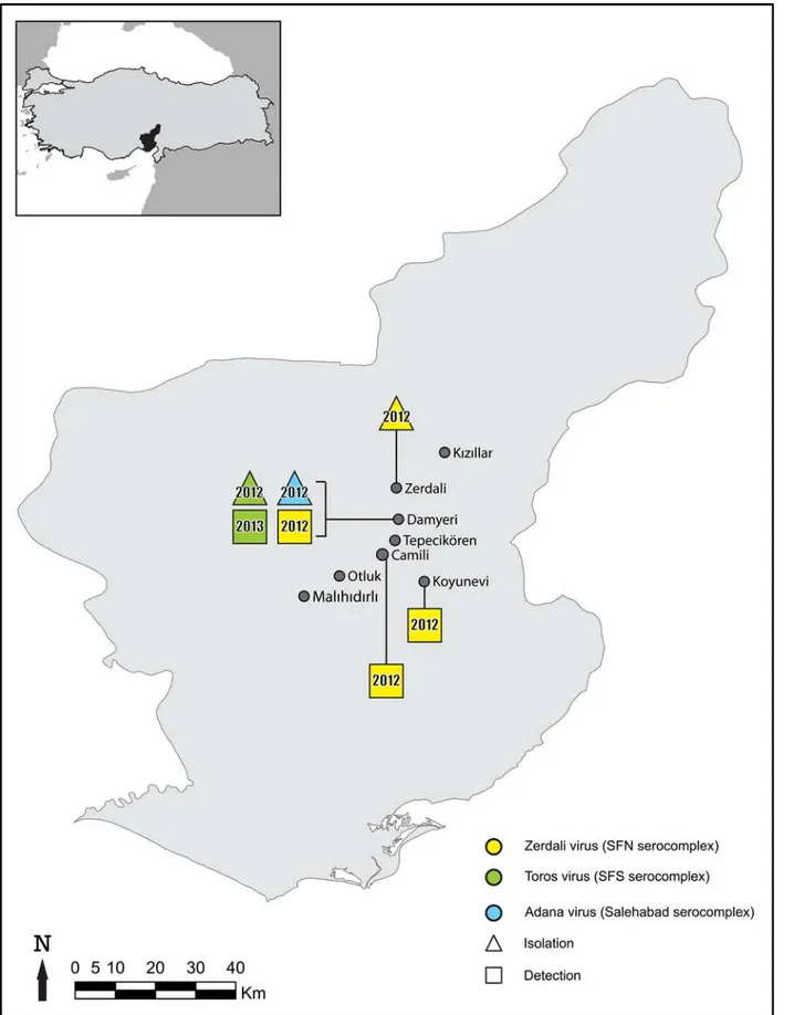
Fig 1. Geographic representation of the results.
Virus isolation
FiftyμL (derived from sandflies ground in the 600μL of EMEM as aforementioned) were
inoc-ulated onto a 12.5 cm2-flask of Vero cells, incubated at room temperature for 1 hr, and supple-mented with 3mL of EMEM (5% FBS, 1% Penicillin/Streptomycin, 1% L-Glutamine 200 mM, 1% Kanamycin, and 3% Fungizone). The flasks were incubated at 37°C in 5% CO2atmosphere
and examined daily for cytopathic effects (CPE).
Complete genome sequencing
For detailed characterisation, Zerdali virus (ZERV) passage 5, Toros virus (TORV) strain 292, passage 3, and TORV strain 213, passage 7 were subjected to complete genome characterisation using Next Generation Sequencing (NGS). Briefly, 140μL of infectious cell culture supernatant
medium was incubated with 30 U of Benzonase (Novagen 70664–3) for 7 hr at 37°C.This
mate-rial was then purified using the Viral RNA Mini Kit (Qiagen). Tagged random primers for reverse transcription (RT) and tag-specific and random-primers were used for PCR (Applied Biosystems). The resulting PCR products were purified (Amicon Ultra Centrifugal filters, Millipore); 200ng of DNA were used for sequencing using the Ion PGM Sequencer (Life Tech-nologies SAS, Saint Aubin, France). NGS reads, of 30 nucleotides minimum length, were trimmed using CLC Genomic Workbench 6.5, with a minimum of 99% quality per base and mapped to reference sequences. Parameters were set such that each accepted read had to map to the reference sequence for at least 50% of its length, with a minimum of 80% identity to the reference. From the contigs obtained, viral sequences were identified by best BLAST similarity against reference databases. Sequence gaps were completed by amplification and sequencing overlapping regions using either Sanger sequencing or NGS. The 5' and 3' extremities of each segment were sequenced using a primer including the 8-nt conserved sequence as previously described [25].
Complete genome sequencing was also performed for Corfou virus (CFUV) strain PaAr814 using the frozen cell culture supernatant medium following the methods above for comparison with the newly discovered TORV strains since they were shown to be closely related but only the complete S [26], the partial L [4] and M [27] genome sequences of the CFUV were known. Ultimately, all complete sequences obtained using NGS were verified by amplification and Sanger sequencing of overlapping regions spanning the entire genome.
Calculation of genetic distances and Phylogenetic Analyses
Complete sequences of each of the 5 genes (L, Gn, Gc, N, Ns) were aligned without indels together with homologous sequences of selected phleboviruses retrieved from the Genbank database using CLUSTAL within the MEGA 5 program [28]. Nucleotide (nt) and amino acid (AA) distances were calculated with the p-distance method. Neighbor-joining (p-distance model) and Maximum likelihood analyses were carried out with AA sequences using MEGA version 5, with 1000 bootstrap pseudoreplications.
Recombination analysis
Genotyping of sandflies in the virus-positive pools
To attempt identification of the sandfly species present in the TORV and ZERV positive pools, PCR was performed using 3-μL of nucleic acid extract of the pool to amplify the cytochrome c
oxidase I (COI) gene using the following primers; LCO1490: GGTCAACAAATCATAAAGA TATTGG and HCO2198: TAAACTTCAGGGTGACCAAAAAATCA [30]. The PCR products were processed and sequenced through NGS as described above. NGS reads were compared with available sequences in Genbank by Blastn using the CLC Genomic Workbench 6.5. For the final determination of the species the sequences were aligned with the reference sequences of regional populations of the sandfly species.
However, we would like to acknowledge that a valid protocol would be to cut off the male genitalia using a cold-stage microscope in the laboratory, so that the specimens can be identi-fied morphologically. This would be faster and cheaper than PCR amplification followed by NGS or Sanger sequencing of the COI gene.
Results
Sandfly trapping and virus detection
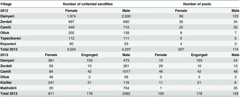
A total of 11,302 (4,513 females and 6,789 males) sandflies were collected in August and Sep-tember 2012 and 2013 from eight villages (Fig 1) located in the surroundings of Adana city (Mediterranean Turkey). They were organized as 797 pools (494 females, 303 males) (Table 1). Two pools, #213 and #292, were positive with primers N-phlebo2S/2R and N-phlebo1S/1R [24], respectively. The 245-nt sequence obtained from pool #213 was most closely related with CFUV (Genbank no: GQ165521; 95% AA identity, 78% nt identity). The 513-nt sequence cor-responding to pool #292 was also closely related with CFUV (Genbank no: GQ165521; 88% and 78% identity at the AA and nt level, respectively). These 2 pools consisted each of 20 male sandflies trapped in Damyeri village in 2012 (36S0733357 North and 4140570 East, altitude 194m).
Table 1. Distribution of the sandfly specimens and pools according to the sampling locations in Adana, Mediterranean region of Turkey in 2012 and 2013.
Village Number of collected sandflies Number of pools
2012 Female Male Female Male
Damyeri 1,974 2,500 99 123
Zerdali 697 692 35 34
Camili 449 712 22 35
Otluk 202 139 9 7
Tepecikoren 112 111 5 5
Koyunevi 90 53 4 3
Total 2012 3,524 4,207 207 174
2013 Female Engorged Male Female Engorged Male
Damyeri 361 103 473 19 103 24
Zerdali 59 10 261 29 10 13
Camili 84 42 1017 46 42 48
Otluk 46 2 26 3 2 3
Kizillar 241 21 116 11 21 6
Malihidirli 20 - 704 1 - 35
Total 2013 811 178 2582 109 178 129
The pool #37 (20 males trapped in Zerdali village in 2013; 36S732947 North and 4142749 East, altitude 238m) was positive using primers SFNV-S1/S2 [23]. The corresponding 390-nt sequence was most closely related to THEV (Genbank no: JF939848; 96% AA identity, 85% nt identity).
Sandfly rates of infection
The TORV specific rt-RT-PCR confirmed that pools #213 and #292 were positive (Ct values<26). Pool #10 (20 females collected in Damyeri in 2013) was also positive for TORV. Four-fold dilutions of the RNA were positive until the dilution 1:256 for the pools #213, #292, and #10.
The ZERV specific rt-RT-PCR confirmed pool #37 was positive (Ct value = 26.33), and detected ZERV RNA in 3 additional pools (#128–20 females, #342–20 males, and #374–29
females). Four-fold dilutions of the pool #37 RNA on the one hand and of pools #128, #342 and #374 on the other were positive until dilutions 1:4,096 and 1:1,1024, respectively. Pools#128, #342 and #374 consisted of sandflies trapped in 2012 in the respective villages of Damyeri, Camili and Koyunevi (Fig 1).
The rate of infection for TORV was 0.026%, for ZERV 0.035%, and for both TORV and ZERV 0.062% assuming that only one sandfly was infected in each pool.
Virus isolation
Vero cells inoculated with pool #292 showed a clear CPE at day 6 post-inoculation (pi). Pool #213-inoculated Vero cells did not produce CPE during 4 serial passages. However, virus repli-cation was demonstrated by RT-PCR (N-phlebo1 system, 24) starting from passage 3. CPE appeared at day 4 pi at passages 4 and 5 and virus replication was confirmed by RT-PCR. In a similar manner, pool #37- inoculated Vero cells provided a clear CPE at day 4 pi of passage 3 (RT-PCR was positive at passage 2). Neither virus isolation nor positive RT-PCR was obtained after 5 serial passages for pools #10, #128, #342, and #374.
Freeze-dried suspensions of ZERV-strain #37 (passage 8), TORV-strain #292 (passage 5) and TORV-strain #213 (passage 8) have been included in the collection of the European Virus Archive (www.european-virus-archive.com/) where they are publicly available for academic research at non-profit costs.
Complete genome sequencing
The complete genomes of both strains (#213 and #292) of the TORV consisted of 6,456 nts, 4,326 nts and 1,702 nts for the L, M and S segment, respectively (Genbank acc. no of the strain 213; KP966619, KP966620, andKP966621; Genbank acc. no of the strain 292;KP966622, KP966623, and KP966624). The polymerase gene encoded a 6,270-nt long ORF (2,090 AA), whereas the glycoprotein gene encoded a 4,077-nt long ORF (1,359AA). The small segment encoded a 738-nt and a 780-nt long ORF which when translated corresponded to the nucleo-capsid protein (246 AA) and a non-structural protein (260 AA), respectively.
The complete genome of the ZERV (strain #37) consisted of 6,403 nts, 4,202 nts and 1,907nts for the L, M and S segment, respectively (Genbank acc. KP966616, KP966617, and KP966618). The polymerase gene encoded a 6,285-nt long ORF (2,095 AA), whereas the glyco-protein gene encoded a 4,002-nt long ORFs (1,334). The small segment encoded a 942-nt and a 762-nt long ORF which were translated to a nucleocapsid protein (314AA) and a non-struc-tural protein (254AA), respectively.
polymerase gene encoded a 6270-nt long ORF (2,090AA), whereas the glycoprotein gene encoded a 4,077-nt long ORFs (1,359 AA). The small segment encoded a 738-nt and a 780-nt long ORF which were translated to a nucleocapsid protein (246AA) and a non-structural pro-tein (260AA), respectively.
Genetic distances
Pairwise distances of the nt- and AA- sequences are presented inS1 Table. The alignment of each gene is also available inS2 Table.
AA distances between TORV and SFSV-like viruses (SFSV, SFTV, SFCV, CFUV) were 25.2% (N),37.8% (NS),43.3% (M),40.7% (Gn),33.7% (Gc) and20.6% (L), whereas AA distances between TORV and other phleboviruses were much higher:42.9% (N), 71.4% (NS),58.7% (M),53.3% (Gn),47.4% (Gc) and43.9% (L).
AA pairwise distances between ZERV and viruses of the SFNV species (TOSV, THEV, SFNV, PUNV, MASV, GRAV) were17.3% (N),58.0% (NS),42.7% (M),41.8% (Gn), 29.5% (Gc) and17.4% (L), whereas AA distances between ZERV and other phleboviruses were much higher:40.6% (N),80.9% (NS),66.3% (M),64.8% (Gn),55.0% (Gc) and 44.5% (L).
Gene by gene comparative analysis showed that distances observed between ZERV and viruses belonging to the SFNV species were consistently lower than the lowest distances observed between ZERV and non SFNV-phleboviruses. The same relationship was observed with distances between TORV and SFSV-like viruses on the one hand, and TORV and non-SFSV-like viruses on the other.
These findings are supportive for (i) the inclusion of TORV in the SFSV species complex (SFSV, SFTV, SFCV, CFUV), (ii) the inclusion of ZERV in the SFNV species complex (TOSV, THEV, SFNV, PUNV, MASV, GRAV).
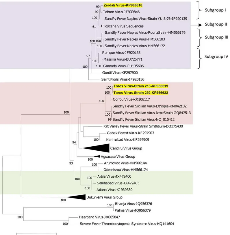
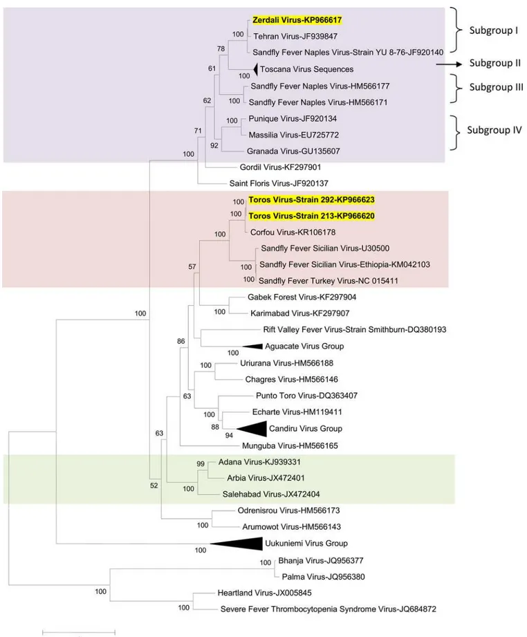
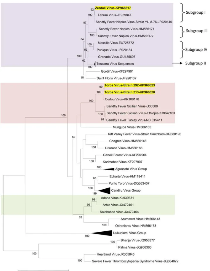
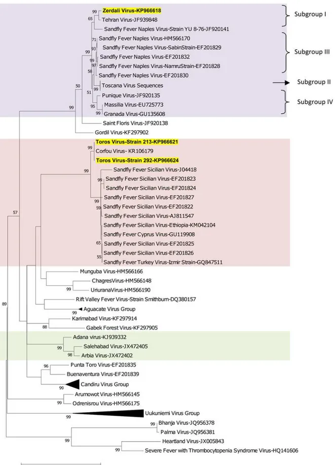
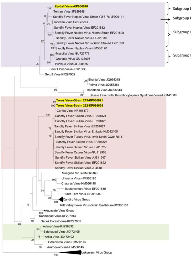
Phylogenetic analysis
Regardless of the gene used for phylogenetic analysis, and the tree-building programme (i.e. NJ or ML) TORV clustered with SFSV, CFUV and the other SFS-like viruses (from Turkey, Cyprus, Ethiopia) with bootstrap values99% (Figs2,3,4,5and6). TORV consistently grouped together with CFUV (99%bootstrap) forming a subgroup within the SFS-like viruses that is distinct from the second subgroup including SFSV and related genotypes origi-nating from Italy, Turkey, Cyprus, and Ethiopia. The stability of the topology and relationships between TORV and most closely related viruses suggested that the TORV genome did not con-tain evidence of genetic recombination or reassortment. Likewise, no recombination events were detected using any of the 6 algorithms implemented in RDP4.
ZERV consistently grouped together with THEV and Naples virus strain Yu_8–76
(sub-group I), with bootstrap values at99 for L, Gn and Gc, and with lower values for N and Ns. Within this species, there were 3 other subgroups: (i) subgroup II: Toscana viruses; (ii) sub-group III: Naples viruses (except for Yu_8–76 included in subgroup I); (iii): subgroup IV:
Gra-nada, Massilia and Punique viruses.
originating from neighbouring localities (Fig 1); this suggests that there may be topotypes that remain to be identified and characterised.
Fig 2. The Maximum likelihood phylogenetic analysis of the phlebovirus polymerase sequences.The Genbank accession numbers of all the phleboviruses included in the analysis can be found inS2 Table.
Fig 3. The Maximum likelihood phylogenetic analysis of the phlebovirus glycoprotein n (Gn) sequences.The Genbank accession numbers of all the phleboviruses included in the analysis can be found inS2 Table.
Fig 5. The Maximum likelihood phylogenetic analysis of the phlebovirus nucleocapsid protein sequences.The Genbank accession numbers of all the phleboviruses included in the analysis can be found inS2 Table.
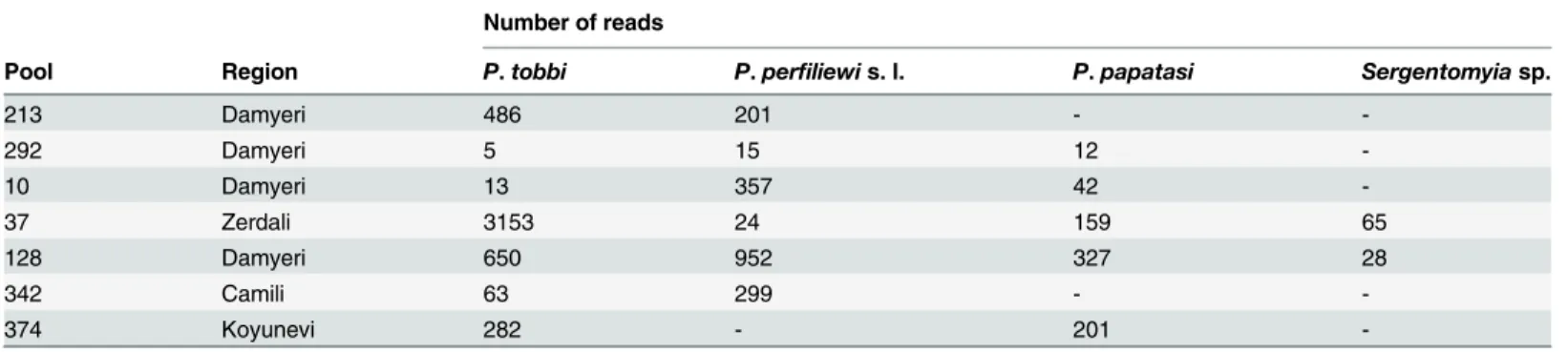
Genotyping the sandflies of virus-positive pools
Genotyping was performed for 7 pools. The species composition of the pools and number of reads are shown inTable 2. NGS reads were compared with available sequences in Genbank (Genbank accession numbers: KT634318, KF483675, KR349298, JQ769142, KF137560, KJ481126) by Blastn using the CLC Genomic Workbench 6.5. The species were determined when the consensus sequences had98% similarity with the regional reference sequences exceptSergentomyia sp. sequences which had85% similarity withS.dentatafrom Adana (Genbank accession numbers: KU659595, KU659596, KU659597, KU659598; release date 01 July 2016). Therefore we indicated these sequences asSergentomyia sp.
Discussion
Although there are published serological data [13] and a recent report of detection of TOSV [19] in the Adana, Mediterranean region of Turkey there have been no previous reports of virus isolation. To further understand the presence of sandfly-borne phleboviruses that circu-late in this endemic region for leishmaniasis [31], close to the border of Syria where many refu-gee camps are settled, we organised field-study campaigns in 2012 and 2013. The TORV and ZERV that were isolated during our study were most closely related to but distinct from SFS-and SFNV- like viruses, respectively. Studies conducted in 2012 led to the isolation of ADAV a novel putative member of theSalehabadspecies [20]. These results demonstrate that 3 phlebo-viruses belonging to 3 different genetic lineages co-circulate in the population of sandflies in this geographic area. Cumulative data resulting from this study and that of [20] enabled esti-mation of infection rates in sandflies (0.07% for sandfly-borne viruses in this region of Turkey) which is in the same order of magnitude as previously reported in France, Tunisia, Spain and Italy [2,23,32,33,34].
Regardless of the gene used for analysis TORV and CFUV were grouped together in a subli-neage that is clearly distinct from that including all other SFS-like viruses. CFUV was isolated fromPhlebotomus majorsensu lato [35] trapped in the eponymous Greek island. Interestingly, TORV has been isolated from two pools that containedP.perfiliewisensu lato andP.tobbi, both belonging to theLarroussiussubgenus asP.majorsensu lato. Similarly, SFTV was detected inP.majorsensu lato [21]. In contrast, other SFSV strains were isolated fromP. papa-tasithat belongs to thePhebotomussubgenus which is distinct from theLarroussiussubgenus [36]. Therefore, the two subgroups of viruses might reflect vector properties, with CFUV/ TORV associated with theLarroussiussubgenus whereas other SFSV viruses are associated withP.papatasiwith the exception of SFTV association withP.majorsensu lato. Vector-virus association needs to be studied in a more detailed manner in order (i) to determine
Table 2. Genotyping of sandflies in the virus positive pools.
Number of reads
Pool Region P.tobbi P.perfiliewis. l. P.papatasi Sergentomyiasp.
213 Damyeri 486 201 -
-292 Damyeri 5 15 12
-10 Damyeri 13 357 42
-37 Zerdali 3153 24 159 65
128 Damyeri 650 952 327 28
342 Camili 63 299 -
-374 Koyunevi 282 - 201
unambiguously the sandfly species transmitting these newly described viruses, (ii) and to verify our hypothesis that virus subgroups within viral species might be linked to specific vectors belonging to distinct taxonomic entities.
Additional field studies combined with experimental studies using sandfly colonies need to be initiated to understand the parameters driving vector capacity and competence for different strains of viruses.
ZERV was consistently grouped together with THEV and Naples virus strain YU-8-76. Interestingly, THEV and the Serbian isolate Yu 8/76 apparently do not require expression of the NSs ORF, since their replication is not impaired by the presence of either an early stop codon or a large truncation [37], whereas there is no such impairment or truncation in the ZERV genome which has a complete NSs ORF. Similar observations were also reported for a naturally attenuated RVFV strain (clone 13) that has a large in-frame deletion in the NSs cod-ing region [37,38].
Within the SFNV species, it is possible to discriminate 4 sublineages (I to IV); we propose to assign ZERV to sublineage I, where it was most closely related with THEV (Figs2,3,4,5and6). THEV was isolated fromP.papatasisandflies in Iran in 1959 [39], whereas YU-8-76 strain of SFNV was isolated fromP.perfiliewisensu lato trapped in Serbia in 1976 [37]. Subgroup III appears to be associated withP.papatasi, whereas subgroups II and IV appear to be associated with vectors belonging to the subgenusLarroussius. Subgroup I may be associated with Larrous-siusexcept THEV isolated inP.papatasi. OnlyP.tobbiwas found to be present in all of the four ZERV positive pools. The same comment formulated above concerning the need for experimen-tal studies to understand species-related competence and specificity of sandflies applies here.
Genetic and phylogenetic analyses support the fact that both ZERV and TORV should be considered as new strains within pre-existing SFNV species and the yet to be recognised species including SFSV and CFUV, respectively.
To date, SFSV and CFUV are listed as tentative species by the ICTV [1]. This study, based on complete genome sequences, suggests that all these viruses should be considered as mem-bers of the same species which could be further subdivided into CFUV / TORV, and SFSV / SFS-like viruses. A written proposal will be submitted to theBunyaviridaeStudy Group of the ICTV.
During this two-year study, 48% of the sandflies were trapped from Damyeri village where the ecological conditions were no different from those observed in other sampling stations. However, in Damyeri, the number of domestic animals (sheep, goats, and cows) was much higher than in other localities and these animals were constantly in the close vicinity of houses producing droppings which are known to be favoured breeding sites for sandflies or we set the traps very close to the possible breeding sites by chance therefore we got higher numbers of sand flies for this village. Thus, human exposure to sandflies might be greater in Damyeri than other sampling stations.
against CFUV / SFSV were reported in humans living in mainland Greece and on Corfu Island using the immunofluorescence assay (IFA) [44]. Viral RNA of Chios virus, closely related to CFUV, was detected in the CSF of a patient presenting with severe encephalitis (Papa and Pav-lidou, 2003, Genbank no, AY293623). In contrast, specific antibodies were never described in humans for THEV [12], although SFNV a close relative of THEV and subsequently ZERV was undoubtedly the cause of explosive outbreaks in newcomers to endemic areas during summer-time [9]. Importantly, although serological studies have not yet been reported we do have sero-logical data to support the concept that the newly isolated ZERV and TORV can infect vertebrates. However, whether or not these vertebrates do play a reservoir or amplifying role is not yet clear. Interestingly, although TORV and ZERV genomic RNA was detected in female pools of sandflies, both viruses were only isolated from male pools. Based on current knowl-edge it is not known how male sandflies become infected. Whilst, transovarial transmission seems a likely possibility it is not yet known how significant or efficient this mechanism of transmission is in natural habitats. However, laboratory experiments have shown that the rates of infection amongst offspring are low and show a decline from the first generation to ongoing generations. Other studies suggest that venereal (horizontal) transmission from infected males to uninfected females by mating and transstadial transmission of TOSV in diapausing Phlebo-tomus perniciosuslarvae [9] may also contribute to long-term virus survival. From what is cur-rently known and in the absence of defined vertebrate reservoirs, maintenance and
transmission of sandfly-borne phleboviruses appears to depend on the abundance and accessi-bility of appropriate vector species. This lack of available knowledge of virus transmission and the virus maintenance mechanisms clearly need to be investigated both in natural habitats and under experimental conditions. Future studies are planned to examine female sandfly salivary glands and heads to look for the presence of infectious virus.
Recent studies have detected sequences compatible with the existence of many putative new phleboviruses transmitted by sandflies in the Old World. In most of the cases, they were only partial sequences in the polymerase or the nucleoprotein genes. To date these limited genetic data, preclude classification by ICTV. This situation applies for viruses that may belong to (i) the SFNV species such as FERV [2], Provincia virus [48], Girne1 virus [18], and Saddaguia virus [45], (ii) the SALV species such as Adria virus [46,47] Olbia virus [48] and Edirne virus [18], and to (iii) the SFSV / CFUV complex such as Chios virus (Papa and Pavlidou, 2003, Gen-bank no, AY293623), Utique virus [4], Girne2 virus [18], SFS-like viruses [4,42,43,49]. Accordingly, although our knowledge of sandfly-borne phleboviruses is more extensive than it was a half-decade ago; efforts to isolate virus strains and determine their complete sequence should continue. Since virus taxonomy for thePhlebovirusgenus still relies on neutralisation-based antigenic relationships, virus isolation is also essential. Nevertheless, the criteria for tax-onomy appear to be evolving towards full-length genome comparative analysis.
Supporting Information
S1 Table. Amino acid and nucleotide pairwise distances between phleboviruses including the Zerdali virus and Toros virus.
(XLSX)
S2 Table. The alignments of L, Gn, Gc, N, and Ns genes of the phleboviruses including the Zerdali virus and Toros virus.
(MAS)
Acknowledgments
We would like to thank Asli Belen Saglam and Mehmet Karakus for their contributions during the sandfly collection campaigns, Géraldine Piorkowski for excellent assistance in the next gen-eration sequencing. We are very grateful to Prof. Ernest Gould for proofreading and editing the final manuscript.
Author Contributions
Conceived and designed the experiments: CA BA XdL RNC OEK. Performed the experiments: CA. Analyzed the data: CA BA XdL RNC. Contributed reagents/materials/analysis tools: CA BA XdL RNC. Wrote the paper: CA BA XdL RNC OEK.
References
1. Plyusnin A, Beaty BJ., Elliott RM, Goldbach R, Kormelink R, Lundkvist A, Schmaljohn CS, and Tesh RB. 2011. Bunyaviridae. In Virus taxonomy: classification and nomenclature of viruses. Ninth Report of the International Committee on Taxonomy of Viruses. San Diego: Elsevier. pp. 693–709.
2. Remoli ME, Fortuna C, Marchi A, Bucci P, Argentini C, Bongiorno G, Maroli M, Gradoni L, Gramiccia M, Ciufolini MG. 2014. Viral isolates of a novel putative phlebovirus in the Marche Region of Italy. Am J Trop Med Hyg. 90(4):760–3. doi:10.4269/ajtmh.13-0457PMID:24534812
3. Collao X, Palacios G, de Ory F, Sanbonmatsu S, Pérez-Ruiz M, Navarro JM, Molina R, Hutchison SK, Lipkin WI, Tenorio A, Sánchez-Seco MP. 2010. Granada virus: a natural phlebovirus reassortant of the sandfly fever Naples serocomplex with low seroprevalence in humans. Am J Trop Med Hyg. 83 (4):760–5. doi:10.4269/ajtmh.2010.09-0697PMID:20889862
4. Zhioua E, Moureau G, Chelbi I, Ninove L, Bichaud L, Derbali M, Champs M, Cherni S, Salez N, Cook S, de Lamballerie X, Charrel RN. 2010. Punique virus, a novel phlebovirus, related to sandfly fever Naples virus, isolated from sandflies collected in Tunisia. J Gen Virol. 91(Pt 5):1275–83. doi:10.1099/vir.0. 019240–0PMID:20089800
5. Wang J, Selleck P, Yu M, Ha W, Rootes C, Gales R, Wise T, Crameri, Chen H, Broz I, Hyatt A, Woods R, Meehan B, McCullough S, Wang LF. 2014. Novel phlebovirus with zoonotic potential isolated from ticks, Australia. Emerg Infect Dis 20:1040–1043. doi:10.3201/eid2006.140003PMID:24856477
6. McMullan LK, Folk SM, Kelly AJ, MacNeil A, Goldsmith CS, Metcalfe MG, Batten BC, Albariño CG, Zaki SR, Rollin PE, Nicholson WL, Nichol ST. 2012. A new phlebovirus associated with severe febrile illness in Missouri. N Engl J Med. 30; 367(9):834–41. doi:10.1056/NEJMoa1203378PMID:22931317
7. Mourya DT, Yadav PD, Basu A, Shete A, Patil DY, Zawar D, Majumdar TD, Kokate P, Sarkale P, Raut CG, Jadhav SM. 2014. Malsoor virus, a novel bat phlebovirus, is closely related to severe fever with thrombocytopenia syndrome virus and heartland virus. J Virol. 88(6):3605–9. doi: 10.1128/JVI.02617-13PMID:24390329
8. Zhao G, Krishnamurthy S, Cai Z, Popov VL, Travassos da Rosa AP, Guzman H, Cao S, Virgin HW, Tesh RB, Wang D. 2013. Identification of novel viruses using VirusHunter-an automated data analysis pipeline. PLoS One. 22; 8(10):e78470.
9. Alkan C, Bichaud L, de Lamballerie X, Alten B, Gould EA, Charrel RN. 2013. Sandfly-borne phlebo-viruses of Eurasia and Africa: epidemiology, genetic diversity, geographic range, control measures. Antiviral Res. 100(1):54–74. doi:10.1016/j.antiviral.2013.07.005Epub 2013 Jul 19. PMID:23872312
10. Charrel R.N., Gallian P., Navarro-Mari J.M., Nicoletti L., Papa A., Sanchez-Seco M.P, Tenorio A., de Lamballerie X., 2005. Emergence of Toscana virus in Europe. Emerg Infect Dis 11, 1657–1663. PMID:
11. Serter D. 1980. Present status of arbovirussero-epidemiology in the Aegean region of Turkey. In: Vesenjak-Hirjan J, Porterfield JS, Arslanagic E. Arboviruses in the Mediterranean Countries. Stuttgart, New York: Gustav Fisher Verlag, 155–161.
12. Tesh RB, Saidi S, Gajdamovic SJ, Rodhain F, Vesenjak-Hirjan J. 1976. Serological studies on the epi-demiology of sandfly fever in the Old World. Bull. World Health Organ. 54: 663–674. PMID:829416
13. Carhan A, Uyar Y, Ozkaya E, Ertek M, Dobler G, Dilcher M, Wang Y, Spiegel M, Hufert F, Weidmann M. 2010. Characterization of a sandfly fever Sicilian virus isolated during a sandfly fever epidemic in Turkey. J. Clin. Virol. 48: 264–269. doi:10.1016/j.jcv.2010.05.011PMID:20579934
14. Guler S, Guler E, Caglayik DY, Kokoglu OF, Ucmak H, Bayrakdar F, Uyar Y. 2012. A sandfly fever virus outbreak in the East Mediterranean region of Turkey. Int. J. Infect. Dis. 16: 244–246.
15. Torun Edis C., YagciCaglayik D., Uyar Y., Korukluoglu G., Ertek M., 2010. [Sandfly fever outbreak in a province at Central Anatolia, Turkey]. MikrobiyolBul 44, 431–439.
16. Ergunay K, Saygan MB, Aydogan S, Lo MM, Weidmann M, Dilcher M, Sener B, Hasçelik G, Pınar A,
Us D. 2011. Sandfly fever virus activity in central/northern Anatolia, Turkey: first report of Toscana virus infections. ClinMicrobiol Infect. 17(4):575–81. doi:10.1111/j.1469-0691.2010.03346
17. Ergunay K, Aydogan S, IlhamiOzcebe O, Cilek EE, Hacioglu S, Karakaya J, Ozkul A, Us D. 2012. Toscana virus (TOSV) exposure is confirmed in blood donors from Central, North and South/Southeast Anatolia, Turkey. Zoonoses Public Health 59, 148–154. doi:10.1111/j.1863-2378.2011.01436.x
PMID:21914151
18. Ergunay K, ErisozKasap O, Orsten S, Oter K, Gunay F, Akkutay AZ, Dincer E, Alten B, Ozkul A, 2014. PhlebovirusandLeishmaniadetection in sandflies from eastern Thrace and northern Cyprus. Parasit Vectors. 12; 7(1):575.
19. Dincer E, Gargari S, Ozkul A, Ergunay K. 2015. Potential Animal Reservoirs of Toscana Virus and Coinfections withLeishmania infantumin Turkey. Am J Trop Med Hyg. pii: 14–0322. [Epub ahead of print].
20. Alkan C, Alwassouf S, Piorkowski G, Bichaud L, Tezcan S, Dincer E, Ergunay K, Ozbel Y, Alten B, Lamballerie X, a RN. 2015. Isolation, genetic characterization and seroprevalence of Adana virus a novel phlebovirus belonging to the Salehabad virus complex in Turkey. J Virol. pii: JVI.03027-14. 89 (8):4080–91. doi:10.1128/JVI.03027-14Epub 2015 Feb 4. PMID:25653443
21. Ergunay K, ErisozKasap O, KocakTufan Z, Turan MH, Ozkul A, Alten B. 2012. Molecular evidence indi-cates thatPhlebotomus majorsensu lato (Diptera: Psychodidae) is the vector species of the recently-identified sandfly fever Sicilian virus variant: sandfly fever turkey virus. Vector Borne Zoonotic Dis. 12: 690–698. doi:10.1089/vbz.2011.0927PMID:22651385
22. Charrel RN, Moureau G, Temmam S, Izri A, Marty P, Parola P, da Rosa AT, Tesh RB, de Lamballerie X. 2009. Massilia virus, a novel Phlebovirus (Bunyaviridae) isolated from sandflies in the Mediterra-nean. Vector Borne Zoonotic Dis. 9: 519–530. doi:10.1089/vbz.2008.0131PMID:19055373
23. Charrel RN, Izri A, Temmam S, Delaunay P, Toga I, Dumon H, Marty P, de Lamballerie X &Parola P. 2007. Cocirculation of 2 genotypes of Toscana virus, southeastern France. Emerg. Infect. Dis. 13: 465–468. PMID:17552102
24. Sánchez-Seco M P, Echevarria JM, Hermndez L, Estévez D, Navarro-Mari JM &Tenorio A. 2003. Detection and identification of Toscana and other phleboviruses by RT-nested-PCR assays with degenerated primers. J. Med. Virol. 71: 140–149. PMID:12858420
25. Palacios G, Savji N, Travassos da Rosa A, Desai A, Sanchez-Seco MP, Guzman H, Lipkin WI, Tesh R. Characterization of the Salehabad virus species complex of the genus Phlebovirus (Bunyaviridae). 2013. J. Gen. Virol. 94(Pt 4):837–42. doi:10.1099/vir.0.048850–0PMID:23239568
26. Xu F, Chen H, Travassos da Rosa AP, Tesh RB, Xiao SY. 2007. Phylogenetic relationships among sandfly fever group viruses (Phlebovirus: Bunyaviridae) based on the small genome segment. J Gen Virol.; 88(Pt 8):2312–9. PMID:17622637
27. Liu DY, Tesh RB, Travassos Da Rosa AP, Peters CJ, Yang Z, Guzman H, Xiao SY. 2003. Phylogenetic relationships among members of the genus Phlebovirus (Bunyaviridae) based on partial M segment sequence analyses. J Gen Virol.; 84(Pt 2):465–73. PMID:12560581
28. Tamura K, Peterson D, Peterson N, Stecher G, Nei M, Kumar S. MEGA5: Molecular Evolutionary Genetics Analysis using maximum likelihood, evolutionary distance, and maximum parsimony meth-ods. MolBiolEvol. 2011; 28:2731–9 Epub 2011 May 4. doi:10.1093/molbev/msr121
29. Martin D, Williamson C, Posada D. 2005. RDP2: recombination detection and analysis from sequence alignments. Bioinformatics 21: 260–262. PMID:15377507
31. Svobodova M, Alten B, Zidkova L, Dvorak V, Hlavackova J, Myskova J, Seblova V, ErisozKasap O, Belen A, Votypka J, and Volf P. 2009. CL caused byLeishmania infantumand transmitted by Phleboto-mus tobbi. Int. J. Parasitol. 39: 251–256.
32. Bichaud L, Dachraoui K, Piorkowski G, Chelbi I, Moureau G, Cherni S, De Lamballerie X, Sakhria S, Charrel RN, Zhioua E. 2013. Toscana virus isolated from sandflies, Tunisia. Emerg Infect Dis. 19 (2):322–4. PMID:23460990
33. Sanbonmatsu-Gámez S, Pérez-Ruiz M, Collao X, Sánchez-Seco MP, Morillas-Márquez F, de la Rosa-Fraile M, Navarro-Mari JM, Tenorio A. 2005. Toscana virus in Spain. Emerg Infect Dis. 11(11):1701–7. PMID:16318721
34. Verani P., Ciufolini M.G., Caciolli S., Renzi A., Nicoletti L., Sabatinelli G., Bartolozzi D., Volpi G., Ama-ducci L., Coluzzi M., et al., 1988. Ecology of viruses isolated from sand flies in Italy and characterized of a new Phlebovirus (Arabia virus). Am J Trop Med Hyg 38, 433–439. PMID:3128131
35. Rodhain F., Madulo-Leblond G., Hannoun C., Tesh R.B., 1985. Le virus Corfou. Un nouveau Phlebo-virus Phlebo-virus isole de Phlebotomes en Grece. Ann Insl Pasteur/Virol 126, 161–166.
36. Nitzulescu V. 1931. Essai de classification des phlébotomes. Ann. Parasitol. Hum. Comp. 9. 271–2 75. 37. Palacios G, Tesh RB, Savji N, Travassos da Rosa AP, Guzman H, Bussetti AV, Desai A, Ladner J,
Sanchez-Seco M, Lipkin WI. 2014. Characterization of the Sandfly fever Naples species complex and description of a new Karimabad species complex (genus Phlebovirus, family Bunyaviridae). J Gen Virol. doi:10.1099/vir.0.056614–0Epub 2013 Oct 4.
38. Muller R., Saluzzo J. F., Lopez N., Dreier T., Turell M., Smith J., Bouloy M. 1995. Characterization of clone 13, a naturally attenuated avirulent isolate of Rift Valley fever virus, which is altered in the small segment. Am J Trop Med Hyg 53, 405–411. PMID:7485695
39. Karabatsos N. 1985. International catalogue of arboviruses including certain other viruses of verte-brates. San Antionio, TX: American Society of Tropical Medicine and Hygiene.
40. Sabin A.B., 1951. Experimental studies on Phlebotomus (pappataci, sandfly) fever during World War II. Arch GesamteVirusforsch 4, 367–410.
41. Tesh R.B., Papaevangelou G., 1977. Effect of insecticide spraying for malaria control on the incidence of sandfly fever in Athens, Greece. Am J Trop Med Hyg 26, 163–166. PMID:190909
42. Konstantinou G.N., Papa A., Antoniadis A., 2007. Sandfly-fever outbreak in Cyprus: are phleboviruses still a health problem? Travel Med Infect Dis 5, 239–242. PMID:17574146
43. Papa A., Konstantinou G., Pavlidou V., Antoniadis A., 2006. Sandflyfever virus outbreak in Cyprus. ClinMicrobiolInfect 12, 192–194.
44. Antoniadis A., Alexiou-Daniel S., Malisiovas N., Doutsos I,. Polyzoni T., Leduc J.W., et al. 1990.Seroe-pidemiological survey for antibodies to arboviruses in Greece. Arch Virol (Suppl. 1: ): 277e85. 45. Fares W, Charrel RN, Dachraoui K, Bichaud L, Barhoumi W, Derbali M, Cherni S, Chelbi I, de
Lamball-erie X, Zhioua E. 2015. Acta Trop. Infection of sand flies collected from different bio-geographical areas of Tunisia with phleboviruses. 141(Pt A):1–6. doi:10.1016/j.actatropica.2014.09.009Epub 2014 Sep 22.
46. Anagnostou V, Pardalos G, Athanasiou-Metaxa M, Papa A., 2011. Novel phlebovirus in febrile child, Greece. Emerg. Infect. Dis. 17: 940–941. doi:10.3201/eid1705.101958PMID:21529422
47. Papa A., Velo E., Bino S., 2011. A novel phlebovirus in Albanian sandflies. ClinMicrobiolInfect 17, 585–587.
48. Peyrefitte CN, Grandadam M, Bessaud M, Andry PE, Fouque F, Caro V, Diancourt L, Schuffenecker I, Pagès F, Tolou H, Zeller H, Depaquit J. 2013. Diversity ofPhlebotomus perniciosusin Provence, southeastern France: Detection of two putative new phlebovirus sequences. Vector Borne Zoonotic Dis. 13(9):630–6. doi:10.1089/vbz.2012.1169Epub 2013 May 24. PMID:23705585