Submitted24 November 2014
Accepted 25 October 2016
Published22 November 2016
Corresponding author
Siouxsie Wiles, [email protected]
Academic editor
Paul Tulkens
Additional Information and Declarations can be found on page 8
DOI10.7717/peerj.2717
Copyright
2016 Dalton et al.
Distributed under
Creative Commons CC-BY 4.0
OPEN ACCESS
Effect of common and experimental
anti-tuberculosis treatments on
Mycobacterium
tuberculosis
growing as biofilms
James P. Dalton1,2,3, Benedict Uy1,2, Narisa Phummarin4, Brent R. Copp3,4, William A. Denny3,5, Simon Swift1and Siouxsie Wiles1,2,3
1Molecular Medicine and Pathology, University of Auckland, Auckland, New Zealand 2Bioluminescent Superbugs Lab, University of Auckland, Auckland, New Zealand 3Maurice Wilkins Centre for Molecular Biodiscovery, Auckland, New Zealand 4School of Chemical Sciences, University of Auckland, Auckland, New Zealand
5Auckland Cancer Society Research Centre, University of Auckland, Auckland, New Zealand
ABSTRACT
Much is known regarding the antibiotic susceptibility of planktonic cultures of
Mycobacterium tuberculosis, the bacterium responsible for the lung disease tuberculosis (TB). As planktonically-grownM. tuberculosisare unlikely to be entirely representative of the bacterium during infection, we set out to determine how effective a range of anti-mycobacterial treatments were against M. tuberculosis growing as a biofilm, a bacterial phenotype known to be more resistant to antibiotic treatment. Light levels from bioluminescently-labelledM. tuberculosisH37Rv (strain BSG001) were used as a surrogate for bacterial viability, and were monitored before and after one week of treatment. After treatment, biofilms were disrupted, washed and inoculated into fresh broth and plated onto solid media to rescue any surviving bacteria. We found that in this phenotypic stateM. tuberculosiswas resistant to the majority of the compounds tested. Minimum inhibitory concentrations (MICs) increased by 20-fold to greater than 1,000-fold, underlying the potential of this phenotype to cause significant problems during treatment.
SubjectsMicrobiology, Infectious Diseases
Keywords Mycobacterium tuberculosis, MTB, TB, Biofilm, Pellicle, Ascorbic acid, Nitroimidazole, Fluoroanthranilic acid, Tryptophan biosynthesis
INTRODUCTION
Bacterial cells present in an infected host can display a range of phenotypes and occupy several divergent physiological niches (Sendi et al., 2009;Tuchscherr et al., 2011). For example, during infection, cells ofM. tuberculosiscan be both replicative and non-replicative (Wayne & Sohaskey, 2001) and occupy a number of different niches, including macrophages (Rengarajan, Bloom & Rubin, 2005) and necrotic and non-necrotic lesions (Fenhalls et al., 2002).M. tuberculosisgrowing in such diverse environments is unlikely to be accurately reflected by planktonically-grown laboratory cultures. Many bacteria can form microcolonies called biofilms, which can contain a mixture of replicating and non-replicating cells, and cells in different metabolic states (Rice, Hamilton & Camper, 2003;Stewart & Franklin, 2008). Bacteria can form biofilms at the interface between a surface and the surrounding air or liquid. Alternatively, floating biofilms can form at a liquid/air interface. These floating biofilms are also known as pellicles. Within a biofilm or pellicle, bacterial cells are more resistant to disinfection and drug treatment and therefore represent a much harder target to sterilise (Kulka, Hatfull & Ojha, 2012;Ceri et al., 1999). As such, the biofilm/pellicle represents a useful model for investigating the efficacy of antibacterial treatments.
M. tuberculosiscan form pelliclesin vitro(Sambandan et al., 2013) and the presence of microcolonies of extracellular M. tuberculosisin animal models has led to speculation that these are biofilms formed in vivo (Lenaerts et al., 2007). Some sources point to the possible presence of pellicles in the lung-air interface present in secondary TB in humans; (Hunter et al., 2006;Hunter et al., 2013;Hunter et al., 2014) and indicate that the susceptibility of this phenotype to antibacterial compounds is of particular relevance from a treatment standpoint. Here we describe the use of bioluminescently-taggedM. tuberculosis
to investigate the susceptibility of pellicle cells to a range of anti-mycobacterial compounds, including those in current clinical use as well as a selection of experimental compounds.
MATERIALS AND METHODS
Strains and growth conditions
In this study we usedM. tuberculosisBSG001 (Wang et al., 2016), a stable bioluminescent derivative of H37Rv transformed with the integrating plasmid pMV306hspLuxABG13CDE (Andreu et al., 2010). Cultures of BSG001 were grown at 37 ◦C with gentle shaking (100 rpm) in Middlebrook 7H9 broth (Fort Richard, Auckland) supplemented with 10% Middlebrook ADC enrichment media (Fort Richard) and 0.5% glycerol (Sigma-Aldrich), or on 7H11 agar (Fort Richard) supplemented with 10% Middlebrook OADC enrichment media (Fort Richard) and 0.5% glycerol. We grew pellicles in sterile, black 96 well plates (Nunc) using a previously described method (Kulka, Hatfull & Ojha, 2012). Briefly, we grewM. tuberculosisBSG001 in liquid culture for 2 weeks at 37◦C and then adjusted the cultures to give an optical density at 600 nm (OD600) of 1.0, before diluting them 1:100
in modified Sauton’s media (0.5 g/L KH2PO4, 0.5 g/L MgSO4, 4 g/L L-Asparagine, 2 g/L
Citric acid, 0.05 g/L Ferric Ammonium Citrate, 60 mL/L glycerol, 0.1% ZnSO4, pH 7.0
Table 1 Minimum inhibitory and bactericidal concentrations of common and experimental anti-tuberculosis treatments againstMycobacterium tuberculosis.
Planktonic MICa Biofilm MICa Planktonic MBCb Biofilm MBCb
Pyrazinamide 50 mg/L >1,000 mg/L 50 mg/L >1,000 mg/L
Rifampicin 0.04 mg/L 4 mg/L 0.04 mg/L 4 mg/L
Isoniazid 0.04 mg/L >256 mg/L 0.08 mg/L >256 mg/L
Ethambutol 1 mg/L >2,000 mg/L 2 mg/L >2,000 mg/L
Streptomycin 0.5 mg/L 125 mg/L 0.5 mg/L 1,000 mg/L
Amikacin 4 mg/L 250 mg/L 4 mg/L 1,000 mg/L
Rifabutin 0.04 mg/L 8 mg/L 0.04 mg/L 16 mg/L
Ascorbic acid 700 mg/L 2,800 mg/L 700 mg/L 2,800 mg/L
Delamanid 0.042 mg/L >53.45 mg/L 0.042 mg/L >53.45 mg/L
Pretomanid 0.011 mg/L >3.6 mg/L 0.011 mg/L >3.6 mg/L
SN30488 0.0056 mg/L >4.2 mg/L 0.0056 mg/L >4.2 mg/L
QOA1 0.5 mg/L >128 mg/L 0.5 mg/L >128 mg/L
QOA2 0.25 mg/L >128 mg/L 0.25 mg/L >128 mg/L
QOA3 0.25 mg/L >128 mg/L 0.25 mg/L >128 mg/L
5-FAAc 9.7 mg/L 19.4 mg/L 19.4 mg/L 19.4 mg/L
6-FAAc 9.7 mg/L 19.4 mg/L 19.4 mg/L 19.4 mg/L
Notes.
aMinimum inhibitory concentrations (MIC).
bMinimum bactericidal concentrations (MBC) for biofilm and planktonic formsof M. tuberculosisBSG001 for a variety of
ex-perimental and non-exex-perimental compounds. cFluoroanthranilic acid.
well plate. We filled the outer wells with 200µL of sterile water to reduce evaporation from theM. tuberculosiscontaining wells. We incubated the cultures without shaking for 8 weeks at 37◦C.
Determination of compound activity
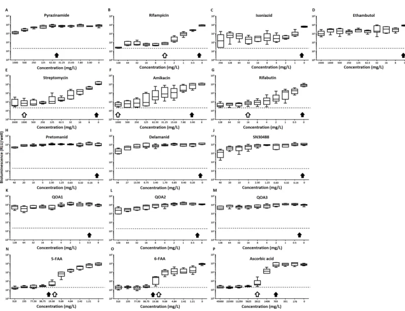
Figure 1 Chemical structures of the experimental compounds used in this study.
defined the MIC as causing a 1 log reduction in light production, as previously described (Andreu et al., 2012).
RESULTS
Decreased susceptibility of pellicle-grownM. tuberculosis to
front-line and experimental compounds
Of the four main first line drugs only rifampicin was seen to inhibit pellicles of
M. tuberculosis at the concentrations tested (Table 1,Figs. 2Cand3B). Isoniazid also led to some inhibition but below the threshold defined (Figs 2Cand3C). In the case of rifampicin the MIC and MBC for pellicle-grown BSG001 were determined to be 4 mg/L, 100 times the concentration required to produce a similar result with planktonic cells (Table 1). High levels of pyrazinamide, isoniazid and ethambutol (20, 6,000 and 1,000 times the MIC’s for planktonic cells, respectively) failed to sufficiently reduce light to be classed as inhibitory (Table 1,Figs. 2A–2Dand3A–3D). More success was observed with non-first line antibiotics, with MICs obtained for pellicle-grown BSG001 for streptomycin (125 mg/L), amikacin (250 mg/L) and rifabutin (8 mg/L) (Table 1,Figs. 2E–2Gand3E–3G). These pellicle-MICs represent an increase of 250, 62.5 and 200 times the MIC’s obtained for planktonic cultures, respectively (Table 1).
When novel and experimental compounds were examined, none of the current derivations of the nitroimidazole based compounds (Olaru et al., 2015;Palmer et al., 2010) (delamanid, pretomanid and SN30488) were able to reduce light from the pellicle-grown
M. tuberculosisat the concentrations tested (Table 1,Figs. 2H–2Jand3H–3J). The same resistance to drug-killing was seen with experimental compounds based on 2-(quinoline-4-yloxy) acetamides (Phummarin et al., 2016) (QOA 1, QOA 2 and QOA 3) (Table 1,
Figs. 2K–2Mand3K–3M). In contrast, the fluoroanthranilic-acid based compounds, 5-fluoroanthranilic acid (5-FAA) and 6-fluoroanthranilic acid (6-FAA), which target the tryptophan biosynthetic pathway (Toyn et al, 2000) were seen to be quite effective at inhibiting light fromM. tuberculosisBCG001 growing as a pellicles (Table 1,Figs. 2N/O
and3N/O). Ascorbic acid was also seen to cause inhibition at 2.8 g/L, 4 times the MIC for planktonically-grownM. tuberculosis(Table 1,Figs. 2Pand3P).
DISCUSSION
Figure 2 The effect of clinically-used and experimental compounds onM. tuberculosis BSG001 pellicles.The inhibitory effect of first line (A– D) and second line (E–G) anti-tuberculosis drugs used in the clinic and experimental compounds (H–P), including those based on nitroimidazole (H–J), 2-(quinoline-4-yloxy) acetamides (K–M) and fluoroanthranilic-acid (N, O), is presented as a reduction in bioluminescence plotted as relative light units (RLU) per well on day 7 of treatment. The dashed line indicates the limit of detection. The solid and open arrows indicate the MBC’s (the concentration which resulted in the recovery of no bacterial colonies) obtained for planktonically-grown cells and pellicles, respectively. All com-pounds were tested in three biological replicates on separate days with multiple technical replicates. Results are given as box whisker plots with the box representing values from the lower to upper quartile and the whiskers representing the range.
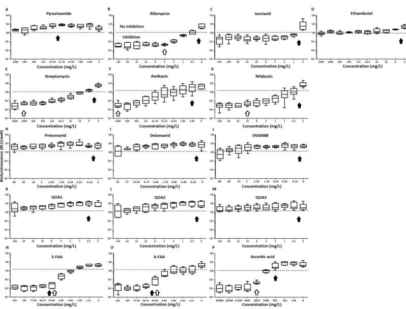
Figure 3 The relative effect of clinically-used and experimental compounds onM. tuberculosisBSG001 pellicles. The relative change in bio-luminescence (relative light units [RLU]) following the treatmentof M. tuberculosisBSG001 biofilms with first line (A–D) and second line (E–G) anti-tuberculosis drugs used in the clinic and experimental compounds (H–P), including those based on nitroimidazole (H–J), 2-(quinoline-4-yloxy) acetamides (K–M) and fluoroanthranilic-acid (N, O), is shown as the ratio of RLU before treatment and RLU after seven days of treatment. The dashed line indicates the level at which no change occurs; values above the dashed line indicate an increase in light levels (and hence survival/-growth) over the course of the experiment, while those below indicate a decrease (and hence inhibition/death). The solid and open arrows indicate the MBC’s (the concentration which resulted in the recovery of no bacterial colonies) obtained for planktonically-grown cells and pellicles, respec-tively. Results are given as box whisker plots with the box representing values from the lower to upper quartile and the whiskers representing the range.
compounds also showed little activity against this biofilm form. The mode of action of these compounds is likely due to electron transport inhibition of cytochrome bc1 oxidase (Phummarin et al., 2016). As the cells are actively metabolising, the lack of an effect from these compounds is most likely due to an inability to access the cells.
week of treatment. The activity of ascorbic acid is thought to be due to the generation of highly reactive hydroxyl radicals via presence of iron and Fenton reaction chemistry (Vilcheze et al., 2013). Killing due to this mechanism would be non-specific and not dependant on uptake. Interestingly the fluoroanthranilic acid tryptophan biosynthesis inhibitors were also seen to be effective at inhibiting and killing pellicles, indicating that this is a pathway worthy of further consideration for drug targeting.
It is possible that the comparative ease in which test compounds can access bacteria within a pellicle, that is from both above and below, as compared to a biofilm attached to a surface which cannot be accessed from the surface side, make this form of biofilm easier to kill. While it is still unknown ifM. tuberculosisforms biofilms/pelliclesin vivo, many mycobacterial species do form complex, secondary structures such as pelliclesin vitro(Ojha & Hatfull, 2012). Researchers have also reported histological evidence for the presence of multicellular structures involving M. tuberculosisoutside of the macrophage (Lenaerts et al., 2007). Others have reported the presence of cells that resembles biofilms/pellicles in the cavities formed during secondary tuberculosis which would indicate that this phenotype is likely to play a role in human disease (Lenaerts et al., 2007;Hunter et al., 2006;Hunter et al., 2014). The biphasic response ofM. tuberculosisinfections, in which a large kill is seen early on in drug treatment followed by a marked reduction in the bactericidal activity of therapeutic agents due to phenotypic rather than genetic resistance, could also be evidence that M. tuberculosis is able to form biofilms/pelliclesin vivo. Such structures could act as a reservoir for drug tolerant bacilli which are responsible for the increased duration of drug treatment required in cases of TB. Regardless, the
M. tuberculosis-pellicle model is a useful multi-phenotypic environment in which a novel compound can be tested against cells with a range of susceptibilities. The susceptibility of
M. tuberculosiswithin this model indicates that drugs which can attack the surface of the cell or can pass through the extracellular matrix of the pellicle represent the best option for treatment. We also saw that the inhibition of tryptophan biosynthesis could be utilised in TB treatment and their design should be further investigated.
ADDITIONAL INFORMATION AND DECLARATIONS
Funding
This work was supported by the Maurice Wilkins Centre for Molecular Biodiscovery and the University of Auckland Vice-Chancellor’s Strategic Development Fund and Faculty Research Development Fund. SW is supported by a Sir Charles Hercus Fellowship (09/099) from the Health Research Council of New Zealand. The funders had no role in study design, data collection and analysis, decision to publish, or preparation of the manuscript.
Grant Disclosures
The following grant information was disclosed by the authors: Maurice Wilkins Centre for Molecular Biodiscovery.
Faculty Research Development Fund. Sir Charles Hercus Fellowship: 09/099.
Competing Interests
Siouxsie Wiles is an Academic Editor for PeerJ.
Author Contributions
• James P. Dalton conceived and designed the experiments, performed the experiments,
analyzed the data, contributed reagents/materials/analysis tools, wrote the paper, prepared figures and/or tables, reviewed drafts of the paper.
• Benedict Uy performed the experiments, contributed reagents/materials/analysis tools,
reviewed drafts of the paper.
• Narisa Phummarin contributed reagents/materials/analysis tools.
• Brent R. Copp and William A. Denny contributed reagents/materials/analysis tools,
reviewed drafts of the paper.
• Simon Swift conceived and designed the experiments, analyzed the data, contributed
reagents/materials/analysis tools, reviewed drafts of the paper.
• Siouxsie Wiles conceived and designed the experiments, analyzed the data, contributed
reagents/materials/analysis tools, wrote the paper, prepared figures and/or tables, reviewed drafts of the paper.
Data Availability
The following information was supplied regarding data availability:
Wiles, Siouxsie; Dalton, James; Uy, Benedict; Copp, Brent; Denny, Bill (2016): Effect of common and experimental anti-tuberculosis treatments onMycobacterium tuberculosis
growing as biofilms. figshare:10.17608/k6.auckland.4097772.v1.
Supplemental Information
Supplemental information for this article can be found online athttp://dx.doi.org/10.7717/ peerj.2717#supplemental-information.
REFERENCES
Andreu N, Fletcher T, Krishnan N, Wiles S, Robertson BD. 2012.Rapid measurement
of antituberculosis drug activityin vitroand in macrophages using bioluminescence.
Journal of Antimicrobial Chemotherapy67:404–414DOI 10.1093/jac/dkr472.
Andreu N, Zelmer A, Fletcher T, Elkington PT, Ward TH, Ripoll J, Parish T, Bancroft
GJ, Schaible U, Robertson BD, Wiles S. 2010.Optimisation of bioluminescent
reporters for use with mycobacteria.PLoS ONE5:e10777
DOI 10.1371/journal.pone.0010777.
Ceri H, Olson ME, Stremick C, Read RR, Morck D, Buret A. 1999.The Calgary biofilm
device: new technology for rapid determination of antibiotic susceptibilities of bacterial biofilms.Journal of Clinical Microbiology37:1771–1776.
Donlan RM, Costerton JW. 2002.Biofilms: survival mechanisms of clinically relevant
Fenhalls G, Stevens L, Moses L, Bezuidenhout J, Betts JC, Helden PV, Lukey PT,
Duncan K. 2002.In situdetection ofMycobacterium tuberculosistranscripts in
human lung granulomas reveals differential gene expression in necrotic lesions.
Infection and Immunity70:6330–6338DOI 10.1128/IAI.70.11.6330-6338.2002.
Hunter RL, Actor JK, Hwang SA, Karev V, Jagannath C. 2014.Pathogenesis of post
primary tuberculosis: immunity and hypersensitivity in the development of cavities.
Annals of Clinical and Laboratory Science44:365–387.
Hunter RL, Armitige L, Jagannath C, Actor JK. 2013.TB Research at UT-Houston; a
review of cord factor: new approaches to drugs, vaccines and the pathogenesis of tuberculosis.Tuberculosis89:S18–S25.
Hunter RL, Olsen MR, Jagannath C, Actor JK. 2006.Multiple roles of cord factor in the
pathogenesis of primary, secondary, and cavitary tuberculosis, including a revised description of the pathology of secondary disease.Annals of Clinical and Laboratory Science36:371–386.
Klopper M, Warren RM, Hayes C, Gey van Pittius NC, Streicher EM, Müller B, Sirgel FA, Chabula-Nxiweni M, Hoosain E, Coetzee G, David van Helden P, Victor TC,
Trollip AP. 2013.Emergence and spread of extensively and totally drug-resistant
tuberculosis, South Africa.Emerging Infectious Diseases19:449–455
DOI 10.3201/eid1903.120246.
Kulka K, Hatfull G, Ojha AK. 2012.Growth ofMycobacterium tuberculosisbiofilms.
Journal of Visualized Experiments: JoVE60:3820DOI 10.3791/3820.
Lenaerts AJ, Hoff D, Aly S, Ehlers S, Andries K, Cantarero L, Orme IM, Basaraba RJ.
2007.Location of persisting mycobacteria in a Guinea pig model of tuberculosis
revealed by r207910.Antimicrobial Agents and Chemotherapy 51:3338–3345
DOI 10.1128/AAC.00276-07.
Ojha AK, Hatfull GF. 2012. Biofilms ofMycobacterium tuberculosis: new perspectives of
an old pathogen. In: Cardona P-J, ed.Understanding tuberculosis—deciphering the secret life of the bacilli. Rijeka: InTech.
Olaru ID, Von Groote-Bidlingmaier F, Heyckendorf J, Yew WW, Lange C, Chang
KC. 2015.Novel drugs against tuberculosis: a clinician’s perspective.European
Respiratory Journal45:1119–1131DOI 10.1183/09031936.00162314.
Palmer BD, Thompson AM, Sutherland HS, Blaser A, Kmentova I, Franzblau
SG, Wan B, Wang Y, Ma Z, Denny WA. 2010.Synthesis and structure–activity
studies of biphenyl analogues of the tuberculosis drug
(6S)-2-nitro-6-{[4-(trifluoromethoxy)benzyl]oxy}-6,7-dihydro-5H-imidazo[2,1-b][1, 3]oxazine (PA-824).Journal of Medicinal Chemistry 53:282–294DOI 10.1021/jm901207n.
Phummarin N, Boshoff HI, Tsang PS, Dalton J, Wiles S, Barry 3rd CE, Copp BR.
2016.SAR and identification of 2-(quinolin-4-yloxy)acetamides asMycobacterium
Rengarajan J, Bloom BR, Rubin EJ. 2005.Genome-wide requirements for Mycobac-terium tuberculosisadaptation and survival in macrophages.Proceedings of the National Academy of Sciences of the United States of America102:8327–8332
DOI 10.1073/pnas.0503272102.
Rice AR, Hamilton MA, Camper AK. 2003.Movement replication and emigration rates
of individual bacteria in a biofilm.Microbial Ecology45:163–172
DOI 10.1007/s00248-002-1028-x.
Safadi RA, Abu-Ali GS, Sloup RE, Rudrik JT, Waters CM, Eaton KA, Manning SD.
2012.Correlation betweenin vivobiofilm formation and virulence gene expression
inEscherichia coliO104:H4.PLoS ONE7:e41628
DOI 10.1371/journal.pone.0041628.
Sambandan D, Dao DN, Weinrick BC, Vilchéze C, Gurcha SS, Ojha A, Kremer L,
Besra GS, Hatfull GF, Jacobs Jr WR. 2013.Keto-mycolic acid-dependent pellicle
formation confers tolerance to drug-sensitiveMycobacterium tuberculosis.MBio
4:e00222–e00213.
Sendi P, Johansson L, Dahesh S, Van Sorge NM, Darenberg J, Norgren M, Sjölin
J, Nizet V, Norrby-Teglund A. 2009.Bacterial phenotype variants in group B
streptococcal toxic shock syndrome.Emerging Infectious Diseases15:223–232
DOI 10.3201/eid1502.080990.
Stewart PS, Franklin MJ. 2008.Physiological heterogeneity in biofilms.Nature Reviews
Microbiology6:199–210DOI 10.1038/nrmicro1838.
Toyn JH, Gunyuzlu PL, White WH, Thompson LA, Hollis GH. 2000.A counterselection
for the tryptophan pathway in yeast: 5-fluoroanthranilic acid resistance.Yeast
16:553–550
DOI 10.1002/(SICI)1097-0061(200004)16:6<553::AID-YEA554>3.0.CO;2-7.
Tuchscherr L, Medina E, Hussain M, Völker W, Heitmann V, Niemann S, Holzinger
D, Roth J, Proctor RA, Becker K, Peters G, Löffler B. 2011.Staphylococcus
aureus phenotype switching: an effective bacterial strategy to escape host immune response and establish a chronic infection.EMBO Molecular Medicine3:129–141
DOI 10.1002/emmm.201000115.
Vilcheze C, Hartman T, Weinrick B, Jacobs Jr WR. 2013.Mycobacterium tuberculosisis
extraordinarily sensitive to killing by a vitamin C-induced Fenton reaction.Nature Communications4:Article 1881DOI 10.1038/ncomms2898.
Wand ME, Bock LJ, Turton JF, Nugent PG, Sutton JM. 2012.Acinetobacter baumannii
virulence is enhanced in Galleria mellonella following biofilm adaptation.Journal of Medical Microbiology 61:470–477DOI 10.1099/jmm.0.037523-0.
Wang J, Pearce AN, Chan ST, Taylor RB, Page MJ, Valentin A, Bourguet-Kondracki
ML, Dalton JP, Wiles S, Copp BR. 2016.Biologically active acetylenic amino alcohol
and n-hydroxylated 1,2,3,4-tetrahydro-beta-carboline constituents of the New Zealand ascidian pseudodistoma opacum.Journal of Natural Products79:607–610.
Wayne LG, Sohaskey CD. 2001.Nonreplicating persistence ofmycobacterium
World Health Organization. 2011.Guidelines for the programmatic management of drug-resistant tuberculosis: 2011 update. World Health Organization, Geneva.
Available athttps:// www.ncbi.nlm.nih.gov/ books/ NBK148644/.
World Health Organization. 2015.Global tuberculosis report 2015. World Health