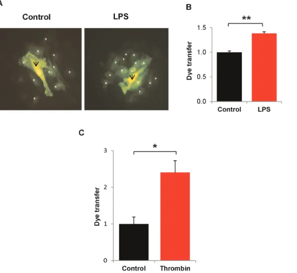
Gap junction protein connexin43 exacerbates lung vascular permeability.
Texto
Imagem
Documentos relacionados
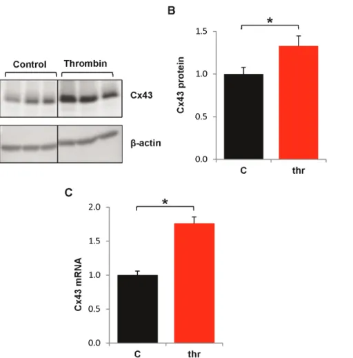
Since prior studies had shown variations in gap junction expression and intercellular communication in endothelial cells depend- ing on cell growth and density (11,12), we studied
We examined the downregulation of survivin mRNA and protein levels in neuroblastoma SH-SY5Y cells after stable survivin siRNA or control siRNA transfection.. RT- PCR and Western
Meanwhile the first World Ocean Assessment Report (WOAI), recently presented and approved by the General Assembly of the United Nations, highlight concern about the impact of human
Extravascular lung water index (EVLWI) and pulmonary vascular permeability index (PVPI) were determined using pulse contour cardiac output (PiCCO) technology, and the oxygenation
A questão da reforma do Conselho de Segurança foi, igualmente, aludida com maior incidência nos oito anos de administração Cardoso, conquanto o tema não tenha
The fact that mRNA and protein patterns of the gap genes Kr , kni , and gt are similar (yet not identical), and show a delay in dynamics with regard to each other, raises
Given that mice deficient in Apoe show vascular permeability, decreased CBF, synapse loss, and cognitive impairments [ 37 , 75 , 76 ], a decrease in Apoe expression in the aging
Although monocytes and macrophages are the predominant HIV-1 infected cells in CNS tissue, studies of brain sections of acquired immune deficiency syndrome (AIDS)
