Rsp5 Ubiquitin Ligase Is Required for Protein Trafficking
in
Saccharomyces cerevisiae
COPI Mutants
Katarzyna Jarmoszewicz1, Katarzyna Łukasiak1, Howard Riezman2, Joanna Kaminska1*
1Department of Genetics, Institute of Biochemistry and Biophysics, Polish Academy of Sciences, Warsaw, Poland,2Department of Biochemistry, University of Geneva, Geneva, Switzerland
Abstract
Retrograde trafficking from the Golgi to the endoplasmic reticulum (ER) depends on the formation of vesicles coated with the multiprotein complex COPI. InSaccharomyces cerevisiaeubiquitinated derivatives of several COPI subunits have been identified. The importance of this modification of COPI proteins is unknown. With the exception of the Sec27 protein (b’COP) neither the ubiquitin ligase responsible for ubiquitination of COPI subunits nor the importance of this modification are known. Here we find that the ubiquitin ligase mutation,rsp5-1, has a negative effect that is additive withret1-1and sec28Dmutations, in genes encodinga- ande-COP, respectively. The doubleret1-1 rsp5-1mutant is also more severely defective in the Golgi-to-ER trafficking compared to the singleret1-1, secreting more of the ER chaperone Kar2p, localizing Rer1p mostly to the vacuole, and increasing sensitivity to neomycin. Overexpression of ubiquitin inret1-1 rsp5-1mutant suppresses vacuolar accumulation of Rer1p. We found that the effect ofrsp5mutation on the Golgi-to-ER trafficking is similar to that ofsla1Dmutation in a gene encoding actin cytoskeleton proteins, an Rsp5p substrate. Additionally, Rsp5 and Sla1 proteins were found by co-immunoprecipitation in a complex containing COPI subunits. Together, our results show that Rsp5 ligase plays a role in regulating retrograde Golgi-to-ER trafficking.
Citation:Jarmoszewicz K, Łukasiak K, Riezman H, Kaminska J (2012) Rsp5 Ubiquitin Ligase Is Required for Protein Trafficking inSaccharomyces cerevisiaeCOPI Mutants. PLoS ONE 7(6): e39582. doi:10.1371/journal.pone.0039582
Editor:Michael Polymenis, Texas A&M University, United States of America
ReceivedNovember 9, 2011;AcceptedMay 24, 2012;PublishedJune 26, 2012
Copyright:ß2012 Jarmoszewicz et al. This is an open-access article distributed under the terms of the Creative Commons Attribution License, which permits unrestricted use, distribution, and reproduction in any medium, provided the original author and source are credited.
Funding:This work was supported by the Ministry of Science and Higher Education of Poland grant N303 101 32/3456 (J.K., K.J. and K.Ł.) and by the Swiss National Science Foundation (H.R.). The funders had no role in study design, data collection and analysis, decision to publish, or preparation of the manuscript.
Competing Interests:The authors have declared that no competing interests exist.
* E-mail: [email protected]
Introduction
The trafficking of proteins between membrane-delimited organelles is mediated by vesicles which form on one membrane and fuse with another. Vesicle formation is mediated by coat proteins that form a lattice on the vesicle surface. One such coat is COPI composed of the Arf1 GTPase and two subcomplexes: F-COPI (b, c, d, and f subunits) and B-COPI (a, b’, e) [1]. Individual COPI components interact with cargo proteins through specific signal sequences located in their cytosolic sequences and target them to appropriate transport vesicles. The best described signal sequence is C-terminal K(X)KXX (di-lysine motif) which interacts with subunits of the B-COPI subcomplex; the coatomer isolated from thesec27-1(b’-COP) orret1-1(a-COP) yeast mutants fails to bind this signal in vitro [1,2]. In vitro cross-linking experiments have also identifiedc-COP, a subunit of the F-COPI subcomplex, as the binding partner for the di-lysine motif [1] and c-COP was also found to bind the cytosolic protein Cdc42 (Rho-related GTPase) [3].
Other proteins, e.g., ER transmembrane proteins, use the receptor protein Rer1 for packing into COPI vesicles. Rer1p interacts with subunits of the COPI coat through its cytoplasmic signals. One of these signals is similar to the di-lysine motif and the other is a tyrosine signal motif [4]. Soluble cargo proteins like the ER chaperone Kar2p, which are unable to interact with the coat, have to use receptors for efficient incorporation into vesicles [1].
In yeast COPI-coated vesicles mediate the retrograde transport from the Golgi apparatus to the endoplasmic reticulum (ER).
There is some evidence suggesting an additional function for a subset of COPI subunits in post-Golgi trafficking steps. It has been found in yeast that endocytic cargo, the uracil permease Fur4p or theafactor receptor Ste2p, accumulates on endosomes in some COPI mutants [5]. Also, the transport of biosynthetic cargo, carboxypeptidase S (CPS), is partially blocked in these COPI mutants. Additionally, some COPI mutants are impaired in the recycling of Snc1p, a v-SNARE (vesicle membrane soluble N -ethylmaleimide-sensitive factor attachment protein receptor), from endosomes to the Golgi [6].
Interactions of the coat with various proteins may regulate coat specificity on different membranes. This specificity is achieved by posttranslational modifications of coat subunits, e.g., phosphory-lation or ubiquitination. A screen for membrane-associated ubiquitinated proteins has identified two of the seven coatomer subunits– a-COP and b’-COP (encoded in yeast by RET1 and SEC27, respectively) [7]. Other studies also found COPI components, such as Ret3p, Sec21p, Sec26p, Sec27p and Sec28p to be ubiquitinated [8]. However the importance of COPI subunit modification with ubiquitin is not well documented.
called multiubiquitination. Proteins are also modified with poly-ubiquitin chains in which subsequent poly-ubiquitin molecules are linked C-terminally to a lysine residue in the preceding ubiquitin. When lysine 48 (K48) of ubiquitin is the site of the linkage, such poly-ubiquitination marks the protein for degradation by 26S proteasome [12]. However, poly-ubiquitin chains can also be formed through other lysines present in ubiquitin, K6, K11, K27, K29, K33 or K63, resulting in various conformations of the ubiquitin chains and as a consequence a range of molecular signals [13]. Ubiquitination is reversible - ubiquitins can be removed by specific deubiquitinating enzymes - ubiquitin proteases (Ubps) and ubiquitin C-terminal hydrolases (Uch) [14].
In the yeastSacharomyces cerevisiaethe ubiquitin ligase Rsp5p has been shown to tag proteins with monoubiquitin or with chains formed through K63 [15]. The Rsp5-dependent modification is important for several processes including inheritance of mito-chondria, chromatin remodelling, and activation of transcription factors. The role of Rsp5 ligase in the endocytosis of several plasma membrane transporters, channels and permeases and intracellular trafficking of proteins has also been documented thoroughly [16]. Rsp5p participates also in the sorting of permeases like Fur4p or the general amino acid permease, Gap1p, at Golgi apparatus and in the sorting of several cargoes in multivesicular bodies (MVB) [16]. This action of Rsp5p at several distinct locations is believed to be achieved by interactions with different adaptor proteins. These adaptors are also required for ubiquitination of those Rsp5p substrates that lack motifs for Rsp5p binding. Such adaptors have been described for endocytic cargoes and for the sorting at the Golgi. Rsp5p can also affect intracellular transport by influence on actin cytoskeleton organization. Rsp5p has several substrates among actin-cytoskeleton proteins. The described in vivo and in vitrosubstrates for Rsp5 are Sla1, Lsb1, Lsb2 - proteins that bind to Las17 (an activator of Arp2/3 complex required for actin polimerization), Rvs167 - a protein required for viability upon starvation and Sla2 [17]. In the case of Sla1 protein Rsp5-dependent ubiquitination causes its processing [18] but the physiological role of ubiquitination of most of actin cytoskeleton proteins is unknown.
Genetic and biochemical evidence indicates that the deubiqui-tinating enzyme Ubp2p antagonizes Rsp5p activity [19]. In contrast, a lack of Ubp3p activity (ubp3Dmutation) seems to have an additive negative effect on the growth of anrsp5 mutant – a double ubp3D rsp5 mutant shows synthetic growth defect [20]. Moreover, Rsp5p cooperates with Ubp3p in the regulation of ribophagy, a specific type of autophagy responsible for degrada-tion of ribosomes [20]. Recently Rsp5p was shown in vitro to ubiquitinate Sec23p, a subunit of COPII coat [21] and Ubp3p is responsible for Sec23p deubiquitination [22]. Ubp3p and its cofactor Bre5p were also shown to be responsible for deubiqui-tination of Sec27p (b’COP). Modulation of Sec27p ubiquitination status has a regulatory role. Only after Ubp3-catalyzed deubiqui-tination is Sec27p able efficiently to bind cargo containing the di-lysine motif [22].
Here we asked if ubiquitin ligase Rsp5p, together with the Ubp3p-Bre5p complex, regulates Golgi-to-ER retrograde trafficking. We found that a lack of the Bre5p cofactor (bre5D) combined with the rsp5-1mutation has an additive negative effect on yeast growth and on the trafficking of GFP-Rer1 marker fusion protein. We also show that Rsp5p is necessary when COPI function is impaired due toret1-1 mutation in the gene encoding itsasubunit. Thersp5-1mutation combined with ret1-1 compromises Golgi-to-ER trafficking as evidenced by vacuolar localization of the Rer1p receptor and secretion of Kar2p (an ER chaperone). Overexpression of ubiquitin was able to suppress the vacuolar accumulation of GFP-Rer1 in the
ret1-1 rsp5-1mutant. However, ubiquitinated forms of Sec27p were still found in thersp5-1mutant suggesting the action of another ubiquitin ligase(s). Further genetic studies suggest that Rsp5p influences Golgi-to-ER trafficking by regulating the actin cytoskel-eton. Taken together, our results show a new role of Rsp5p ligase together with Bre5p and actin cytoskeleton in the regulation of trafficking from the Golgi to the ER.
Results
bre5Dandrsp5Mutations have Additive Effect on
Trafficking from Golgi to ER
It has been reported that the double rsp5 ubp3D mutant is inviable at a temperature permissive for the singlersp5andubp3D mutants and thersp5andubp3Dmutations have an additive effect on ribophagy [20]. Additionally, Ubp3p, together with its cofactor Bre5p, is involved in deubiquitination of Sec23p, a subunit of COPII coat. Recently it was shown that Sec23p is ubiquitinated by Rsp5p [21]. The singleubp3Dandbre5Dmutants were shown to accumulate ubiquitinated Sec27p, a b’-COPI subunit [22]. Therefore, we asked if Rsp5p also acts with the Ubp3p-Bre5p complex in retrograde Golgi-to-ER trafficking. First we tested the genetic interaction between rsp5-1 mutation, which carries an amino acid substitution in the catalytic Hect domain, and deletion of theBRE5 gene (bre5D) in adoa4Dbackground (see below for explanation). A comparison of growth of strains doa4D bre5D, doa4DHA-rsp5-1anddoa4Dbre5DHA-rsp5-1on YPD at different temperatures revealed that the doa4D bre5D HA-rsp5-1 mutant grew worse at 35uC than did thedoa4Dbre5Dordoa4DHA-rsp5-1 mutants, indicating a very weak genetic interaction betweenbre5D andrsp5-1(not shown). The same genetic interaction was visible when all above strains were transformed with a plasmid bearing theDOA4gene (Figure 1A). This interaction is in agreement with data from a genetic interaction map [23].
Next we asked if genetic interaction betweenbre5Dandrsp5-1was reflected in compromised Golgi-to-ER trafficking. To probe the latter we used GFP-tagged Rer1p. Rer1p is a Golgi membrane protein that is required for the retrieval of escaped ER membrane proteins back from the Golgi [4]. Vacuolar accumulation of Rer1p is characteristic for mutants defective in the retrograde trafficking from Golgi to ER [4]. As shown in Figure 1B, indoa4Dcells GFP-Rer1p exhibits a Golgi pattern of fluorescence typical for wild type yeast. In about 10% of the doa4Dbre5D cells the GFP-Rer1 signal overlapped with vacuole staining by the vacuolar marker, blue CMAC, suggesting vacuolar accumulation of Rer1. This confirms that the bre5D mutant is defective in the Golgi-to-ER retrograde trafficking. In contrast, the doa4DHA-rsp5-1mutant showed localization of GFP-Rer1 similar to that ofdoa4Dcells. In about 18% of thedoa4Dbre5DHA-rsp5-1mutant cells the GFP-Rer1 fusion protein was found in vacuoles. Additional punctuate and diffuse GFP fluorescence was also observed in those cells (Figure 1B). This suggests that the defect in retrograde trafficking in thedoa4Dbre5DHA-rsp5-1mutant is slightly stronger than indoa4D bre5D. The more prevalent diffuse distribution of GFP-Rer1 in the triple mutant indicates that the delivery of GFP-Rer1 to the vacuole at trans-Golgi or endosomes may also be partially defective.
The above data show that Rsp5p ligase acts together with Bre5p in retrograde Golgi-to-ER trafficking.
Thersp5-1Mutation Shows Genetic Interaction with ret1-1andsec28D
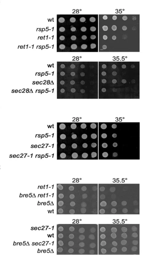
rsp5-1 6 sec27-1, and rsp5-1 6 sec28D was tested and the phenotypes of the respective double mutants were scored. As shown in Figure 2A, the doubleret1-1 rsp5-1mutant stops growing at 35uC, a temperature permissive for the singleret1-1andrsp5-1 mutants. This genetic interaction was not-allele specific. Otherrsp5 alleles such asrsp5-w1,rsp5-w2,rsp5-w3[24] andrsp5-19[25] with mutations in the WW domains responsible for Rsp5p interaction with other proteins, also had additive negative effects on ret1-1 growth (not shown). A negative genetic interaction was also observed between sec28D and rsp5-1. The double sec28D rsp5-1 mutant failed to grow at 35.5uC (Figure 2A). Interestingly, the sec27-1 rsp5-1mutant grew as well as did the singlesec27-1mutant at all temperatures tested (Figure 2B). Thus, variousrsp5mutations show a genetic interaction with mutated alleles of some genes encoding COPI subunits.
A negative genetic interaction between deletion of theBRE5 gene and ret1-1 or sec27-1 mutations was previously described [26,27]. However, we observed only an interaction betweenbre5D and ret1-1 and no such interaction between bre5D and sec27-1 (Figure 2C). Thus, thersp5-1and bre5Dmutations displayed the same genetic interaction spectrum. This fact together with the additive effects ofrsp5-1andbre5Don retrograde trafficking shows that both ubiquitination and deubiquitination processes are important for COPI-dependent trafficking.
Defect in Golgi-to-ER Trafficking of Cargo Proteins in ret1-1 rsp5-ret1-1Mutant
Rsp5 participates in the sorting of several cargoes in multive-sicular bodies (MVB). It is also known that in some COPI mutants the transport of a biosynthetic cargo sorted via the MVB, carboxypeptidase S (CPS), is partially blocked at a post-Golgi step [5]. So we first investigated if the observed genetic interaction betweenrsp5-1and ret1-1 could be due to an additive effect on cargo sorting in endosomes affecting MVB formation. To test this possibility the trafficking of the vacuolar carboxypeptidase Y (CPY) was monitored. In mutants defective in MVB formation CPY is released to the extracellular space. Thesec27-1mutant was characterized as secreting more CPY compared to wild type cells [5]. The level of CPY secretion by spore clones from theret1-16 rsp5-1andsec27-16rsp5-1crosses was monitored, but neither of the double mutants showed enhanced secretion of CPY compared with the single ret1-1, sec27-1 or rsp5-1 mutants (Figure 3A). Another type of cargo known to require Rsp5p for proper segregation into the lumen of MVB is Sna3p. Sna3p binds to Rsp5p and its sorting into MVB is affected by rsp5-1mutation [28]. The localization of Sna3-GFP and the level of free GFP released from Sna3-GFP in vacuole lumen were monitored in spore clones from theret1-16rsp5-1cross. As shown in Figure 3B in the wild type,ret1-1,rsp5-1andret1-1 rsp5-1strains Sna3-GFP Figure 1.rsp5andbre5mutations have additive effect on growth and mislocalization of GFP-Rer1.(A)bre5DandHA-rsp5-1mutations show weak genetic interaction. Strainsdoa4D, doa4Dbre5D, doa4DHA-rsp5-1anddoa4Dbre5DHA-rsp5-1were transformed with plasmid encoding
DOA4. Serial 1:10 dilutions of transformants were spotted on YPD and incubated at indicated temperatures. (B)doa4Dbre5DHA-rsp5-1mutant accumulates GFP-Rer1 in vacuole. Plasmid encoding GFP-Rer1 fusion was transformed into same mutants as in panel A. Transformants were grown on SC -ura at 28uC and GFP-Rer1 was observed by fluorescence (GFP). Cells were stained with CMAC to visualize vacuole. Percentage of cells accumulating GFP in vacuole is given.
doi:10.1371/journal.pone.0039582.g001
Figure 2. Genetic interaction betweenrsp5-1orbre5Dand mutations in genes encoding COPI subunits.(A) Negative genetic interaction betweenrsp5-1andret1-1orsec28Dmutations. (B) No genetic interaction betweenrsp5-1andsec27-1mutation. (C) Negative genetic interaction betweenbre5Dandret1-1and no genetic interaction betweenbre5Dandsec27-1. Serial 1:10 dilutions of spore clones from crossesret1-16rsp5-1 (RH30426FW1808),sec28D6rsp5-1(KJK396FW1808),sec27-16rsp5-1(RH359-7D6FW1808),bre5D6ret1-1(JK140-5A6JK82-4B) andbre5D6
was present in the vacuole. However, inrsp5-1and ret1-1 rsp5-1 more intact Sna3-GFP was present (39% and 36% respectively) than inret1-1and wild type cells (13.5% and 19%) as assessed by western blotting with anti-GFP antibody (Figure 3C). After a shift to 35uC for 2 hours, a temperature restrictive for theret1-1 rsp5-1 mutant, more Sna3-GFP was present in cytoplasmic foci inrsp5-1 andret1-1 rsp5-1mutants. Western blotting analysis revealed that in these strains, respectively, 77% and 57% of total GFP signal comes from the intact Sna3-GFP fusion (Figure 3C). This result shows that addition of theret1-1mutation torsp5-1does not cause a stronger defect in Sna3-GFP transport. From these experiments we conclude that the observed genetic interaction between the ret1-1 and rsp5-1 mutations is probably not due to an additive negative effect on the sorting into the MVB. Rsp5p ligase is apparently necessary when COPI is defective but at a trafficking step other than MVB sorting. Recently the ubiquitination of Sec23p, a subunit of COPII coat has been shown to be mediated by Rsp5p [21]. Thus, the genetic interaction betweenret1-1and rsp5-1could be a result of their additive effect on the anterograde transport between ER and Golgi. Alternatively, Rsp5 could regulate Golgi-to-ER transport. The first possibility was ruled out. We tested the activity of extracellular invertase. As shown in Figure 3D neither singleret1-1 orrsp5-1mutant nor the double ret1-1 rsp5-1had lowered activity of extracellular invertase at 30uC nor after a shift to 35uC, a temperature restrictive for doubleret1-1 rsp5-1mutant.
The possibility that Rsp5p influences Golgi-to-ER trafficking was verified by monitoring the transport of three different types of cargo in the rsp5-1mutant. To study the trafficking of proteins containing the di-lysine motif we tested sensitivity to neomycin, an aminoglycoside antibiotic which competes with the di-lysine motif for coatomer binding [29]. We assumed that when the interaction of the cargo with COPI is impaired, but not abolished, due to the ret1-1mutation, addition of neomycin should cause an additional defect in retrograde trafficking and thus should be deleterious to the ret1-1 mutant. Indeed, as shown in Figure 4A, the ret1-1 mutant is more sensitive to neomycin than the wild type. We also tested if the rsp5-1 mutation further intensified the neomycin sensitivity caused by theret1-1mutation. We found that thersp5-1 mutation had an additive effect to that caused by the ret1-1 mutation. In contrast there was no additive effect on the growth on neomycin-containing media between sec27-1 and rsp5-1 (not shown). Because neomycin interferes with other processes in the cell besides affecting coatomer we cannot exclude other reasons of the observed effect of rsp5-1 on neomycin sensitivity. However, these results are at least in agreement with the idea that Rsp5p ligase is needed for proper di-lysine motif interaction with the COPI complex.
The Rer1-dependent retrograde trafficking was monitored by observing Rer1 localization. There was no defect in GFP-Rer1 localization in thersp5-1mutant even after a shift to 37uC (Figure 1B, 4B, 4C and not shown). Thus, the rsp5-1 mutation alone does not block retrograde trafficking. We then checked if
rsp5-1enhances the defect in retrograde trafficking caused by the ret1-1orsec27-1mutations. A vacuolar localization of GFP-Rer1 had already been described for the singleret1-1mutant [4]. In our strain this was visible only in a minor fraction of cells. In contrast, almost all double rsp5-1 ret1-1 mutant cells had the GFP-Rer1 fusion protein localized to vacuoles even at 28uC, a temperature permissive for grow of this mutant (Figure 4D). In principle, this enhanced vacuolar accumulation of GFP-Rer1 in theret1-1 rsp5-1 strain could be a consequence of thersp5-1 mutation or of the genetic background of the double mutant. To distinguish between these two possibilities and to quantify the effect of Rsp5p on the trafficking, a centromeric vector encoding HA-RSP5 was trans-formed intoret1-1 rsp5-1expressingGFP-RER1; as a control empty vector was used. Protein extracts were obtained from the transformants and analyzed by Western blotting with anti-GFP antibody. The ratio of free GFP, liberated from the GFP-Rer1 fusion in the vacuole, to that of total GFP (free GFP and GFP-Rer1) was calculated. We found that there is ca. 15% more free GFP in theret1-1 rsp5-1mutant compared toret1-1 (Figure 4D). So, thersp5-1mutation has an additive effect to theret1-1mutation on the trafficking of the Rer1p cargo receptor. Interestingly, the double mutantrsp5-1 sec27-1also accumulated GFP-Rer1 fusion in the vacuole (Figure 4C).
Next, we analyzed the trafficking of Kar2p. Kar2p is an ER-resident protein which, when misaddressed to the Golgi, interacts with its receptor Erd2p and returns to the ER in a COPI-dependent manner. Blocking the retrograde Golgi-to-ER traffick-ing causes Kar2p secretion to the medium. We found that the percentage of total Kar2p that is secreted is enhanced in the doubleret1-1 rsp5-1mutant compared with the single mutants and wild type cells (Figure 4E). No such effect was observed in the sec27-1 rsp5-1mutant (not shown). Thus, the effect of the double ret1-1 rsp5-1mutation on Kar2p secretion corroborates the genetic interaction of the two mutations described above.
To answer a question how Rsp5 regulates trafficking from the Golgi to ER the binding of Rsp5 to coatomer was tested. For this purpose thersp5D strain was transformed with empty vector or vector bearing taggedRSP5(HA-RSP5). When HA-tagged Rsp5 was immunoprecipitated from cells and analyzed by immunoblot, we observed the presence of COPI subunits together with HA-Rsp5. There were no COPI proteins in the control immunopre-cipitation from rsp5D cells transformed with empty vector (Figure 4F).
We conclude that Rsp5 is found in a complex together with the COPI coat and Rsp5p ligase is necessary for trafficking of different cargo types from the Golgi to the ER when this transport is defective due to mutations in genes encoding COPI subunits.
Sec27 Protein is Ubiquitinated inrsp5-1 Mutant
Finding that Rsp5 ligase and COPI proteins could be co-immunoprecipitated suggests that Rsp5 ligase might be responsible for ubiquitination of some COPI subunits. Sec27p had been found to be deubiquitinated by the Ubp3p-Bre5p enzyme, which Figure 3. CPY secretion and Sna3-GFP trafficking and invertase activity are the same inret1-1 rsp5-1mutant and in singlersp5-1.(A) Double mutantsret1-1 rsp5-1andsec27-1 rsp5-1do not secrete more CPY compared with the singleret1-1,sec27-1orrsp5-1mutants. Spore clones (as in Figure 2) were replica-plated onto nitrocellulose filters and grown on solid YPD for 1 day at 28uC. Cells secreting CPY were identified by Western blotting with anti-CPY antibody. (B) Sna3-GFP trafficking defect caused byrsp5-1mutation is not augmented byret1-1. Plasmid encoding Sna3-GFP was transformed into spore clones as in Figure 2A. Transformants were grown to mid logarithmic phase on SC -ura at 30uC or shifted to 35uC for 1 hour. Sna3-GFP was observed by fluorescence (GFP). Cells were stained with CMAC to visualize vacuole and viewed with Nomarski optics (NOM). (C) Whole cell lysates form transformants from Figure 3B were analyzed by Western blotting with anti-GFP antibody. Percentage of Sna3-GFP in total GFP signal (GFP and Sna3-GFP) is given. (D) Invertase activity was assayed in spore clones as in Figure 2A. Cultures were grown to mid logarithmic phase at 30uC and shifted or not to 35uC for 30 minutes. The proportion between activity of secreted invertase to the total invertase activity (invertase secretion index) is shown.
prompted the question whether the observed effect of the rsp5 mutation on retrograde trafficking could be a result of Rsp5-dependent ubiquitination of Sec27p. To test this we used four strains: doa4D, doa4D bre5D, doa4D HA-rsp5-1, and doa4D bre5D HA-rsp5-1. In these strains theDOA4gene, which encodes one of the deubiquitinating enzymes, was deleted. Thedoa4D mutation decreases the level of free ubiquitin, which can be suppressed by ectopically expressed tagged version of ubiquitin. This allows easier detection of proteins modified with ubiquitin. The bre5D mutation was introduced to prevent deubiquitination in order to facilitate detection of ubiquitinated Sec27. Strains were trans-formed with a plasmid expressing His-tagged ubiquitin from the CUP1promoter. Cells were grown to mid-exponential phase and half of the culture was shifted to 37uC for 4 hours. Next, protein extracts were prepared from cultures grown at both temperatures and His6-Ubi was pulled down on Ni-NTA beads. The total
protein extracts and the material bound to the beads were analyzed by Western blotting with anti-Sec27 antibody. As shown in Figure 5A, at 30uC in all strains tested Sec27p was present in the bound fraction. When extracts were made from cultures incubated at 37uC the Sec27p protein was also recovered in all of the strains but accumulation of slower-migrating forms of Sec27p was visible in doa4D HA-rsp5-1, and doa4D bre5D HA-rsp5-1 mutants (Figure 5A). This result can be interpreted in several ways: Sec27p is not ubiquitinated by Rsp5p, is ubiquitinated by Rsp5p and another ligase, or overexpression of ubiquitin suppresses the defect of rsp5-1 mutation. Indeed ubiquitin is a multicopy suppressor of the temperature sensitivity of thersp5-1mutant [30]. The accumulation of Sec27 protein in polyubiquitinated form, observed inrsp5-1mutant, might change COPI function and in consequence could cause the defect in the COPI trafficking which is additive to the defect caused byret1-1mutation. If this is true, overexpression ofSEC27should reduce the defect in the Golgi-to-ER trafficking in theret1-1 rsp5-1mutant. To validate this idea we tested if overexpression ofSEC27is able to attenuate accumulation of GFP-Rer1 in vacuole in theret1-1 rsp5-1mutant. The double ret1-1 rsp5-1 mutant was transformed with empty plasmid or plasmid overexpressing SEC27 (Figure 5B). Unexpectedly the overproduction of Sec27 caused fragmentation of vacuoles. The GFP-Rer1 was still in small vacuoles, but some bright punctuate structures were also visible (Figure 5B).
Overexpression of Ubiquitin Suppresses Some Defects of ret1-1 rsp5-1Mutant
To test if a lack of Rsp5-dependent ubiquitination is a reason for the observed interaction between ret1-1 and rsp5-1 we checked if overexpression of ubiquitin suppresses the growth defect at 35.5uC and GFP-Rer1 accumulation in the vacuole of the doubleret1-1 rsp5-1 mutant. Mutated variants of ubiquitin with only a single lysine, 48 (K48) or 63 (K63) present, were also tested. Theret1-1 rsp5-1mutant grew better at the nonpermissive temperature when transformed with
a multicopy plasmid encoding ubiquitin but even better when the K63 variant was used (Figure 6A). Overexpression of native ubiquitin changed the GFP-Rer1 localization from vacuoles to numerous punctate structures (Figure 6B), but expression of the only K48 or only K63 ubiquitin variants did not abolish the vacuolar accumulation of GFP-Rer1 (Figure 6B). The level of free GFP accumulated in the vacuole in theret1-1 rsp5-1mutant was tested by Western blotting. In this mutant as much as 93% of total GFP signal derives from free GFP, indicating an almost exclusive vacuolar localization (and cleavage) of GFP-Rer1. Overexpression of wild type ubiquitin decreased the vacuolar cleavage of GFP-Rer1 to about 62% (Figure 6C). Overex-pression of alleles encoding ubiquitin variants showed that only K48 or only K63 had a minor effect on GFP-Rer1 integrity (85% or 78% free GFP). These results suggest that ubiquitination regulates the trafficking of GFP-Rer1 to the vacuole and that formation of differently coupled ubiquitin chains (via both K48 and K63) is important.
Ubiquitination of proteins is a signal for their sorting in endocytic or MVB pathways [16]. Fusion of ubiquitin to these proteins results in their proper sorting in mutants defective in their ubiquitination [31]. In contrast there is no evidence that sorting of protein to COPI vesicles requires their ubiquitination. However, Rer1p has been shown to be ubiquitinated [8], so it is possible that its ubiquitination is a signal for sorting. Additionally, if Rer1 is a substrate for Rsp5p it is easy to explain why overexpression of ubiquitin prevents vacuolar accumulation of GFP-Rer1 inret1-1 rsp5-1mutant. Moreover, this also explains a defect in GFP-Rer1 trafficking inret1-1 rsp5-1 mutant as a result of additive effect – impaired function of COPI complex caused by ret1-1 mutation and lack of GFP-Rer1 ubiquitination due torsp5-1mutation.
To test this presumption we addressed two questions: (1) Is GFP-Rer1 ubiquitinated in Rsp5-dependent manner? (2) Is covalent attachment of ubiquitin to GFP-Rer1 sufficient to prevent its accumulation in a vacuole in theret1-1 rsp5-1mutant? First we checked if Rsp5 is responsible for Rer1 ubiquitination. Wild type and rsp5-1 mutants strains were transformed with a plasmid expressing His-tagged ubiquitin from theCUP1promoter and with a plasmid expressing GFP-RER1. Additionally as a control wild type strain was transformed with plasmid expressing His-tagged ubiquitin and with empty vector. Transformants were grown to mid-exponential phase and half of the culture was shifted to 37uC for 1 hour. Next, protein extracts were prepared from cultures grown at both temperatures and His6-Ubi was pulled
down on Ni-NTA beads. The total protein extracts and the material bound to the beads were analyzed by Western blotting with anti-GFP antibody. As shown in Figure 6D, the single band probably corresponding to monoubiquitinated GFP-Rer1p was detected in extracts from cells expressingGFP-RER1, which grew at 30uC and at 37uC regardless of the tested strain. This result suggests that vacuolar localization of GFP-Rer1p inret1-1 rsp5-1 Figure 4. Retrograde trafficking from Golgi to ER is impaired in doubleret1-1 rsp5-1mutant.(A) Double mutantret1-1 rsp5-1is sensitive to neomycin. Spore clones fromret1-16rsp5-1cross (as in Figure 2) were serially diluted 1:10, spotted on YPD or YPD containing 1 mM neomycin (Neo) and grown for 1 day at 28uC. (BandC) Defect of GFP-Rer1 trafficking inret1-1 rsp5-1andsec27-1 rsp5-1mutants. Spore clones (as in Figure 2 and 3B) fromret1-16rsp5-1andsec27-16rsp5-1crosses were transformed with plasmid encodingGFP-RER1. Transformants were grown on SC -ura at 28uC. GFP-Rer1 was localized by fluorescence (GFP) and cells were viewed with Nomarski optics (NOM). (D)rsp5-1mutation is responsible for additional defect in GFP-Rer1 trafficking caused byret1-1. Centromeric vector encodingHA-RSP5or empty vector ([-]) were transformed intoret1-1 rsp5-1expressingGFP-RER1. Whole cell protein extracts from transformants were analysed by Western blotting with anti-GFP antibody. (E) Secretion of Kar2p is enhanced inret1-1 rsp5-1mutant. Spore clones fromret1-16rsp5-1cross (as in B) were grown at 28uC in YPD, transferred to fresh medium and incubated at 28uC for 1 h. Whole cell protein extracts and proteins TCA-precipitated from medium were analyzed by Western blotting with anti-Kar2 and anti-PGK antibody. The latter was to control cell integrity. (F) HA-Rsp5 binds COPI complex. The extracts fromrsp5Dstrain transformed with empty vector ([-]) or with centromeric plasmid YCpHA-RSP5 ([HA-RSP5]) were used for immunoprecipitation using anti-HA antibody (16B12). Total extracts (T) and immunoprecipitated fraction (IP) were analysed by Western blotting with anti-HA and with anti-coatomer antibody.
mutant is not a result of deficiency in ubiquitination of Rer1p caused byrsp5-1mutation.
Next we tested if fusion of ubiquitin to the GFP-Rer1 protein changes its localization. Plasmid encoding Ub-GFP-Rer1 was introduced intorer1-1 rsp5-1mutant cells. The localization of Ub-GFP-Rer1 was mostly vacuolar and similar to the localization of GFP-Rer1 (Figure 6D). The observed difference was in the intensity of fluorescence, the signal was stronger for a
Ub-GFP-Rer1. Thus ubiquitination of the cargo protein (GFP-Rer1) seems not be important for its proper sorting at the Golgi.
Rsp5 may Influence Retrograde Golgi-to-ER Trafficking via the Actin Cytoskeleton
Several types of actin and actin-related proteins are found on Golgi membranes, including the GTPase Cdc42p which modu-Figure 5.rsp5-1mutation does not abolish accumulation of polyubiquitinated Sec27p.(A) Mutantsdoa4D, doa4Dbre5D, doa4DHA-rsp5-1,
anddoa4Dbre5DHA-rsp5-1were transformed with plasmid expressingHis6-UBI. Transfromants were grown to mid-logarithmic phase at 30uC and
shifted or not to 35uC for 4 hours. His6-Ubi was pulled down on Ni-NTA beads. Total fraction and fraction bound to beads were analysed by Western
blotting with anti-Sec27 or with anti-PGK antibody for control. (B)SEC27is overproduced. Multicopy vector encodingSEC27or empty vector ([-]) were transformed intoret1-1 rsp5-1strain expressingGFP-RER1. Whole cell protein extracts from transformants were analysed by Western blotting with anti-Sec27 or with anti-PGK antibody for a control. (C) Overexpression ofSEC27causes fragmentation of vacuoles. The same transformants as in A were grown on SC -ura -trp at 28uC. GFP-Rer1 was localized by fluorescence (GFP) and cells were viewed with Nomarski optics (NOM).
doi:10.1371/journal.pone.0039582.g005
lates actin cytoskeleton formation via the actin nucleating complex Arp2/3 interacts with Sec21p (cCOP) [3]. The rsp5 mutations show a genetic interaction with thearp2-1mutation (ARP2encodes a subunit of the Arp2/3 complex), and with mutations in thePAN1 gene or with deletions ofLAS17(PAN1andLAS17encode Arp2/3 complex activators) [17,32,33]. Therefore, we asked the question if Rsp5p acts in retrograde trafficking indirectly by influencing formation of the actin cytoskeleton. To test this hypothesis we first monitored the trafficking from Golgi-to-ER inarp2-1mutant. As shown in Figure 7Aarp2-1mutant alone does not have defect in trafficking of GFP-Rer1, is not sensitive to neomycin and does not secrete Kar2p asrsp5-1cells do. The effect of botharp2-1and rsp5-19 was also monitored. As shown in Figure 7A in doublearp2-1 rsp5-19mutant the GFP-Rer1 fusion was accumulated in vacuole. We did not observe an enhanced sensitivity to neomycin compared to wild type or to single mutants (Figure 7B), but Kar2p was secreted in the double mutant (Figure 7C). To further support the hypothesis that Rsp5p may influence Golgi-to-ER trafficking by regulating actin cytoskeleton organization we also tested genetic interaction betweenarp2-1and ret1-1. The double arp2-1 ret1-1mutant exhibited the same phenotypes asret1-1 rsp5-1. It accumulated GFP-Rer1 in the vacuole (Figure 7D), was more sensitive to neomycin (Figure 7E) and secreted Kar2p (Figure 7F). Together these results shows that Arp2p and Rsp5p are important for the transport form the Golgi to the ER and support the hypothesis that Rsp5p influences trafficking from Golgi-to-ER indirectly by regulation of actin cytoskeleton dynamics.
Sla1, an Actin Cytoskeleton Protein, is Important for the Golgi-to-ER Trafficking
If our hypothesis that Rsp5 influences retrograde Golgi-to-ER trafficking by regulation of actin cytoskeleton dynamics is correct we should be able to find an actin cytoskeleton protein which is a substrate for Rsp5 and is necessary in retrograde trafficking. The mutation in a gene encoding such a protein should also have negative genetic interaction with ret1-1 mutation. We tested genetic interaction betweensla1D, rvs167D,lsb1D,lsb2Dmutations and ret1-1. The additive growth defect was observed between mutationssla1Dand ret1-1 (Figure 8A). The doublesla1D ret1-1 mutant accumulated GFP-Rer1 in a vacuole (Figure 8B) and was more sensitive to neomycin (Figure 8A) compared to the single mutants sla1D and ret1-1. Thus, sla1D has the same impact on retrograde trafficking form Golgi-to-ER as rsp5-1. If Rsp5 participates together with Sla1 in the investigated trafficking we expected that the double mutant sla1D rsp5 has the same phenotypes as each of the single mutants in regard to the GFP-Rer1 localization, neomycin sensitivity and Kar2 secretion. Indeed in the doublesla1Drsp5-19mutant strain there were no changes in
GFP-Rer1 localization, Kar2 secretion or neomycin sensitivity compared to single mutants sla1D or rsp5-19 (Figure 8D–E). Moreover, HA-Sla1 was co-immunoprecipitated with COPI subunits as was Rsp5 (Figure 8C). Together this results support the hypothesis that Rsp5 might participate in retrograde trafficking from the Golgi-to-ER by its participation in regulation of actin cytoskeleton.
Discussion
In this work, we present evidence that Rsp5p ubiquitin ligase, besides its well documented role in the entry of proteins into endocytic or MVB vesicles, also regulates the trafficking in the early secretory pathway between the Golgi apparatus and the ER. This is in addition to the recently published data that Rsp5p can, at leastin vitro,ubiquitinate Sec23p, a subunit of COPII coat [21]. Our data provide different lines of evidence indicating that Rsp5p regulates retrograde trafficking to ER. First, there is genetic interaction between thersp5-1mutation andret1-1orsec28Dboth affecting Golgi-to-ER transport. Second, the double mutantret1-1 rsp5-1shows enhanced phenotypes characteristic for mutants with defective Golgi-to-ER trafficking (accumulation of GFP-Rer1 in the vacuole, secretion of Kar2p) and added sensitivity to neomycin. It can thus be concluded that Rsp5p regulates COPI operation at the Golgi. Cooperation of COPI and Rsp5p in MVB formation cannot be completely excluded, but some findings argue against it. The level of CPY secretion is not increased in theret1-1 rsp5-1double mutant relatively to that in ret1-1. CPY sorting is regarded as an indicator of endosomal function. Partial sorting defects, like in the class E vps mutant vps4, cause a substantial fraction of CPY to be secreted [34]. The lack of an effect of the rsp5-1mutation on endosomal sorting is in agreement with the results of Katzman and co-workers who found that the rsp5-1 mutant did not secrete CPY [35]. Thus, the endosomal function seems not to be perturbed by thersp5-1mutation. Second, Rer1p is a transmembrane protein and has to be sorted in the MVB. Cleaved GFP from the GFP-Rer1 fusion was found in the vacuolar lumen in the ret1-1 rsp5-1 mutant suggesting unperturbed trafficking to the vacuole. This is in agreement with the suggestion that ubiquitination by Rsp5p ligase is required at this stage of trafficking for selective cargo recognition rather than for MVB formation. Moreover, the mutant used here,rsp5-1, was described earlier as showing no defect in the ubiquitination of carboxypep-tidase S precursor (pCPS) [35]. Also the defect in Sna3-GFP fusion protein sorting into the vacuole caused by thersp5-1mutation is not enhanced byret1-1. Another possibility is that the observed genetic interaction is a result of an additive defect in anterograde trafficking caused byret1-1andrsp5-1. Theret1-1mutation inhibits the transport of Gas1p, a glycosylphosphatidylinositol (GPI)-Figure 6. Overexpression of ubiquitin suppressesret1-1 rsp5-1mutant defects.(A) Growth defect ofret1-1 rsp5-1mutant is suppressed by overexpression of ubiquitin or its variants.ret1-1 rsp5-1mutant was transformed with empty vector [-] or with plasmids encoding wild type ubiquitin [UBI], ubiquitin with only single lysine 48 [K48] or 63 [K63] present and all other lysines replaced with arginine. Serial 1:10 dilutions of transformants were spotted on YPD medium and incubated for 2 days at indicated temperatures. (B) Localization of GFP-Rer1 to vacuole inret1-1 rsp5-1mutant is suppressed by overexpression of ubiquitin. Transformants from Figure 6A were additionally transformed with plasmid encoding GFP-Rer1 and were grown on SC -ura -leu at 28uC. Expression of ubiquitin variants was induced by addition of 100mM CuSO4for 2 hours before observations. GFP-Rer1
was localized by fluorescence (GFP) and cells were viewed with Nomarski optics (NOM). (C) Whole cell lysates form transformants from Figure 6B were analyzed by Western blotting with anti-GFP antibody. Percentage of GFP-Rer1 in total GFP signal (GFP and GFP-Rer1) in each lane is given. (D) Wild type orrsp5-1mutant were transformed with a plasmid expressingHis6-UBIand with empty vector [-] or a plasmid encoding GFP-Rer1. Transfromants
were grown to mid-logarithmic phase at 30uC. Expression of ubiquitin was induced by addition of 100mM CuSO4for 2 hours before cultures were
shifted or not to 37uC for 1 hour. His6-Ubi was pulled down on Ni-NTA beads. Total fraction and fraction bound to beads were analysed by Western
blotting with anti-GFP antibody. (E) Fusion protein Ub-GFP-Rer1 is targeted to vacuole. Therer1-1 rsp5-1strain was transformed with plasmid encoding one of the fusionsGFP-RER1orUb-GFP-RER1. The GFP-Rer1 and Ub-GFP-Rer1 proteins were localized by fluorescence (GFP) and cells were viewed with Nomarski optics (NOM). (F) Ub-GFP-Rer1 protein is expressed. Total protein extracts from the same transformants as in Figure 6E were analyzed by Western blotting with anti-GFP antibody.
doi:10.1371/journal.pone.0039582.g006
anchored protein, and other GPI-anchored proteins [36], while the transport of CPY [2] or invertase proceed with wild type kinetics [36]. However, strong inhibition of Gas1 transport is also observed in thesec21-1mutant, but there is no genetic interaction betweensec21-1andrsp5-1(J.K unpublished data). Moreover, ret1-1 rsp5-ret1-1 double mutant secretes invertase normally. So it seems that the defect in transport of GPI-anchored proteins and invertase is not the reason of the observed growth defect of theret1-1 rsp5-1 mutant.
The idea that Rsp5p regulates, together with COPI, trafficking at the early Golgi is further supported by the finding that Bsd2p, an adaptor protein for Rsp5p, competes with Rer1p for transmembrane proteins [37]. In addition we were able to co-immunoprecipitate COPI subunits together with HA-Rsp5.
By what mechanism does Rsp5p influence the retrograde trafficking to the ER? There are at least four possibilities: (1) Rsp5 could ubiquitinate cargo proteins to target them into COPI vesicles; (2) Rsp5 might regulate Sec27 function; (3) Rsp5 might ubiquitinate COPI proteins other than Sec27; (4) Rsp5 regulates Figure 7. Retrograde trafficking from the Golgi to the ER is impaired in doublearp2-1 rsp5-1andarp2-1 ret1-1mutants.(A-C) Analysis of transport from the Golgi to ER in spore clones from crossarp2-16ret1-1.(A) Defect of GFP-Rer1 trafficking inarp2-1 rsp5-1mutants. Spore clones were transformed with plasmid encodingGFP-RER1. Transformants were grown and analyzed similarly as spore clones in Figure 4B. (B) Double mutantarp2-1 rsp5-1is not sensitive to neomycin. Spore clones were serially diluted 1:10, spotted on YPD or YPD containing 1 mM neomycin (Neo) and grown for 1 day at 28uC. (C) Secretion of Kar2 is enhanced inarp2-1 rsp5-1mutant. Spore clones were replica-plated onto nitrocellulose filters and grown on solid YPD for 1 day at 28uC. Cells secreting Kar2 were identified by Western blotting with anti-Kar2 antibody. (D–F) Analysis of transport from the Golgi to ER in spore clones from crossarp2-16ret1-1.(D) Defect of GFP-Rer1 trafficking inarp2-16ret1-1mutant. The spore clones were transformed with plasmid encodingGFP-RER1. Transformants were grown and analyzed similarly as spore clones in Figure 4B. (E) Double mutantarp2-1 ret1-1is sensitive to neomycin. The sensitivity to neomycin was tested as in B. (F) Secretion of Kar2p is enhanced inarp2-1 ret1-1
the Golgi-to-ER trafficking indirectly, for example by influencing proteins regulating later steps in COPI vesicle biogenesis, its fission, transport of fusion.
The first explanation is unlikely because even though the Rer1p receptor is known to be ubiquitinated [8], the ubiquitinated form of Rer1 is detected in rsp5-1 mutant and fusion of ubiquitin to GFP-Rer1 does not changes its localization inret1-1 rsp5-1mutant. Additionally, the additive sensitivity to neomycin of the rsp5-1 mutation withret1-1supports the idea of a general role of Rsp5 in the regulation of coatomer function.
The second explanation would be consistent with the genetic data ifsec27-1 is defective in the Rsp5-dependent regulation. In this case there would not be a synthetic interaction between the two alleles. The presence of a genetic interaction between mutations in two of the three genes encoding subcomplex B subunits (ret1-1andsec28Dwithrsp5-1, would be consistent as well. This interpretation is also supported by the finding that there is a strong genetic interaction between mutated alleles of RET1and SEC27 genes, similar to the ret1-1 rsp5-1 one ([38], our unpublished observation). On the other hand, the compromised growth of theret1-1 rsp5-1mutant can be due to a defect of Kar2p transport and probably other -HDEL motif-containing proteins from the Golgi to the ER. Saturation of this system inhibits growth [39]. No Kar2p secretion is observed in thesec27-1 rsp5-1mutant, which correlates with the lack of an additive growth defect of the sec27-1 and rsp5-1 mutations. Still, the double sec27-1 rsp5-1 mutant has an additive defect in GFP-Rer1 retrieval to the ER compared with the single mutants, suggesting that Rsp5p regulates retrograde transport.
The obtained results suggest a role of Rsp5p in changing the mode of Sec27p action in the COPI complex. In the doubleret1-1 rsp5-1mutant the changes in Sec27p operation caused by the rsp5-1 mutation (accumulation of polyubiquitinated Sec27p) would have an additive effect with that caused by ret1-1 and in consequence would enhance the defect in retrograde trafficking. The experiment designed to test this hypothesis – testing the effect of overexpression ofSEC27on GFP-Rer1 trafficking inret1-1 rsp5-1mutant, did not give an answer. The suppression of vacuolar accumulation of GFP-Rer1 and of the temperature sensitivity of ret1-1 rsp5-1by overexpression of ubiquitin suggests that a process defective in the double mutant relies on ubiquitination. Further studies are needed to establish the type of ubiquitination affected. Testing GFP-Rer1 localization in strains defective in the formation of specific ubiquitin chains (SUB strains) failed to provide an answer – in all these strains the localization of GFP-Rer1 was unperturbed (J.K., unpublished data). The decreased ability of ubiquitin with only K48 or only K63 to suppress theret1-1 rsp5-1 mutant defects suggests the action of more than one ligase similarly as in the case of Rbp1p [40]. This raises the possibility that Sec27p is ubiquitinated by Rsp5 even though ubiquitinated Sec27p is still detected in the bre5D rsp5-1 mutant and can explain why we observe changes in ubiquitination pattern in the rsp5-1 mutant after shift to nonpermissive temperature.
The third possibility is that Rsp5 influences ubiquitination of other COPI subunits or other protein regulating the formation of COPI vesicles, because subunits Ret1p, Ret3p, Sec21p, Sec26p and Sec28p were also found to be ubiquitinated [8]. Also the regulatory effect of Rsp5p on COPI function might be connected with the ability of WD40 domains of Sec27 or Ret1 to bind ubiquitin [41].
The fourth possibility is that Rsp5 affects trafficking from Golgi to ER by influencing formation of the actin cytoskeleton. This hypothesis is supported by our finding that thearp2-1mutation has similar effect on traffic as rsp5-1 and that arp2-1 ret1-1 mutant accumulates GFP-Rer1 in the vacuole, is more sensitive to neomycin and has enhanced secretion of Kar2p. This hypothesis is also supported by the finding that Rvs167p, a protein involved in actin cytoskeleton dynamics and a substrate for Rsp5p [42], has been found in complexes with Sec21p [43] that Arp2 is required for efficient retrograde traffic, as is Sla1, a multi-domain protein and a substrate for Rsp5 [42]. The doubleret1-1 sla1Dmutant has similar phenotypes as ret1-1 rsp5-1 with respect to GFP-Rer1 trafficking, Kar2 secretion and neomycin sensitivity. Moreover, the doublesla1D rsp5-19 mutant has no enhanced defect in the Golgi-to-ER trafficking compared to the single mutants. The interpretation of these genetic relationships is that Sla1 and Rsp5 regulate retrograde trafficking through the same pathway. Further work should clarify the molecular mechanism by which Rsp5p participates in COPI-dependent Golgi-to-ER trafficking.
Materials and Methods
Strains, Media and Growth Conditions
The Escherichia coli strain DH5aF’ [F’ supE44 DlacU169 (W80 lacZDM15) hsdR17 recA1 endA1 gyrA96 thi-1 relA1] was used for cloning and plasmid propagation. The plasmids used in this study were: pSKY5/RER1-0 (GFP-RER1, CEN, URA3) [44], YEp96, pTer78 and pTer79 (PCUP1-myc-UBI, -ubi K48or -ubi K63, where all other lysines are replaced with arginines) (gift from M.J. Ellison), PCUP1-HIS6-UBI, [45], YCp33-HA-RSP5 [24], and YCpJYS-22 (DOA4, CEN, URA3) [46]. Plasmid pRS424 PADH1
-SEC27 was created by amplification ofSEC27gene from plasmid BG1805-SEC27 (Open Biosystems) with primers having addition-al overhangs addition-allowing for cloning of PCR product into PstI Saddition-alI restriction sites of pRS424 PADH1.
To obtain the gene fusion UBI-GFP-RER1plasmid pSK5 was digested with NotI enzyme and religated to obtain plasmid pSK5DNotI withoutTDH3promoter sequence. NextTDH3-UBI fusion was constructed by fusion-PCR. Plasmid Yep96 was used as a template to amplify ubiquitin gene and to add 21 bp overhang of TDH3promotor at 59end and NotI site in the 39end.TDH3promotor was amplify on pSK5 plasmid and 19 bp corresponding to ubiquitin gene was added to 39end. Next fusion-PCR was performed. The TDH3-UBIfragment was ligated into NotI site of pSK5DNotI.
TheS. cerevisiaestrains used are listed in Table 1. Experiments done on spore clones were always performed on two independent tetra type tetrads, the representative results done on one of them Figure 8. Retrograde trafficking from Golgi to ER is impaired in doubleret1-1 sla1Dmutant, but not inrsp5-19 sla1D.(A) Negative genetic interaction betweenret1-1andsla1Dand no such a genetic interaction betweensec27-1andsla1Dmutations. Serial 1:10 dilutions of spore clones from crossesret1-16sla1D(JK82-4B6sla1D) andsec27-16sla1D(JK84-3C6sla1D) were spotted on YPD medium or YPD containing 1 mM neomycin (NEO) and incubated for 2 or 3 days at indicated temperatures. (B) The spore clones from crossesret1-16sla1Dwere transformed with plasmid expressing GFP-Rer1 and localization of GFP was monitored by fluorescence. (C) Sla1 is in complex with COPI proteins. Thesla1Dmutant was transformed with empty vector ([-]) or with plasmid expressingHA-SLA1. Protein extracts were prepared from transformants and HA-Sla1 was immunoprecipitated with anti-HA antibody. The total protein extracts (T) and immunprecipitated materials (IP) were analyzed by Western blotting with anti-HA and anti-COPI antibody. (D)rsp5-19andsla1Dmutations have no additive effect on GFP-Rer1 localization. The spore clones from crosses
are shown. Yeast growth followed standard procedures [47]. YPD (1% yeast extract, 1% peptone, 2% glucose), synthetic drop out (SC -ura, SC -trp or SC -trp -ura), SC+59fluorouracil (59FOA)
and synthetic minimal medium (SM) were used [47]. Yeast strains were transformed as in [48]. The KJK74 strain was obtained by deletion of the BRE5 gene in MHY623 using a PCR product
Table 1.S. cerevisiaestrains used in this study.
Strain Genotype Source
MHY500 MATahis3D-200 leu2-3, 112 ura3–52 lys2–801 trp1–1 [52]
MHY623 MATadoa4-D1::LEU2 his3D-200 leu2-3, 112 ura3–52 lys2–801 trp1–1 [53]
FW1808 MATarsp5-1 his4-912dR5 lys2-128Dura3-52 F. Winston
RH359-7D MATasec27-1 his4 ura3 leu2 bar1 Laboratory collection
RH3042 MATaret1-1 his4 ura3 leu2 trp1 Laboratory collection
RH2948 MATahis1 Laboratory collection
PC4 MATarsp5-w1 his3D-200 leu2-3, 112 ura3–52 lys2–801 trp1–1 P. Chołbin´ski
PC7 MATarsp5-19 his3D-200 leu2-3, 112 ura3–52 lys2–801 trp1–1 P. Chołbin´ski
YMW82 MATaade2-101 his3D-200 leu2-D1 lys2-801 trp1-D63 ura3-52 arp2-1 [54]
KJK39 MATamet15-Dura3-Dhis3-Dleu2-DSEC28::kanMX OpenBiosystems
KJK74 MATadoa4-D1::LEU2 his3D-200 leu2-3, 112 ura3–52 lys2–801 trp1–1 bre5::kanMX This study
KJK76 MATadoa4-D1::LEU2 his3D-200 leu2-3, 112 ura3–52 lys2–801 trp1–1 HA–rsp5-1 This study
KJK82 MATadoa4-D1::LEU2 his3D-200 leu2-3, 112 ura3–52 lys2–801 trp1–1 bre5::kanMX HA-rsp5-1 This study
JK39-2A MATaade2 ura3 his3-D200 lys2 leu- trp- Spore clones from cross YMW826T82-14C [30]
JK39-2B MATaade2 ura3 lys2 trp- [30]
JK39-2C MATaade2 ura3 lys2 leu- trp- [30]
JK39-2D MATaade2 MOD5 SUP11 ura3 his3-D200 lys2 leu- [30]
JK82-2A MATahis4 ura3 ret1-1 Spore clone from cross FW18086RH3042
JK82-2B MATaleu2 leu2 his4 ura3 rsp5-1 Spore clone from cross FW18086RH3042
JK82-2C MATalys2 his4 ura3 Spore clone from cross FW18086RH3042
JK82-2D MATalys2 leu2 his4 ura3 ret1-1 rsp5-1 Spore clone from cross FW18086RH3042
JK84-3A MATalys2 leu2 his4 ura3 sec27-1 rsp5-1 Spore clone from cross FW18086RH359-7D
JK84-3B MATahis4 ura3 Spore clone from cross FW18086RH359
JK84-3C MATahis4 ura3 sec27-1 Spore clone from cross FW18086RH359
JK84-3D MATalys2 leu2 his4 ura3 rsp5-1 Spore clone from cross FW18086RH359
JK107-1D MATarsp5-w1 his3D-200 leu2-3, 112 ura3–52 lys2–801 trp1–1 Spore clone from cross MHY5006PC4
JK139-1A MATaade2-1 lys2 leu2 ura3 trp1 his3 his4 arp2-1 Spore clone from cross JK82-2A6YMW82
JK139-1B MATalys2 leu2 ura3 Spore clone from cross JK82-2A6YMW82
JK139-1C MATalys2 leu2 ura3 trp1 ret1-1 arp2-1 Spore clone from cross JK82-2A6YMW82
JK139-1D MATaade2-1 lys2 leu2 ura3 his3 his4 ret1-1 Spore clone from cross JK82-2A6YMW82
JK140-5A MATa leu2 trp1-1 ura3-52 lys2 his3 bre5::kanMX Laboratory collection
KJK135 MATalys2 leu2 his4 ura3 trp1 ::kanMX ret1-1 rsp5-1 Derivative of JK82-2D
JK164-4A MATasla1::kanMX ret1-1 lys2 his ura3 leu2 spore clone from cross JK82-4B6sla1D (OpenBiosystems)
JK164-4B MATamet15 his ura3 leu2 spore clone from cross JK82-4B6sla1D
(OpenBiosystems)
JK164-4C MATaret1-1 met15 lys2 his ura3 leu2 spore clone from cross JK82-4B6sla1D (OpenBiosystems)
JK164-4D MATasla1::kanMX ura3 leu2 spore clone from cross JK82-4B6sla1D
(OpenBiosystems)
JK187-1A MATaura3 leu2 his3 spore clone from cross PC76sla1D
(OpenBiosystems)
JK187-1B MATaura3 leu2 his3 met15 lys2 trp1 sla1::kanMX rsp5-19 spore clone from cross PC76sla1D (OpenBiosystems)
JK187-1C MATaura3 leu2 his3 met15 rsp5-19 spore clone from cross PC76sla1D (OpenBiosystems)
JK187-1D MATaura3 leu2 his3 lys2 trp1 sla1::kanMX spore clone from cross PC76sla1D (OpenBiosystems)
doi:10.1371/journal.pone.0039582.t001
according to the method of [49]. KJK76 and KJK82 were obtained by RSP5 allele replacement in MHY623 and KJK74, respectively. YIP-HA-rsp5-1 was linearized with PstI. Integrants were selected on SC-ura dropout plates and then replica-plated on 59FOA plates and on YPD incubated at 37uC to select for cells that had lost theURA3 marker and were temperature sensitive. The allele replacement and the presence of the HA tag was confirmed by PCR.
Total Protein Extracts, Immunoprecipitations and Western Blot Analysis
Extracellular Kar2p secretion was analyzed as described in [50]. Protein extracts to monitor the GFP-Rer1 or Sna3-GFP process-ing in the vacuole were prepared as described in [32]. The immunoprecipitation was done as in [32] The rabbit polyclonal antibodies used in the study were: Kar2 (from M. Rose), anti-CPY [51], anti-COPI (from A. Spang) and anti-Sec27 (from F. Letourneur). Mouse monoclonal antibodies were: anti-GFP (Roche), anti-HA (Babco) and anti-PGK (Invitrogen). Secondary anti-mouse or anti-rabbit HRP-conjugated antibodies were from DACO. The Westerns were developed with an enhanced chemiluminescence kit from Millipore. The intensity of bands was calculated with ImageQuant 5.2 software.
His6-Ubi Pull Down
The pull down of His6-tagged ubiquitinated proteins was
performed two times as described in [28] with some modifications. For testing of Sec27p ubiquitination strains MHY623, KJK74, KJK76 and KJK82 were used and for GFP-Rer1 ubiquitination strains MHY501 and PC10 All above strains were transformed with plasmid PCUP1HIS6-UBIand MHY501 and PC10 addition-ally with plasmid pSKY5. Transformants were grown to mid-exponential phase at 30uC. Next, Cu2+ was added to a final concentration of 100mM. To monitor Sec27p ubiquitination half of each culture was incubated at 30uC and half at 37uC for 4 hours. To monitor GFP-Rer1 ubiquitination cultures were first incubated with Cu2+at 30
uC for 2 hours and half of the culture was shifted to 37uC for 1 hour. The same number of cells from
each culture was harvested and disrupted with glass beads in lysis buffer (100 mM NaPOi pH 8.0 10 mM Tris pH 8, 6 M
guanidine, 5 mM imidazole, 10 mM mercaptoethanol, 0.1% Triton X-100). The lysate was incubated with Ni-NTA beads for 2 hours and washed with lysis buffer and with washing buffer (100 mM NaPOipH 6.4 10 mM Tris pH 6.4, 8 M urea, 10 mM
mercaptoethanol, 0.1% Triton X-100). Fraction bound to beads was eluted with sample buffer. All buffers except the latter were supplemented with protease and proteasome inhibitors.
Invertase Activity Assay
Invertase activity was assayed as in [52]. The activity of invertase was assayed twice from three independent cultures for each strain.
Fluorescence Microscopy
For GFP fluorescence yeast were grown to the logarithmic phase in indicated medium at indicated temperature. Staining with CellTrackerTM Blue CMAC (7-amino-4-chloromethylcou-marin) was performed as in [28]. Cells were mounted on a slide and were viewed with an Eclipse fluorescence microscope (Nikon) equipped with an ORCA (Nikon) camera. Images were collected using Lucia General 5.1 software (Laboratory Imaging Ltd.). The percentage of cells accumulating GFP-Rer1 fusion in the vacuole was counted for 150–250 cells.
Acknowledgments
We would like to thank T. Z˙ oła˛dek, P. Chołbin´ski, M. Mun˜iz, M.J. Ellison, B. Andre´ and S. Friant for plasmids; P. Chołbin´ski for strains; F. Letourneur, A. Spang and M. Rose for antibodies; M. Mun˜iz, A. Spang, and R. Haguenauer-Tsapis for discussion and advice.
Author Contributions
Conceived and designed the experiments: JK. Performed the experiments: KJ KŁ JK. Analyzed the data: KŁ HR JK. Contributed reagents/ materials/analysis tools: HR JK. Wrote the paper: JK. Preparation of figures: KJ KŁ JK. Critical reading: KJ HR JK.
References
1. Lee M, Miller E, Goldberg J, Orci L, Schekman R (2004) Bi-directional protein transport between the ER and Golgi. Annu Rev Cell Dev Biol 20: 87–123. 2. Letourneur F, Gaynor E, Hennecke S, De´mollie`re C, Duden R, et al. (1994)
Coatomer is essential for retrieval of dilysine-tagged proteins to the endoplasmic reticulum. Cell 79: 1199–1207.
3. Wu W, Erickson J, Lin R, Cerione R (2000) The gamma-subunit of the coatomer complex binds Cdc42 to mediate transformation. Nature 405: 800– 804.
4. Sato K, Sato M, Nakano A (2001) Rer1p, a retrieval receptor for endoplasmic reticulum membrane proteins, is dynamically localized to the Golgi apparatus by coatomer. J Cell Biol 152: 935–944.
5. Gabriely G, Kama R, Gerst J (2007) Involvement of specific COPI subunits in protein sorting from the late endosome to the vacuole in yeast. Mol Cell Biol 27: 526–540.
6. Robinson M, Poon P, Schindler C, Murray L, Kama R, et al. (2006) The Gcs1 Arf-GAP mediates Snc1,2 v-SNARE retrieval to the Golgi in yeast. Mol Biol Cell 17: 1845–1858.
7. Hitchcock A, Auld K, Gygi S, Silver P (2003) A subset of membrane-associated proteins is ubiquitinated in response to mutations in the endoplasmic reticulum degradation machinery. Proc Natl Acad Sci U S A 100: 12735–12740. 8. Peng J, Schwartz D, Elias J, Thoreen C, Cheng D, et al. (2003) A proteomics
approach to understanding protein ubiquitination. Nat Biotechnol 21: 921–926. 9. Haglund K, Di Fiore P, Dikic I (2003) Distinct monoubiquitin signals in receptor
endocytosis. Trends Biochem Sci 28: 598–603.
10. Di Fiore P, Polo S, Hofmann K (2003) When ubiquitin meets ubiquitin receptors: a signalling connection. Nat Rev Mol Cell Biol 4: 491–497. 11. Mukhopadhyay D, Riezman H (2007) Proteasome-independent functions of
ubiquitin in endocytosis and signaling. Science 315: 201–205.
12. Hershko A, Ciechanover A (1998) The ubiquitin system. Annu Rev Biochem 67: 425–479.
13. Ikeda F, Dikic I (2008) Atypical ubiquitin chains: new molecular signals. ‘Protein Modifications: Beyond the Usual Suspects’ review series. EMBO Rep 9: 536– 542.
14. Hochstrasser M (1996) Ubiquitin-dependent protein degradation. Annu Rev Genet 30: 405–439.
15. Kim HC, Huibregtse JM (2009) Polyubiquitination by HECT E3s and the determinants of chain type specificity. Mol Cell Biol 29: 3307–3318. 16. Lauwers E, Erpapazoglou Z, Haguenauer-Tsapis R, Andre´ B (2010) The
ubiquitin code of yeast permease trafficking. Trends Cell Biol 20: 196–204. 17. Kaminska J, Spiess M, Stawiecka-Mirota M, Monkaityte R, Haguenauer-Tsapis
R, et al. (2011) Yeast Rsp5 ubiquitin ligase affects the actin cytoskeletonin vivo
andin vitro. Eur J Cell Biol 90: 1016–1028.
18. Lu JY, Lin YY, Qian J, Tao SC, Zhu J, et al. (2008) Functional dissection of a HECT ubiquitin E3 ligase. Mol Cell Proteomics 7: 35–45.
19. Kee Y, Lyon N, Huibregtse J (2005) The Rsp5 ubiquitin ligase is coupled to and antagonized by the Ubp2 deubiquitinating enzyme. EMBO J 24: 2414–2424. 20. Kraft C, Peter M (2008) Is the Rsp5 ubiquitin ligase involved in the regulation of
ribophagy? Autophagy 4: 838–840.
21. Ossareh-Nazari B, Cohen M, Dargemont C (2010) The Rsp5 ubiquitin ligase and the AAA-ATPase Cdc48 control the ubiquitin-mediated degradation of the COPII component Sec23. Exp Cell Res 316: 3351–3357.
22. Cohen M, Stutz F, Dargemont C (2003) Deubiquitination, a new player in Golgi to endoplasmic reticulum retrograde transport. J Biol Chem 278: 51989–51992. 23. Collins SR, Miller KM, Maas NL, Roguev A, Fillingham J, et al. (2007) Functional dissection of protein complexes involved in yeast chromosome biology using a genetic interaction map. Nature 446: 806–810.
25. Kaliszewski P, Ferreira T, Gajewska B, Szkopinska A, Berges T, et al. (2006) Enhanced levels of Pis1p (phosphatidylinositol synthase) improve the growth of
Saccharomyces cerevisiaecells deficient in Rsp5 ubiquitin ligase. Biochem J 395: 173–181.
26. Schuldiner M, Collins SR, Thompson NJ, Denic V, Bhamidipati A, et al. (2005) Exploration of the function and organization of the yeast early secretory pathway through an epistatic miniarray profile. Cell 123: 507–519. 27. Costanzo M, Baryshnikova A, Bellay J, Kim Y, Spear ED, et al. (2010) The
genetic landscape of a cell. Science 327: 425–431.
28. Stawiecka-Mirota M, Pokrzywa W, Morvan J, Zoladek T, Haguenauer-Tsapis R, et al. (2007) Targeting of Sna3p to the endosomal pathway depends on its interaction with Rsp5p and multivesicular body sorting on its ubiquitylation. Traffic 8: 1280–1296.
29. Hudson R, Draper R (1997) Interaction of coatomer with aminoglycoside antibiotics: evidence that coatomer has at least two dilysine binding sites. Mol Biol Cell 8: 1901–1910.
30. Krsmanovic´ T, Ko¨lling R (2004) The HECT E3 ubiquitin ligase Rsp5 is important for ubiquitin homeostasis in yeast. FEBS Lett 577: 215–219. 31. Reggiori F, Pelham HR (2001) Sorting of proteins into multivesicular bodies:
ubiquitin-dependent and -independent targeting. EMBO J 20: 5176–5186. 32. Kamin´ska J, Gajewska B, Hopper A, Zoladek T (2002) Rsp5p, a new link
between the actin cytoskeleton and endocytosis in the yeastSaccharomyces cerevisiae. Mol Cell Biol 22: 6946–6948.
33. Zoladek T, Tobiasz A, Vaduva G, Boguta M, Martin N, et al. (1997)MDP1, a
Saccharomyces cerevisiae gene involved in mitochondrial/cytoplasmic protein distribution, is identical to the ubiquitin-protein ligase geneRSP5. Genetics 145: 595–603.
34. Babst M, Sato T, Banta L, Emr S (1997) Endosomal transport function in yeast requires a novel AAA-type ATPase, Vps4p. EMBO J 16: 1820–1831. 35. Katzmann DJ, Sarkar S, Chu T, Audhya A, Emr SD (2004) Multivesicular body
sorting: ubiquitin ligase Rsp5 is required for the modification and sorting of carboxypeptidase S. Mol Biol Cell 15: 468–480.
36. Su¨tterlin C, Doering T, Schimmo¨ller F, Schro¨der S, Riezman H (1997) Specific requirements for the ER to Golgi transport of GPI-anchored proteins in yeast. J Cell Sci 110 (Pt 21): 2703–2714.
37. Hettema E, Valdez-Taubas J, Pelham H (2004) Bsd2 binds the ubiquitin ligase Rsp5 and mediates the ubiquitination of transmembrane proteins. EMBO J 23: 1279–1288.
38. Eugster A, Frigerio G, Dale M, Duden R (2004) The alpha- and beta’-COP WD40 domains mediate cargo-selective interactions with distinct di-lysine motifs. Mol Biol Cell 15: 1011–1023.
39. Townsley FM, Frigerio G, Pelham HR (1994) Retrieval of HDEL proteins is required for growth of yeast cells. J Cell Biol 127: 21–28.
40. Harreman M, Taschner M, Sigurdsson S, Anindya R, Reid J, et al. (2009) Distinct ubiquitin ligases act sequentially for RNA polymerase II polyubiquityla-tion. Proc Natl Acad Sci U S A 106: 20705–20710.
41. Pashkova N, Gakhar L, Winistorfer SC, Yu L, Ramaswamy S, et al. (2010) WD40 repeat propellers define a ubiquitin-binding domain that regulates turnover of F box proteins. Mol Cell 40: 433–443.
42. Stamenova S, Dunn R, Adler A, Hicke L (2004) The Rsp5 ubiquitin ligase binds to and ubiquitinates members of the yeast CIN85-endophilin complex, Sla1-Rvs167. J Biol Chem 279: 16017–16025.
43. Bon E, Recordon-Navarro P, Durrens P, Iwase M, Toh-E A, et al. (2000) A network of proteins around Rvs167p and Rvs161p, two proteins related to the yeast actin cytoskeleton. Yeast 16: 1229–1241.
44. Sato K, Sato M, Nakano A (1997) Rer1p as common machinery for the endoplasmic reticulum localization of membrane proteins. Proc Natl Acad Sci U S A 94: 9693–9698.
45. Morvan J, Froissard M, Haguenauer-Tsapis R, Urban-Grimal D (2004) The ubiquitin ligase Rsp5p is required for modification and sorting of membrane proteins into multivesicular bodies. Traffic 5: 383–392.
46. Springael JY, Galan JM, Haguenauer-Tsapis R, Andre´ B (1999) NH4+-induced down-regulation of the Saccharomyces cerevisiae Gap1p permease involves its ubiquitination with lysine-63-linked chains. J Cell Sci 112 (Pt 9): 1375–1383. 47. Sherman F (2002) Getting started with yeast. Methods Enzymol 350: 3–41. 48. Chen D, Yang B, Kuo T (1992) One-step transformation of yeast in stationary
phase. Curr Genet 21: 83–84.
49. Longtine M, McKenzie Ar, Demarini D, Shah N, Wach A, et al. (1998) Additional modules for versatile and economical PCR-based gene deletion and modification inSaccharomyces cerevisiae. Yeast 14: 953–961.
50. Belden W, Barlowe C (2001) Deletion of yeast p24 genes activates the unfolded protein response. Mol Biol Cell 12: 957–969.
51. Dulic´ V, Riezman H (1989) Characterization of theEND1gene required for vacuole biogenesis and gluconeogenic growth of budding yeast. EMBO J 8: 1349–1359.
52. Bankaitis VA, Malehorn DE, Emr SD, Greene R (1989) TheSaccharomyces cerevisiae SEC14gene encodes a cytosolic factor that is required for transport of secretory proteins from the yeast Golgi complex. J Cell Biol 108: 1271–1281. 53. Chen P, Johnson P, Sommer T, Jentsch S, Hochstrasser M (1993) Multiple
ubiquitin-conjugating enzymes participate in thein vivodegradation of the yeast MAT alpha 2 repressor. Cell 74: 357–369.