Phosphoproteomic Analysis of KSHV-Infected
Cells Reveals Roles of ORF45-Activated RSK
during Lytic Replication
Denis Avey, Sarah Tepper, Wenwei Li, Zachary Turpin, Fanxiu Zhu*
Department of Biological Science, Florida State University, Tallahassee, Florida, United States of America
Abstract
Kaposi’s Sarcoma-Associated Herpesvirus (KSHV) is an oncogenic virus which has adapted unique mechanisms to modulate the cellular microenvironment of its human host. The pathogenesis of KSHV is intimately linked to its manipulation of cellular signaling path-ways, including the extracellular signal-regulated kinase (ERK) mitogen-activated protein kinase (MAPK) pathway. We have previously shown that KSHV ORF45 contributes to the sustained activation of both ERK and p90 ribosomal S6 kinase (RSK, a major functional mediator of ERK/MAPK signaling) during KSHV lytic replication. ORF45-activated RSK is required for optimal KSHV lytic gene expression and progeny virion production, though the underlying mechanisms downstream of this activation are still unclear. We hypothesized that the activation of RSK by ORF45 causes differential phosphorylation of cellular and viral substrates, affecting biological processes essential for efficient KSHV lytic replication. Accordingly, we observed widespread and significant differences in protein phosphorylation upon induction of lytic replication. Mass-spectrometry-based phosphoproteomic screening identified putative substrates of ORF45-activated RSK in KSHV-infected cells. Bioinformatic analyses revealed that nuclear proteins, including several transcriptional regulators, were overrepresented among these candidates. We validated the ORF45/RSK-dependent phos-phorylation of several putative substrates by employing KSHV BAC mutagenesis, kinase inhibitor treatments, and/or CRISPR-mediated knockout of RSK in KSHV-infected cells. Furthermore, we assessed the consequences of knocking out these substrates on ORF45/ RSK-dependent regulation of gene expression and KSHV progeny virion production. Finally, we show data to support that ORF45 regulates the translational efficiency of a sub-set of viral/cellular genes with complex secondary structure in their 5’UTR. Altogether, these data shed light on the mechanisms by which KSHV ORF45 manipulates components of the host cell machinery via modulation of RSK activity. Thus, this study has important implications for the pathobiology of KSHV and other diseases in which RSK activity is dysregulated.
OPEN ACCESS
Citation:Avey D, Tepper S, Li W, Turpin Z, Zhu F (2015) Phosphoproteomic Analysis of KSHV-Infected Cells Reveals Roles of ORF45-Activated RSK during Lytic Replication. PLoS Pathog 11(7): e1004993. doi:10.1371/journal.ppat.1004993
Editor:Paul M Lieberman, Wistar Institute, UNITED STATES
Received:April 29, 2015
Accepted:June 2, 2015
Published:July 2, 2015
Copyright:© 2015 Avey et al. This is an open access article distributed under the terms of the
Creative Commons Attribution License, which permits unrestricted use, distribution, and reproduction in any medium, provided the original author and source are credited.
Data Availability Statement:All relevant data are within the paper and its Supporting Information files.
Funding:FZ received funding from the National Institute of Dental and Craniofacial Research (http:// www.nidcr.nih.gov/R01DE016680). DA received funding from the National Cancer Institute (http:// www.cancer.gov/F31CA183250). The funders had no role in study design, data collection and analysis, decision to publish, or preparation of the manuscript.
Author Summary
Kaposi’s sarcoma-associated herpesvirus (KSHV) is a human tumor virus which hijacks the host signaling pathways in order to maintain persistent infection. We previously dis-covered that the KSHV protein ORF45 binds to and activates the cellular kinase RSK (p90 ribosomal S6 kinase), and that this activation is vital for optimal KSHV gene expression and virion production. Here, we performed a phosphoproteomic analysis of KSHV-infected cells to further characterize the specific substrates of ORF45-activated RSK. Bioin-formatic analyses provided insights into the functional roles of these substrates. We veri-fied the ORF45/RSK-dependent phosphorylation of a subset of these substrates by various means. Finally, we used genome editing to knock out RSK, as well as several cellular sub-strates identified by our screening, and characterized the consequent effect(s) on regula-tion of gene expression and virion producregula-tion. Thus, this work further elucidates one of the key signaling nodes modulated by KSHV, and implicates ORF45-mediated activation of RSK in the regulation of viral and host gene expression during KSHV lytic replication.
Introduction
The dysregulation of kinase signal transduction pathways is the basis for a variety of illnesses, including infectious diseases and multiple forms of cancer. Kaposi’s sarcoma-associated her-pesvirus (KSHV), or human herher-pesvirus 8 (HHV-8), is a human oncogenic virus and the etio-logical agent of primary effusion lymphoma, multicentric Castleman’s disease, and Kaposi’s sarcoma (KS) [1–3]. The incidence and severity of these diseases is more pronounced in immu-nocompromised individuals, including organ transplant recipients and HIV/AIDS patients [4,5]. KS remains the most common AIDS-associated malignancy, and there is substantial experimental and epidemiological evidence to suggest a co-regulatory relationship between KSHV and HIV [6–10]. As obligate intracellular parasites, KSHV and other viruses must mod-ulate their hosts’cellular signaling pathways in order to evade host antiviral immune responses, express viral genes and efficiently produce and disseminate progeny virions. The remarkable ability of herpesviruses to establish lifelong infection is due to their distinctive life cycle, com-prised of a latent replicative cycle with periodic reactivation of lytic replication. Several KSHV genes, both latent and lytic, have been shown to manipulate various cellular signal transduction pathways (reviewed in [11,12]).
We have previously shown that expression of the KSHV lytic protein ORF45 causes sus-tained activation of p90 ribosomal S6 kinase (RSK), and that this activation is critical for opti-mal lytic gene expression [13,14]. Importantly, a single point mutation of ORF45 (F66A) abolishes binding to and activation of RSK [15]. To assess the significance of the ORF45/RSK signaling axis to KSHV replication, we introduced the F66A point mutation into the KSHV genome and found that upon lytic reactivation, this mutant is deficient in RSK activation. This results in reduced phosphorylation of putative RSK substrates, decreased lytic gene expression, and sub-optimal progeny virion production [15].
activates the HIV-1 long terminal repeat (LTR) [17]. This is reminiscent of previous observa-tions that ORF45 synergizes with HIV Tat to transcriptionally activate the HIV-1 LTR [7]. This is significant because HIV co-infection increases the incidence of KS 10,000-fold, an increase which cannot be explained solely by HIV-induced immunodeficiency, and is likely attributable to direct interactions between the two viruses [5,18–20]. Furthermore, work by Chang and Ganem has shown that ORF45 expression in KSHV-infected lymphatic endothelial cells contributes to RSK-dependent mTOR activation, sensitizing cells to rapamycin-induced apoptosis [21,22]. Taken together, these studies point to an integral role for ORF45-mediated RSK activation in KSHV replication and pathogenesis.
We postulate that ORF45 functions through regulatory mechanisms as diverse as the func-tional roles of RSK substrates, by affecting various cellular processes such as growth, prolifera-tion, survival/apoptosis, cell cycle regulaprolifera-tion, transcription and translation [23,24]. Here, we show widespread increases in protein phosphorylation following the induction of KSHV lytic reactivation at a putative RSK phosphorylation motif (RxRxxS
/T
) [25,26]. Furthermore, a subset of these changes was found to be dependent on ORF45-mediated RSK activation. We performed a mass-spectrometry-based phosphoproteomic analysis of KSHV-infected cells and identified several hundred putative substrates of ORF45-activated RSK. The phosphorylation of putative substrates of ORF45-activated RSK, many of which play significant roles in tran-scriptional/translational regulation, was further validated. Pharmacological inhibitor treatment or CRISPR-mediated knockout of RSK confirmed that RSK activity is crucial for optimal KSHV lytic replication, and CRISPR-mediated knockout of several downstream substrates shed light on their significance to ORF45/RSK-dependent transactivation, as well as to KSHV progeny virion production. Finally, we discovered a new role of the ORF45/RSK/eIF4B signal-ing axis in the translation of mRNAs with highly structured 5’-untranslated regions (UTRs) which has important implications for KSHV pathobiology.
Results
KSHV lytic replication induces widespread changes in the
phosphorylation of AGC kinase substrates
The ERK/MAPK and PI3K/Akt/mTOR signaling pathways are intimately involved in diverse cellular processes, and their dysregulation has been implicated in the progression of several human diseases, including KSHV-related illnesses [27–32]. A major functional mediator of ERK/MAPK signaling, RSK, as well as Akt and p70 S6 kinase (S6K), are members of the AGC kinase family known to phosphorylate their substrates at the consensus motif RxRxxS
/T (R, arginine; S, serine; T, threonine; and x, any amino acid;Fig 1) [25,33,34]. Because of the estab-lished roles of these kinase signaling pathways in KSHV replication and pathogenesis, we set out to characterize the extent to which their substrates are phosphorylated throughout the KSHV life cycle. We first analyzed the overall pattern of phosphorylation at the RxRxxS
A subset of proteins is phosphorylated during KSHV lytic replication in a
manner dependent on KSHV ORF45-mediated activation of RSK
Since we have previously shown that ORF45 binds to and activates RSK, and that RSK activity is important for efficient KSHV lytic gene expression, we were curious as to the significance of ORF45-mediated RSK activation to the observed differences in phosphorylation induced by KSHV. Notably, a single point mutation in ORF45 (F66A) abolishes the binding to and activa-tion of RSK [15]. We used BAC mutagenesis to introduce this mutation into the KSHV genome in BAC16, transfected this BAC into KSHV-negative iSLK cells, and selected for stably express-ing cells, designated iSLK.BAC16 F66A. We also introduced the revertant mutation back to the wild-type, iSLK.BAC16 A66F. Importantly, the F66A mutation is accompanied by a ~10-fold reduction in progeny virion production [15].
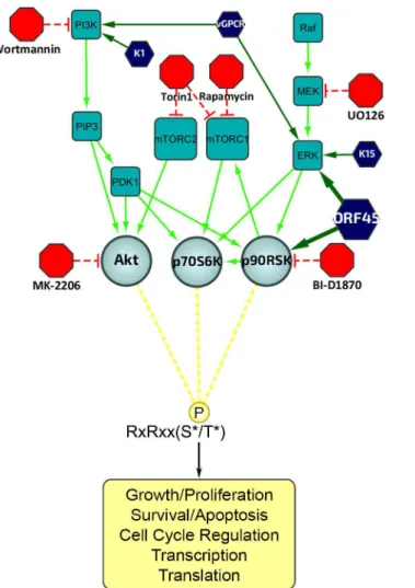
As expected, the F66A mutant virus is deficient in RSK activation, as evidenced by decreased levels of pRSK (Fig 2). We reasoned that the reduced KSHV lytic gene expression and virion production of the F66A mutant could be explained by the decreased phosphorylation and Fig 1. Diagram of the upstream signaling pathways that converge on activation of AGC kinases.In dark blue are the KSHV proteins that have been shown to affect various nodes of the PI3K/Akt/mTOR and/or Raf/MEK/ERK signaling pathways. In red are the kinase inhibitors used in this study. AGC kinases, including Akt, p70S6K (S6K), and p90RSK (RSK), are shown in light blue, and phosphorylate their substrates at the RxRxxS*/T*motif, leading to regulation of key biological processes.
consequent functional regulation of certain RSK substrates, and therefore sought to determine the contributions of KSHV ORF45-activated RSK to the differential phosphorylation induced by KSHV lytic replication. We predicted that a subset of the KSHV-induced changes in phos-phorylation would be dependent on ORF45-activated RSK, and thus be diminished in cells infected by the F66A mutant virus. Indeed this was the case, as overall phosphorylation at the RxRxxS
/T
motif was dramatically decreased in iSLK.BAC16 F66A cells compared to the revertant, especially at 24 and 48 hpi (Fig 2). As expected, we observed a dramatic decrease in ERK and RSK phosphorylation in the iSLK.BAC16 F66A cells. Moreover, the levels of phos-phorylation of Akt and S6K were only moderately affected by the F66A mutation, suggesting that the considerable variation in the phosphoproteomic profiles of these cells could be attrib-uted to the difference in RSK activity. Importantly, a similar phenomenon could be induced by infection of SLK cells with a lentivirus expressing ORF45 wild-type or F66A, suggesting that ORF45 is capable of inducing phosphorylation of cellular proteins independently of other viral factors (S1 Fig).
A phosphoproteomic screening identified proteins exhibiting differential
phosphorylation upon induction of KSHV lytic replication
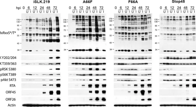
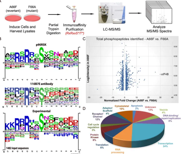
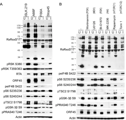
In order to further investigate the abundant KSHV-induced phosphorylation of putative RSK substrates, we performed a phosphoproteomic screening by employing Cell Signaling Technol-ogy’s PhosphoScan service [35]. We compared the phosphoproteomic profiles of iSLK.BAC16 F66A and its revertant (A66F) in order to determine which proteins are phosphorylated in an ORF45/RSK-dependent manner. Importantly, cell lysates were immunoaffinity purified with the anti-RxRxxS/Tmotif antibody prior to analysis by LC-MS/MS (Fig 3A). A substantial amount of manual vetting was undertaken to ensure that any conclusions drawn from the MS Fig 2. The increased phosphorylation of putative RSK substrates induced by KSHV lytic reactivation is dramatically reduced by ORF45 mutation or deletion.Stable iSLK.219 or iSLK.BAC16 cells carrying the indicated BAC (F66A, Stop45, or the F66A revertant (A66F)), were uninduced (U) or induced (I) with doxycycline and butyrate. Cell lysates were harvested at 0, 6, 12, 24, 48, and 72 hours post-induction (hpi) and analyzed by western blot using the indicated antibodies.
spectra could be made with high confidence. We ignored MS spectra assigned to peptides with a DeltaCN of less than 0.1, the general rule of thumb for what constitutes a“good hit”from a SEQUEST search [36]. We identified 1482 total peptides representing 1094 unique phospho-peptides of 479 proteins that met this cutoff. Of these, 1265 phosphophospho-peptides (85.4%) con-tained at least the minimal RxxS/Tmotif. The motif derived from this experimental data correlates well with the expected specificity of the antibody we used, as well as the previously characterized RSK phosphorylation motif (Fig 3B) [24–26,37].
Fig 3Cshows a scatterplot representation of the unique phosphopeptides identified by the screening arranged by average intensity in the A66F (y-axis) and fold-change between the A66F and F66A (x-axis). These IDs met an additional threshold for intensity (100,000) and spectral counts (2), leaving 1000 unique peptides. Of those that had at least a 2.5-fold reduc-tion in phosphorylareduc-tion in the F66A mutant (121 unique peptides; 98 proteins), we analyzed Fig 3. Phosphoproteomic analysis allows for the identification of putative substrates of ORF45-activated RSK during KSHV lytic replication.(A) Flowchart of the experimental design. (B) Phosphorylation motif for RSK, the 110B7E antibody used for the PhosphoScan, and the experimental data. (C) Scatterplot representation of the unique phosphopeptides identified by the screening that met the cutoff for deltaCN, intensity, and spectral counts. (D) Pie chart of the protein type/function distribution among the unique phosphopeptides with at least a 2.5-fold change (A66F:F66A).
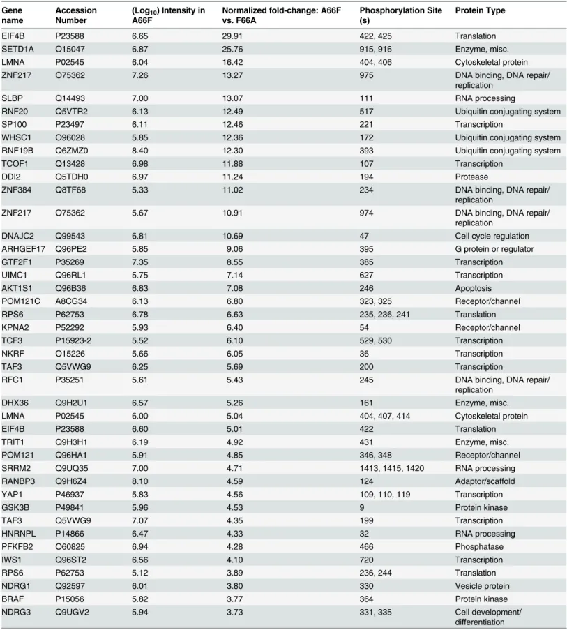
the distribution of the major protein types represented (Fig 3D). Significantly, peptides map-ping to proteins with roles in transcriptional regulation accounted for about a quarter of these putative substrates of ORF45-activated RSK.Table 1shows a list of the 50 most significant phosphopeptides identified, arranged according to fold-change of phosphorylation. A com-plete list of the phosphopeptides identified can be found in the supplemental material (S1 File). We were able to identify several proteins previously described as RSK substrates, confirming the validity of the experimental design and the phosphoproteomic screening (S1 File:‘RSK substrates’).
Data from the PhosphoScan was then subjected to DAVID bioinformatic analysis [38,39]. Our goal was to ascertain the most biologically relevant functions of potential substrates of ORF45-activated RSK. To address this, we assessed the enrichment of Gene Ontology (GO) terms, functional categories, and protein domains associated with this dataset (121 unique phosphopeptides with2.5-fold change in phosphorylation) in the background of the human proteome.Fig 4summarizes the results of this analysis, and the complete functional annotation chart can be found inS2 File. Among the most enriched categories were phosphoproteins, pro-teins with nuclear localization, cell cycle regulators, and propro-teins involved in RNA binding/ processing (Fig 4). A similar analysis was performed using the total IDs from our phosphopro-teomic screening as the background gene list, and yielded comparable results (S2 Fig). As a whole, these analyses are consistent with the notion that ORF45-activated RSK plays a major role in the nuclei of KSHV-infected cells, by regulating DNA/RNA-binding proteins, histones and histone-modifying enzymes, and transcription factors. Due to the low number of proteins with roles in translation whose phosphorylation was affected by F66A mutation (only two were identified in the screening–rpS6 and eIF4B), this group was not enriched in the DAVID analy-sis. However, because of their dramatic difference in phosphorylation, and the established effect of phosphorylation at the identified residues on these proteins’activities, we would argue that ORF45/RSK also plays a significant role in translational regulation.
KSHV ORF45/RSK-dependent phosphorylation of a subset of
substrates was validated
In order to verify the site-specific phosphorylation of the proteins identified in the PhosphoS-can study, we analyzed the lysates of KSHV-infected cells by western blotting with phospho-specific antibodies. We focused on: (1) previously identified substrates of RSK, Akt, and/or S6K, (2) proteins with physiologically relevant roles to the pathogenesis of KSHV or other her-pesviruses, (3) proteins with the most significant fold-changes in phosphorylation, and (4) phosphorylation sites that could be detected by commercially available phospho-specific anti-bodies. We were able to confirm that the site-specific phosphorylation of multiple proteins is induced by KSHV lytic replication (Fig 5A). Moreover, many of these, including eIF4B, 40S ribosomal protein S6 (rpS6 or S6), tuberous sclerosis complex (TSC2), glycogen synthase kinase 3 beta (GSK-3β), and proline-rich Akt substrate of 40 kDa (PRAS40), exhibited dimin-ished phosphorylation in the iSLK.BAC16 F66A and/or Stop45 cells, indicating that their phosphorylation may be dependent on ORF45-activated RSK. The RSK-dependent phosphory-lation of eIF4B at Ser422 is consistent with previous work published by our lab and others [16,40–42].
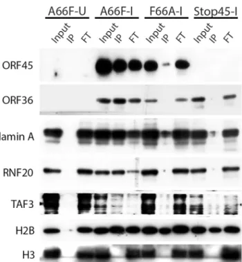
Because there appeared to be an enrichment of nuclear substrates of ORF45-activated RSK upon KSHV lytic reactivation (Table 1,Fig 4), and phospho-specific antibodies were unavail-able, we sought to validate these putative substrates by an alternative approach. We employed nuclear fractionation of iSLK.BAC16 A66F, F66A, and Stop45 cells at 2 dpi, followed by immu-noprecipitation with magnetic beads conjugated to the anti-RxRxxS
/T
Table 1. List of genes representing the phosphopeptides identified by the phosphoproteomic screening with the highest fold-change in phosphor-ylation between A66F and F66A.
Gene name
Accession Number
(Log10) Intensity in
A66F
Normalized fold-change: A66F vs. F66A
Phosphorylation Site (s)
Protein Type
EIF4B P23588 6.65 29.91 422, 425 Translation
SETD1A O15047 6.87 25.76 915, 916 Enzyme, misc.
LMNA P02545 6.04 16.42 404, 406 Cytoskeletal protein
ZNF217 O75362 7.26 13.27 975 DNA binding, DNA repair/
replication
SLBP Q14493 7.00 13.07 111 RNA processing
RNF20 Q5VTR2 6.13 12.49 517 Ubiquitin conjugating system
SP100 P23497 6.11 12.46 221 Transcription
WHSC1 O96028 5.85 12.36 172 Ubiquitin conjugating system
RNF19B Q6ZMZ0 8.40 12.30 393 Ubiquitin conjugating system
TCOF1 Q13428 6.98 11.88 107 Transcription
DDI2 Q5TDH0 6.97 11.24 194 Protease
ZNF384 Q8TF68 5.33 11.02 234 DNA binding, DNA repair/
replication
ZNF217 O75362 5.67 10.91 974 DNA binding, DNA repair/
replication
DNAJC2 Q99543 6.81 10.69 47 Cell cycle regulation
ARHGEF17 Q96PE2 5.85 9.06 395 G protein or regulator
GTF2F1 P35269 7.35 8.55 385 Transcription
UIMC1 Q96RL1 5.75 7.14 627 Transcription
AKT1S1 Q96B36 6.83 7.08 246 Apoptosis
POM121C A8CG34 6.13 6.80 323, 325 Receptor/channel
RPS6 P62753 6.78 6.63 235, 236, 241 Translation
KPNA2 P52292 5.93 6.40 54 Receptor/channel
TCF3 P15923-2 5.52 6.10 529, 530 Transcription
NKRF O15226 5.66 6.05 36 Transcription
TAF3 Q5VWG9 6.25 5.69 200 Transcription
RFC1 P35251 5.61 5.43 245 DNA binding, DNA repair/
replication
DHX36 Q9H2U1 6.57 5.26 161 Enzyme, misc.
LMNA P02545 6.00 5.04 404, 407, 414 Cytoskeletal protein
EIF4B P23588 6.60 5.01 422 Translation
TRIT1 Q9H3H1 6.19 4.92 431 Enzyme, misc.
POM121 Q96HA1 5.91 4.85 346, 348 Receptor/channel
SRRM2 Q9UQ35 7.00 4.71 1413, 1415, 1420 RNA processing
RANBP3 Q9H6Z4 8.10 4.59 124 Adaptor/scaffold
YAP1 P46937 5.83 4.56 109, 110, 119 Transcription
GSK3B P49841 5.96 4.53 9 Protein kinase
TAF3 Q5VWG9 7.07 4.35 199 Transcription
HNRNPL P14866 6.47 4.33 32 RNA processing
PFKFB2 O60825 6.94 4.28 466 Phosphatase
IWS1 Q96ST2 6.56 4.10 720 Transcription
RPS6 P62753 5.12 3.89 236, 244 Translation
NDRG1 Q92597 6.01 3.80 330 Vesicle protein
BRAF P15056 5.82 3.77 364 Protein kinase
NDRG3 Q9UGV2 5.94 3.73 331, 335 Cell development/
differentiation
this approach, we found that viral ORF45 and ORF36, and cellular Lamin A, RNF20, and TAF3 are all phosphorylated in an ORF45/RSK-dependent manner (Fig 6). While H2B phos-phorylation was robustly induced by lytic reactivation, it was only mildly reduced by ORF45 mutation or deletion. Significantly, all of these substrates are known or suspected to be involved in transcriptional regulation. The phosphorylation of ORF45 itself was further con-firmed by immunoprecipitation with an anti-ORF45 antibody (S3 Fig).
Pharmacological RSK inhibition or CRISPR-mediated RSK knockout
reduces RSK substrate phosphorylation and progeny virion production
To reinforce our conclusion that RSK activity is required for the differential phosphorylation of a subset of proteins during KSHV lytic replication, and to delineate the involvement of other AGC kinases, we treated KSHV-infected iSLK.BAC16 A66F cells with a panel of kinase inhibi-tors, including the RSK inhibitor BI-D1870 (Fig 5B) [43]. The apparent change in the pattern Table 1. (Continued)Gene name
Accession Number
(Log10) Intensity in
A66F
Normalized fold-change: A66F vs. F66A
Phosphorylation Site (s)
Protein Type
UBR4 Q5T4S7 8.47 3.59 1760 Receptor/channel
YAP1 P46937 6.15 3.59 127, 143 Transcription
TPX2 Q9ULW0 5.27 3.49 499 Cell cycle regulation
DDX17 Q92841 7.05 3.45 571 Transcription
H2B P33778 6.94 3.42 37 DNA binding, DNA repair/
replication
EIF4B P23588 6.38 3.23 406 Translation
PDCD4 Q53EL6 5.66 2.93 67, 76 Apoptosis
Unique phosphopeptides identified by LC-MS/MS werefiltered by deltaCN (0.1), intensity (100,000), and spectral counts (2), then ordered according to fold-change phosphorylation (A66F vs. F66A). The top 50 hits are shown, as well as the phosphorylation site(s) and protein type.
doi:10.1371/journal.ppat.1004993.t001
Fig 4. DAVID bioinformatic analysis of the phosphoproteomic screening results.The enrichment of Gene Ontology (GO) terms associated with the putative substrates of ORF45-activated RSK (121 unique phosphopeptides with2.5-fold change in phosphorylation between A66F and F66A) was assessed in the background of the human proteome. The x-axes values denote the percentage of the 98 unique proteins that were enriched (black bars) or the fold enrichment (gray bars) for the indicated GO term, functional category, or protein domain. P-values were all<0.05. The exact p-values and additional information can be found in S2 File.
of overall phosphorylation at the RxRxxS/Tmotif upon treatment with inhibitors of RSK, Akt (MK-2206), or mTOR (Rapamycin and Torin 1) hints at unique and shared substrates of AGC kinases. The lack of any discernable consequence of the MEK inhibitor U0126 (compare lanes 2 and 6) is consistent with ORF45 activating the downstream ERK and RSK indepen-dently of MEK [13]. In congruence with previous results, we found that the phosphorylation of eIF4B, TSC2, GSK-3β, and PRAS40 were all reduced by RSK inhibition. The phosphorylation of eIF4B was especially and uniquely sensitive to treatment with the RSK inhibitor. In contrast, the sensitivities of S6 (to MK-2206, Rapamycin, and Torin 1) and PRAS40 (to MK-2206 and Torin 1) suggest that in this system of KSHV lytic reactivation they are predominantly phos-phorylated by S6K/Akt and Akt, respectively. The mobility shift of ORF45 upon treatment with BI-D1870 is supportive of its RSK-dependent phosphorylation. The phosphorylation of TSC2 at Ser1798 also appears to be highly sensitive to RSK inhibition, which is consistent with previous findings [44]. Moreover, since this phosphorylation is known to relieve TSC2-me-diated inhibition of mTOR, the implication is that this is a mechanism by which KSHV Fig 5. The phosphorylation of several substrates with roles in translational regulation is mediated by ORF45-activated RSK.(A) The indicated cell lines were uninduced (U) or induced (I) as described
previously. Two days post-induction (dpi), cells lysates were collected and subjected to western blot analysis with the indicated antibodies. (B) Pharmacological inhibition confirms that KSHV-induced phosphorylation of eIF4B is dependent on RSK activity. iSLK.BAC16 A66F cells were uninduced (U) or induced (I) as described previously. At 46 hpi, cells were treated for 2 h with the indicated kinase inhibitor (the target of which is listed above in parentheses), then cells were harvested and lysates were analyzed by western blot with the indicated antibodies.
ORF45-activated RSK regulates Akt/mTOR/S6K activity [44]. We consistently observed multi-ple bands using the anti-pTSC2 Ser1798 antibody, which we believe represent differential extents of PTMs.
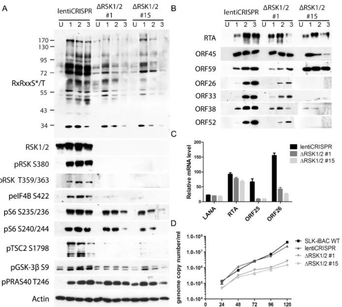
We previously found that pharmacological inhibition or siRNA-mediated RSK1/2 knock-down in 293T [13] or BCBL-1 [14,16] cells suppressed TPA-induced KSHV lytic reactivation, as well as phosphorylation of eIF4B [16]. To improve and expand upon our previous studies, we knocked out both RSK1 and RSK2 in a new system, SLK-iBAC cells, using the CRISPR/ Cas9 lentiviral delivery method [45]. SLK-iBAC cells are derived from the parental SLK cell line, but are stably transfected with a BAC16 mutant (iBAC) that has been engineered to express RTA from a doxycycline-inducible promoter, thereby allowing for robust lytic reactiva-tion in a wide range of transfecreactiva-tion-competent cell lines and negating the need for exogenous RTA induction and puromycin selection (seeMaterials and Methods). We induced lytic reacti-vation in two single cell clones with RSK1/2 knocked out, as well as in cells transduced with the empty lentiCRISPR, and compared phosphorylation at the RxRxxS/T motif. We observed dra-matically reduced phosphorylation at this motif as a consequence of RSK knockout, similar to that seen upon ORF45 mutation or pharmacological inhibition (Fig 7A). Additionally, although inducibility of these cells was not dramatically affected, late lytic protein levels were substantially reduced by RSK1/2 ablation (Fig 7B). The noticeable difference in KSHV late lytic protein levels between the two clones could be attributed to the slightly lower induction effi-ciency of clone #15 (Fig 7B; compare RTA level of clone #1 and #15). Relative mRNA levels of ORF25 and ORF26 (capsid components) were significantly lower in RSK knockout cells, whereas that of LANA or RTA were not dramatically affected, confirming that late lytic gene expression is compromised by RSK knockout (Fig 7C). We also measured extracellular viral genome copy number during a time course of lytic reactivation and found that RSK knockout Fig 6. Nuclear IP further validated ORF45/RSK-dependent phosphorylation of a subset of substrates.
Nuclei were isolated from the indicated cell lines at 48 hpi, then subjected to IP using anti-RxRxxS*/T*
magnetic beads. The input, eluate (IP), and flow-through (FT) were analyzed by western blot with the indicated antibodies.
significantly inhibits KSHV progeny virion production (Fig 7D). There is a ~10-fold decrease in virion production by 5 dpi, which is comparable to the defect of the ORF45 F66A mutant virus, and consistent with that observed upon treatment with the RSK inhibitor BI-D1870 [15].
Assessment of the roles of ORF45/RSK substrates in the regulation of
gene expression and progeny virion production
We next attempted to ascertain the contributions of the various transcriptional regulators iden-tified by our screening in ORF45/RSK-dependent transactivation. To do so, we adopted a lucif-erase reporter system recently used by Karijolich et al. to describe an ORF45/RSK2-dependent Fig 7. CRISPR/Cas9-mediated knockout of RSK1/2 results in the reduction of RSK substrate phosphorylation, viral late lytic gene expression, and virion production.(A and B) SLK-iBAC cells transduced with empty lentiCRISPR or single cell clones with RSK1/2 knockout (#1 and #15) were uninduced (U) or induced (I) with dox/butyrate, and cells were harvested at the indicated dpi. Lysates were analyzed by western blot with the indicated antibodies. (C) The indicated cell lines were induced as in (A and B). Twenty-four hpi, cells were harvested and RNAs were extracted, reverse transcribed, and subjected to qRT-PCR analysis using the indicated primers. Relative mRNA level is shown normalized toβ-Actin. (D) KSHV progeny virion production is compromised by RSK knockout. SLK-iBAC cells were induced as described in (A and B). At the indicated hpi, media were collected from cells, extracellular virion DNA was extracted, and viral genome copy number was measured by qPCR.
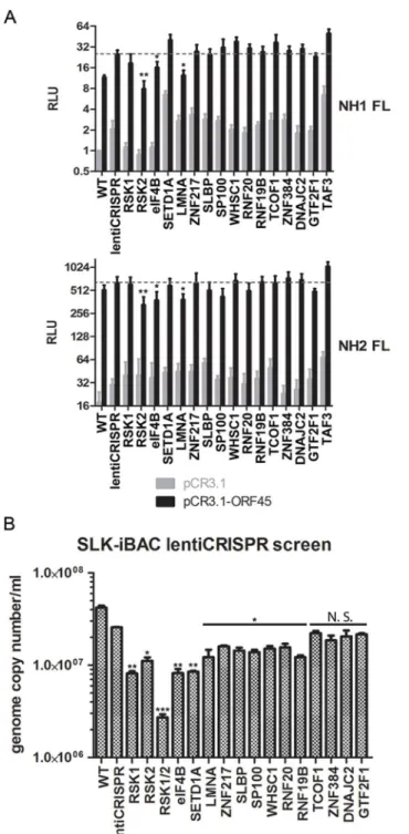
mechanism for increased transactivation of the HIV-1 LTR [18]. We performed a CRISPR screen using both NH1 (containing the integrated HIV LTR fused to firefly luciferase (FL)) and NH2 (containing the LTR-FL, as well as constitutive expression of HIV Tat) cells. We targeted RSK1, RSK2, and each putative RSK substrate identified by our screening with a>10-fold dif-ference in phosphorylation and/or a well-established role in transcriptional regulation (14 total). We then generated stably transduced knockout cell lines and performed luciferase assays in the presence and absence of exogenous ORF45 expression. Thus, the readout for this assay should give an indication of which genes are essential for ORF45-mediated transactivation of the HIV LTR, a phenomenon that is physiologically relevant in the context of KSHV/HIV co-infection in natural populations [6–10].
In concordance with the findings of Karijolich et al. [18], we found that knockout of RSK2, but not RSK1, significantly diminished the ORF45-dependent induction of luciferase expres-sion (Fig 8A). Of the putative RSK substrates analyzed, only eIF4B or LMNA knockout had a significant effect on ORF45-mediated LTR transactivation (Fig 8A). We next performed the same CRISPR screen in SLK-iBAC cells, then induced lytic reactivation and measured progeny virion production. We detected a significant, albeit mild reduction for most genes assessed (Fig 8B). The observed effect supports the hypothesis that multiple substrates downstream of ORF45-activated RSK contribute to the optimal efficiency of the KSHV lytic cycle.
The ORF45/RSK/eIF4B signaling axis is critical for efficient translation of
mRNAs with highly structured 5
’
untranslated regions (UTRs)
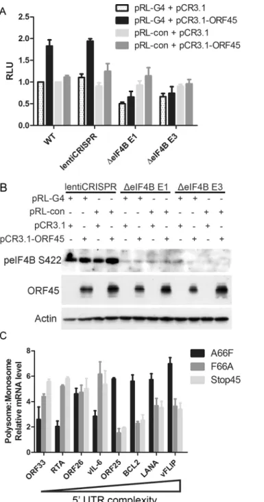
All of our data indicated that eIF4B is robustly phosphorylated during viral lytic reactivation, and that it represents one of the most specific and functionally significant substrates of ORF45-activated RSK. Thus, we aimed to further characterize the role(s) of the ORF45/RSK/ eIF4B signaling axis in translational regulation. Recent reports that the RNA helicase eIF4A regulates the translation of mRNAs with G-quadruplex (G4) structures in their 5’UTR [46], in addition to the known role of eIF4B phosphorylation in stimulating the helicase activity of eIF4A [42,47,48], led us to hypothesize that eIF4B may also contribute to this phenomenon. To test this, we transfected 293T (WT or eIF4B-knockout) cells with a reporter construct contain-ing G4 structures in the 5’UTR G4) or random sequence with equal G/C content (pRL-con) in the presence or absence of ORF45 overexpression (Fig 9A and 9B). Interestingly, we found that ORF45 significantly increased luciferase activity of pRL-G4, and that loss of eIF4B almost completely abrogated this effect (Fig 9A and 9B). The ORF45-dependent induction of luciferase activity of the negative control (pRL-con) is mild by comparison and insensitive to eIF4B knockout. Thus, this difference probably represents transcriptional regulation by ORF45, whereas the more significant effect on the G4-containing reporter can be attributed primarily to translational regulation.
Fig 8. CRISPR screen characterizes the contributions of putative RSK substrates to ORF45/RSK-mediated transactivation and KSHV progeny virion production.(A) NH1 (top) and NH2 (top) reporter cells were stably transduced with the indicated lentiCRISPR, transfected with pCR3.1 or pCR3.1-ORF45, and subjected to luciferase assays. Shown are the relative light units (RLU) for firefly luciferase (FL) normalized to the NH1 FL WT sample. Values shown are the average of three biological replicates. (B) SLK-iBAC cells were stably transduced with the indicated lentiCRISPR, then induced as described previously. Extracellular virions were collected from the media at 5 dpi, and virion DNA was extracted and quantified by qPCR. Values shown are the average of three technical replicates from each of two biological replicates. Knockout of TAF3 in SLK-iBAC cells was not viable. (A and B) For RSK substrate knockout, values shown are the average of knockout cells generated from two independent gRNAs. Significance compared to empty lentiCRISPR sample:*p<0.05;**p<0.01;***p<0.001; N.S.–not significant.
Fig 9. ORF45/eIF4B-dependent translational control of mRNAs with complex 5’UTR structure.(A and B) 293T WT cells or cells stably transduced with the indicated lentiCRISPR were transfected with the indicated plasmids, as well as HCV IRES-FL (firefly luciferase), and subjected to luciferase assays (A) or western blot analysis (B) at 24 hpt. Values for (A) are represented as the renilla/firefly luciferase ratio normalized to the WT sample. (C) iSLK.BAC16 A66F, F66A, or Stop45 cells were uninduced or induced with dox/butyrate, and cells were harvested at 48 hpi. Lysates were fractionated by sucrose gradient
centrifugation and RNAs were extracted. RNAs associated with either monosome or polysome fractions were pooled, reverse transcribed, and subjected to qRT-PCR using the indicated primers. Values shown are for mRNA level relative to the uninduced control, as a ratio of polysome:monosome (normalized toβ-Actin). Genes are arranged in order of decreasing minimum free energy (left to right), as predicted by RNAFold software [109].
energy (MFE)) were detected at higher levels in polysome fractions in an ORF45/RSK-depen-dent manner (Fig 9C). Among these were viral ORF25, LANA and vFLIP, and cellular BCL2. Altogether, these data suggest that the ORF45/RSK/eIF4B signaling axis is important for the efficient translation of a subset of viral and cellular mRNAs containing highly structured 5’ UTRs. These findings have important implications for KSHV replication and pathogenesis (discussed below).
Discussion
KSHV relies on manipulating the host kinase signaling pathways in order to express viral genes, evade host antiviral responses, and assemble and release progeny virions with maximum efficiency. The exact mechanisms by which specific viral proteins contribute to these various aspects of KSHV lytic replication remain unclear. The elucidation of these mechanisms is of critical importance to completely understand KSHV pathogenesis and oncogenesis, and could be useful in the development of novel therapies to treat or prevent KSHV-related ill-nesses. The PI3K/Akt/mTOR and ERK/MAPK signaling pathways are perhaps the two most intensively studied signaling pathways with regard to KSHV. They each have well-established roles in the progression of diverse human diseases, including various cancers, and targeted inhibition of these pathways is a common therapeutic approach with expanding applications [12,29,32,51,52]. In the context of KSHV, several viral proteins have been implicated in the dysregulation of at least one of these pathways, most notably LANA [53], vGPCR [54–62], K1 [63,64], K15 [65,66] and ORF45 [13–16].
The multiple functions of ORF45
It is clear that KSHV ORF45 performs at least four distinct functions throughout viral lytic rep-lication: (1) It inhibits IRF7-mediated induction of type 1 interferon by acting as a more effi-cient substrate of TBK1/IKKε[67–69], (2) binds to and activates ERK and RSK, resulting in the differential regulation of their various downstream substrates [13,14,16], (3) binds to the kinesin-2 motor protein KIF3A to facilitate transport of virions during egress [70], and (4) binds to and stabilizes KSHV ORF33, an event that appears to be crucial for efficient tegumen-tation [71]. The ORF45 F66A mutant virus, which is deficient only in the second function listed above, exhibits greater than a 10-fold reduction in progeny virion production, suggesting that the activation of ERK and RSK during lytic replication is critical for optimal lytic replication [15]. Interestingly, ORF45 possesses no inherent kinase activity, but appears to sustain ERK/ RSK kinase activity by preventing their dephosphorylation [14]. The activation of RSK by ORF45 has been shown to increase the phosphorylation of the RSK substrate eIF4B and thereby enhance its assembly into translation initiation complexes, ultimately contributing to the increased translation of viral mRNAs during KSHV lytic replication [16]. Here, we further illuminate the roles of ORF45 during lytic replication.
A phosphoproteomic analysis of KSHV-infected cells identified several putative substrates of ORF45-activated RSK. We found dramatic differences in the phosphorylation of diverse substrates which can be attributed to ORF45. The picture that emerges from these data sub-stantiates the claim that the activation of RSK by ORF45 is of critical importance during KSHV lytic replication. In the following sections, we will discuss the possible consequences of the phosphorylation of specific proteins by ORF45-activated RSK.
ORF45/RSK-dependent phosphorylation of KSHV proteins
between A66F and F66A were not especially dramatic. This could be explained by the transient nature of these phosphorylation events during the lytic life cycle and the limitation that we only analyzed the phosphoproteome at one time point, 48 hpi. The increased presence of newly synthesized, virion-contained, or otherwise unmodified viral proteins in the iSLK. BAC16 A66F lysates (compared to the replication-deficient F66A mutant) may partially explain why the fold-changes in phosphorylation were not deemed significant. A phosphopro-teomic screening of iSLK.219 cells (uninduced vs. 24 hpi) did identify phosphopeptides of all of the viral proteins mentioned above, which were significantly enriched in the induced sample (S1 File). This data lends credence to the supposition that multiple viral proteins are phosphor-ylated at putative RSK phosphorylation motifs during KSHV lytic replication.
ORF45 itself was found to be phosphorylated at residue 303, which not only falls within the putative RSK phosphorylation motif, but is directly adjacent to ORF45’s nuclear localization signal (NLS),297KRKR300[72]. It is well-established that phosphorylation, especially near an NLS or NES, can affect a protein’s subcellular localization [73–75]. We have previously shown the presence of distinct mobility shifts in ORF45 protein, indicative of dramatically increased phosphorylation of the protein in the cytoplasm compared to the nucleus, and postulated that both populations of ORF45 are critical for its various functions [72]. We hypothesize that ORF45 binding to and activation of RSK increases phosphorylation of ORF45 at Ser303, thereby regulating its localization during KSHV lytic replication. This would offer an attractive explanation for the ability of ORF45 to perform several diverse roles throughout lytic replica-tion. We have confirmed that ORF45 WT, but not the F66A mutant, is phosphorylated at the RxRxxS
/T
motif during KSHV lytic replication (Figs6andS3).
ORF36 was another viral protein identified by the PhosphoScan, and phosphorylation at the RxRxxS
/T
motif was reduced in iSLK.BAC16 F66A cells. ORF36 is the only serine/threo-nine protein kinase encoded by KSHV and is conserved throughout herpesviruses [76–78]. We confirmed that ORF36 is indeed phosphorylated at its RxRxxS
/T
motif (Fig 6). Furthermore, while investigating the phosphorylation of ORF36, we discovered a ternary interaction between ORF36, ORF45, and RSK that is dependent on ORF36 kinase activity. Based on these data, we believe that ORF45 and RSK may regulate ORF36 activity during KSHV lytic replication.
Elucidating the mechanisms by which KSHV ORF45 regulates gene
expression
episomes. The finding that RSK inhibition reduces LANA binding to H2B is consistent with this notion [53].
We previously found that mRNA levels of several KSHV genes are reduced in induced iSLK.BAC16 F66A and Stop45 cells compared to their revertants, indicating a crucial role for ORF45-activated RSK in transcription of viral genes during KSHV lytic replication [15]. A more recent study offered a potential mechanistic explanation for this effect, showing that ORF45/RSK-mediated phosphorylation and accumulation of c-Fos contributes to the
increased transcription of viral genes [83]. Work by Dr. Glaunsinger’s group has elucidated an additional role for ORF45/RSK2 in transcriptional activation of the HIV-1 LTR [17]. Here, we report that the phosphorylation of numerous transcriptional regulators is significantly affected by ORF45 F66A mutation (Fig 3andTable 1). This is consistent with a major role of ORF45/ ERK/RSK in the nuclei of infected cells, which appears to be conserved amongst gammaherpes-viruses [84,85]. We validated the ORF45/RSK-dependent phosphorylation of Lamin A, RNF20, and TAF3 (Fig 6). Lamin A/C has well-established roles in transcriptional regulation [86,87], and phosphorylation of the nuclear lamina is particularly relevant during herpesviral life cycles, as it has been shown to promote nuclear egress of virions [88–92]. Our CRISPR/ Cas9-based screenings indicate that Lamin A (LMNA) is involved in ORF45/RSK dependent transactivation (Fig 8A), and is important for optimal progeny virion production (Fig 8B). Fur-ther studies are required to determine the consequences of site-specific phosphorylation of the nuclear lamina to KSHV replication and pathogenesis.
We also validated the ORF45/RSK-dependent phosphorylation of multiple proteins with established roles in the regulation of translation, including eIF4B, S6, TSC2, and GSK-3β. Fig 10shows the established nodes of PI3K/Akt/mTOR and ERK/MAPK signaling which
Fig 10. Summary of the mechanisms by which KSHV ORF45 manipulates ERK/RSK signaling to regulate translation.ORF45 regulates translation at multiple levels through direct and indirect mechanisms, including: (1) RSK-dependent phosphorylation of eIF4B and S6, (2) ERK/RSK-mediated inhibition of TSC2, relieving its inhibition of mTORC1, and (3) crosstalk with Akt/mTOR, thereby enhancing S6K-dependent phosphorylation of S6. Several other factors, such as GSK-3β, PRAS40, and PDCD4 (all of which were identified by our analyses to have decreased phosphorylation at key regulatory residues upon ORF45 F66A mutation), also contribute to this phenomenon.
contribute to regulation of translation. Significantly, the activity of TSC2 is of critical impor-tance to KSHV pathogenesis [59,62]. Although several kinases can phosphorylate TSC2, and thereby contribute to mTOR activation, we show that the KSHV-induced phosphorylation of Ser1798 is dependent on ORF45-mediated RSK activation (Figs5and7A). Notably, we did not detect any significant difference in the Akt-dependent phosphorylation of TSC2 at Ser939 or Thr1462 [93]. Our results also clearly implicate ORF45-activated RSK in phosphorylation of the translation initiation factor eIF4B during KSHV lytic reactivation. This finding corrobo-rates previous work by our laboratory, in which we reported that KSHV-induced phosphoryla-tion of eIF4B at Ser422 is insensitive to U0126 and rapamycin, and promotes progeny virion production [16]. Taken together, these results are remarkably consistent with what has recently been observed by Chang and Ganem. They showed that in KSHV-infected lymphatic endothe-lial cells (LECs), but not blood endotheendothe-lial cells (BECs), there was an increase in pERK, pRSK, peIF4B (Ser422), pS6, and pTSC2 (Ser1798 but not Thr1462). Presumably, this is due to ORF45 expression in LECs, since siRNA-mediated ablation of ORF45 expression abolished many of these changes [21].
It has been known that phosphorylation of eIF4B at the residues identified by our MS analy-sis (Ser406 and Ser422) are involved in its ability to enhance the RNA helicase activity of eIF4A [42,47,48]. The Ser422 residue can be phosphorylated by Akt [42], S6K [40], and/or RSK [41,94]. However, our data show that in the context of KSHV lytic reactivation, the upregula-tion of eIF4B phosphorylaupregula-tion at this site is uniquely sensitive to RSK inhibiupregula-tion/ablaupregula-tion (Figs 5Band7A). Interestingly, eIF4B activity has been shown to be especially critical for the transla-tion of mRNAs with highly ordered 5’UTRs [50,95]. Knockdown of eIF4B led to the discovery of several eIF4B-dependent transcripts that have roles in cell proliferation and survival, includ-ing c-MYC and BCL2 [50,96]. Recently, it was found that RNA G-quadruplexes are a key fea-ture present within the 5’UTRs of eIF4A-dependent transcripts, many of which are bona fide oncogenes [46]. This was discovered by sequencing the ribosome-protected mRNAs (ribosome profiling; Ribo-Seq [97]) of cells treated with a selective inhibitor of eIF4A. We used a luciferase reporter assay to show that ORF45 increases the translation of a G4-containing construct in a manner dependent on eIF4B (Fig 9A and 9B). It has been reported that G4 binding by EBV EBNA1 is important for viral DNA replication and episome maintenance [98]. More recently, it was found that G-quadruplexes regulate EBNA1 mRNA translation [99]. A simple sequence motif analysis identifies several putative quadruplex forming G-rich sequences (QGRS) in the KSHV genome [100]. Thus, it is likely that G-quadruplex structures are important for KSHV replication and pathogenesis. Growing evidence for thein vivorelevance of G4s opens up excit-ing prospects for the development and application of innovative diagnostics and therapeutic agents [101,102].
understanding of the regulatory mechanisms governing changes in viral/host gene expression in KSHV-infected cells [79]. The development of innovative tools and techniques, coupled with unprecedented advances in sequencing technology, have facilitated the characterization of epigenetic, transcriptional, post-transcriptional, and translational levels of regulation. Compre-hensive gene expression profiling of KSHV-infected cells is fundamental to further our under-standing of how KSHV modulates the host cellular machinery, and, more importantly, how these changes contribute to KSHV pathogenesis. By aspiring to more completely characterize the complex workings of cellular signaling pathways, we will uncover shared and unique mech-anisms of dysregulation by human pathogens, which could lead to the future development of novel, targeted therapies to prevent or treat a wide variety of human diseases.
Materials and Methods
Antibodies, chemicals, and vectors
Anti-RxRxxS/T
antibody was ordered from Cell Signaling Technology (#110B7E). Anti-RTA (ORF50) monoclonal mouse antibody was given by Dr. Ke Lan (Institut Pasteur of Shanghai). Anti-ORF59 antibody was given by Dr. Robert Ricciardi. Monoclonal antibodies against ORF26, ORF52, ORF33, ORF38 and ORF45 were generated by the Florida State University hybridoma facility. pTSC2 (Ser1798) was ordered from LifeSpan Biosciences. pAkt, pS6K, pGSK-3β, and pPRAS40 antibodies, anti-RxxS/T-conjugated magnetic beads, Wortmannin (#9951), and Rapamycin (#9904) were ordered from Cell Signaling Technology. MK-2206 dihydrochloride was ordered from Santa Cruz Biotechnology (sc-364537), Torin 1 was ordered from Tocris Bioscience (#4247). The pCDH lentiviral expression construct and pPACK pack-aging plasmids were ordered from System Biosciences. The lentiCRISPR-puro vector was ordered from Addgene (#49535) [45]. The pRL-G4 and pRL-con vectors were graciously pro-vided by Dr. Hans-Guido Wendel and previously described [46]. All other antibodies and chemicals used in this study have been described previously [13,14,72,103].
Cell culture
SLK, 293T, NH1, and NH2 cells were cultured in Dulbecco's modified Eagle's medium (DMEM) containing 10% FBS and antibiotics. NH1 and NH2 cells were graciously provided by Dr. Britt Glaunsinger [17]. iSLK.219 and iSLK.BAC16 cells were cultured in DMEM con-taining 10% FBS, antibiotics, 450μg/ml G418, 400μg/ml hygromycin B and 1μg/ml puromy-cin [104]. The induction conditions were as follows: Cells were seeded so that they reached 90– 95% confluence one day later. The media was replaced with DMEM containing 1% FBS with or without doxycycline (0.2μg/ml for iSLK.219; 2μg/ml for iSLK.BAC16) and sodium butyrate (1 mM). Cells were harvested at the indicated times post-induction.
iBAC construction
SLK-iBAC are a cell line recently developed by our laboratory derived from the parental SLK cells and stably transfected with a BAC16 mutant which contains a doxycycline-inducible RTA. This BAC contains the TET-on transactivator (Tet3G) and the tet-responsive elements (PTre3G; inserted upstream of RTA). This therefore bypasses the requirement for inducible
was 0.05μg/ml (because the dox-inducible promoter is on the BAC, induction is much more efficient).
Immunoprecipitation and western blot analysis
Western blot analysis was performed as previously described [13,14,16]. For immunoprecipita-tion of ORF45 from iSLK.BAC16 cells, we used a monoclonal anti-ORF45 antibody (8B8) con-jugated to CNBr-Activated Sepharose 4B (GE Life Sciences). Clarified lysates were bound to the beads for 2 h at 4°C, washed three times each with lysis buffer and TBS, and bound com-plexes were eluted by boiling. For nuclear IP, cells were harvested in Tween 20 lysis buffer (25 mM Tris/Hepes pH 8.0, 20 mM NaCl, 2 mM EDTA, 1 mM PMSF, 0.5% Tween 20) for 15 min on ice, then nuclei were pelleted by centrifugation at 6,000× gfor 1 min at 4°C. The pellet was resuspended in Tween 20 lysis buffer containing 500 mM NaCl and incubated on ice for 15 min, then fresh lysis buffer (without NaCl) was added to bring the final salt concentration to 250 mM. This was centrifuged at 10,000× gfor 15 min and the supernatant was saved as the soluble nuclear extract for IP. The IP was performed according to the manufacturer’s instruc-tions (Cell Signaling Technology). For western blot, about 20μg of proteins were resolved by SDS-PAGE and transferred to nitrocellulose membranes. The membranes were blocked in 5% dried milk in 1× PBS plus 0.2% Tween 20 and then incubated with diluted primary antibodies for 2 h at room temperature or overnight at 4°C. Anti-rabbit or anti-mouse IgG antibodies con-jugated to horseradish peroxidase (Pierce) were used as the secondary antibodies. SuperSignal chemiluminescence reagents (Pierce) were used for detection.
ORF45 lentiviral transduction
293T cells were co-transfected with pCDH-CMV-MCS-EF1-copGFP (CD511B-1; containing either no insert, ORF45 WT, or ORF45 F66A) and the pPACK packaging plasmid mix accord-ing to the manufacturer’s instructions. Media containing pseudoviral particles were collected at 48 and 72 hpt, and clarified by centrifugation. SLK cells were transduced using a standard spin-fection protocol. Briefly, lentivirus-containing media was added to SLK cells in the presence of 4μg/ml polybrene. Cells were immediately centrifuged at 800× gfor 1 h at 37°C, and cultured under normal conditions for 24 h. The media was aspirated, fresh DMEM containing 1% FBS was added, and cells were cultured overnight. At 48 hpi, cells were harvested and lysates were subjected to western blot analysis.
Phosphoproteomic analysis
Approximately 2 × 10⁸iSLK.BAC16 F66A or A66F cells were induced as described previously. At 48 hpi (when we observed the most dramatic effect of ORF45/RSK-mediated substrate phosphorylation), cells were washed twice with PBS, and lysed in freshly prepared urea lysis buffer (20 mM HEPES pH 8.0, 9 M urea, 1 mM sodium orthovanadate, 2.5 mM sodium pyro-phosphate, 1 mMβ-glycerophosphate; 1.5-ml per plate). Lysates were sonicated and clarified by centrifugation, and the lysate supernatant was flash-frozen in liquid nitrogen and stored at -80°C. Samples were then sent to Cell Signaling Technology (CST) and subjected to their PTMScan discovery service [35].
The complete protocol, from sample prep to MS analysis, can be found on the CST website. Briefly, proteins in lysates were carboxamidomethylated to inactivate enzymatic activity (addition of DTT, then iodoacetamide), then digested with LysC. The digested cell lysate was acidified by addition of TFA to a final volume of 1%, and peptides were purified using a Sep-Pak C18column. Subsequently, immunoaffinity purification (IAP) was performed using
the PTMScan Phospho Akt Substrate Motif (RxxS /T
concentrated and purified on ZipTip, followed by a final trypsin digestion step before liquid chromatography-tandem mass spectrometry (LC-MS/MS). Two technical replicates were run for each biological sample. The spreadsheet with the total IDs is available inS1 File.
Bioinformatic analyses
The experimental motif (Fig 3B) was generated using the Sequence Logo Generator in the Motif & Logo Analysis Tools on PhosphoSitePlus [105]. The scatterplot inFig 3Cshows all of the unique phosphopeptides identified that met a cutoff for deltaCN (0.1), intensity (100,000), and spectral counts (2). The pie chart inFig 3Dclusters the 121 unique phos-phopeptides that met a cutoff of 2.5-fold-change in phosphorylation (A66F:F66A), in addition to all of the aforementioned criteria, by their protein type. DAVID functional annotation was performed by uploading a list of all unique gene names with at least one phosphopeptide exhib-iting 2.5-fold change in phosphorylation (A66F:F66A), and analyzing this list in the back-ground of either the entire human genome (Fig 4) or a list of the total unique gene names identified by the PhosphoScan (S2 Fig) [38,39].
Kinase inhibitor assays
iSLK.BAC16 cells were induced as described previously. At 46 hpi, one of the following inhibi-tors was added at the indicated concentration: Wortmannin (PI3K; 200 nM), U0126 (MEK; 10μM), BI-D1870 (RSK; 10μM), MK-2206 (Akt; 1μM), Rapamycin (mTORC1; 10 nM), Torin 1 (mTORC1/2; 250 nM), or DMSO control. Lysates were collected 2 h later (48 hpi) and analyzed by western blotting.
Genome editing
Sucrose density gradient fractionation and polysome analysis
The cells were washed once in cold phosphate-buffered saline (PBS) containing 100μg/ml cycloheximide and scraped off the plate into 1 ml of the same solution. The cells were centri-fuged for 10 min at 1,200 rpm and resuspended in 425μl hypotonic lysis buffer (5 mM Tris-HCl [pH 7.5], 2.5 mM MgCl2, 1.5 mM KCl). The lysates were transferred to a prechilled tube,
incubated with 100μg/ml cycloheximide, 2 mM dithiothreitol (DTT), and 2μl RNasin (40 U/ μl; Stratagene), kept on ice for 5 min, and vortexed. To each 425μl of lysate, 25μl of 10% Triton X-100 and 25μl of 10% sodium deoxycholate were added. The samples were vortexed again and incubated on ice for 5 min. Cell extracts were centrifuged for 5 min at 14,000 rpm; the supernatant was collected, and protein concentration was determined. Equal amounts of pro-tein were loaded onto prechilled 10 to 50% sucrose gradients. Each gradient was formed in a Beckman centrifuge tube (14 by 89 mm) (catalog no. 3311372; Beckman Instruments, CA) using a BioComp gradient master according to the manufacturer’s instructions. The tubes were centrifuged in a Beckman SW40Ti rotor at 35,000 rpm for 3 h at 4°C. Fractions (0.5 ml) were collected and RNA concentration and quality was assessed by Nanodrop and denatured aga-rose gel electrophoresis. RNAs in the monosomal or polysomal fractions were extracted and analyzed by qRT-PCR (see below).
Reverse transcription and RT-PCR
Sucrose gradient fractions were subjected to RNA extraction using TRIzol (Invitrogen). Reverse transcription was performed using a SuperScript III reverse transcriptase kit (Invitro-gen) according to the manufacturer’s instructions. PCRs were carried out as described below. PCR was performed using 1μl of RT reaction mixture in a total reaction mixture volume of 20μl containing reverse and forward primers (1μl of 10μM solutions). Primer sequences used for BCL2 are TTTCTCATGGCTGTCCTTCAGGGT and AGGTCTGGCTTCATACCACA GGTT. Primer sequences for all other genes analyzed in this study were previously described [15,108].
Quantification of extracellular virion genomic DNA by real-time qPCR
Viral DNA was isolated from supernatant medium of induced iSLK.BAC16 and derivative cells as previously described [72,103]. The DNAs were used in SYBR green real-time PCRs with KSHV-specific primers: ORF73-LCN (5’-CGCGAATACCGCTATGTACTCA-3’) and ORF73-LCC (5’-GGAACGCGCCTCATACGA-3’) with Bio-Rad C1000TM Thermal Cycler and CFX96TM Real-Time Detection System. Viral DNA copy numbers were calculated with external standards of known concentrations of serially diluted BAC16 DNA ranging from 1 to 107genome copies per reaction.Luciferase assay
gRNAs to target eIF4B exon 1 (E1) or exon 3 (E3) as described above. Stable cell lines were seeded to 24-well plates and co-transfected with pCR3.1/pCR3.1-ORF45 and pRL-G4/pRL-con, as well as HCV IRES-FL (firefly luciferase; negative control). Cells were harvested and luminescence was detected as described above. Values shown are the relative ratio of Renilla luciferase (RL) to firefly luciferase (FL), averaged from three biological replicates.
Supporting Information
S1 File. CST PhosphoScan data.The raw spreadsheet obtained from the CST PhosphoScan, including all of the information acquired from the phosphoproteomic screening.
(XLSX)
S2 File. Details of DAVID Bioinformatic analysis.DAVID functional annotation analyses featuring the complete list as well as clustering of GO terms, functional categories, and protein domains that were enriched among the phosphopeptides with at least a 2.5-fold change (A66F: F66A).
(XLSX)
S1 Fig. ORF45 WT but not the F66A mutant induces phosphorylation of RSK substrates independently of the viral background.SLK cells were transduced with lentiviral particles containing no expression vector, ORF45 wild-type, or the F66A mutant. At 48 hpi, cells were lysed, and the lysates were analyzed by western blot with the indicated antibodies.
(TIF)
S2 Fig. DAVID bioinformatic analysis.DAVID functional annotation analyses were per-formed using the same parameters as inFig 4, except that the total unique IDs from the Phos-phoScan were used as the background gene list.
(TIF)
S3 Fig. ORF45 WT but not the F66A mutant is phosphorylated at the RxRxxS/Tmotif
during KSHV lytic replication.ORF45 was immunoprecipitated from iSLK.BAC16 A66F or iSLK.BAC16 F66A cells using 8B8 monoclonal antibody as described previously [15]. The inputs, eluates, and flow-throughs were analyzed by western blot with the indicated antibodies. (TIF)
S4 Fig. NH1 and NH2 lentiCRISPR screen–Renilla luciferase.The same samples as in
Fig 8Awere analyzed for renilla luciferase activity. (TIF)
S1 Table. List of primers used to clone gRNAs for CRISPR screening. (XLS)
S1 References. (DOCX)
Acknowledgments
We thank Dr. Hans-Guido Wendel for sharing the G4 luciferase reporter constructs. We thank members of the Zhu laboratory for critical reading of the manuscript and for helpful discus-sions. We also thank Jen Kennedy at the Florida State University for excellent editorial assistance.
Author Contributions
Conceived and designed the experiments: DA FZ. Performed the experiments: DA ST WL ZT. Analyzed the data: DA FZ. Wrote the paper: DA ST FZ.
References
1. Chang Y, Cesarman E, Pessin MS, Lee F, Culpepper J, et al. (1994) Identification of herpesvirus-like DNA sequences in AIDS-associated Kaposi's sarcoma. Science 266: 1865–1869. PMID:7997879
2. Cesarman E, Chang Y, Moore PS, Said JW, Knowles DM (1995) Kaposi's sarcoma-associated her-pesvirus-like DNA sequences in AIDS-related body-cavity-based lymphomas. N Engl J Med 332: 1186–1191. PMID:7700311
3. Soulier J, Grollet L, Oksenhendler E, Cacoub P, Cazals-Hatem D, et al. (1995) Kaposi's sarcoma-associated herpesvirus-like DNA sequences in multicentric Castleman's disease. Blood 86: 1276– 1280. PMID:7632932
4. Dezube BJ (1996) Clinical presentation and natural history of AIDS—related Kaposi's sarcoma. Hematol Oncol Clin North Am 10: 1023–1029. PMID:8880194
5. Ganem D (2010) KSHV and the pathogenesis of Kaposi sarcoma: listening to human biology and medicine. J Clin Invest 120: 939–949. doi:10.1172/JCI40567PMID:20364091
6. Guo HG, Pati S, Sadowska M, Charurat M, Reitz M (2004) Tumorigenesis by human herpesvirus 8 vGPCR is accelerated by human immunodeficiency virus type 1 Tat. J Virol 78: 9336–9342. PMID: 15308728
7. Huang LM, Chao MF, Chen MY, Shih H, Chiang YP, et al. (2001) Reciprocal regulatory interaction between human herpesvirus 8 and human immunodeficiency virus type 1. J Biol Chem 276: 13427– 13432. PMID:11154704
8. Chen X, Cheng L, Jia X, Zeng Y, Yao S, et al. (2009) Human immunodeficiency virus type 1 Tat accel-erates Kaposi sarcoma-associated herpesvirus Kaposin A-mediated tumorigenesis of transformed fibroblasts in vitro as well as in nude and immunocompetent mice. Neoplasia 11: 1272–1284. PMID: 20019835
9. Xue M, Yao S, Hu M, Li W, Hao T, et al. (2014) HIV-1 Nef and KSHV oncogene K1 synergistically pro-mote angiogenesis by inducing cellular miR-718 to regulate the PTEN/AKT/mTOR signaling pathway. Nucleic Acids Res.
10. Zhu X, Guo Y, Yao S, Yan Q, Xue M, et al. (2014) Synergy between Kaposi's sarcoma-associated herpesvirus (KSHV) vIL-6 and HIV-1 Nef protein in promotion of angiogenesis and oncogenesis: role of the AKT signaling pathway. Oncogene 33: 1986–1996. PMID:23604117
11. Jarviluoma A, Ojala PM (2006) Cell signaling pathways engaged by KSHV. Biochim Biophys Acta 1766: 140–158. PMID:16828973
12. Bhatt AP, Damania B (2012) AKTivation of PI3K/AKT/mTOR signaling pathway by KSHV. Front Immunol 3: 401. doi:10.3389/fimmu.2012.00401PMID:23316192
13. Kuang E, Tang Q, Maul GG, Zhu F (2008) Activation of p90 ribosomal S6 kinase by ORF45 of Kapo-si's sarcoma-associated herpesvirus and its role in viral lytic replication. J Virol 82: 1838–1850. PMID:18057234
14. Kuang E, Wu F, Zhu F (2009) Mechanism of sustained activation of ribosomal S6 kinase (RSK) and ERK by kaposi sarcoma-associated herpesvirus ORF45: multiprotein complexes retain active phos-phorylated ERK AND RSK and protect them from dephosphorylation. J Biol Chem 284: 13958– 13968. doi:10.1074/jbc.M900025200PMID:19304659
15. Fu B, Kuang E, Li W, Avey D, Li X, et al. (2014) Activation of p90 Ribosomal S6 Kinases (RSKs) by ORF45 of Kaposi Sarcoma-Associated Herpesvirus is Critical for Optimal Production of Infectious Viruses. J Virol.
17. Karijolich J, Zhao Y, Peterson B, Zhou Q, Glaunsinger B (2014) Kaposi's Sarcoma-Associated Her-pesvirus ORF45 Mediates Transcriptional Activation of the HIV-1 Long Terminal Repeat via RSK2. J Virol 88: 7024–7035. doi:10.1128/JVI.00931-14PMID:24719417
18. Goedert JJ (2000) The epidemiology of acquired immunodeficiency syndrome malignancies. Semin Oncol 27: 390–401. PMID:10950365
19. Ganem D (1995) AIDS. Viruses, cytokines and Kaposi's sarcoma. Curr Biol 5: 469–471. PMID: 7583090
20. Ganem D (2006) KSHV infection and the pathogenesis of Kaposi's sarcoma. Annu Rev Pathol 1: 273–296. PMID:18039116
21. Chang HH, Ganem D (2013) A unique herpesviral transcriptional program in KSHV-infected lymphatic endothelial cells leads to mTORC1 activation and rapamycin sensitivity. Cell Host Microbe 13: 429– 440. doi:10.1016/j.chom.2013.03.009PMID:23601105
22. Yogev O, Boshoff C (2013) Redefining KSHV latency. Cell Host Microbe 13: 373–374. doi:10.1016/j. chom.2013.04.003PMID:23601098
23. Carriere A, Ray H, Blenis J, Roux PP (2008) The RSK factors of activating the Ras/MAPK signaling cascade. Front Biosci 13: 4258–4275. PMID:18508509
24. Romeo Y, Zhang X, Roux PP (2012) Regulation and function of the RSK family of protein kinases. Biochem J 441: 553–569. doi:10.1042/BJ20110289PMID:22187936
25. Leighton IA, Dalby KN, Caudwell FB, Cohen PT, Cohen P (1995) Comparison of the specificities of p70 S6 kinase and MAPKAP kinase-1 identifies a relatively specific substrate for p70 S6 kinase: the N-terminal kinase domain of MAPKAP kinase-1 is essential for peptide phosphorylation. FEBS Lett 375: 289–293. PMID:7498520
26. Galan JA, Geraghty KM, Lavoie G, Kanshin E, Tcherkezian J, et al. (2014) Phosphoproteomic analy-sis identifies the tumor suppressor PDCD4 as a RSK substrate negatively regulated by 14-3-3. Proc Natl Acad Sci U S A 111: E2918–2927. doi:10.1073/pnas.1405601111PMID:25002506
27. Inoki K, Corradetti MN, Guan KL (2005) Dysregulation of the TSC-mTOR pathway in human disease. Nat Genet 37: 19–24. PMID:15624019
28. Laplante M, Sabatini DM (2012) mTOR signaling in growth control and disease. Cell 149: 274–293. doi:10.1016/j.cell.2012.03.017PMID:22500797
29. Porta C, Paglino C, Mosca A (2014) Targeting PI3K/Akt/mTOR Signaling in Cancer. Front Oncol 4: 64. doi:10.3389/fonc.2014.00064PMID:24782981
30. Roberts PJ, Der CJ (2007) Targeting the Raf-MEK-ERK mitogen-activated protein kinase cascade for the treatment of cancer. Oncogene 26: 3291–3310. PMID:17496923
31. Kim EK, Choi EJ (2010) Pathological roles of MAPK signaling pathways in human diseases. Biochim Biophys Acta 1802: 396–405. doi:10.1016/j.bbadis.2009.12.009PMID:20079433
32. Romeo Y, Roux PP (2011) Paving the way for targeting RSK in cancer. Expert Opin Ther Targets 15: 5–9. doi:10.1517/14728222.2010.531014PMID:20958120
33. Frodin M, Antal TL, Dummler BA, Jensen CJ, Deak M, et al. (2002) A phosphoserine/threonine-bind-ing pocket in AGC kinases and PDK1 mediates activation by hydrophobic motif phosphorylation. EMBO J 21: 5396–5407. PMID:12374740
34. Manning BD, Tee AR, Logsdon MN, Blenis J, Cantley LC (2002) Identification of the tuberous sclero-sis complex-2 tumor suppressor gene product tuberin as a target of the phosphoinositide 3-kinase/akt pathway. Mol Cell 10: 151–162. PMID:12150915
35. Stokes MP, Farnsworth CL, Moritz A, Silva JC, Jia X, et al. (2012) PTMScan direct: identification and quantification of peptides from critical signaling proteins by immunoaffinity enrichment coupled with LC-MS/MS. Mol Cell Proteomics 11: 187–201. doi:10.1074/mcp.M111.015883PMID:22322096
36. Eng JK, McCormack AL, Yates JR (1994) An approach to correlate tandem mass spectral data of pep-tides with amino acid sequences in a protein database. J Am Soc Mass Spectrom 5: 976–989. doi: 10.1016/1044-0305(94)80016-2PMID:24226387
37. Moritz A, Li Y, Guo A, Villen J, Wang Y, et al. (2010) Akt-RSK-S6 kinase signaling networks activated by oncogenic receptor tyrosine kinases. Sci Signal 3: ra64.
38. Huang da W, Sherman BT, Lempicki RA (2009) Systematic and integrative analysis of large gene lists using DAVID bioinformatics resources. Nat Protoc 4: 44–57. doi:10.1038/nprot.2008.211PMID: 19131956
40. Raught B, Peiretti F, Gingras AC, Livingstone M, Shahbazian D, et al. (2004) Phosphorylation of eucaryotic translation initiation factor 4B Ser422 is modulated by S6 kinases. EMBO J 23: 1761– 1769. PMID:15071500
41. Shahbazian D, Roux PP, Mieulet V, Cohen MS, Raught B, et al. (2006) The mTOR/PI3K and MAPK pathways converge on eIF4B to control its phosphorylation and activity. EMBO J 25: 2781–2791. PMID:16763566
42. van Gorp AG, van der Vos KE, Brenkman AB, Bremer A, van den Broek N, et al. (2009) AGC kinases regulate phosphorylation and activation of eukaryotic translation initiation factor 4B. Oncogene 28: 95–106. doi:10.1038/onc.2008.367PMID:18836482
43. Sapkota GP, Cummings L, Newell FS, Armstrong C, Bain J, et al. (2007) BI-D1870 is a specific inhibi-tor of the p90 RSK (ribosomal S6 kinase) isoforms in vitro and in vivo. Biochem J 401: 29–38. PMID: 17040210
44. Roux PP, Ballif BA, Anjum R, Gygi SP, Blenis J (2004) Tumor-promoting phorbol esters and activated Ras inactivate the tuberous sclerosis tumor suppressor complex via p90 ribosomal S6 kinase. Proc Natl Acad Sci U S A 101: 13489–13494. PMID:15342917
45. Shalem O, Sanjana NE, Hartenian E, Shi X, Scott DA, et al. (2014) Genome-scale CRISPR-Cas9 knockout screening in human cells. Science 343: 84–87. doi:10.1126/science.1247005PMID: 24336571
46. Wolfe AL, Singh K, Zhong Y, Drewe P, Rajasekhar VK, et al. (2014) RNA G-quadruplexes cause eIF4A-dependent oncogene translation in cancer. Nature 513: 65–70. doi:10.1038/nature13485 PMID:25079319
47. Korneeva NL, First EA, Benoit CA, Rhoads RE (2005) Interaction between the NH2-terminal domain of eIF4A and the central domain of eIF4G modulates RNA-stimulated ATPase activity. J Biol Chem 280: 1872–1881. PMID:15528191
48. Bi X, Ren J, Goss DJ (2000) Wheat germ translation initiation factor eIF4B affects eIF4A and eIFiso4F helicase activity by increasing the ATP binding affinity of eIF4A. Biochemistry 39: 5758–5765. PMID: 10801326
49. Shahbazian D, Parsyan A, Petroulakis E, Hershey J, Sonenberg N (2010) eIF4B controls survival and proliferation and is regulated by proto-oncogenic signaling pathways. Cell Cycle 9: 4106–4109. PMID:20948310
50. Shahbazian D, Parsyan A, Petroulakis E, Topisirovic I, Martineau Y, et al. (2010) Control of cell sur-vival and proliferation by mammalian eukaryotic initiation factor 4B. Mol Cell Biol 30: 1478–1485. doi: 10.1128/MCB.01218-09PMID:20086100
51. Anjum R, Blenis J (2008) The RSK family of kinases: emerging roles in cellular signalling. Nat Rev Mol Cell Biol 9: 747–758. doi:10.1038/nrm2509PMID:18813292
52. Meier F, Schittek B, Busch S, Garbe C, Smalley K, et al. (2005) The RAS/RAF/MEK/ERK and PI3K/ AKT signaling pathways present molecular targets for the effective treatment of advanced melanoma. Front Biosci 10: 2986–3001. PMID:15970553
53. Woodard C, Shamay M, Liao G, Zhu J, Ng AN, et al. (2012) Phosphorylation of the chromatin binding domain of KSHV LANA. PLoS Pathog 8: e1002972. doi:10.1371/journal.ppat.1002972PMID: 23093938
54. Sodhi A, Montaner S, Patel V, Zohar M, Bais C, et al. (2000) The Kaposi's sarcoma-associated herpes virus G protein-coupled receptor up-regulates vascular endothelial growth factor expression and secretion through mitogen-activated protein kinase and p38 pathways acting on hypoxia-inducible factor 1alpha. Cancer Res 60: 4873–4880. PMID:10987301
55. Montaner S, Sodhi A, Pece S, Mesri EA, Gutkind JS (2001) The Kaposi's sarcoma-associated herpes-virus G protein-coupled receptor promotes endothelial cell survival through the activation of Akt/pro-tein kinase B. Cancer Res 61: 2641–2648. PMID:11289142
56. Pati S, Foulke JS Jr., Barabitskaya O, Kim J, Nair BC, et al. (2003) Human herpesvirus 8-encoded vGPCR activates nuclear factor of activated T cells and collaborates with human immunodeficiency virus type 1 Tat. J Virol 77: 5759–5773. PMID:12719569
57. Cannon ML, Cesarman E (2004) The KSHV G protein-coupled receptor signals via multiple pathways to induce transcription factor activation in primary effusion lymphoma cells. Oncogene 23: 514–523. PMID:14724579