Gene-Gene Associations with the
Susceptibility of Kawasaki Disease and
Coronary Artery Lesions
Ho-Chang Kuo1,2,3*, Jen-Chieh Chang4, Mindy Ming-Huey Guo1,3, Kai-Sheng Hsieh1,3, Deniz Yeter3, Sung-Chou Li4, Kuender D. Yang5,6*
1Department of Pediatrics, Kaohsiung Chang Gung Memorial Hospital, Kaohsiung, Taiwan,2Chang Gung University College of Medicine, Taoyuan, Taiwan,3Kawasaki Disease Center, Kaohsiung Chang Gung Memorial Hospital, Kaohsiung, Taiwan,4Genomic & Proteomic Core Laboratory, Department of Medical Research, Kaohsiung Chang Gung Memorial Hospital, Kaohsiung, Taiwan,5Department of Pediatrics, Mackay Memorial Hospital, Taipei, Taiwan,6Institute of Clinical Medicine, National Yang Ming University, Taipei, Taiwan
*erickuo48@yahoo.com.tw(HCK);dr.hckuo@gmail.com(HCK);yangkd.yeh@hotmail.com(KDY)
Abstract
Kawasaki disease (KD) is a systemic vasculitis primarily affecting children<5 years old.
Genes significantly associated with KD mostly involve cardiovascular, immune, and inflam-matory responses. Recent studies have observed stronger associations for KD risk with multiple genes compared to individual genes. Therefore, we investigated whether gene combinations influenced KD susceptibility or coronary artery lesion (CAL) formation. We examined 384 single-nucleotide polymorphisms (SNPs) for 159 immune-related candidate genes in DNA samples from KD patients with CAL (n = 73), KD patients without CAL (n = 153), and cohort controls (n = 575). Individual SNPs were first assessed by univariate analy-sis (UVA) and multivariate analyanaly-sis (MVA). We used multifactor dimensionality reduction (MDR) to examine individual SNPs in one-, two-, and three-locus best fit models. UVA identified 53 individual SNPs that were significantly associated with KD risk or CAL forma-tion (p<0.10), while 35 individual SNPs were significantly associated using MVA (p 0.05). Significant associations in MDR analysis were only observed for the two-locus mod-els after permutation testing (p0.05). In logistic regression, combined possession of
PDE2A(rs341058) andCYFIP2(rs767007) significantly increased KD susceptibility (OR = 3.54;p= 4.14 x 10⁻⁷), while combinations ofLOC100133214(rs2517892) andIL2RA (rs3118470) significantly increased the risk of CAL in KD patients (OR = 5.35;p= 7.46 x 10⁻⁵). Our results suggest varying gene-gene associations respectively predispose individ-uals to KD risk or its complications of CAL.
OPEN ACCESS
Citation:Kuo H-C, Chang J-C, Guo MM-H, Hsieh
K-S, Yeter D, Li S-C, et al. (2015) Gene-Gene Associations with the Susceptibility of Kawasaki Disease and Coronary Artery Lesions. PLoS ONE 10 (11): e0143056. doi:10.1371/journal.pone.0143056
Editor:Weili Zhang, Chinese Academy of Medical Sciences, CHINA
Received:April 13, 2015
Accepted:September 30, 2015
Published:November 30, 2015
Copyright:© 2015 Kuo et al. This is an open access article distributed under the terms of theCreative Commons Attribution License, which permits unrestricted use, distribution, and reproduction in any medium, provided the original author and source are credited.
Data Availability Statement:All relevant data are within the paper and its Supporting Information files.
Introduction
Kawasaki disease (KD) is an acute febrile illness that predominately affects children under 5 years of age. KD is characterized by the development of an autoimmune-like vasculitis involv-ing the small- to medium-sized arteries, and has a predilection for the coronary arteries. KD patients present with marked elevation of various circulating immune and inflammatory cells, which infiltrate pass activated endothelial cells and into the vascular wall. As a result, up to 25 to 30% of untreated KD patients develop coronary artery lesions (CAL) including coronary artery dilation, aneurysms, or fistula formation. In rare cases, cardiac failure or thrombosis can occur and may result in sudden death (1 to 2%)[1,2]. Therefore, prompt detection of acute KD is crucial and must be followed by timely treatment before the 10th day after disease onset, as delayed treatment with intravenous immunoglobulin (IVIG) is significantly associated with an increased risk of CAL formation in KD patients. In addition, approximately 10% of all KD patients do not respond to IVIG treatment, which is significantly associated with a higher risk of CAL formation[3].
To fulfill a diagnosis of KD, patients must develop a high-grade fever lasting longer than five days that does not respond to either antibiotics or antipyretics, in addition to four out of the following five principal diagnostic features: 1) conjunctivitis; 2) changes in the extremities; 3) oral changes; 4) polymorphous rash; and 5) cervical lymphadenitis[4].
The cause of KD remains unknown despite several decades of extensive international inves-tigation. Genetic investigations are now primarily used to identify pathways involved in KD so that its cause may ultimately be discovered. This has led to a wealth of reports largely regarding single-nucleotide polymorphisms (SNP) that are associated with cardiovascular, inflammatory, or immune responses. However, numerous genetic findings are later found to be inconsistent or conflicting in KD upon replication.
The genetic propensity to develop KD seems to be greatly influenced by ethnicity. Not only are Asian children 10 to 20 times more likely to develop KD when compared to other ethnic groups[5], the genes associated with KD and the degree of their expression appears to differ among varying ethnic populations, including ethnic Han Chinese, Korean, or Japanese children[6]. In response to this situation, genome-wide association studies (GWAS) and their meta-analyses are now employed in differing ethnic populations to determine the sta-tistical significance of genetic associations. This has led to the apparent confirmation of sev-eral susceptibility loci in KD, including SNPs for theFCGR2A,BLK,CD40,ITPKC, and CASP3genes[7–9]. However, these are fairly modest genetic findings of an increased risk for KD susceptibility and do not reveal any primary genes that are involved in the development of KD.
Several authors have recently begun to investigate potential gene-gene interactions in KD patients, and have found that gene-gene associations may have a greater predictive value for the development and prognosis of KD when compared to individual SNPs alone. For example, prior studies have found that patients who possess the susceptibility allele SNPs for both ITPKCandCASP3were more significantly associated with IVIG resistance when compared with those with only one susceptible SNP[10,11]. Therefore, we examined 159 immune-related candidate genes using a commercialized 384-SNP multiplex microarray to identify potential gene-gene interactions associated with KD risk or subsequent CAL formation. In addition, we also collected the plasma levels for certain inflammatory and immune markers in KD patients to correlate the functional effect of significantly identified gene-gene associations.
Competing Interests:The authors have declared
Materials and Methods
Study participants
Our study received the approve consent procedure from the Institutional Review Board of the Chang Gung Memorial Hospital in Taiwan. We collected blood samples after written informed consent had been obtained from either the parents or guardians. For patients with KD, blood samples were collected before IVIG treatment. Our study participants included Taiwanese chil-dren who completely fulfilled the diagnostic criteria for KD according to the American Heart Association guidelines and were admitted to the Kaohsiung Chang Gung Memorial Hospital for IVIG treatment between 2001 and 2006. Previously, we had investigated this sample of KD patients for biomarkers of IVIG treatment resistance[12] and CAL formation[13].
KD patients were treated with a single dose of IVIG (2 g/kg) during a 12-hour period. Aspi-rin was administered until all of the signs of inflammation resolved and CAL regressed as detected with two-dimensional (2D) echocardiography. Principle clinical features of KD that occur during the acute stage of the illness were recorded and coded for analysis. Each patient with KD underwent 2D echocardiography of the coronary arteries before treatment with IVIG. Two subsequent echocardiograms were performed within the 4 weeks following IVIG treat-ment. CAL were defined as the internal diameter of the coronary artery being at least 3 mm for KD patients aged 0 to 5 years (4 mm in patients>5 years of age) or if the internal diameter of
a coronary artery segment was at least 1.5 times larger than an adjacent segment as detected by echocardiogram[14].
According to PASS 2008 Statistical Software (Utah, USA), we initially estimated the sample size at 59 patients with CAL formation in KD subjects based on a study power of 0.8, with sig-nificance at<0.05 for two-sided alternative hypothesis, the 25 to 30% of untreated KD patients
may develop CAL, and 10% of all KD patients do not respond to IVIG whose risk of CAL for-mation are very high[3]. We finally included 73 children with CAL formation (32.3%) in the study from 226 KD subjects. 575 control subjects for KD susceptibility were obtained from our previous investigations of a Taiwanese birth cohort examining gene-gene interactions with IgE production until the sixth year of age[15,16]. Our cohort controls were confirmed to have no history of KD, while more than 90% of KD cases occur by the sixth year of age. We used KD patients without CAL as controls to investigate the risk of CAL formation.
Collection of plasma and DNA from peripheral blood samples
We collected blood samples from KD patients and cohort controls in heparin tubes. Plasma was prepared from the blood samples by centrifuge at 3,000 rpm for 10 minutes and then stored in six aliquots. DNA samples were harvested from total leukocytes in the plasma using cell lyses buffer according to instructions from the manufacturer (GENETRA DNA extraction kit, Minneapolis, MN).
Amplification of genomic DNA for oligonucleotide-based 384-SNP
microarray
with representative SNPs in the NCBI Genome Build 36.3 (dbSNP build 129) were genotyped with the Illumina BeadStation 500GX. DNA samples were quantified with a PicoGreen dsDNA Quantitation Kit (Molecular Probes, Eugene, OR, USA). After DNA quantification, DNA sam-ples were adjusted to 50 ng/μL in a TE buffer (Tris-HCl, 10 mM, EDTA 1 mM, pH 8.0) for
PCR amplification in Oligo Pool All (OPA), which contained a set of all the primers for every individual SNP. The PCR products were hybridized and then analyzed using BeadStudio soft-ware (version 2.1.10) for genotyping. We conducted the genotyping in accordance with the manufacturer's recommendations and as outlined in our previous investigations of the allergy cohort[15,16].
Verification of 384-SNP microarray data accuracy through repeat
measurements and concurrent experiments of restriction fragment
length polymorphisms
To further verify the accuracy of the 384-SNP multiplex microarray, we performed a parallel genotyping experiment to validate significantly identified gene-gene associations for the PDE2A(rs341058),CYFIP2(rs767007), andIL2RA(rs3118470) SNPs. Restriction fragment length polymorphism methods were conducted using DNA samples (n = 98) from our KD patient sample. TheLOC100133214(rs2517892) SNP was respectively validated via pyrose-quencing assay using a PyroMark Q24 instrument (Qiagen, Valencia, CA, USA). The genotyp-ing accuracy for thePDE2A,CYPIP2,IL2RAandLOC100133214SNPs between these two methods was 100%.
Measurement of plasma cytokines in KD patients
A total of n = 73 KD patients, including n = 35 with CAL formation, from our sample were fur-ther enrolled to examine cytokines levels in the illness. Plasma concentrations of 2, 3, IL-4, IL-5, IL-6, IL-8, IL-10, IL-17A, and IFN-γin KD patients were assessed using the Upstate Beadlyte Human Cytokine Beadmates System (Upstate Group, Inc.) in association to high-risk genotypes for our identified gene-gene combinations. In summary, we mixed 50μl plasma
samples with multiplexed antibody-conjugated beads, which were then subjected to a multi-channel detection of their bead-array. Acquired fluorescence data was assessed by Master-PlexTM QT software (Ver. 1.2; MiraiBio, Inc.). We determined the calibration of cytokine concentrations in KD patients through the interpolation of a series of well-known standard samples in accordance to the manufacturer’s recommendation. The plasma levels of TGF-β1 in KD patients were determined with ELISA using a commercial kit (R&D Systems). This study method was modified from our previous report examining risk factors for CAL formation in KD patients.
Data analysis and statistics
Individual SNPs were initially examined among KD patients and cohort controls by univariate analysis (UVA) to identify associations with the risk of KD or CAL formation. We then used multivariate analysis (MVA) to investigate the respective allele combinations for individual SNPs among KD patients and controls in association with the development of KD or CAL for-mation. The variables for MVA consisted of a dominant (AA/AB vs. BB), co-dominant (AB vs. AA/BB), recessive (BB vs. AB/AA), and additive genotype (number of A or B alleles). Using these variables, we determined the best fitting genetic model for the allele variations of each individual SNP. For our study purposes, ap-value<0.10 was considered to be statistically
To investigate potential gene-gene interactions associated with either KD risk or CAL for-mation, we used a multifactor dimensionality reduction (MDR) method to examine each indi-vidual SNP in a one-, two-, and three-locus best fit model. Statistically significant best fit models were further analyzed in logistic regression. We classified statistically significant gene-gene associations into high- and low-risk genotypes using the varying combinations of alleles for identified SNPs. High- and low-risk groups were then analyzed in the Chi-square test. We conducted our analysis with MDR software (version 1.1.0), which is a freely available program that is part of a collaborative open-source project (sourceforge.net/projects/mdr/).
Several methods to correct for multiple testing are useful for candidate gene studies, espe-cially for GWAS. The Bonferroni correction and permutation are common adjustments. In contrast to the Bonferroni correction, permutation tests can give the optimal exact threshold and are considered the gold standard in multiple testing adjustments for genetic association studies[17]. The MDR analysis incorporates a cross-validation/permutation procedure to mini-mize the rate of false positive findings that may otherwise result from tests involving multiple variables or comparisons[18]. In this study, the predictive performance of the best model is then assessed through 20-fold cross-validation and its significance determined through Monte Carlo permutation testing[19,20]. Calculatep-value by comparing where the observed test sta-tistic value lies in the permuted distributed of test stasta-tistics as following description. To identify the best fit models, we performed 20 cross-validation runs of permutation testing to calculate the cross-validation consistency (CVC) and prediction errors for every pooled combination of individual SNPs. Pooled SNPs were chosen in a cross-validation run when they presented with the highest training-balanced accuracy [(Sensitivity + Specificity) / 2], while the CVC involved the number of times a group of pooled SNPs was selected in a cross-validation run. Individual groups of pooled SNPs with the highest CVC following 20 cross-validation runs were selected as the best fit for the one-, two-, and three-locus models. Statistical significance was determined by comparing average the prediction errors from our observed data to the average prediction errors under the null hypothesis of no association, which we derived empirically from 10,000 permutations. The null hypothesis was rejected when thep-value derived from our permuta-tion testing was0.05.
Results
Clinical features of KD patients
A total of n = 801 cases were enrolled in our current investigation, including n = 226 patients with KD and n = 575 cohort controls. There were n = 230 female (40.0%) and n = 345 male (60.0%) subjects in the control group. Among the KD group, males accounted for n = 153 cases (67.6%) and females accounted for n = 73 cases (33.4%). All of our patients with KD presented with fever (100%), while most of our KD patients also presented with conjunctivitis (96.5%), fissured lips (92.9%), a strawberry tongue (87.6%), changes in the extremities (86.3%), and polymorphous skin rashes (88.5%). Approximately half of these KD patients presented with lymphadenopathy. For children in the KD group, n = 73 cases (32.3%) developed CAL formation.
Excluded SNPs in candidate genes
A total of 345 SNPs assessed in DNA samples from KD patients (n = 226) and cohort controls (n = 575) were included in our final analysis. We excluded a total of 39 SNPs from our analyses that possessed either a call rate<90% or were beyond the Hardy-Weinberg equilibrium
the HWE were located in HLA-DR regions that have been previously reported to be distributed beyond the HWE[15].
Univariate and multivariate analysis
We identified 31 individual SNPs for 27 genes that were significantly associated with KD sus-ceptibility in UVA (p<0.10), as shown inS2 Table. After using MVA to investigate allele
vari-ations, we identified 23 individual SNPs for 22 genes that demonstrated associations with increased KD risk in a statistically significant manner (p0.05), as shown inTable 1. The majority of these best fit allele variations identified in MVA demonstrated a protective effect against KD development (60.9%). More than half of protective alleles were recessive (57.1%), while only one susceptibility allele was recessive (7.1%). The recessive T allele of an SNP for the SPP1gene (rs2853744) demonstrated the greatest protective effect against the development of KD (OR = 0.04;p= 0.028), while the dominant T allele of an SNP for thePDGFRAgene (rs4358459) demonstrated the highest risk of KD development (OR = 4.24;p= 0.027).
We identified 22 individual SNPs for 21 genes that were significantly associated with CAL formation in KD patients using UVA (p<0.10), as shown inS3 Table. After using MVA to
investigate allele variations, 12 SNPs for 12 genes that were significantly associated with CAL formation in KD patients (p0.05), as shown inTable 2. The majority of these allele varia-tions were significantly associated with an increased risk of CAL formation in patients with KD Table 1. Multivariate analysis of 345 SNPs associated with KD susceptibility in patients and cohort controls (p0.05).
Gene dbSNP Bestfitting genetic model p-value OR (95% CI)
Innate immunity
SPP1 rs2853744 Recessive (TT vs. TG/GG) 0.028 0.04 (0.00–0.72)
CLEC4C rs10845821 Recessive (TT vs. TC/CC) 0.011 0.45 (0.25–0.83)
SPP1 rs2728127 Additive (number of G alleles) 0.011 0.89 (0.82–0.97)
COLEC11 rs10210631 Dominant (AA/AG vs. GG) 0.030 1.51 (1.04–2.19)
C5 rs17611 Recessive (AA vs. AG/GG) 0.012 1.67 (1.12–2.49)
CD209 rs2287886 Dominant (AA/AG vs. GG) 0.002 3.50 (1.58–7.76)
Adaptive immunity
HLA-DQA1 rs2040410 Recessive (AA vs. AG/GG) 0.035 0.12 (0.02–0.86)
TBX21 rs2240017 Co-dominant (CG vs. CC/GG) 6.92*10−5 0.37 (0.23
–0.61)
TAP1 rs2071541 Recessive (TT vs. TC/CC) 0.011 0.59 (0.40–0.89)
LY75 rs2042772 Recessive (TT vs. TC/CC) 0.031 0.66 (0.46–0.96)
IL13 rs1800925 Additive (number of C alleles) 0.003 0.94 (0.91–0.98)
HLA-DPB1 rs3097671 Additive (number of G alleles) 2.10*10−4 1.10 (1.05
–1.16)
IL5RA rs340833 Co-dominant (AG vs. AA/GG) 0.014 1.61 (1.10–2.34)
Stress and response
ADAM33 rs3918400 Recessive (TT vs. TC/CC) 0.018 0.24 (0.07–0.78)
ELF5 rs836145 Recessive (TT vs. TG/GG) 0.009 0.54 (0.34–0.86)
PIK3CD rs11121484 Co-dominant (TC vs. TT/CC) 0.043 0.57 (0.33–0.98)
PDE2A rs341058 Recessive (AA vs. AG/GG) 0.040 0.68 (0.47–0.98)
CYFIP2 rs767007 Additive (number of G alleles) 0.042 0.96 (0.92–1.00)
PEX6 rs2274514 Additive (number of G alleles) 0.005 0.98 (0.97–0.99)
SELP rs6128 Additive (number of G alleles) 0.016 1.02 (1.00–1.04)
ADRB2 rs1042713 Co-dominant (AG vs. AA/GG) 0.014 1.59 (1.10–2.29)
PIM1 rs262918 Dominant (TT/TC vs. CC) 0.021 2.07 (1.11–3.83)
PDGFRA rs4358459 Dominant (TT/TG vs. GG) 0.027 4.24 (1.18–15.31)
(75.0%). Half of the identified susceptibility alleles were co-dominant (50.0%), while only one protective allele was co-dominant (25.0%). The dominant T allele of an SNP for theIL4gene (rs2243250) demonstrated the greatest protective effect against the development of CAL in KD patients (OR = 0.03;p= 0.006), while the dominant A allele of an SNP for theCD14gene (rs2569190) demonstrated the highest risk of CAL formation in KD patients (OR = 5.72; p= 0.005).
Multifactor dimensionality reduction analysis
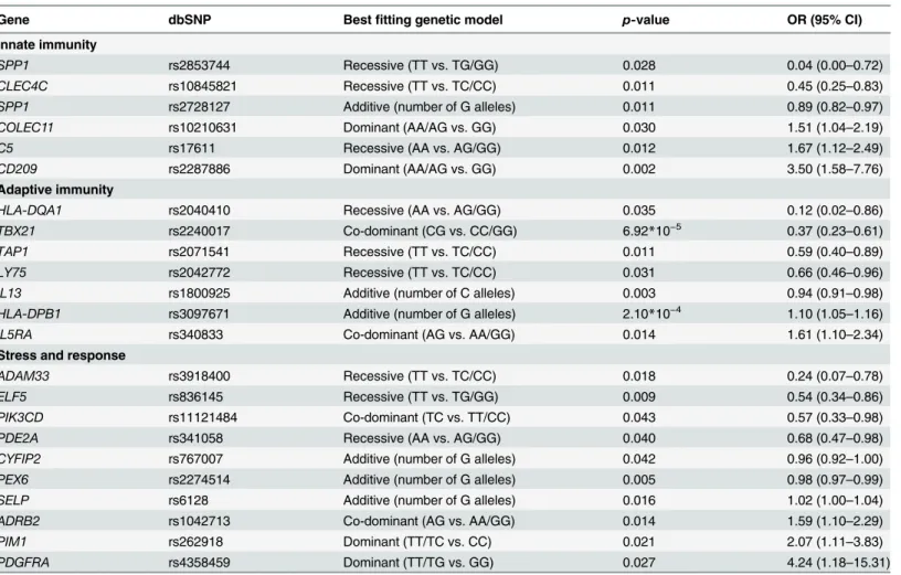
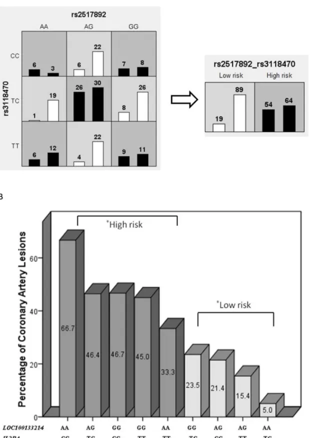
We identified pooled SNPs with the highest training-balanced accuracy in one-, two-, and three-way best fit models. Significant associations were only observed with two-locus models after permutation testing (p0.05). For the increased risk of KD susceptibility, two SNPs of PDE2A(rs341058) andCYFIP2(rs767007) revealed the highest training-balanced accuracy average (53.7%) in the two-way model (Table 3), with an OR = 3.54 in logistic regression (p= 4.14 x 10⁻⁷). For the subsequent risk of CAL formation in KD patients, two SNPs of LOC100133214(rs2517892) andIL2RA(rs3118470) revealed the highest average training-bal-anced accuracy (53.6%) in the two-way model (Table 4), with an OR = 5.35 in logistic regres-sion (p= 7.46 x 10⁻⁵).
The MDR results of these significant associations in the two-locus model were further ana-lyzed using the Chi-square test. As shown inFig 1A and 1B, our classification of varying allele combinations for thePDE2AandCYFIP2SNPs identified in MDR revealed four low-risk geno-types (n = 358) and five high-risk genogeno-types (n = 443) that significantly differed between the KD group and cohort controls (p= 9.71 x 10⁻⁷). As shown inFig 2A and 2B, our classification of varying allele combinations for theLOC100133214andIL2RASNPs revealed four low-risk genotypes (n = 118) and five high-risk genotypes (n = 108) that differed significantly between KD patients with subsequent CAL formation and KD patients without CAL (p= 3.36 x 10⁻⁶). Among the nine allele classifications, we observed that combinations of the AG allele for PDE2Aand the CC allele forCYFIP2conferred the highest risk of KD susceptibility (42.9%), while combinations of the CC allele forCYFIP2and the GG allele forPDE2Aconferred the lowest risk (15.0%). No significant associations were observed between these genotype Table 2. Multivariate analysis of 345 SNPs associated with CAL formation in KD patients (p0.05).
Gene dbSNP Bestfitting genetic model p-value OR (95% CI)
Innate immunity
CD209 rs12611071 Recessive (AA vs. AC/CC) 0.007 0.20 (0.06–0.64)
NOD2 rs2111235 Additive (number of C alleles) 0.006 1.28 (1.07–1.53)
CLEC2D rs1863873 Co-dominant (TC vs. TT/CC) 0.032 2.36 (1.08–5.16)
CXCL10 rs867562 Co-dominant (AG vs. AA/GG) 0.002 3.41 (1.55–7.49)
CCL24 rs2302004 Co-dominant (TC vs. TT/CC) 0.001 3.64 (1.64–8.07)
CD14 rs2569190 Dominant (AA/AG vs. GG) 0.005 5.72 (1.67–19.60)
Adaptive immunity
IL4 rs2243250 Dominant (TT/TC vs. CC) 0.006 0.03 (0.00–0.38)
CD80 rs1485332 Additive (number of G alleles) 0.001 1.22 (1.09–1.38)
MS4A2 rs2583476 Co-dominant (TC vs. TT/CC) 0.001 3.81 (1.69–8.57)
Stress and response
LTC4S rs730012 Co-dominant (AC vs. AA/CC) 0.027 0.29 (0.10–0.87)
ADAM33 rs3918400 Additive (number of C alleles) 0.015 0.91 (0.84–0.98)
EHF rs286902 Recessive (AA vs. AG/GG) 0.003 3.31 (1.51–7.26)
combinations of theCYFIP2andPDE2ASNPs with the development of either CAL formation or IVIG resistance in KD (p<0.10). We also found that combinations of the AA allele for
LOC100133214and the CC allele forIL2RAconferred the highest risk of CAL formation in KD patients (66.7%), while combinations of the TC allele ofIL2RAand the AA allele of LOC100133214conferred the lowest risk (5.0%). We did not observe any significant associa-tions for these genotypes with either KD risk or the development of IVIG resistance (p<0.10).
High-risk allele combinations of thePDE2AandCYFIP2SNPs accounted for 67.1% of our KD cases. As shown inFig 3A, we observed that these high-risk genotypes in KD patients were significantly associated with reduced plasma levels of TGF-β1 (9489 ± 1605 vs. 16133 ± 3015 pg/ml;p= 0.036) compared to KD patients in the low-risk group. High-risk allele combina-tions of theLOC100133214andIL2RASNPs accounted for 47.9% of our KD cases. We found significantly elevated plasma levels of interleukin (IL)-2 (14.1 ± 1.6 vs. 9.6 ± 1.2 pg/ml;p= 0.028), IL-6 (51.0 ± 14.3 vs. 18.4 ± 3.7 pg/ml;p= 0.033), and Interferon-γ(119.2 ± 15.2 vs. 81.8 ± 10.1 pg/ml;p= 0.041) in KD patients with the high-risk genotypes of CAL formation compared to KD patients in the low-risk CAL formation genotype group, as shown inFig 3B, 3C and 3D. No significant differences were found between high- and low-risk genotypes in KD patients with levels of IL-3, IL-4, IL-5, IL-10, or IL-17A.
Table 3. Best fit results using multifactor dimensionality reduction analysis of one-, two-, and three-locus models for KD susceptibility in patients and cohort controls.
Gene (polymorphism) aAverage testing balanced
accuracy
bAverage cross validation
consistency
cp-value
TBX21 (rs2240017) 50.32% 12/20 0.868
PDE2A (rs341058) and CYFIP2 (rs767007) 53.73% 17/20 0.021**
STAT3 (rs1026916), CLEC7A (rs2078178), and PSMB8 (rs3763364)
43.24% 6/20 0.999
a
Average testing balanced accuracy is the accuracy of classifications for cases and controls in the testing dataset (one-twentieth of the data) calculated as (Sensitivity+Specificity)/2.
b
Average cross validation consistency is the number of times the model was selected as the best model after 20 cross-validation runs. c
Significance of accuracy (empirical p-value based on 10,000 permutations). **p-value0.05
doi:10.1371/journal.pone.0143056.t003
Table 4. Best fit results using multifactor dimensionality reduction analysis of one-, two-, and three-locus models for CAL formation in KD patients.
Gene (polymorphism) aAverage testing balanced
accuracy
bAverage cross validation
consistency
cp-value
LY75 (rs2042772) 45.59% 12/20 0.588
LOC100133214 (rs2517892) and IL2RA (rs3118470) 53.60% 13/20 0.021**
FGF1 (rs249923), CLEC2D (rs1863873), and CCL2 (rs2857656)
43.42% 5/20 0.942
aAverage testing balanced accuracy is the accuracy of classi
fications for cases and controls in the testing dataset (one-twentieth of the data) calculated as (Sensitivity+Specificity)/2.
bAverage cross validation consistency is the number of times the model was selected as the best model after 20 cross-validation runs. cSigni
ficance of accuracy (empirical p-value based on 10,000 permutations). **p-value0.05
Fig 1.PDE2A(rs341058) andCYFIP2(rs767007) gene-gene interaction in a two-way mode of MDR analysis.The interaction ofPDE2AandCYFIP2
Discussion
To investigate gene-gene associations for the development of KD or its outcomes, we used an MDR method of analysis to establish the best fit models after repeated permutation testing for either individual or multiple genes. The cross-validation/permutation procedure incorporated in our MDR analysis minimizes false positive rates that result from multiple comparisons. This method of MDR analysis is able to identify a high order of evidence for gene-gene interactions in diseases when there is a lack of any primary individual genes that influence susceptibility [21], including autoimmune disorders and certain malignancies[22]. Therefore, MDR is of potential value in genetic studies of KD, which lacks a primary susceptibility marker. Numer-ous genes to date have been found to be associated with the development or prognosis of KD, which probably reflects the multi-genetic nature of the disease. Our approach using MDR anal-ysis more closely matches the seemingly complex nature of genetics in KD and allows us to examine genes in a more unbiased manner, by taking into account the effect of gene-gene inter-actions, as opposed to the effect of one candidate gene alone. As reported in previous studies, we observed much stronger associations with KD and its outcomes for multiple genes com-pared to individual genes. We found that the two-locus models were significantly associated with either the development of KD or the subsequent formation of CAL (p0.05), although no statistically significant associations for any of the individual genes in our one-locus model after repeated permutation testing. This was seen as a surprising development given the fact that we identified allele variations in nearly three dozen individual SNPs that were significantly associated with KD risk or CAL formation.
We also observed that varying alleles of individual SNPs or gene-gene combinations were associated with either the development of KD or its subsequent complications. We also found that KD patients who possessed the high-risk genotypes of our identified gene-gene associa-tions also had significantly different levels of the immune and inflammatory markers that were tested in this study. Lastly, our analyses using an MDR method yielded the most significant results with the lowestp-value compared to our analyses using univariate or multivariate methods.
In our initial investigations using UVA and MVA, we found several dozen individual SNPs and allele variations that were significantly associated with either an increased risk of KD sus-ceptibility or the formation of CAL. Among our results, we found that the dominant A allele of an SNP for theDC-SIGN(CD209) promoter gene (rs2287886) was significantly associated with an increased risk of KD susceptibility (OR = 3.50;p= 0.002). Previously, Portmanet al. reported that the major A allele of the rs2287886 SNP for theCD209gene was significantly associated with IVIG-treatment resistance during acute KD among Asian children in a US pop-ulation (OR = 1.76;p= 0.04)[23]. The authors did not find a similar association among either Caucasian or Hispanic children, for whom the A allele was only of minor frequency. We recently reported that haplotypes ofCD209polymorphisms were significantly associated with an increased risk of KD susceptibility in Taiwanese children[24], which included the major A allele of the rs2287886 SNP (OR = 1.61;p= 0.0002). However, we did not observe significant associations between any polymorphisms ofCD209polymorphisms and IVIG-treatment response for Taiwanese children with KD.
In addition, we found that KD patients with the dominant A allele of an SNP forCD14 (rs2569190) were at greatest risk for the development of CAL formation (OR = 5.76,
odds ratio of 3.54 (95% CI: 2.17–5.78) and ap-value of 4.14 x 10⁻⁷. (A) MDR classified the nine interactive items of allele combinations into high- or low-risk
KD groups, which were significantly different in our further analysis using the Chi-square test (p = 9.71 x 10⁻⁷). (B).
Fig 2.LOC100133214(rs2517892) andIL2RA(rs3118470) gene-gene interaction in a 2-way mode of MDR analysis.The interaction ofLOC100133214
p= 0.005). Previously, an investigation in Japan reported that KD patients with the T allele of theCD14C(−159)T polymorphism (rs2569190) were significantly more likely to develop CAL
(OR = 2.20;p<0.05), although no associations were found with KD susceptibility[25]. We
also observed that KD patients with the dominant T allele of an SNP for theIL-4gene (rs2243250) had the lowest risk of developing CAL formation (OR = 0.03;p= 0.006). Previ-ously, Burnset al. reported the significant asymmetrical transmission of alleles for theIL-4C (-589)T polymorphism (rs2243250) from parents to their children with KD (p= 0.05) in a US population, although no association was observed with CAL formation[26]. However, SNPs of theIL-4gene have not been found to be associated with KD susceptibility or subsequent CAL formation among children in Taiwan[27,28].
and KD patients without CAL (n = 153), with an odds ratio of 5.35 (95% CI: 2.33–12.25) and ap-value of 7.46 x 10⁻⁵. (A) MDR classified the nine interactive
items into high- or low-risk CAL groups, which were significantly different in our further analysis using the Chi-square test (p= 3.36 x 10⁻⁶). (B).
doi:10.1371/journal.pone.0143056.g002
Fig 3. Comparison cytokines levels between KD patients with high-risk genotypes and low-risk genotypes.KD patients possessing the high-risk (KD risk: 1)PDE2A(rs341058) andCYFIP2(rs767007) genotypes of KD susceptibility (n = 49) presented with significantly lower plasma levels of TGF-β1 (9489±1605 vs. 16133±3015) compared to KD patients in the low-risk group (KD risk: 0, n = 24), with an odds ratio of 0.59 (p= 0.036). (A) KD patients possessing the high-riskLOC100133214(rs2517892) andIL2RA(rs3118470) genotypes of CAL formation (CAL risk: 1, n = 35) presented with significantly elevated plasma levels of IL-2 (14.1±1.6 vs. 9.6±1.2) compared to KD patients in the low-risk group (CAL risk: 0, n = 38), with an odds ratio of 1.47
(p= 0.028). (B) KD patients possessing the high-riskLOC100133214(rs2517892) andIL2RA(rs3118470) genotypes of CAL formation (CAL risk: 1, n = 35) presented with significantly elevated plasma levels of IL-6 (51.0±14.3 vs. 18.4±3.7) compared to KD patients in the low-risk group (CAL risk: 0, n = 38), with an odds ratio of 2.77 (p= 0.033). (C) KD patients possessing the high-riskLOC100133214(rs2517892) andIL2RA(rs3118470) genotypes of CAL formation (CAL risk: 1, n = 35) presented with significantly elevated plasma levels of IFN-γ(119.2±15.2 vs. 81.8±10.1) compared to KD patients in the low-risk group
(CAL risk: 0, n = 38), with an odds ratio of 1.46 (p= 0.041). (D).
In our two-locus model, combined possession of SNPs for thePDE2A(rs341058) and the CYFIP2(rs767007) gene were significantly associated with increased KD susceptibility in logis-tic regression (OR = 3.54;p= 4.14 x 10⁻⁷), although no associations were found for the risk of CAL formation or responsiveness to IVIG treatment. Respectively, we observed that KD patients with both SNPs forLOC100133214(rs2517892) andIL2RA(rs3118470) were signifi-cantly more likely to develop CAL formation in logistic regression (OR = 5.35;p= 7.46 x 10⁻⁵), although no associations were found with the risk of developing KD or the response to IVIG treatment during the disease course. We observed even lowerp-values in the Chi-square test for both of the identified gene-gene associations after we had separated individuals into and low-risk genotype groups by their respective allele combinations. KD patients with high-risk genotypes for thePDE2AandCYFIP2SNPs had significantly reduced plasma levels of TGF-β1 (OR = 0.59;p= 0.036) compared to the low-risk KD group, while KD patients with high-risk allele combinations for theLOC100133214andIL2RASNPs had significantly ele-vated plasma IL-2 (OR = 1.47;p= 0.028), IL-6 (OR = 2.77;p= 0.033), and IFN-γ(OR = 1.46; p= 0.041) compared to the low-risk KD group.
In our current investigation,PDE2A(rs341058) andCYFIP2(rs767007) were the only SNPs significantly associated in UVA, MVA, and MDR analysis. To our knowledge, no other study to date has found a link betweenCYFIP2SNPs and the development or prognosis of KD, although SNPs in the geneCYFIP2has been found to be associated with allergic disease. In a study of 492 Mexican children, rs17599222 in the geneCYFIP2was found to be associated with childhood asthma[29]. Similarly, in our previous study, we found that the combination of PDE2A(rs341058) andCYFIP2(rs767007) plus an SNP ofIL-13(rs1800925) in a three-locus model was significantly associated with increased IgE production in Taiwanese children who lacked a history of KD[15]. Recent populations studies have also found that patients with Kawasaki disease appear to have a higher subsequent risk of developing atopic dermatitis and other allergic diseases[30,31], suggesting that both KD and allergic disease may share a similar immune response. It is possible that CYFIP2 is implicated in a common pathway shared by both KD and allergy, although more research regarding the functional effect ofCYFIP2 (rs767007) SNP mutations would be required to confirm this hypothesis.
Likewise, the link betweenPDE2A(rs341058) SNP and Kawasaki disease found in this study, is to our knowledge, a novel finding. ThePDE2Agene encodes for phosphodiesterase 2 (PDE2), which increases the hydrolysis of cAMP after being activated by cGMP. Overexpres-sion of PDE2 has been found in human myocytes of patients with heart failure, and bluntsβ -adrenergic responses via decreasing the cAMP stimulation of the L-type Ca2+current[32]. This finding suggests that development of Kawasaki disease may be associated with differences in myocardial calcium current conduction; of note, previous studies have linked KD susceptibility to the C allele of theITPKCSNP (rs28493229) a gene associated with the Ca2+/NFAT pathway [33,34]. Tumor necrosis factor-alpha, a cytokine that is critical in the development coronary artery lesions in KD, has been found to upregulate PDE2 in cultivated human umbilical vein endothelial cells, and may play a role in increased endothelial permeability[35].
system, which results in an elevation of immune and inflammatory cells. As genetic studies are largely used to identify pathways involved in KD so that its environmental causes are ultimately being discovered, it is therefore concluded that theITPKCsusceptibility in KD sensitizes indi-viduals to xenobiotics that induce calcium influx[33,34].
In conclusion, our findings indicate that differing gene-gene interactions appear to be respectively associated with predisposition for the development of KD or CAL formation. Varying gene-gene interactions may account for why individual susceptibility loci in KD appear to be fairly modest or inconsistent upon larger replication and meta-analysis, while stronger associations are observed when these genes are present in combination. This suggests a fairly high order of genetic variation for KD and may reflect a multifactorial etiology in the disease process that impacts the same general pathways. The highest incidence of KD in Tai-wan is 69 cases per 100,000 children under 5 years of age[37]. The major limitation of this study is that the low sample size, which reflected the difficulties inherent in recruiting patients with such a rare disease. Replication of the findings in large, well-powered independent sam-ples is crucial if this problem is to be overcome, and will likely require a multicenter collabora-tion to provide strong evidence on validacollabora-tion of the novel gene to gene interaccollabora-tions discovered the susceptibility of Kawasaki disease and coronary artery lesions. This study only analyzed patients with KD in our current investigation without a formal assessment of population struc-ture of the sampled population; there is still a possibility that the observed positive association is over-represented. These limitations should be considered in future studies.
Supporting Information
S1 Table. The SNPs excluded for the final analysis due to call rate<90% or their genotypes’ distributions beyond Hardy Weinberg equilibrium (HWE)≦0.001.
(DOC)
S2 Table. Association of 31 SNPs in 27 innate, adaptive and stress/response genes with CB and KD cohort UVA analysis (p<0.1).
(DOC)
S3 Table. Association of 22 SNPs in 21 innate, adaptive and stress/response genes with/ without CAL in KD cohort by UVA analysis (p<0.1).
(DOC)
Acknowledgments
The authors would also like to convey thanks to the Genomic and Proteomic Core Laboratory, Department of Medical Research, Kaohsiung Chang Gung Memorial Hospital for providing the laboratory facilities.
Author Contributions
Conceived and designed the experiments: HCK KDY. Performed the experiments: MMHG JCC SCL. Analyzed the data: JCC MMHG. Contributed reagents/materials/analysis tools: JCC HCK KDY. Wrote the paper: HCK MMHG KSH KDY DY.
References
2. Newburger JW, Takahashi M, Gerber MA, Gewitz MH, Tani LY, Burns JC, et al. Diagnosis, treatment, and long-term management of Kawasaki disease: a statement for health professionals from the Com-mittee on Rheumatic Fever, Endocarditis and Kawasaki Disease, Council on Cardiovascular Disease in the Young, American Heart Association. Circulation. 2004; 110(17):2747–71. Epub 2004/10/27. doi: 110/17/2747 [pii] doi:10.1161/01.CIR.0000145143.19711.78PMID:15505111.
3. Burns JC, Glode MP. Kawasaki syndrome. Lancet. 2004; 364(9433):533–44. Epub 2004/08/11. doi: 10.1016/S0140-6736(04)16814-1S0140-6736(04)16814-1 [pii]. PMID:15302199.
4. Wang CL, Wu YT, Liu CA, Kuo HC, Yang KD. Kawasaki disease: infection, immunity and genetics. Pediatr Infect Dis J. 2005; 24(11):998–1004. Epub 2005/11/12. doi: 00006454-200511000-00015 [pii]. PMID:16282937.
5. Holman RC, Curns AT, Belay ED, Steiner CA, Effler PV, Yorita KL, et al. Kawasaki syndrome in Hawaii. Pediatr Infect Dis J. 2005; 24(5):429–33. Epub 2005/05/07. doi: 00006454-200505000-00007 [pii]. PMID:15876942.
6. Onouchi Y. Genetics of Kawasaki disease: what we know and don't know. Circ J. 2012; 76(7):1581–6. Epub 2012/07/14. doi: DN/JST.JSTAGE/circj/CJ-12-0568 [pii]. PMID:22789975.
7. Onouchi Y, Ozaki K, Burns JC, Shimizu C, Terai M, Hamada H, et al. A genome-wide association study identifies three new risk loci for Kawasaki disease. Nat Genet. 2012; 44(5):517–21. Epub 2012/03/27. doi:10.1038/ng.2220ng.2220 [pii]. PMID:22446962.
8. Lee YC, Kuo HC, Chang JS, Chang LY, Huang LM, Chen MR, et al. Two new susceptibility loci for Kawasaki disease identified through genome-wide association analysis. Nat Genet. 2012; 44(5):522– 5. Epub 2012/03/27. doi:10.1038/ng.2227ng.2227 [pii]. PMID:22446961.
9. Khor CC, Davila S, Breunis WB, Lee YC, Shimizu C, Wright VJ, et al. Genome-wide association study identifies FCGR2A as a susceptibility locus for Kawasaki disease. Nat Genet. 2011; 43(12):1241–6. Epub 2011/11/15. doi:10.1038/ng.981ng.981 [pii]. PMID:22081228.
10. Kuo HC, Hsu YW, Wu CM, Chen SH, Hung KS, Chang WP, et al. A Replication Study for Association of ITPKC and CASP3 Two-Locus Analysis in IVIG Unresponsiveness and Coronary Artery Lesion in Kawasaki Disease. PLoS One. 2013; 8(7):e69685. Epub 2013/07/31. doi:10.1371/journal.pone. 0069685PONE-D-12-35484 [pii]. PMID:23894522; PubMed Central PMCID: PMC3722201.
11. Onouchi Y, Suzuki Y, Suzuki H, Terai M, Yasukawa K, Hamada H, et al. ITPKC and CASP3 polymor-phisms and risks for IVIG unresponsiveness and coronary artery lesion formation in Kawasaki disease. Pharmacogenomics J. 2013; 13(1):52–9. Epub 2011/10/12. doi:10.1038/tpj.2011.45tpj201145 [pii]. PMID:21987091.
12. Kuo HC, Yang KD, Liang CD, Bong CN, Yu HR, Wang L, et al. The relationship of eosinophilia to intra-venous immunoglobulin treatment failure in Kawasaki disease. Pediatr Allergy Immunol. 2007; 18 (4):354–9. Epub 2007/06/23. doi: PAI516 [pii] doi:10.1111/j.1399-3038.2007.00516.xPMID: 17584314.
13. Kuo HC, Wang CL, Liang CD, Yu HR, Huang CF, Wang L, et al. Association of lower eosinophil-related T helper 2 (Th2) cytokines with coronary artery lesions in Kawasaki disease. Pediatr Allergy Immunol. 2009; 20(3):266–72. Epub 2009/05/15. doi: PAI779 [pii] doi:10.1111/j.1399-3038.2008.00779.xPMID: 19438983.
14. Guidelines for diagnosis and management of cardiovascular sequelae in Kawasaki disease (JCS 2008)—digest version. Circ J. 2010; 74(9):1989–2020. Epub 2010/08/21. doi: JST.JSTAGE/circj/CJ-10-74-0903 [pii]. PMID:20724794.
15. Yang KD, Chang JC, Chuang H, Liang HM, Kuo HC, Lee YS, et al. Gene-gene and gene-environment interactions on IgE production in prenatal stage. Allergy. 2010; 65(6):731–9. Epub 2009/12/09. doi: ALL2260 [pii] doi:10.1111/j.1398-9995.2009.02260.xPMID:19968631.
16. Chang JC, Kuo HC, Hsu TY, Ou CY, Liu CA, Chuang H, et al. Different Genetic Associations of the IgE Production among Fetus, Infancy and Childhood. PLoS One. 2013; 8(8):e70362. Epub 2013/08/13. doi:10.1371/journal.pone.0070362PONE-D-12-39897 [pii]. PMID:23936416; PubMed Central PMCID: PMC3731352.
17. Gao X. Multiple testing corrections for imputed SNPs. Genet Epidemiol. 2011; 35(3):154–8. Epub 2011/01/22. doi:10.1002/gepi.20563PMID:21254223; PubMed Central PMCID: PMC3055936.
18. Hahn LW, Ritchie MD, Moore JH. Multifactor dimensionality reduction software for detecting gene-gene and gene-environment interactions. Bioinformatics. 2003; 19(3):376–82. Epub 2003/02/14. PMID: 12584123.
20. Ritchie MD. Bioinformatics approaches for detecting gene-gene and gene-environment interactions in studies of human disease. Neurosurg Focus. 2005; 19(4):E2. Epub 2005/10/26. PMID:16241104.
21. Moore JH, Gilbert JC, Tsai CT, Chiang FT, Holden T, Barney N, et al. A flexible computational frame-work for detecting, characterizing, and interpreting statistical patterns of epistasis in genetic studies of human disease susceptibility. J Theor Biol. 2006; 241(2):252–61. Epub 2006/02/07. doi: S0022-5193 (05)00521-7 [pii] doi:10.1016/j.jtbi.2005.11.036PMID:16457852.
22. Gui J, Moore JH, Kelsey KT, Marsit CJ, Karagas MR, Andrew AS. A novel survival multifactor dimensionality reduction method for detecting gene-gene interactions with application to bladder can-cer prognosis. Hum Genet. 2011; 129(1):101–10. Epub 2010/10/29. doi:10.1007/s00439-010-0905-5 PMID:20981448; PubMed Central PMCID: PMC3255326.
23. Portman MA, Wiener HW, Silva M, Shendre A, Shrestha S. DC-SIGN gene promoter variants and IVIG treatment response in Kawasaki disease. Pediatr Rheumatol Online J. 2013; 11(1):32. Epub 2013/09/ 07. doi:10.1186/1546-0096-11-321546-0096-11-32 [pii]. PMID:24006904; PubMed Central PMCID: PMC3847673.
24. Kuo HC, Huang YH, Chien SC, Yu HR, Hsieh KS, Hsu YW, et al. Genetic variants of CD209 associated with Kawasaki disease susceptibility. PLoS One. 2014; 9(8):e105236. Epub 2014/08/26. doi:10.1371/ journal.pone.0105236PONE-D-14-16880 [pii]. PMID:25148534; PubMed Central PMCID: PMC4141786.
25. Nishimura S, Zaitsu M, Hara M, Yokota G, Watanabe M, Ueda Y, et al. A polymorphism in the promoter of the CD14 gene (CD14/-159) is associated with the development of coronary artery lesions in patients with Kawasaki disease. J Pediatr. 2003; 143(3):357–62. Epub 2003/10/01. doi: S0022-3476(03)00330-5 [pii] doi:10.1067/S0022-3476(03)00330-5PMID:14517520.
26. Burns JC, Shimizu C, Shike H, Newburger JW, Sundel RP, Baker AL, et al. Family-based association analysis implicates IL-4 in susceptibility to Kawasaki disease. Genes Immun. 2005; 6(5):438–44. Epub 2005/05/13. doi: 6364225 [pii] doi:10.1038/sj.gene.6364225PMID:15889128.
27. Weng KP, Ho TY, Chiao YH, Cheng JT, Hsieh KS, Huang SH, et al. Cytokine genetic polymorphisms and susceptibility to Kawasaki disease in Taiwanese children. Circ J. 2010; 74(12):2726–33. Epub 2010/11/05. doi: JST.JSTAGE/circj/CJ-10-0542 [pii]. PMID:21048327.
28. Huang FY, Chang TY, Chen MR, Lee HC, Chiu NC, Chi H, et al. The -590 C/T and 8375 A/G interleu-kin-4 polymorphisms are not associated with Kawasaki disease in Taiwanese children. Hum Immunol. 2008; 69(1):52–7. Epub 2008/02/26. doi: S0198-8859(07)00468-5 [pii] doi:10.1016/j.humimm.2007. 11.002PMID:18295676.
29. Wu H, Romieu I, Shi M, Hancock DB, Li H, Sienra-Monge JJ, et al. Evaluation of candidate genes in a genome-wide association study of childhood asthma in Mexicans. J Allergy Clin Immunol. 2010; 125 (2):321–7 e13. Epub 2009/11/17. doi:10.1016/j.jaci.2009.09.007PMID:19910030; PubMed Central PMCID: PMC2823974.
30. Kuo HC, Chang WC, Yang KD, Yu HR, Wang CL, Ho SC, et al. Kawasaki disease and subsequent risk of allergic diseases: a population-based matched cohort study. BMC Pediatr. 2013; 13:38. Epub 2013/ 03/26. doi:10.1186/1471-2431-13-381471-2431-13-38 [pii]. PMID:23522327; PubMed Central PMCID: PMC3614461.
31. Brosius CL, Newburger JW, Burns JC, Hojnowski-Diaz P, Zierler S, Leung DY. Increased prevalence of atopic dermatitis in Kawasaki disease. Pediatr Infect Dis J. 1988; 7(12):863–6. Epub 1988/12/01. PMID:3211629.
32. Mehel H, Emons J, Vettel C, Wittkopper K, Seppelt D, Dewenter M, et al. Phosphodiesterase-2 is up-regulated in human failing hearts and blunts beta-adrenergic responses in cardiomyocytes. J Am Coll Cardiol. 2013; 62(17):1596–606. Epub 2013/07/03. doi:10.1016/j.jacc.2013.05.057PMID:23810893.
33. Yeter D, Deth R. ITPKC susceptibility in Kawasaki syndrome as a sensitizing factor for autoimmunity and coronary arterial wall relaxation induced by thimerosal's effects on calcium signaling via IP3. Auto-immun Rev. 2012; 11(12):903–8. Epub 2012/04/14. doi:10.1016/j.autrev.2012.03.006S1568-9972 (12)00075-4 [pii]. PMID:22498790.
34. Rowley AH. Kawasaki disease: novel insights into etiology and genetic susceptibility. Annu Rev Med. 2011; 62:69–77. Epub 2010/08/10. doi:10.1146/annurev-med-042409-151944PMID:20690826; PubMed Central PMCID: PMC3021097.
35. Seybold J, Thomas D, Witzenrath M, Boral S, Hocke AC, Burger A, et al. Tumor necrosis factor-alpha-dependent expression of phosphodiesterase 2: role in endothelial hyperpermeability. Blood. 2005; 105 (9):3569–76. Epub 2005/01/15. doi:10.1182/blood-2004-07-2729PMID:15650061.
36. Onouchi Y, Gunji T, Burns JC, Shimizu C, Newburger JW, Yashiro M, et al. ITPKC functional polymorphism associated with Kawasaki disease susceptibility and formation of coronary artery aneurysms. Nat Genet. 2008; 40(1):35–42. Epub 2007/12/18. doi: ng.2007.59 [pii] doi:10.1038/ng.2007.59PMID:18084290.