REGULAR ARTICLE
MYCOTOXINS CONTAMINATION IN EDIBLE LAND SNAIL AT GRAZING
PADDOCK ENVIRONMENT
Ime Ebenso*1, Uyimeobong Ekwere1, Nkoyo Isong2
Address: 1 Department of Animal Science,University of Uyo, Nigeria.
2
Department of Human Ecology, Nutrition and Dietetics, University of Uyo, Nigeria.
*Corresponding author: [email protected]
ABSTRACT
Mycotoxins contamination of animal products is under reported. Juvenile edible land
snails (Archachatina marginata) were exposed as sentinels in bottomless metal drums for 1
week at abandoned, new and reference sites respectively at grazing paddock environment, to
assess the presence of foodborne microbiological mycotoxins contamination during the dry
season. Mycological analysis of A. marginata samples revealed high (p<0.05) contamination
at all paddocks ranged from 1.2-1.3 x 10⁵ cfu-g. Results revealed values that were found to be
unacceptable by FAO/WHO standards. The presence of Aspergillus niger, A. fumigatus and
Penicillum expansum were noted as potential toxicogenic mycoflora. Snails were tolerant to
all levels of contamination with no clinical signs of infection or mortality. This finding could
serve as basis for assessing pre-slaughter microbial contamination of livestock farm/field
environment in order to establish data with comparative epidemiological value, which could
highlight early warning signals of food safety risk and cross-contamination of mycotoxins in
the food chain.
INTRODUCTION
Giant African land snails (GALS) are relished as delicacy by indigenes of Niger Delta
region of Nigeria (Ebenso and Ebenso, 2011). Previous study provides evidence of edible
snail as a bioindicator of transfer of foodborne (bacterial) pathogens within the food chain and
for food safety assessment (Ebenso et al., 2012). Hence, edible snail provides early warning
signal of toxins and pollutants in the environment (Ebenso, 2012).
Used as sentinels, snails are the representative primary consumers in the terrestrial
ecosystem (Naaem et al., 1994). Snails are involved in many aspects of organic matter
decomposition, potential regulation of microbial activities, nutrient cycles and crumby
structures (Cortert et al., 1999).
Mycotoxins comprise a family of fungal toxins, many of which have been implicated
as chemical progenitors of toxicity to man and animals (Zaki et al., 2012). Mycotoxins are
regarded as extrolites (secondary metabolites of fungal origin) are odourless, tasteless and
colourless (Fapohunda, 2012). Mycotoxins are toxic to humans and animals, which explains
the major concern of food and feed industry in preventing them from entering the food chain
(Pierre, 2007). Mycotoxins have been associated with a number of human diseases. Beardall
and Miller, (1994) have given detailed account of human illnesses that have been associated
with mycotoxin ingestion. Chronic intake is the most widespread form of human health
exposure. Bryden, (2007) reported that the impact of regular low level of mycotoxins on
human health is likely to be significant with the possible consequences including impaired
growth and development, immune dysfunction and the disease consequences of alleviation in
DNA metabolism.
Human food can be contaminated by mycotoxins at the various stages in the food
chain from the genera Aspergillus (aflatoxins) and Penicillum (ochratoxins). The disease
resulting from mycotoxin exposure is mycotoxicosis (Bryden, 2007). Human exposure may
result from the carry over of mycotoxins and metabolites into animal products such as milk,
meat and eggs (CAST, 2003). According to Pestka, (1995) trace levels of mycotoxins and
their metabolites may carryover into edible tissue (meat) of food producing animals.
There is now overwhelming epidemiological evidence (Wild and Hall, 1996) that
aflatoxin consumption contributes significantly to the high incidence of human liver cancer
(Henry et al., 1999) and ochratoxin is nephrotoxic, a possible cause of urinary tract tumour
(Peraica et al., 1999). Consumption of a mycotoxin contaminated diet may induce acute and
suppressive effects. Gong et al., (2002) revealed a strong association between exposure to
aflatoxin in children and both stunting and being underweight.
The objective of this study was aimed at using juvenile edible snail (Archachatina
marginata) as sentinels at grazing paddocks to determine microscopic fungi involved in
pre-slaughter contamination and to demonstrate potential adverse effects of mycotoxins within the
livestock farm environment.
MATERIALS AND METHODS
Animal Management
The design of the experiment was completely randomized design. The experimental
area was the Teaching and Research Farm, University of Uyo, within latitude 4o31´N and
4o45´N, longitude 7o31´E and 45o51´E with mean temperature of 30oC and rainfall of 2000 –
3000mm per annum.
The 135 juvenile A. marginata snails 100±5.00g were randomly assigned to 3
treatment sites of grazing paddocks within the cattle unit namely, old (abandoned, no longer
used for 1 year), new (currently in use) and reference (control) paddocks respectively, using
45 snails replicated 3 times.
The microcosm was bottomless metal drums (0.6m diameter and 0.6m high) with
perforated lids for air, light and protects snails against predators. The snails as sentinels (from
uncontaminated laboratory stock) were transferred to the bottomless microcosm (suitable for
contact with soil and vegetation). These microcosms were positioned 20m apart per paddock.
During the experimental period of 1 week in October (dry season), snails consumed food (soil
and vegetation) in situ and ad libitum.
Sample Collection
Fresh snail samples for microbiological analyses were collected in labelled sterile
isotherm polythene bags and then transported to the laboratory. The snails were extensively
washed with water and rinsed with normal saline to remove all surface contaminants. The
edible parts of the snails were dissected to remove internal extracts from the proximal gut for
Identification and Enumeration of Bacteria
One gram of snail extract was diluted serially in ten fold dilution blanks and properly
mixed with sterile glass rod. The 0.1 ml of diluted sample was pipetted into sterile plate and molten sterile agar medium (45°C) was poured. The media used were plate count agar (PCA, Biotech), nutrient agar (NA Biotech), xylose lysine desoxycholate agar (XLD, Biotech) and
DeMan Rogosa Sharpe agar (MRS, Biotech). The plates were rotated gently to disperse
inoculum in medium and allowed to solidify. This was done in triplicates and plates were incubated at 37°C.
Mycoflora Identification
The snail extracts were transferred to a 0.9% saline solution (10 ml) and vortexed for 3
min. Then, 0.1 ml of this solution was transferred to Potato Dextrose Agar (PDA) plates.
Later, the collected snail extracts were surface-sterilized by consecutive washing in sterile
distilled water and 70% alcohol for 1 min (Pereira et al., 2009). Each snail was immersed in
10 ml of phosphate saline buffer. Dilutions x1000 of each sample were seeded onto PDA
plates. Plates were incubated at 28ºC.
Cultures growing on Potato Dextrose Agar (PDA) and Malt Extract Agar (Difco) were
identified according to microscopic observations such as morphological characters of
mycelium and conidia. Observations were made by staining the isolated fungus using
lactophenol cotton blue and examination under low-power microscope. The species were
identified according to Von Arx, (1981); Hasenekoglu, (1991); Domsch et al., (1993);
Watanabe, (2002). The organisms were maintained on PDA slants at 40C.
Statistical Analysis
Enumeration of samples, with mycoflora (n=100), were performed in triplicates. Data
were subjected to one way analysis of variance (ANOVA), using statistical analysis system
RESULTS AND DISCUSSION
The A. marginata positioned as sentinels in metal drums in this study did not record
any mortality or clinical infection; they were tolerant of high load of microbes. According to
Bryden, (2007) the effect of immunity and resistance are often difficult to recognize in the
field because signs of disease are associated with the infection rather than the toxin that
predispose the individual to infection through decreased resistance. May be the experimental
period of this study was not long enough for clinical signs in A. marginata, it was contrary to
reports of Bryden, (1982) that available evidence suggests that tissue accumulation of
mycotoxins or their metabolites was very low and that residues are excreted in a few days.
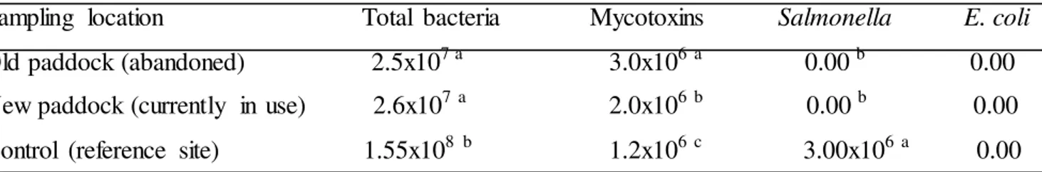
In Table 1, E. coli and Salmonella in A. marginata were not detected at the new and
old paddocks. According to Wilson et al., (2002) E. coli and Salmonella are gram negative
bacteria, and are infrequently recovered at feedlots, possibly because of their rapid
inactivation by UV irradiation (from the sun). Reports of previous study by Sproston et al.,
(2006) with slugs have shown no relationship between E. coli and Salmonella carriage, as
microbial analysis failed to detect pathogens. In fact, Smith et al., (2001) reported that the
prevalence of E. coli and Salmonella in cattle was greater in paddocks that were muddy and
wet. Callaway et al., (2004) indicated that detection of E. coli and Salmonella is also
complicated by the fact that, fecal shedding can be very sporadic, with an animal testing
positive one day, but not again for several days or even weeks.
Levels of mycotoxins in A. marginata in this study (Table 1), were higher (p<0.05)
and unacceptable compared with FAO/WHO mycotoxins standards/levels in fresh meat
(JECFA, 2001) consequently, the presence of moulds in meat and meat products could cause
a decrease in their biological value due to the enzymatic degradation of meat components
(Okin et al., 2001). According to Zaki et al., (2012) mycotoxins interfere with protein
formation. Kan and Meijer, (2007) reported that aflatoxin binds both RNA and DNA and
blocks transcription, while ochratoxin blocks RNA synthesize and thus blocks translation.
Mycotoxins are non-antigenic, but an antibody response can be elicited to the toxin after
conjugation to a protein or polypeptide carrier. In Table 1, the high total bacteria count is
well above limit of 104 by HPA, (2009); ICMSF, (1986). Melabolic interactions of
mycotoxins with bacterial pathogens has played important role in the outbreak of foodborne
illnesses.
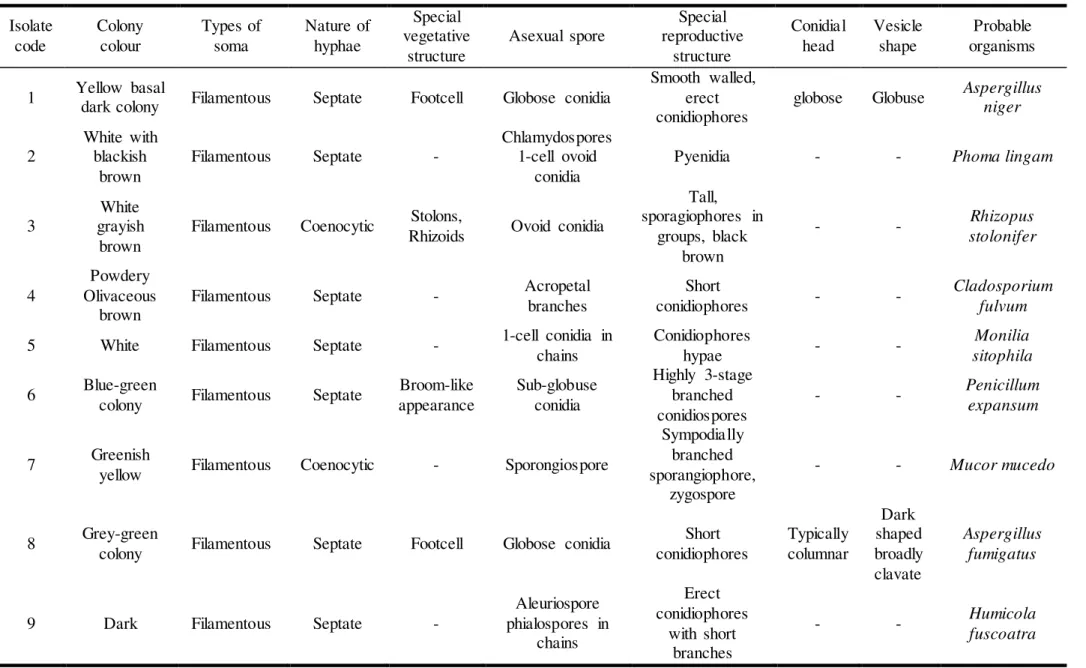
From analysed positive samples (Table 2 and 3), there were 9 fungi isolated from gut
This compare with report of Zaki et al., (2012) that concurrent exposure to multiple
mycotixins is more likely in the livestock industry. According to Erber and Binder, (2004)
mycotoxin contaminations are due to combinations of two or more than two toxins.
Co-occurrence of mycotoxins is of special concern, for instance, in the case of furmonisins (a
potent cancer promoter) and aflatoxin (a potent human carcinogen) where a complimentary
toxicity mechanism of actions occurs (Riley, 1998). In African and Asia, the co-occurrence
of these mycotoxins is common and a significant percentage of the population is infected with
Hepatitis B or C which leads to the conclusion that mycotoxins in this region can have a
devastating human effect (Bryden, 2007). In developing countries it is likely that consumers
will be confronted with a diet that contains a low level of toxin and in many cases there may
be other toxins present (Bryden et al., 2001). Suppression of cellular immune system is a
known result after ingestion of several mycotoxins Stoev et al., (2000).
During the dry season (period of experiementation), ambient temperature is high and
the dry paddock environment is predisposed to dust (fungi mycoflora are easily dispersed
through dust), hence snails could be contaminated by air containing fungal spores. Inhalation
is a route of toxic bioaccumulation (Ebenso and Ologhobo, 2008). The potential health
hazards of mycoflora need not be overemphasized, because, fungi following inhalation are
capable of surviving in the respiratory and intestinal tract, where they may continue to grow
and produce their toxins (Oyero and Oyefolu, 2011; Frisvad et al., 2007). According to
Ebenso and Ebenso, (2011) children who eat more than 0.18kg/meat of snail meat, and
above African maximum permissible limit for aflatoxin of 20mg-kg (Oyero and Oyefolu,
2011) will be at risk of additional chance of developing adverse health conditions. Reports of
Hendrickse, (1991) linked kwashiorkor to aflatoxin exposure.
A big setback is that most mycotoxins are heat resistant within the range of
conventional food processing temperature (80-120oC) of normal cooking conditions such as
boiling and frying or even following pasteurization (Zaki, 2012) every effort to prevent
mycotoxins should be integrative of physical, chemical and biological systems.
It can be speculated from the present study that livestock that grazed the paddocks
could present animal products of inferior market quality that could attract discount prices
(such as meat, animal skin and hide), could be carriers of sub-clinical mycotoxic illnesses, and
Table 1 The pathogenic profile (cfu-g) isolated from proximal gut of A. marginata at grazing paddocks
Sampling location Total bacteria Mycotoxins Salmonella E. coli
Old paddock (abandoned) 2.5x10⁷ a 3.0x10⁶ a 0.00 b 0.00
New paddock (currently in use) 2.6x10⁷a 2.0x10⁶ b 0.00 b 0.00
Control (reference site) 1.55x10⁸ b 1.2x10⁶ c 3.00x10⁶a 0.00
Legend: abc means with different alphabets in a column are significantly different by Duncan Multiple Range Test α = 0.05
Table 2 Positive samples (n=100) of microscopic fungal isolates (cfu-gx 105) from proximal gut of A. marginata at grazing paddocks
Fungi Control (reference site) Old paddock (abandoned) New paddock (currently in use)
Aspergillus niger 4 7 2
Phoma lingam 0 0 5
Rhizopus stolonifer 2 5 2
Cladosporium fulvum 1 2 0
Monilia sitophila 0 5 0
Penicillium expansum 3 0 9
Mucor mucedo 1 0 2
Aspergillus fumigatus 1 7 0
Table 3 Morphological examination and names of fungal isolates from proximal gut of A. marginata at grazing paddocks Isolate code Colony colour Types of soma Nature of hyphae Special vegetative structure
Asexual spore
Special reproductive structure Conidial head Vesicle shape Probable organisms
1 Yellow basal
dark colony Filamentous Septate Footcell Globose conidia
Smooth walled, erect conidiophores
globose Globuse Aspergillus niger
2
White with blackish
brown
Filamentous Septate -
Chlamydospores 1-cell ovoid
conidia
Pyenidia - - Phoma lingam
3
White grayish
brown
Filamentous Coenocytic Stolons,
Rhizoids Ovoid conidia
Tall, sporagiophores in
groups, black brown
- - Rhizopus
stolonifer
4
Powdery Olivaceous
brown
Filamentous Septate - Acropetal
branches
Short
conidiophores - -
Cladosporium fulvum
5 White Filamentous Septate - 1-cell conidia in chains
Conidiophores
hypae - -
Monilia sitophila
6 Blue-green
colony Filamentous Septate
Broom-like appearance
Sub-globuse conidia
Highly 3-stage branched conidiospores
- - Penicillum
expansum
7 Greenish
yellow Filamentous Coenocytic - Sporongiospore
Sympodially branched sporangiophore,
zygospore
- - Mucor mucedo
8 Grey-green
colony Filamentous Septate Footcell Globose conidia
Short conidiophores Typically columnar Dark shaped broadly clavate Aspergillus fumigatus
9 Dark Filamentous Septate -
Aleuriospore phialospores in
chains
Erect conidiophores
with short branches
- - Humicola
CONCLUSION
Aspergillus and Penicillum were detected as potential toxicogenic mycoflora in the
study of epidemiology of livestock environment. These foodborne mycotoxins are food safety
risk which could pose adverse health effects to both human and livestock as moulds
frequently occur in animal feed ingredients and related farm commodities.
REFERENCES
BEARDALL, J. M. -MILLER, J. D.1994. Diseases in humans with mycotoxins as possible
causes. In: Miller, J. D. and Trenholm, H. L. (eds.) Mycotoxins in grains. Compounds other
than aflatoxin. In Egan Press: Minnesota 1994, pp. 487-539.
BRYDEN, W. L.1982. Aflatoxin and animal production: An Australian perspective. In Food
Technology of Australia vol. 34, 1982, pp. 216-223.
BRYDEN, W. L. 2007. Mycotoxins in the food chain: Human health implications. In Asian
Pacific Journal of Clinical Nutrition, vol.16, 2007, no. 1, pp. 95-101.
BRYDEN, W. L. LOGRIECO, A. ABBAS, H. K. PORTER, J. K. VESONDER, R. F.
-RICHARD, J. L. -COLE, R. J. 2001. Other significant fusarium mycotoxins. In APS Press:
Minnesota 2001, pp.360-392.
CALLAWAY, I. R. ANDERSON, R. C. EDRINGTON, T. S. GENOVESE, K. J.
-BISCHOFF, K. M. -BOLE, T. L. -JUNG, Y. S. -HARVEY, R. B. -NISBET, D. J. 2004.
What are we doing about Escherchia coli O157 in cattle. In Journal of Animal Science, vol.
82, 2004, pp. 93-99.
CAST (COUNCIL FOR AGRICULTURAL SCIENCE AND TECHNOLOGY) 2003.
Mycotoxins: Risk in plants, animal and human systems. CAST Task Force Report: Iowa
2003.
CORTET, J. -GOMOT-DE VAUFLEUR, A. -PAIN-SOT-BALAGUER, N. -GOMOT, L.-
TEXIE, C. -CLUZEAU, D. 1999. The use of invetebrate soil farmer monitoring pollution
effects. In European Journal of Soil Biology, vol. 35, 1999, pp.115-134.
DOMSCH, K.H. - GAMS, W. - ANDERSON, T.H. 1993. Key to the genera,
Compendium of soil fungi. In IHW: Eching 1993.
EBENSO, I. E. 2012. Snails – warnings of an oracle. In Proceedings of 1st international giant
EBENSO, I. E. -EBENSO, G. I. 2011. Childhood risk estimation of lead metal poisoning
from edible land snail at abandoned battery factory environment. In Ethiopian Journal of
Environmental Studies and Management, vol. 4, 2011, no.3, pp. 73-78.
EBENSO, I. E. -OLOGHOBO, A. D. 2008. Effect of land pollution from vehicular exhaust
fumes against sentinel juvenile Achatina achatina. In Bulletin of Environmental
Contamination and Toxicology vol. 82, 2008, no. 5, pp. 513-515.
EBENSO, I. -EKWERE, A. -AKPAN, B. -OKON, B. -INYANG, U. -EBENSO, G. 2012.
Edible land snail as vector of foodborne pathogenic bacteria of public health challenge. In
The Journal of Microbiology, Biotechnology and Food Sciences, vol. 2, 2012, no. 2, pp.
610-618.
ERBER, E. -BINDER, E. M. 2004. Managing the risk of mycotoxins in modern feed
production. In Korean feed ingredient symposium, Korea 2004, pp. 21-45.
FAPOHUNDA, S. O. 2012. Impact of mycotoxins on sub-Saharan Africa: Nigeria a case
study. Retrieved from world wide web on 23rd July 2012.
FRISVAD, J. C. - THRANE, U. - SAMSON, R. A. 2007. Mycotoxin producers. In
DIJKSTERHUIS, J. - SAMSON, R. A. (eds.): Food mycology a multifaceted approach to
fungi and food. In CRC Press: London, vol. 25, 2007, pp.403-414.
GONG, Y. Y. CARDWELL, K. HOUNSA, A. EGAL, S. TURNER, P. C. HALL, A. J.
-WILD, C. P. 2002. Dietary aflatoxin exposure and impaired growth in young children from
Benin and Togo: Cross sectional study. In British Medical Journal, vol. 325, 2002, pp. 20-21.
HASENEKOGLU, I. 1991. Toprak mikrofungusları. In Atatürk Üniversitesi Yayınları, vol.
IVI., 1991, no. 689, pp. 23-29.
HENDRICKSE, R. C. 1991. Kwashiorkor. The hypothesis that incriminates aflatoxin. In
Paediatrics, vol. 88, 1991, pp.376-379.
HENRY, S. H. -BOSCH, F. X. -TROXELL, I. C. -BOLGER, P. M. 1999. Reducing liver
cancer – global control of aflatoxin. In Science, 1999, pp. 2453-2454.
HPA (HEALTH PROTECTION AGENCY) 2009. Guidelines for assessing the
microbiological safety of ready-to-eat foods. In HPA: London 2009.
ICMSF (INTERNATIONAL COMMISSION OF MICROBIOLOGY STANDARDS FOR
FOOD) 1980. Recommended microbiological units in sea foods: Microorganisms in food, 2nd
edn. In University of Toronto Press: Buffalo 1980.
JECFA (JOINT FAO/WHO EXPERT COMMITTEE ON FOOD ADDITIVES) 2001. Safety
KAN, C. A. -MEIJER, G. A. L. 2007. The risk of contamination of food with toxic
substances present in animal feed. In Animal Feed Science and Technology, vol.133, 2007,
pp. 84-108.
NAEEM, S. -THOMPSON, L. J. -LOWLER, S. P. -LAWTON, J. H. -WOOLFIN, R. M.
1994. Declining biodiversity can alter the performance of ecosystems. In Nature, vol. 365,
1994, pp. 734-737.
OKIN, P. M. -DEVERENT, R. B. -NIEMINEN, M. S. 2001. Relationship of the
electrocardiographic strain pattern in hypertensive patients. In Journal of American College
of Cardiology, vol. 38, 2001, pp. 514-515.
OYERO, O. G. -OYEFOLU, A. B. 2011. Natural occurrence of aflatoxin residues in fresh
and sun-dried meat in Nigeria. In Pan African Medical Journal, vol.7, 2011, pp. 14-22.
PERAICA, M. -RADIC, B. -LUCIC, A. -PAVLOVIC, M. 1999. Toxic effects of mycotoxins
in human health. In Bulletin of World Health Organization, vol. 77, 1999, pp. 754-766.
PEREIRA, E. D. S. - SARQUIS, M. I. M. - FERREIRA-KEPPLER, R. L. - HAMADA,
N. -ALENCAR, Y. B. 2009. Filamentous fungi associated with mosquito larvae
(Diptera: Culicidae) in municipalities of the Brazilian Amazon. In Neotropical Entomology,
vol. 38, 2009, no. 3, pp. 352-359.
PESTKA, S. J. 1995. Fungal toxins in raw and fermented meats. In: Campbell-Platt, G. and
Coole, P. E. (eds.) Fermented meats. In Blackie Academic: Glasgow 1995.
PIERRE, J. J. 2007. Methods of preventing, decontaminating and minimizing the toxicity of
mycotoxins in feeds. In Animal Feed Science and Technology, vol.137, 2007, no,3-4, pp.
342-362.
RILEY, R. T. 1998. Mechanistic interacting of Mycotoxins: Theoretical considerations.
Sinha, K. K. and Bhatnagar, D. (eds.). Mycotoxins in agriculture and food safety. In Markel
Dekker: New York 1998, pp. 227-253.
SAS (STATISTICAL ANALYSIS SYSTEM) 1992. SAS User’s guide. In SAS: New
Carolina 1992.
SMITH, D. M. -BLACKFORD, M. -YOUNTS, S. -MOXLEY, R. -GRAY,
J.-HUNGERFORD, L. -MILTON, T. -KLOPFENSTEIN, T. 2001. Ecological relationships
between the prevalence of cattle shedding Escherichia coli O157: and characteristics of the
cattle or conditions of the feedlot pen. In Journal of Food Protection vol.64, 2001, pp.
SPRONSTON, E. I. -MACRAE, M. -OGDEN, I. D. -WILSON, M. J. -STRACHAN, N. J.
2006. Slugs: Potential vector of Escherichia coli O157 In Applied Environmental
Microbiology vol.72, 2006, no.1, pp. 144-149.
STOEV, S. D. -ANGUELOV, G. -IVANOV, I. -PAVLOV, D. 2000. Influence of ochratoxin
A and extract of artichoke on the vicinal immunity and health in broiler chicks. In
Experimental Toxicology and Pathology vol. 52, 2000, no. 1, pp. 43-45.
VON ARX, J. A. 1981. Key to the orders of fungi, the genera of fungi sporulating in
pure culture. In Cramer: Hirschberg 1981.
WATANABE, T. 2002. Pictoral atlas of soil and seed fungi: morphologies of cultured
fungi and key to species. 2nd edn. In CRC Press: New York 2002.
WILD, C. P. -HALL, A. J. 1996. Epidemiology of mycotoxin related disease. In: Howard, D.
H. and Miller, J. D. (eds.) The mycotoxin. In Springer: Berlin vol. 6, 1996, pp. 213-227.
WILSON, S. C. -MORROW – FESCH, J. STRAUS, D. C. COILEY, J. D. WONG, W. C.
-MITOLOHNER, F. M. -MCGLONE, J. J. 2002. Airborne microbial flora in a cattle feedlot.
In Applied Environmental Microbiology vol.68, 2002, pp. 3238-3242.
ZAKI, M. M. -EL-MIDANY, S. A. -SHAHEEN, H. M. -RIZZI, L. 2012. Mycotoxins in
animals: Occurrence, effects, prevention and management. In Journal of Toxicology and