Submitted5 May 2016
Accepted 5 June 2016
Published14 July 2016
Corresponding authors
Dan Xu, dan.xu@xjtu.edu.cn Yongping Shao,
yongping.shao@xjtu.edu.cn
Academic editor
Jiajie Diao
Additional Information and Declarations can be found on page 16
DOI10.7717/peerj.2178
Copyright
2016 Xu et al.
Distributed under
Creative Commons CC-BY 4.0
OPEN ACCESS
Characterization of a biofilm-forming
Shigella flexneri
phenotype due to
deficiency in Hep biosynthesis
Dan Xu1,*, Wei Zhang1,*, Bing Zhang1, Chongbing Liao1,2and Yongping Shao1,2
1Key Laboratory of Biomedical Information Engineering of the Ministry of Education, School of Life Science and Technology, Xi’an Jiaotong University, Xi’an, Shaanxi, China
2Center for Translational Medicine, Frontier Institute of Science and Technology, Xi’an Jiaotong University, Xi’an, Shaanxi, China
*These authors contributed equally to this work.
ABSTRACT
Deficiency in biosynthesis of inner core of lipopolysaccharide (LPS) rendered a characteristic biofilm-forming phenotype inE. coli. The pathological implications of
this new phenotype in Shigella flexneri, a highly contagious enteric Gram-negative
bacteria that is closely related toE. coli, were investigated in this study. The 1rfaC
(also referred aswaaC) mutant, with incomplete inner core of LPS due to deficiency in Hep biosynthesis, was characteristic of strong biofilm formation ability and exhibited much more pronounced adhesiveness and invasiveness to human epithelial cells than the parental strain and other LPS mutants, which also showed distinct pattern of F-actin recruitment. Failure to cause keratoconjunctivitis and colonize in the intestine in guinea pigs revealed that the fitness gain on host adhesion resulted from biofilm formation is not sufficient to offset the loss of fitness on survivability caused by LPS deletion. Our study suggests a clear positive relationship between increased surface hydrophobicity and adhesiveness ofShigella flexneri, which should be put into consideration of virulence of Shigella, especially when therapeutic strategy targeting the core oligosaccharide (OS) is considered an alternative to deal with bacterial antibiotics-resistance.
SubjectsBioengineering, Microbiology, Molecular Biology Keywords Shigella flexneri, Biofilm formation, Enhanced adhesion
INTRODUCTION
Shigella flexneri is a highly contagious facultative intracellular pathogen causing acute inflammatory enteritis in human. Following infection of epithelial cells and macrophages,
(Brotcke Zumsteg et al.,2014;Carayol & Tran Van Nhieu, 2013; Phalipon & Sansonetti,
2007;Pizarro-Cerdá & Cossart,2006).
LPS is a glycolipid located in the outer membrane of Gram-negative bacteria. It is composed of three covalently linked domains: lipid A, which is embedded in the outer membrane; the oligosaccharide core including inner and outer parts; and repeats of the O-polysaccharide or O-antigen, which cover the bacterial surface. As an essential pathogenic component, LPS of gram-negative bacteria triggers strong immune responses, which are directly related to the adverse clinical outcomes (Alexander & Rietschel,2001;
Wang & Quinn,2010). In contrast to the highly variable and antigenic O-antigen portion, the core oligosaccharide (OS), especially the inner (lipid A-proximal) core, composed of two 3-deoxy-Dmanno-oct-2-ulosonic acids (Kdo) and three L-glycero-D-mannoheptose (Hep), called HepI, HepII, and HepIII, is conserved acrossE. coli,ShigellaandSalmonella
and possesses limited structural variation. Therefore, targeting the core OS for general therapeutic application has been considered as an alternative strategy against antibiotics-resistant Gram-negative bacterial infection (Desroy et al.,2009;Di Padova et al.,1993;
Moreau et al.,2008). Although inhibition of Kdo biosynthesis is usually lethal to bacteria, a defect in Hep biosynthesis results in a viable bacterial cell with a characteristic ‘‘deep rough’’ phenotype (Grizot et al.,2006;Klena et al.,2005).
Biofilm, a microorganism community formed on an environmental surface and by cell aggregation has been implicated in around 80% of microbial infectionsin vivo( Hall-Stoodley, Costerton & Stoodley,2004) (Davies,2003). Nevertheless, the relevance of biofilm
formation toShigellavirulence has not been thoroughly interrogated. Recent studies have
shown that LPS composition regulates biofilm formation besides the previously reported association with variations in salt concentration, starvation or changes in pH (Ellafi et al.,2012). For example, deficiency in Hep synthesis inE. coliresulted in dramatic biofilm formation on abiotic surface, which is caused by enhanced hydrophobicity of the bacteria surface due to the loss of oligosaccharide (Nakao et al.,2012). AlthoughShigellaLPS has been extensively studied through deletion of a serious of genes involved in LPS synthesis, disruption of Hep biosynthesis has never been reported inShigellaso far (Hong & Payne,
1997;Martini et al.,2011;Sandlin et al.,1995).
Table 1 Strains and plasmids in this study.
Strains Relevant genetype, phenotype and description Reference
Shigella flexneri
Sf301 Shigella flexneri2a strain (Jin et al.,2002)
3011icsA Sf3011icsA(+51 to+3255): KRC This work
3011wzy Sf3011wzy(+26 to+1075): KRC This work
3011waaL Sf3011waaL(+6 to+1155): KRC This work
3011rfaC Sf3011rfaC(+33 to+832): KRC This work
Plasmids
pKD4 Template plasmid forλRed recombination system (Datsenko & Wanner,2000) pKD46 λRed recombinase expression plasmid (Datsenko & Wanner,2000) pCP20 λRed FLP-recombinase expression plasmid (Datsenko & Wanner,2000)
MATERIALS & METHODS
Bacterial strains and plasmids
Bacterial strains and plasmids used in this study are listed inTable 1.Shigellastrains were cultured aerobically at 37◦
C in Tryptic Soy (TS) broth (Aoboxing, Beijing, China) or on TS agar plates with 0.1% Congo Red. Antibiotics (Sigma) were used as follows: ampicillin
100µg/ml; kanamycin 100µg/ml.
Strain construction
Bacterial gene knockout was performed using theλRed recombination system (Datsenko & Wanner,2000). Briefly, bacterial cells transformed with pKD46 were grown in the presence of L-arabinose to induce the expression of the lambda Red recombinase. A linear PCR product, amplified using the primers listed inTable 2, containing a kanamycin-resistance
cassette (KRC) flanked by FLP and 50 bp of the 5′
- and 3′
-end homologous sequences of the target gene was electroporated into the bacterial cells and kanamycin was used to select
the transformants. The plasmid pKD46 was eliminated by incubation at 37◦
C. To cure the kanamycin maker, pCP20 was introduced into kanamycin-resistant cells to elicit the recombination of flanking FLP sequences at both ends of the kanamycin cassettes. PCR screening for cured colonies were performed using specific primers listed inTable 2.
LPS preparation and electrophoresis
LPS was prepared fromShigellastrains as described byHitchcock & Brown(1983). Bacteria grown overnight on TSA were resuspended in PBS to an OD600 of 0.8. The bacterial pellet from 1.5 ml of the suspension was then resuspended in 125 ml lysis buffer (0.1 M Tris/HCl
pH 6.8, 2% SDS, 4%β-mercaptoethanol and 10%, v/v, glycerol) and boiled for 10 min.
Proteinase K (50 mg, Sigma) was added and the mixture incubated for 1 h at 60◦
C. Samples were run on a Tris-Tricine gel) and visualized by silver staining (Hitchcock & Brown,1983).
Autoaggregation assay
Overnight-cultured bacteria were harvested by centrifugation (10,000×g for 2 min) and
Table 2 Oligonucleotides used in this study.
Name Sequence Target
icsARedF ATGAATCAAATTCACAAATTTTTTTGTAATATGACCCAAT
GTTCACAGGGGTGTAGGCTGGAGCTGCTTC
KRC targetingicsA
icsARedR AAGGTATATTTCACACCCAAAATACCTTGGGTGTCTCTGT
AACTGTTATTATGGGAATTAGCCATGGTCC
KRC targetingicsA
icsAF GACCCAATGTTCACAGGG icsA
icsAR TGGGTGTCTCTGTAACTG icsA
wzyRedF ATAACTTCCCTATTTTTAACATCCTTTATTTTGCTCCAGAA
GTGAGGTTAGTGTAGGCTGGAGCTGCTTC
KRC targetingwzy
wzyRedR ATAACATTTTTATGTATTGAACTGATTATTGGTGGTGGTGG
AAGATTACTATGGGAATTAGCCATGGTCC
KRC targetingwzy
wzyF TTGCTCCAGAAGTGAGG wzy
wzyR GTGGTGGTGGAAGATTAC wzy
waaLRedF CTCAACATTATTTTTCTCTCTCGAGAAAAAAAACTGGATAGC
GTACTGGAGTGTAGGCTGGAGCTGCTTC
KRC targetingwaaL
waaLRedR TTGTTTTTCATCGCTAATAATAAGCCGGCGTAAACGCCTAAT
AAATTTGGATGGGAATTAGCCATGGTCC
KRC targetingwaaL
waaLF ACTGGATAGCGTACTGG waaL
waaLR TAAGCCGGCGTAAACGC waaL
rfaCRedF CACTGATGCCCAGCAGGCAATCCCAGGGATTAAGTTTGACTG
GGTGGTGGGTGTAGGCTGGAGCTGCTTC
KRC targetingrfaC
rfaCRedR AAGAGACATACTTGTAGAACGACACTCTACTTGATTCTTCCCA
TACCCACATGGGAATTAGCCATGGTCC
KRC targetingrfaC
rfaCF GGGATTAAGTTTGACTGG rfaC
rfaCR GTAGAACGACACTCTAC rfaC
14-ml polyethylene tube and incubated at 4◦
C under a static condition. The OD600 of the
phase above the sediment by aggregation was recorded at different time points.
Biofilm formation assays
Biofilm formation by Shigella strains was assayed as previously described with some
modifications. For biofilm analysis on polystyrene surface, 107CFU ofShigellain 100µl of TSB broth was inoculated into the wells of a 96-well flat-bottom polystyrene microtiter plate. The bacterial strains were grown at 37◦
C for 48 h under a static condition and the planktonic cells in liquid medium were discarded. The plate or tube was washed twice with distilled water and air-dried. Attached biofilms were stained with 0.1% crystal violet for 20 min. Then, the plates were rinsed twice with distilled water to remove excess stain and air-dried. In order to quantify the amount of biofilm on a 96-well plate, all stain associated
with the attached biofilms was dissolved with 95% ethanol, then OD595absorbance was
measured using a microplate reader.
To prepare the biofilm sample for Confocal laser scanning microscopy (CLSM) analysis, GFP-expressing plasmid was electroporated intoShigellastrains. 108CFU of GFP-Shigella
was added in 1 ml of TSB broth per well and incubated at 37◦
C sunder a static condition.
Biofilms were grown on cover glass (ϕ=14 mm) placed in 24-well polystyrene cell culture
cells. After washing, the cells were fixed in 3% paraformaldehyde/PBS and mounted with Anti-Fade solution (Invitrogen) containing DAPI onto glass slides. After the preparation, the samples were examined under the confocal laser scanning microscope ZEISS LSM 710 (Carl-Zeiss), and images were processed by LSM software ZEN (Carl-Zeiss, Oberkochen, Germany).
To prepare the biofilm sample for scanning electron microscopy (SEM), the biofilms grown in the cover glass dehydrated in graded ethanol, critical point dried with CO2 and coated with gold-palladium beads with a diameter of 15 nm. Samples were photographed using a Philips XL-30 scanning electron microscope at 20 kV.
In vitroadhesion and invasion assays and microscopy
One day before the assays, HeLa cells (ATCC CCL-2) were seeded into 24-well plates at a density of∼105cells per well. One hour before the infection, cell culture medium were
changed into serum-free medium and ∼106CFU Sf301 or its LPS mutants from
mid-exponential phase was added to the cells together. Bacteria were centrifuged (2,000 rpm, 10 min, RT) onto HeLa cells (moi 10:1, or indicated moi) to synchronize the infection. For adhesion assay, after washing, the cells were lysed with distilled water and the CFU was enumerated after plating. For invasion and proliferation assays, bacteria/HeLa mixtures were incubated for 40 min after centrifugation and then washed, treated with
gentamycin-containing (25µg/ml) medium for another 1 h (invasion) or 4 h (proliferation) before
lysed for plating. Adhesion was defined as the total number of HeLa cell-associated bacteria and is shown as the percentage of input. Invasion and proliferation was defined as the total number of intracellular bacteria in cells (extracellular bacteria was killed by gentamycin, a cell-impermeable antibiotic). Average results of three independent experiments are shown
as mean±SD.
For fluorescent microscopy of bacterial adhesion to HeLa cells, cells were plated onto glass coverslips and adhesion assays were performed as described using Sf301 harboring the
GFPUV-expressing plasmid (moi 100:1). Cells were fixed in 3% paraformaldehyde/PBS at
room temperature for 15 min, washed in PBS and mounted with Anti-Fade solution (Invitrogen) containing DAPI onto glass slides and visualized under Zeiss confocal microscope. For scanning electron microscopy (SEM), the cover glass was dehydrated in graded ethanol, critical point dried with CO2 and coated with gold-palladium beads with a diameter of 15 nm. Samples were photographed using a Philips XL-30 scanning electron microscope at 20 kV.
Lactate dehydrogenase (LDH) activity assay
The LDH assay was performed using the LDH cytotoxicity assay detection kit (Beyotime, China), according to the manufacturer’s instructions. The assay measures the conversion of a tetrazolium salt to a red formazan product, detectable by absorbance measurement at 490 nm. For this, a SpectraMax 190 Microplate Reader (Molecular Devices) was used.
Phalloidin staining assay
the GFPUV-expressing plasmid (moi 100:1) for two hours. Cells were fixed in 3%
paraformaldehyde/PBS at room temperature for 15 min, washed in PBS and permeabilized in 0.1% Triton-X100 in PBS for 1 min. F-actin were stained with 80nM TRITC-phalloidin (Yeasen, Shanghai, China) for 30 min. The coverslips was mounted with Anti-Fade solution (Invitrogen) containing DAPI onto glass slides and visualized under Zeiss confocal microscope.
Plaque assay
This was carried out according toOaks, Wingfield & Formal(1985) using confluent HeLa cell monolayers. Briefly, HeLa cells to be used in the plaque assay were grown to 100% confluency in 6-well polystyrene plates (Corning) in DMEM supplemented with 10% FBS. One hour before the infection, cell culture medium were changed into fresh medium and Sf301 or its LPS mutants from mid-exponential phase (m.o.i 100:1) was added to
the cells and subsequently incubated at 37◦
C for 90 min. During this adsorption or attachment phase, the plates were rocked every 30 min to assure uniform distribution of the plaque-forming bacteria. Next, an agarose overlay (5 ml) consisting of DMEM, 5%FBS,
25µg of gentamicin per ml, and 0.5% agarose was added to each plate. The plates were
incubated at 37◦
C in a humidified 5% CO2and examined daily for up to 3 days for plaque
formation. The agar was carefully removed three days later and the HeLa monolayers were stained by Giemsa staining kit (Yeasen, Shanghai, China) in order to visualize the plaques.
Sereny test
Female Hartley guinea pigs, aged 6–8 weeks, weighing 200–300 g, were inoculated with 107
CFU/eye of mid-log phase Sf301 and its LPS mutants via conjunctival route as described, with 3 animals in each group. The protocol has been approved by the Animal Research Ethical Committee of School of Life Science and Technology the Xi’an Jiaotong University (Approval No. 201411). Inoculated animals were observed and scored for 7 consecutive days for development of the conjunctivitis. Eyes were blindly scored by three individuals (DX, YPS, YC) on a scale of 0–3 defined as follows: grade 0 (no disease or mild irritation), grade 1 (mild conjunctivitis or late development and/or rapid clearing of symptoms), grade 2 (keratoconjunctivitis without purulence), grade 3 (fully developed keratoconjunctivitis with purulence).
Guinea pigs were anesthetized using chloral hydrate (10 mg/kg of body weight) before being inoculated via an intrarectal (i.r.) route with 108CFU of Sf301 and its LPS mutants in
100µl PBS, with 3 animals in each group. The protocol has been approved by the Animal
Research Ethical Committee of the Xi’an Jiaotong University. Three pieces of feces of each inoculated animal were collected at the same time of the day for 18 days. Each piece of the feces were dispersed and 1 ml PBS. After centrifuge, 100µl of the supernatant was plated
onto MaConkey plates. Shigellabacteria were recognized as smooth, white colonies on
MaConkey plates and further confirmed by PCR ofspa33gene.
Statistical analysis
All data are presented as means±SEMs. Statistical analysis was performed within the
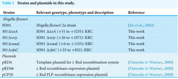
Figure 1 LPS structures, growth and TEM analysis of theShigella flexneriLPS mutants in this study.
(A) Illustrative drawing of LPS structures of Sf301,1wzy,1waaLand1rfaCstrains is showed in the up-per panel. Kdo: 3-deoxy-D-mannooct-2-ulosonic acid; Hep, L-glycero-D-manno-heptose phosphate; PEtN, O-phosphoryl-ethanolamine; Glc, D-glucose; Gla, d-galactose; GlcNAc, N-acetyl-D-glucosamine; Rha, L-rhamnose. The LPS of these strains was analyzed by silver staining of polyacrylamide gel after SDS-PAGE. (B) Growth of Sf301 and the LPS mutants. Each strain was grown in TSB broth under shaking con-ditions at 37◦C. Absorbance at OD600 was measured at different time points. (C) Representative trans-mission electron microphotographs (TEM) of each strain are shown. Overnight culture of each strains were collected and stained by with 1.5% phosphotungstic acid for 90 s and examined under TEM. Arrows indicate the dark spots on the surface of1rfaCstrain.
in vitroadhesion/invasion assays and LDH assays in this study.pvalues of less than 0.05 were regarded as statistically significant. Statistical analysis was performed with GraphPad Instat 3 software (GraphPad Software, Inc, La Jolla, Calif).
RESULTS
Generation and characterization of the LPS mutants ofShigella flexneri
To generate ShigellaLPS mutants of different chain length, we knocked outwzy,waaL
andrfaC individually using the lambda red system (Fig. S1). As shown inFig. 1A, thewzy
mutant contains a complete core and one copy of the O-antigen; thewaaLmutant has a
complete core but no O-antigen; therfaC mutant is deficient of Hep but retains the Kdo.
SDS-PAGE analysis confirmed that the all mutant strains produced proper LPS variants as
expected (Fig. 1A). The1wzy and1waaLstrains formed WT-like colonies on Tryptone
Soya Agar, while the1rfaC strain grew round and smooth colonies with smaller size.
In accordance with this, the1rfaC mutant grew slightly slower in liquid tryptone soya
broth medium than other strains (Fig. 1B). LPS truncation had no apparent impact on the
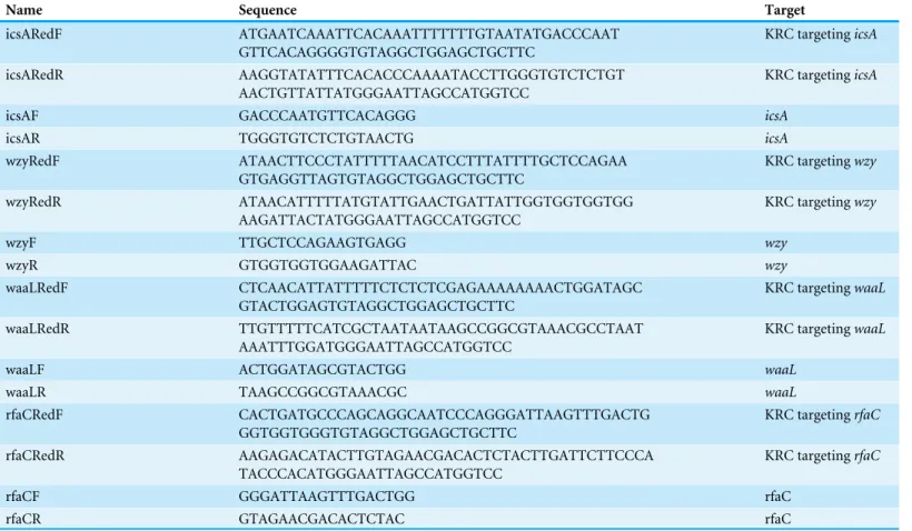
Figure 2 Analysis of biofilm formation of LPS mutants of Sf301.(A) Autoaggregation phenotype by LPS mutants. Each strain standardized at OD600 =1.0 in PBS was used for autoaggregation assay. The
value at OD600after an18-hour incubation is shown as the mean±SD of results from three independent
Figure 2 (...continued)
The mean±SD of results from three independent experiments are shown. Statistical analysis was
per-formed using studentt-test. *,P <0.01, against biofilm formation level of strain Sf301. (C) Biofilms on glass cover slides of LPS mutants stained by DAPI. (D) CLSM and SEM analysis of biofilms formed by 1rfaCstrain. A section which has representative signals in the defined area is shown in the left column (X–Y). The overview of biofilms in the same area of each X–Y section is shown as 3D image in the mid-dle column (3D). Biofilms under SEM are shown in the right column. (E) Influence of DNase I treatment on the biofilm formation of1rfaCstrain.1rfaCstrain was grown in presence of different concentrations of DNase I or in presence of pre-heated DNase I or without DNase I for 48 h under static conditions at 37◦C. Statistical analysis was performed using studentt-test. *,P
<0.01, against the biofilm formation by 1rfaCstrain without DNase I treatment. (F) biofilm formed by1rfaCstrain under high-magnification (10000X) SEM. Arrows indicate the fibrous connections among bacteria.
Biofilm formation is enhanced in the rfaC-deletedShigellastrain
The aggregation tendency of LPS mutants was initially evaluated in PBS and the results
showed that LPS truncation led to enhanced bacterial aggregation (Fig. 2A). Next, we
examined the biofilm formation of the LPS mutants on the polystyrene surface under a static culture condition. While the WT strain had a poor ability to form biofilm, the
LPS mutants showed improved potential to do so (Fig. 2B). Moreover, the ability to
form biofilm appeared to be negatively correlated with the LPS length with the1rfaC
strain (shortest LPS) being the most effective one (Fig. 2B). Fluorescence microscopy and scanning electron microscopy (SEM) further confirmed the enhanced biofilm formation by the1rfaCstrain on glass surface (Figs. 2C–2D). To test the involvement of extracellular
DNA in the biofilm formation by the1rfaC strain, DNase I was added when the bacteria
were seeded into the polystyrene plates. Disruption of extracellular DNA significantly abolished the formation of biofilm without affecting the viability of the bacteria (Fig. 2E
andFig. S2). In addition, SEM analysis demonstrated that there were fibrous connections among bacterial cells in the biofilm (Fig. 2F).
In vitroadhesiveness and thus invasiveness is promoted in
biofilm-forming rfaC-deletedShigella flexneri strain independent of activation of type III secretion system
Since biofilm formation is often associated with bacterial virulence, we set out to evaluate the pathogenic activities of the biofilm-forming LPS mutants using the gentamicin protection
assay. The1wzy and1waaLstrains showed almost identical host invasion efficiency to
the WT strain. By contrast, the1rfaC strain showed a much stronger invasiveness and
intracellular proliferation than wild type (Fig. 3A). Further dissection of time-dependent infection process revealed that the improved invasiveness came from enhanced adhesion during the initial host cell-Shigellacontact (within 10 min), which was further confirmed by fluorescence microscopy and scanning electron microscopy (Figs. 3B–3D). To be specific,
1rfaCmutant exhibited pronounced adhesiveness even when the type III secretion system (T3SS), responsible for bacterial invasion and virulence, was inactivated transcriptionally
at low growth temperature (28–30◦
Figure 3 Invasion and adhesion ability ofShigella flexneriLPS mutants in comparison with the parental Sf301 strain.(A) Invasion and intercellular proliferation of Sf301 and its LPS mutants. (B–C) Initial adhesion of Sf301 and its LPS mutants at 30◦
C and 37◦
Figure 3 (...continued)
The mean±SD of results from three independent experiments are shown. Statistical analysis was
per-formed using studentt-test. *,P<0.01. For fluorescence microscopy in (C), GFP-expressing plasmid was electroporated into theShigellastrains, and adhesion assay was performed as described above, after wash-ing, the cells were fixed in 3% paraformaldehyde/PBS and mounted with Anti-Fade solution (Invitrogen) containing DAPI onto glass slides.Shigellastrains are in green and nuclei are blue. (D) SEM analysis of en-hanced adhesion of1rfaC S.flexneristrain to HeLa cells. Bars in both images represent 30µm. (E) Lev-els of LDH in HeLa cell culture supernatant, 3 h post-infection withS. flexneri. The mean±SD of results from three independent experiments are shown. Statistical analysis was performed using studentt-test. *,P<0.01.
on loss of membrane integrity. The LDH level in the supernatant of1rfaC-infected HeLa
cells was more than twice that of the M90TS-infected cell supernatant (Fig. 3E).
rfaC-deleted strain showed different actin-based motility from other LPS-truncated strains
To examine the distribution of IcsA in the LPS-truncated mutants, intracellular F-actin of the infected host cells were visualized by fluorescent phalloidin, which binds to F-actin of
mammalian cells. Although F-actins could be recruited by all theShigellastrains except
theicsA-deleted strain (Fig. 4AandFig. S3), their cellular distribution varies between WT and LPS mutants. Typical ‘‘actin comets’’ and long protrusion were observed with the wild type strain (Fig. 4A), indicating a normal polar distribution of IcsA, which was further confirmed by the competence to form regular plaques on HeLa cell monolayer in a plaque assay (Fig. 4B). By contrast, the1wzy and1waaLmutants assembled F-actins all around the bacteria cell (Fig. S3), implying a circumferential distribution of IcsA, which caused aberrant actin-based motility (ABM) and thus failure to form plaques on HeLa cells monolayer (Fig. 4B). The1rfaC bacteria also recruited F-actin in a circumferential pattern as other LPS-truncated strains do. Nevertheless, a small amount of bacteria could assemble short and curly ‘‘actin-tail’’ behind the bacteria (as arrows indicated inFig. 4A). Although
1rfaC mutant could not form classical plaques on HeLa cell monolayer in 3 days, the integrity of the monolayer were destroyed due to massive cell detachment (Fig. 4B).
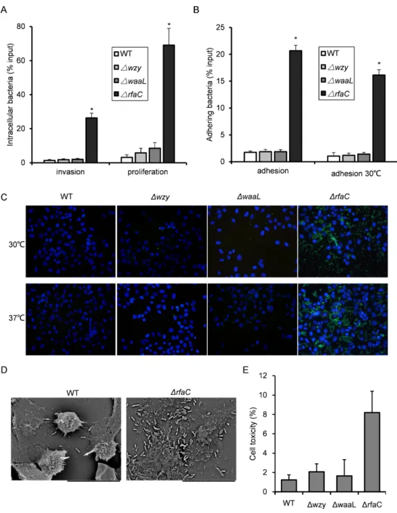
Truncation of LPS rendered bacteria susceptible in vitroandin vivo LPS, especially long-chain LPS of gram-negative bacteria, provides protection for the bacteria against unfavorable environment. While loss of LPS may contribute to biofilm formation and pathogenic activities such as adhesion and invasion, the adverse effects associated with LPS deletion must also be taken into consideration to accurately gauge the overall influences on bacterial fitness. To do this, we measured the susceptibility of the LPS mutants to distilled water (to mimic the low osmotic shock in environment) and
10% pooled human serum (to mimic the adverse environmentin vivo). While WT bacteria
well survived the low osmotic shock (water) during the experiment, all three LPS mutants
showed severely compromised viabilities after two hours in water. The1rfaC mutant in
particular, even begun to exhibit significantly reduced viability within 30 min (Fig. 5A).
Human serum showed much stronger killing activity than water toward1wzy,1waaLand
1rfaC strains (Table 3). It is interesting to note that although most of the bacteria were
Figure 4 Actin-based motility ofShigella flexneriLPS mutants in comparison with the parental Sf301 strain. (A) Fluorescence microscopy analysis of F-actin in HeLa infected by Sf301 and its1rfaC mu-tant strain. Invasion assay was performed as previously described. F-actin is stained by TRITC-phalloidin (red),Shigellabacteria are green and nuclei are blue (DAPI). White arrows in the WT panel indicate the long protrusion with actin comets assembled by Sf301. White arrows in the (triangle)rfaC panel indicate the short actin-tail formed by the1rfaCmutant bacteria. (B) Plaque formation of Sf301 and its LPS mu-tants on HeLa cell monolayer. Plaque assays were performed as described in Methods and Materials.
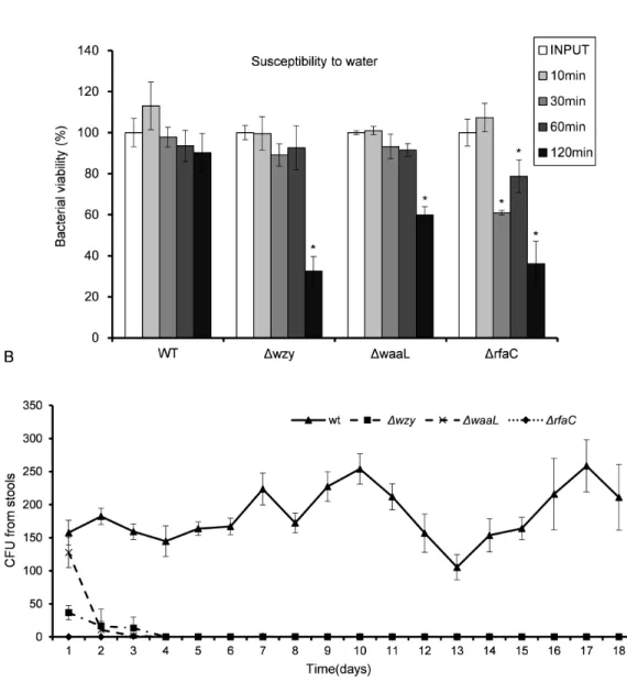
Table 3 Bacterial viability of the strains after two-hour serum killing.
Treatment Heated inactivated serum (%) Pooled human serum (%)
WT 90.6±13.5 63.4±6.4
1wzy 85.5±7.6 0
1waaL 111.1±11.4 0
1rfaC 89.9±10.5 0.0023±0.0004
no colony for1wzy and1waaLstrains (Fig. 5B). Whether this improved survivability of the1rfaC strain against serum is related to its biofilm-forming ability warrants further investigation.
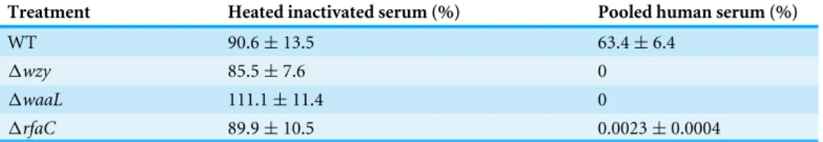
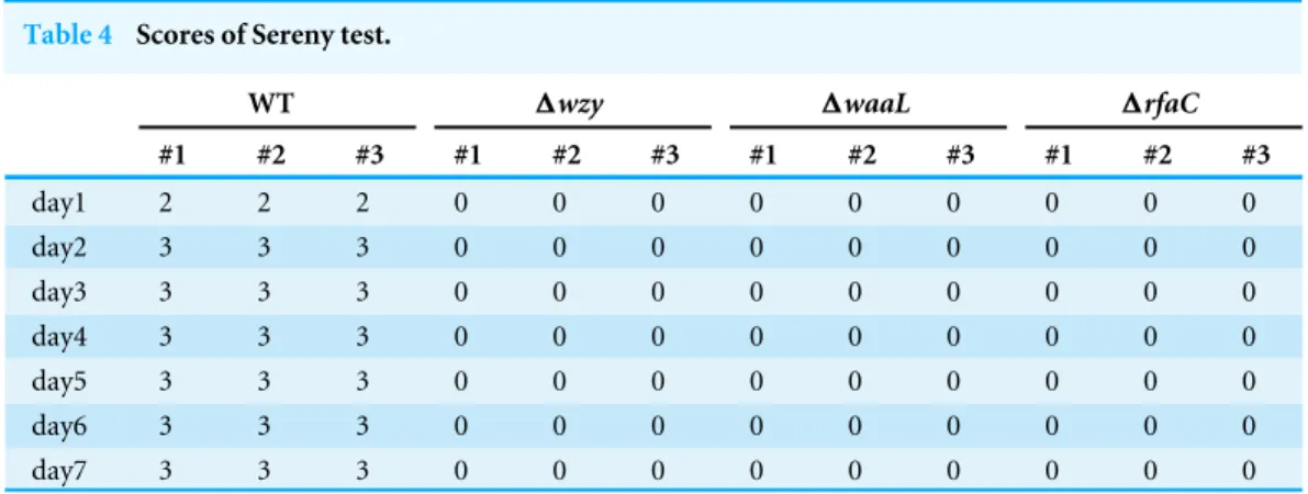
Given the dramatic virulence-boosting phenotype of the1rfaCmutantin vitro, we next
examined thein vivoeffects of LPS mutants on virulence using two independent animal
models. The Sereny test (guinea pig keratoconjunctivitis) indicated that1wzy,1waaL
Figure 5 In vitrosusceptibility to water (A) andin vivocolonization ability (B) ofShigella flexneri LPS mutants in comparison with the parental Sf301 strain. (A) Bacteria from mid- exponential phase were collected and resuspended into distilled water at the density of 107CFU/ml, bacterial viability was
evaluated as output CFU/input CFU X 100% at indicated time intervals. The mean±SD of results from
three independent experiments are shown. Statistical analysis was performed using studentt-test. *,P< 0.01, against bacterial survival level of sf301 wild type. (B) To assess the colonization ability of theShigella strains, 108CFU were inoculated into the rectum of guinea pigs, and bacterial load in the feces was
de-termined by the number of colonies on MacConkey Agar as described by Methods and Materials. The mean±SD of results from three individual animals are shown.
2 weeks’ infection. Wild type, on the other hand, provoked a typical keratoconjunctivitis 72 h post-infection (Table 4). To test the colonizing ability of the ‘‘super-adhesive’’1rfaC
Table 4 Scores of Sereny test.
WT 1wzy 1waaL 1rfaC
#1 #2 #3 #1 #2 #3 #1 #2 #3 #1 #2 #3
day1 2 2 2 0 0 0 0 0 0 0 0 0
day2 3 3 3 0 0 0 0 0 0 0 0 0
day3 3 3 3 0 0 0 0 0 0 0 0 0
day4 3 3 3 0 0 0 0 0 0 0 0 0
day5 3 3 3 0 0 0 0 0 0 0 0 0
day6 3 3 3 0 0 0 0 0 0 0 0 0
day7 3 3 3 0 0 0 0 0 0 0 0 0
Nevertheless, animals inoculated with wild type stably excreted bacteria in 18 tested days, albeit without observed symptoms either (Fig. 5B).
DISCUSSION
In the present study, we described a new and unusual phenotype ofShigella flexnerithat
is characteristic of enhanced biofilm formation. Deficiency in Hep synthesis in LPS due
to deletion of rfaC gene resulted in ‘‘deep-rough’’ LPS with an incomplete inner core
containing only the Kdo moieties. Although various LPS truncations have been studied for their effects on the biological activities ofShigella, the deep-rough mutant has never
been tested. In this work, we found that the deep-rough Shigella (1rfaC) exhibited
significantly enhanced biofilm-forming ability and strong adhesiveness toward host cells, which is in sharp contrast to the WT strain (Brotcke Zumsteg et al.,2014;Kline et al.,2009;
Mahmoud et al.,2016;Schroeder & Hilbi,2008). Besides, this deep-rough mutant could efficiently proliferate in the host cells and cause extensive cell lysis in few hours after
infection. However, the pronouncedin vitrovirulence observed with this mutant was not
recapitulatedin vivo, most likely due to its increased vulnerability to environmental stress caused by LPS loss.
The biofilm of1rfaC Shigellashowed a typical mesh-like structure and its formation
is dependent on extracellular DNA as proven by DNase I treatment experiment. Previous
studies have shown that LPS truncations in E. coliand Porphyromonas gingivalisalso
promoted biofilm formation (Nakao et al., 2012; Nakao, Senpuku & Watanabe,2006), suggesting that the correlation between LPS loss and biofilm-forming capacity might be a common feature in gram-negative bacteria. Bacterial surface hydrophobicity is a major determinant for biofilm formation (Donlan,2002;Mitzel et al.,2016). LPS truncation reduced the hydrophilic sugar moieties from bacteria surface and increased the exposure of the hydrophopic outer membrane lipid layer, thus enhancing the hydrophobicity on bacteria surface and facilitating biofilm formation.
WT Shigella flexneri adheres to host cells much less efficientlyin vitro than other enterobacteria due to the lack of general adhesion apparatus like fimbriae (Edwards & Puente,1998;Pizarro-Cerdá & Cossart,2006;Snellings, Tall & Venkatesan,1997). To our
normal invasion and proliferation capabilities as well. The molecular mechanism of the
hyperadhesiveness of1rfaC mutant remains unclear. Interestingly, we noticed that the
1rfaC Shigellatended to form clusters on abiotic surface or host cells. Whether this cell–cell clustering is also a result of the improved hydrophobicity caused by complete LPS shedding requires further investigation. Nevertheless, it is conceivable that the clusteredShigellamay adhere to host cells as one entity, in which numerous weak contacts contributed by each individual bacterium are pooled to improve the overall avidity of the bacterial cluster toward the host cell.
Previous studies have shown that the long-chain LPS was essential for maintaining polar distribution of IcsA (Doyle et al.,2015;Morona, Daniels & Den Bosch,2003;Sandlin et al.,
1995). Indeed, the1wzyand1waaLmutants with shortened LPS displayed circumferential
distribution of IcsA. Interestingly, a small amount of 1rfaC bacteria could assemble
atypical, short and curly actin-tail, implying the heterogeneous distribution of IcsA in
this new mutant. Results from plaque assay showed that1rfaC bacteria failed to form
typical plaque on HeLa monolayer as the WT does, although it did caused mass host cell death eventually. This could be caused by a limited number of correctly assembled ABM in function or by premature killing of the host cell before the actin protrusions could reach
adjacent cells due to much enhanced intracellular proliferation. How the1rfaC mutant
retains certain level of functional IcsA warrants further investigation.
Although the1rfaC mutant showed a significantly boosted virulencein vitro, it failed to colonizein vivo. Susceptibility test against low osmotic shock or serum revealed that this mutant is highly sensitive to environmental stress. Clearly, the virulence benefit gained from biofilm formation and/or improved adhesion is not enough to compensate for the loss of fitness on survivabilityin vivo.
In conclusion, this study characterized a newShigella flexnerimutant with deficiency in Hep synthesis of LPS. This mutant was capable of forming biofilm on abiotic surface and manifested extraordinary adhesiveness to host cells. Our study established a clear positive relationship between increased surface hydrophobicity and adhesiveness ofShigella flexneri. Although it is not realistic for a pathogenic bacteria to shed off LPS to gain hydrophobicity
in vivo, our study raised a possibility that the host adhesiveness ofShigellamay be modulated by altering the hydrophobicity of the bacteria.
ACKNOWLEDGEMENTS
We thank Dr. J Yu from Strathclyde Institute of Pharmacy and BioScience, University of Strathclyde (Glasgow, UK) for providing theShigella flexneri2a strain Sf301 and plasmids
for λRed recombination system. We thank P Wang from the Laboratory of Electron
ADDITIONAL INFORMATION AND DECLARATIONS
Funding
This work was supported by grants from the Natural Science Foundation of China (31401211 to DX), China Postdoctoral Science Foundation (2014M552428 and 2015T81014 to DX). The funders had no role in study design, data collection and analysis, decision to publish, or preparation of the manuscript.
Grant Disclosures
The following grant information was disclosed by the authors: Natural Science Foundation of China: 31401211.
China Postdoctoral Science Foundation: 2014M552428, 2015T81014.
Competing Interests
The authors declare there are no competing interests.
Author Contributions
• Dan Xu conceived and designed the experiments, performed the experiments, analyzed
the data, contributed reagents/materials/analysis tools, wrote the paper, prepared figures and/or tables, reviewed drafts of the paper.
• Wei Zhang performed the experiments, analyzed the data, reviewed drafts of the paper.
• Bing Zhang and Chongbing Liao performed the experiments, reviewed drafts of the
paper.
• Yongping Shao conceived and designed the experiments, analyzed the data, contributed
reagents/materials/analysis tools, wrote the paper, prepared figures and/or tables, reviewed drafts of the paper.
Animal Ethics
The following information was supplied relating to ethical approvals (i.e., approving body and any reference numbers):
The protocol has been approved by the Animal Research Ethical Committee of School of Life Science and Technology the Xi’an Jiaotong University. Approval No. 201411.
Data Availability
The following information was supplied regarding data availability:
The raw data has been supplied asSupplemental Dataset.
Supplemental Information
Supplemental information for this article can be found online athttp://dx.doi.org/10.7717/ peerj.2178#supplemental-information.
REFERENCES
Alexander C, Rietschel ET. 2001.Invited review: bacterial lipopolysaccharides and innate
immunity.Journal of Endotoxin Research7:167–202
Brotcke Zumsteg A, Goosmann C, Brinkmann V, Morona R, Zychlinsky A. 2014.IcsA Is aShigella flexneriadhesin regulated by the type III secretion system and required for pathogenesis.Cell Host & Microbe15:435–445DOI 10.1016/j.chom.2014.03.001. Carayol N, Tran Van Nhieu GT. 2013.Tips and tricks aboutShigellainvasion of
epithe-lial cells.Current Opinion in Microbiology 16:32–37DOI 10.1016/j.mib.2012.11.010. Datsenko KA, Wanner BL. 2000.One-step inactivation of chromosomal genes in
Escherichia coliK-12 using PCR products.Proceedings of the National Academy of Sciences of the United States of America97:6640–6645DOI 10.1073/pnas.120163297. Davies D. 2003.Understanding biofilm resistance to antibacterial agents.Nature Reviews
Drug Discovery 2:114–122DOI 10.1038/nrd1008.
Desroy N, Moreau F, Briet S, Fralliec GL, Floquet S, Durant L, Vongsouthi V, Gerusz V, Denis A, Escaich S. 2009.Towards gram-negative antivirulence drugs: new inhibitors of HldE kinase.Bioorganic & Medicinal Chemistry 17:1276–1289
DOI 10.1016/j.bmc.2008.12.021.
Di Padova FE, Brade H, Barclay GR, Poxton IR, Liehl E, Schuetze E, Kocher HP, Ram-say G, Schreier MH, McClelland DB. 1993.A broadly cross-protective monoclonal antibody binding toEscherichia coliand Salmonella lipopolysaccharides.Infection and Immunity61:3863–3872.
Donlan RM. 2002.Biofilms: microbial life on surfaces.Emerging Infectious Diseases
8:881–890DOI 10.3201/eid0809.020063.
Doyle MT, Grabowicz M, May KL, Morona R. 2015.Lipopolysaccharide surface
structure does not influence IcsA polarity.FEMS Microbiology Letters362(8)
DOI 10.1093/femsle/fnv042.
Edwards RA, Puente JL. 1998.Fimbrial expression in enteric bacteria: a critical step in intestinal pathogenesis.Trends in Microbiology6:282–287
DOI 10.1016/S0966-842X(98)01288-8.
Ellafi A, Lagha R, Abdallah FB, Bakhrouf A. 2012.Biofilm production, adherence and hydrophobicity of starvedShigellain seawater.African Journal of Microbiology Research6:4355–4359.
Grizot S, Salem M, Vongsouthi V, Durand L, Moreau F, Dohi H, Vincent S, Escaich S, Ducruix A. 2006.Structure of theEscherichia coliheptosyltransferase WaaC: binary
complexes with ADP and ADP-2-deoxy-2-fluoro heptose.Journal of Molecular
Biology363:383–394DOI 10.1016/j.jmb.2006.07.057.
Hall-Stoodley L, Costerton JW, Stoodley P. 2004.Bacterial biofilms: from the Natural
environment to infectious diseases.Nature Reviews Microbiology2:95–108
DOI 10.1038/nrmicro821.
Hitchcock PJ, Brown TM. 1983.Morphological heterogeneity among Salmonella lipopolysaccharide chemotypes in silver-stained polyacrylamide gels.Journal of Bacteriology154:269–277.
Hong M, Payne SM. 1997.Effect of mutations inShigella flexnerichromosomal and plasmid-encoded lipopolysaccharide genes on invasion and serum resistance.
Jin Q, Yuan Z, Xu J, Wang Y, Shen Y, Lu W, Wang J, Liu H, Yang J, Yang F, Zhang X, Zhang J, Yang G, Wu H, Qu D, Dong J, Sun L, Xue Y, Zhao A, Gao Y, Zhu J, Kan B, Ding K, Chen S, Cheng H, Yao Z, He B, Chen R, Ma D, Qiang B, Wen Y, Hou Y, Yu J. 2002.Genome sequence ofShigella flexneri2a: insights into pathogenicity through
comparison with genomes ofEscherichia coliK12 and O157.Nucleic Acids Research
30:4432–4441DOI 10.1093/nar/gkf566.
Klena J, Zhang P, Schwartz O, Hull S, Chen T. 2005.The core lipopolysaccharide ofEscherichia coliis a ligand for the dendritic-cell-specific intercellular adhesion
molecule nonintegrin CD209 receptor.Journal of Bacteriology187:1710–1715
DOI 10.1128/JB.187.5.1710-1715.2005.
Kline KA, Fälker S, Dahlberg S, Normark S, Henriques-Normark B. 2009. Bac-terial adhesins in host-microbe interactions.Cell Host & Microbe5:580–592
DOI 10.1016/j.chom.2009.05.011.
Lett MC, Sasakawa C, Okada N, Sakai T, Makino S, Yamada M, Komatsu K. 1989. virG, a plasmid-coded virulence gene ofShigella flexneri: identification of the virG
protein and determination of the complete coding sequence.Journal of Bacteriology
171:353–359.
Mahmoud RY, Stones DH, Li W, Emara M, El-domany RA, Wang D, Wang Y, Krachler AM, Yu J. 2016.The multivalent adhesion molecule SSO1327 plays a key role inShigellasonnei pathogenesis.Molecular Microbiology 99:658–673
DOI 10.1111/mmi.13255.
Martini M, Hoare A, Contreras I, Álvarez SA. 2011.Contribution of the lipopolysac-charide to resistance ofShigella flexneri2a to extreme acidity.PLoS ONE 6:e25557
DOI 10.1371/journal.pone.0025557.
Mitzel MR, Sand S, Whalen JK, Tufenkji N. 2016.Hydrophobicity of biofilm coatings influences the transport dynamics of polystyrene nanoparticles in biofilm-coated
sand.Water Research92:113–120DOI 10.1016/j.watres.2016.01.026.
Moreau F, Desroy N, Genevard JM, Vongsouthi V, Gerusz V, Fralliec GL, Oliveira C, Floquet S, Denis A, Escaich S, Wolf K, Busemann M, Aschenbrenner A. 2008. Dis-covery of new Gram-negative antivirulence drugs: structure and properties of novel
E. coliWaaC inhibitors.Bioorganic & Medicinal Chemistry Letters18:4022–4026
DOI 10.1016/j.bmcl.2008.05.117.
Morona R, Daniels C, Van Den Bosch L. 2003.Genetic modulation ofShigella flexneri2a lipopolysaccharide O antigen modal chain length reveals that it has been optimized
for virulence.Microbiology149:925–939DOI 10.1099/mic.0.26141-0.
Nakao R, Ramstedt M, Wai SN, Uhlin BE. 2012.Enhanced biofilm formation by
Escherichia coliLPS mutants defective in Hep biosynthesis.PLoS ONE7:e51241
DOI 10.1371/journal.pone.0051241.
Nakao R, Senpuku H, Watanabe H. 2006.Porphyromonas gingivalis galE is involved
in Lipopolysaccharide O-Antigen synthesis and biofilm formation.Infection and
Immunity 74:6145–6153DOI 10.1128/IAI.00261-06.
Oaks EV, Wingfield ME, Formal SB. 1985.Plaque formation by virulentShigella flexneri.
Phalipon A, Sansonetti PJ. 2007.Shigella/’s ways of manipulating the host intestinal
innate and adaptive immune system: a tool box for survival?Immunology and Cell
Biology85:119–129DOI 10.1038/sj.icb7100025.
Philpott DJ, Edgeworth JD, Sansonetti PJ. 2000.The pathogenesis ofShigella flexneri
infection: lessons fromin vitroandin vivostudies.Philosophical Transactions of the Royal Society of London B: Biological Sciences355:575–586
DOI 10.1098/rstb.2000.0599.
Pizarro-Cerdá J, Cossart P. 2006.Bacterial adhesion and entry into host cells.Cell 124:715–727DOI 10.1016/j.cell.2006.02.012.
Sandlin RC, Lampel KA, Keasler SP, Goldberg MB, Stolzer AL, Maurelli AT. 1995. Avirulence of rough mutants ofShigella flexneri: requirement of O antigen for correct unipolar localization of IcsA in the bacterial outer membrane.Infection and Immunity 63:229–237.
Schroeder GN, Hilbi H. 2008.Molecular pathogenesis ofShigellaspp.: controlling host cell signaling, invasion, and death by type III secretion.Clinical Microbiology Reviews
21:134–156DOI 10.1128/CMR.00032-07.
Snellings NJ, Tall BD, Venkatesan MM. 1997.Characterization ofShigellatype 1
fimbriae: expression, FimA sequence, and phase variation.Infection and Immunity
65:2462–2467.
Suzuki T, Sasakawa C. 2001.Molecular basis of the intracellular spreading ofShigella.
Infection and Immunity69:5959–5966DOI 10.1128/IAI.69.10.5959-5966.2001. Wang X, Quinn PJ. 2010. Endotoxins: lipopolysaccharides of gram-negative bacteria. In:
Wang X, Quinn PJ, eds.Endotoxins: structure, function and recognition. Dordrecht: