! "# " $ "%& ' (&)&
*"+, (" & &+ ) - . / %0& ) " "&$&+" + $' &1 %0& & 2 /$&
ii
! "# " $ "%& ' (&)&
*"+, (" & &+ ) - . / %0& ) " "&$&+" + $' &1 %0& & 2 /$&
&# 78 9 )1 & 4556
:::::::::::::::::::::::::::: ::::::::::::::::::::::::::::
; "< = $) " >& ; )1 " ? &.& " & @ ? &.& " & @
:::::::::::::::::::::::::::: ::::::::::::::::::::::::::::
/9") ) & "$# &+; "& " "1
:::::::::::::::::::::::::::: (& & "& $"# &
iv AGRADECIMENTOS
À Universidade Federal de Viçosa, a todo o Departamento de Biologia Vegetal e ao
programa de Pós%graduação em Fisiologia Vegetal;
Ao Conselho Nacional de Desenvolvimento Científico e Tecnológico (CNPq);
À SAMARCO MINERAÇÃO S.A. UBU – ES, principalmente ao Luíz;
À EPAMIG, principalmente ao Felipe Silva, Jardel e ao Plínio Soares;
Ao professor Marco Antonio Oliva;
À professora Andréa Miyasaka;
Ao professor José Cambraia;
À professora Luzimar Campos;
Ao grande amigo e professor Rogério Ribas;
À professora Marília Ventrella;
À professora Kacilda Naomi Kuki;
À professora Rosane Aguiar;
Ao Rogério Gomide;
Ao João Bosco de Freitas, José Antônio Bhering, Maria Mercês Gomes, Celso Costa,
José do Carmo, Ângelo Lopes, Francine; Luciene Fernandes, José Cardoso e José
Carlos;
Em especial à Laíse Rosado e também ao Carlos Stopato, Pitt Wehr e Daniel Pinheiro;
A todos os amigos da Unidade de Crescimento de Plantas % UCP, especialmente
Cláudio Meira, Caroline Müller, Advânio Silva, Claudinéia Santos, Alice Godoy,
Thiago, Letícia dos Anjos, Letícia Nalon, Diego Carretero, Clenilso Mota, Maione
Franco, Michelle, Patrícia Mendes, Adriana Magalhães;
Aos demais amigos que tive o prazer de desfrutar do agradável convívio nesses quatro
anos, em especial Ricardo Montanari, Lílian Hasegawa, Ronnie Veloso, Ricardo
Zanquetin, Leonardo Lucas, Élcio Meira, Gládis Jucosky, Werner Antunes, Danilo
Daloso, Tales, Carla e Cléberson Ribeiro, Giselle Mendes, Denise Colares, Alan Costa,
Michellia Pereira, Dayana Francino, Rosilene Mesquita;
A todos os colegas, funcionários e professores do Programa de Pós%graduação em
Fisiologia Vegetal % UFV;
Aos meus irmãos, Silvana, Cézar, Neide e meus sobrinhos João Vitor e Mariana;
Aos meus pais, Levi Barbosa e Maria dos Reis;
À Elisa, pelo amor e companhia;
v BIOGRAFIA
Eduardo Gusmão Pereira, filho de Levi Barbosa Pereira e Maria dos Reis
Gusmão V. Pereira, nasceu em Montes Claros – MG, no dia 15 de março de 1977.
Concluiu o curso superior de Bacharelado em Ciências Biológicas em dezembro de
2003 pela Universidade Estadual de Montes Claros. Em fevereiro de 2006 recebeu o
título de Mestre em Fisiologia Vegetal pela Universidade Federal de Viçosa. Em março
de 2006 iniciou o curso de Doutorado na mesma universidade, no Programa de Pós%
Graduação em Fisiologia Vegetal do Departamento de Biologia Vegetal. Em novembro
de 2009 assumiu o cargo de Professor de 3° Grau, Assistente I, da Universidade Federal
vi ÍNDICE
Página
RESUMO ... xii
ABSTRACT ... xiv
1. INTRODUÇÃO GERAL ... 16
Referências ... 18
2. OBJETIVO GERAL ... 21
3. CAPÍTULO 1 ... 22
Evaluation of iron toxicity in rice genotypes by non%invasive physiological tools [Avaliação da toxidez por ferro em genótipos de arroz mediante metodologias fisiológieas não invasivas] ... 22
Abstract ... 22
Resumo ... 23
Introduction ... 24
Material and methods ... 25
Plant materials and experimental eonditions ... 25
Morphologieal parameters and iron eontent into tissues ... 26
Chlorophyll eontent (SPAD index)... 26
Gas exehange measurements ... 26
Chlorophyll fluoreseenee measurements ... 27
Assessing photorespiration in vivo... 29
Results ... 30
Iron eontent, morphologieal and visual symptoms ... 30
Chlorophyll eontent index ... 31
Gas exehange parameters and photorespiration ... 32
Chlorophyll a fluoreseenee parameters ... 35
Discussion ... 44
References ... 49
4.CAPÍTULO 2 ... 53
Iron plaque formation and nutritional disorders in tropical rice genotypes from lowland and upland cropping systems [Formação da plaea de ferro e desordens nutrieionais em genótipos tropieais de arroz oriundos de sistemas de eultivo de várzea e terras altas] ... 53
Abstract ... 53
Resumo ... 54
Introduction ... 55
Material and methods ... 56
Plant materials and experimental eonditions ... 56
Iron histoehemistry and seanning eleetron mieroseopy ... 57
Plant growth, evaluation of root iron plaque formation and determination of minerals eontents ... 58
Chlorophyll a fluoreseenee measurements ... 58
Results ... 59
Iron plaque formation, SEM and iron histoehemistry ... 59
vii
Fe and others nutrients in iron plaque, uptake and transloeation by riee ... 65
Chlorophyll a fluoreseenee ... 70
Discussion ... 72
References ... 75
5. CAPÍTULO 3 ... 79
Alterações fotossintéticas em resposta ao excesso de ferro e dióxido de enxofre em plantas de restinga [Changes in photosynthesis due to iron exeess and sulphur dioxide in restinga plant speeies] ... 79
Resumo ... 79
Abstract ... 81
Introdução ... 82
Material e métodos ... 84
Emissões de MSPFe e SO2 em C. spiritu sanetensis e A. parvifolium sob eondições de eampo ... 84
Deposição de material partieulado de ferro e da neblina áeida simulada em C. spiritu sanetensis e A. parvifolium ... 85
Avaliação da deposição partieulada de Fe e SO2 ... 86
Avaliação das troeas gasosas ... 86
Avaliação da fluoreseêneia da elorofila a ... 87
Avaliação dos teores de pigmentos ... 88
Avaliação da permeabilidade de membranas ... 88
Determinação dos teores de malonaldeído (MDA) ... 88
Deteeção histoquímiea de ferro e mieroseopia eletrôniea de varredura (MEV) 89 Determinação do teor de ferro, enxofre e nutrientes minerais ... 89
Resultados ... 90
Emissões de MSPFe e SO2 em C. spiritu sanetensis e A. parvifolium sob eondições de eampo ... 90
Deposição de material partieulado de ferro e da neblina áeida simulada em C. spiritu sanetensis e A. parvifolium ... 96
Discussão ... 107
Referências ... 111
viii LISTA DE FIGURAS E TABELAS
CAPÍTULO 1
Figure 1: Iron content in roots (inner panel) and shoots of four rice genotypes treated witht Fe%EDTA in nutrient solution. Data are means±SE of four replicates. ... 31 Figure 2: Changes in chlorophyll content index (SPAD units) in leaves of four rice genotypes treated with Fe%EDTA in nutrient solution.. ... 33 Figure 3: Changes in photosynthetic rate (A), stomatal conductance (gs), transpiration (E) and Ci/Ca ratio in four rice genotypes treated with Fe%EDTA in nutrient solution.. ... 34 Figure 4: Light response curve of net photosynthesis (A) of the rice genotypes BR IRGA 409 (circles) and BRA 041171 (inverted triangles) under control conditions (Fe%EDTA 0.019mM; open symbols) and under 7mM Fe%EDTA (closed symbols) in nutrient solution. ... 35 Figure 5: Chlorophyll a fluorescence images of minimal fluorescence (F0), maximal PSII quantum yield (Fv/Fm), photochemical (qL) and non%photochemical quenching (NPQ/4), quantum yield of photochemical energy conversion in PSII (ϕΙΙ), quantum yield of regulated (ϕNPQ) and non%regulated (ϕNO) energy dissipation in PSII of four O. sativa genotypes treated with Fe%EDTA (4 and 9 mM) in nutrient solution... 37 Figure 6: Minimal fluorescence (F0), maximal PSII quantum yield (Fv/Fm), coefficient of photochemical quenching (qL), non%photochemical quenching (NPQ) and apparent electron transport rate (ETR) in four rice genotypes treated with Fe%EDTA in nutrient solution. ... 38 Figure 7: Spatial variation in maximal PSII quantum yield (Fv/Fm) across a transect in the leaf blade in four rice genotypes under different Fe%EDTA doses in nutrient solution ... 40 Figure 8: Relationship among quantum yield of photochemical energy conversion in PSII (ϕΙΙ), quantum yield of regulated (ϕNPQ) and non%regulated (ϕNO) energy dissipation in PSII of four rice genotypes treated with Fe%EDTA in nutrient solution ... 42 Figure 9: Light response curve of fluorescence parameters of the rice genotypes BR IRGA 409 (circles) and BRA 041171 (inverted triangles) under control conditions (Fe%EDTA 0.019 mM; open symbols) and under 7 mM Fe%EDTA (closed symbols) in nutrient solution ... 43
ix CAPÍTULO 2
Figure 1: Iron plaque experiment. A: Rice seeds germination procedure in the germination chamber; B: Seedlings transferred to plastic pots (3.8 L) containing nutrient solution; C and D: Disposition of the pots on the bench in the greenhouse; E: Control treatment without iron plaque; F: 4 mM FeSO4 treatment with iron plaque formation on root surface of the genotype Nipponbare. ... 60 Figure 2: Scanning electron micrographs of rice roots surface. Genotypes: BRS Atalanta (A and B), EPAGRI 107 (C and D), Canastra (E and F) e BRSMG Curinga (G e H). Control treatment (A, C, E and G) e FeSO4 treatment (B, D, F and H), after 168 h of exposure. ... 62 Figure 3: Iron histochemistry in rice roots transversal sections. Genotypes: BRS Atalanta (A and B), EPAGRI 107 (C and D), Canastra (E and F) and BRSMG Curinga (G and H). Control treatment (A, C, E and G). ... 63 Figure 4: Time effect on length (left panel) and dry weights (right panel) of shoots (circles) and roots (inverted triangles) in iron treated plants (4mM FeSO4; closed symbols) and control (open symbols) during 168 h. ... 64 Figure 5: Iron content in iron plaque on root surface of rice plants exposed to 4mM FeSO4 (closed symbols) and control treatment (open symbols) in nutrient solution during 168 h. ... 66 Figure 6: Correlation among iron and other nutrients content in root plaque (right panel) and in shoots (right panel) of five rice genotypes exposed to 4mM FeSO4 (closed symbols) or control treatment (open symbols) in nutrient solution... 69 Figure 7: Chlorophyll a fluorescence images (A) and graphic representations (B) of minimal fluorescence (F0), maximal PSII quantum yield (Fv/Fm), photochemical (qP) and non%photochemical quenching (NPQ), and electron transport rate through PSII (ETR) of five rice genotypes exposed to 4 mM of FeSO4 (closed columns) or control treatment (open columns) in nutrient solution ... 71
Table 1: Analysis of variance of data depicted in the figure 4. ... 66 Table 2: Mineral nutrient concentration (mg kg−1rootDW) in iron plaques of roots of five rice genotypes treated with 4 mM FeSO4 and control. ... 67 Table 3: Root iron content and shoot mineral nutrient concentration (mg kg−1DW) in five rice genotypes treated with 4 mM FeSO4 and control. ... 68 Table 4: Analysis of variance of data depicted in the figure 7. Effect of iron plaque formation on chlorophyll a fluorescence parameters. ... 70
CAPÍTULO 3
x esquerda) e Aspidosperma parvifolium (painéis da direita) ao longo de um ano de exposição aos poluentes em área próxima a usina de beneficiamento de minério de ferro e em área referência. ... 94 Figura 4: Alterações nos teores de clorofila a (Chl a), clorofila b (Chl b) e carotenóides (Car) em Clusia spiritu sanetensis (painéis da esquerda) e Aspidosperma parvifolium (painéis da direita) ao longo de um ano de exposição em área próxima a usina de beneficiamento de minério de ferro e em área referência. ... 95 Figura 5: Alterações no extravasamento de eletrólitos (EL) em Clusia spiritu sanetensis (painéis da esquerda) e Aspidosperma parvifolium (painéis da direita) ao longo de um ano de exposição em área próxima a usina de beneficiamento de minério de ferro e em área referência. ... 96 Figura 6: Superfície foliar adaxial (A, D, F e H) e abaxial (B, C, E, G, I e J) de Clusia spiritu sanetensis após 100 dias de aplicação de neblina ácida simulada e MSPFe. Tratamento controle (A % C); com aplicação de MSPFe ao solo (D e E); com aplicação de neblina ácida simulada pH 3,0 (F e G) e com combinação entre neblina ácida simulada e MSPFe no solo (H % J). ... 97 Figura 7: Superfície foliar adaxial (A, D, F, G e I) e abaxial (B, C, E, H, J e K) de Aspidosperma parvifolium após 30 dias com aplicação de neblina ácida simulada e MSPFe. Tratamento controle (A % C), com deposição de MSPFe no solo (D e E), com aplicação de neblina ácida simulada pH 3,0 (F % H) e a combinação entre neblina ácida simulada e MSPFe no solo (I % K)... 98 Figura 8: Detecção histoquímica de ferro em cortes transversais da lâmina foliar de Clusia spiritu sanetensis após 100 dias com aplicação de neblina ácida simulada e MSPFe. Tratamento controle (B), com deposição de MSPFe no solo (C e D), com aplicação de neblina ácida simulada pH 3,0 (E e F) e a combinação entre neblina ácida simulada e MSPFe no solo (G e H)... 100 Figura 9: Detecção histoquímica de ferro em cortes transversais da lâmina foliar de Aspidosperma parvifolium após 30 dias com aplicação de neblina ácida simulada e MSPFe. Tratamento controle (B), com deposição de MSPFe no solo (C e D), com aplicação de neblina ácida simulada pH 3,0 (E e F) e a combinação entre neblina ácida simulada e MSPFe no solo (G e H)... 102 Figura 10: Alterações na taxa fotossintética (A; A e B), condutância estomática (gs; C e D); razão entre a concentração interna e externa de CO2 (Ci/Ca; E e F) em Clusia spiritu sanetensis (painéis da esquerda) e Aspidosperma parvifolium (painéis da direita) em resposta a aplicação de neblina ácida simulada e MSPFe. ... 103 Figura 11: Imagens dos parâmetros de fluorescência da clorofila (A) e representações gráficas (B) da eficiência quântica máxima do PSII (Fv/Fm), fluorescência mínima (F0), quenehing fotoquímico (qP) e não%fotoquímico (NPQ) e taxa aparente de transporte de elétrons no PSII (ETR) em Clusia spiritu sanetensis (painéis da esquerda) e Aspidosperma parvifolium (painéis da direita) em resposta a aplicação de neblina ácida simulada e MSPFe. ... 106 Figura 12: Alterações no rendimento quântico efetivo do PSII (ϕII; A e B) e nos rendimentos quânticos de dissipação não%fotoquímica regulada (ϕNPQ; C e D) e não% regulada (ϕNO; E e F) em Clusia spiritu sanetensis (painéis da esquerda) e Aspidosperma parvifolium (painéis da direita) em resposta a aplicação de neblina ácida simulada e MSPFe. ... 107
xii RESUMO
PEREIRA, Eduardo Gusmão; D.S., Universidade Federal de Viçosa, dezembro de 2009.
Efeitos tóxicos do ferro: alterações fisiológicas e morfológicas em plantas cultivadas e de restinga. Orientador: Marco Antonio Oliva Cano. Co%orientadores: Andréa Miyasaka de Almeida, José Cambraia.
O ferro (Fe) é um micronutriente essencial envolvido em vários processos do
metabolismo vegetal como fotossíntese, respiração, fixação de nitrogênio, entre outros.
Entretanto, em altas concentrações torna%se tóxico. O aumento exagerado da
disponibilidade de Fe para as plantas pode ser observado naturalmente em solos ácidos
constantemente alagados ou em eventos antrópicos como a mineração e beneficiamento
de minério de ferro. A toxidez por ferro é o principal fator abiótico que limita a
produtividade de arroz em cultivo alagado. Por outro lado, o crescente número de usinas
do setor de mineração e beneficiamento de Fe pode constituir um risco ambiental para
espécies vegetais em fragmentos de restinga próximos a fontes poluidoras, lançando
nessas áreas considerável quantidade de Fe na forma de material sólido particulado de
Fe (MSPFe) juntamente com precipitações ácidas caracterizadas por alta concentração de
SO2. Desta forma, o objetivo do presente estudo foi caracterizar fisiologicamente o
processo de toxidez por ferro em plantas, abrangendo causas antrópicas, como em
regiões sujeitas à poluição por MSPFe e SO2, e causas naturais, como no caso dos solos
ácidos inundados onde o arroz é cultivado. Em condições de cultivo hidropônico, os
efeitos do Fe%EDTA em genótipos de arroz foram examinados utilizando parâmetros
fisiológicos não invasivos. O aumento no teor foliar de Fe levou a alterações nos
diversos parâmetros avaliados, podendo ser empregados na prévia detecção dos efeitos
tóxicos do ferro, antes do aparecimento de sintomas visuais. A redução na taxa
fotossintética nos genótipos de arroz pode ser atribuída a componentes estomáticos e a
componentes não%estomáticos em resposta ao aparecimento de estresse oxidativo.
Reduções nos teores de clorofila e distúrbios nos processos de captura e utilização da
energia luminosa foram constantemente verificados nos genótipos mais sensíveis. Os
efeitos prejudiciais do ferro em excesso também ocorreram com a formação de uma
placa de ferro na superfície radicular, alterando processos nutricionais em plantas. Tais
efeitos foram investigados em genotipos tropicais de arroz provenientes de cultivo
xiii aparecimento da placa de ferro, com coloração alaranjada, em todos os genótipos
estudados, porém em maior intensidade nos genótipos de várzea. Observou%se redução
significativa no teor de P, Mg e Mn na parte aérea em decorrência da formação da placa
de ferro nas raízes. Frequentemente foram observadas rupturas das células epidérmicas
da raiz. Quanto aos aspectos da toxidez por ferro e outros elementos imposta por fontes
de mineração, observou%se que a deposição de MSPFe e SO2 causou reduções em
diversos parâmetros fotossintéticos em duas espécies de restinga: Clusia spiritu
sanetensis e Aspidosperma parvifolium, sob condições de campo e em casa de
vegetação. As plantas expostas aos poluentes apresentaram acúmulo significativo de
ferro nos tecidos. Os maiores teores desse elemento foram observados em A.
parvifolium que também apresentou incremento nos teores de enxofre sob condições de
campo, sendo observados sintomas mais severos nessa espécie. Desta forma, pode%se
concluir que a toxidez por Fe pode limitar o desempenho fotossintético e nutricional de
genótipos de arroz sensíveis ao excesso de Fe e que as emissões de MSPFe e outros
poluentes por fontes mineradoras podem causar desequilíbrio na dinâmica sucessional
xiv ABSTRACT
PEREIRA, Eduardo Gusmão; D.S., Universidade Federal de Viçosa, december, 2009.
Iron toxic effects: physiological and morphological changes in cultivated and restinga plants. Adviser: Marco Antonio Oliva Cano. Co%advisers: Andréa Miyasaka de Almeida, José Cambraia.
Iron (Fe) is an essential micronutrient for several processes in plant metabolism,
such as photosynthesis, respiration, nitrogen fixation and others. However, reaching
high levels it became toxic. The exaggerated increase in Fe availability to plants can be
found naturally in waterlogged acidic soils or due anthropic consequences, such as iron
ore processing and beneficiation factories. In lowland rice, Fe is the major abiotic factor
limiting plant productivity. In other hand, the increasing number of iron ore factories
near the restinga, may constitute an environmental hazard. These factories release
considerable amounts of sulfur dioxide (SO2) and iron ore dust into the air over the
restinga as iron solid particulate matter (SPMFe) and acid precipitation. Thus, the
purpose of this study was characterize with physiological evaluations the different
processes of iron toxicity in plants, covering anthropic causes, such as SPMFe and SO2
pollution, and natural causes, such as iron toxicity in waterlogged soils. Using
hydroponic culture the effects of Fe%EDTA on rice genotypes were evaluated by non%
invasive methodologies. The rise in shoot iron content led to changes in most of non%
invasive physiological parameters used. These parameters may be used in early
detection of iron toxicity prior to visual symptoms. Reduction in rice genotypes
photosynthesis can be attributed to stomatal and non%stomatal limitations in response to
the oxidative stress. Chlorophyll content reduction and impairment in light capture and
utilization were commonly observed in sensitive genotypes. The damaging effects of
iron also occured with iron plaque formation on the root surface, changing nutritional
processes. Those effects were investigated in tropical rice genotypes from lowland and
upland cropping systems. FeSO4 application in nutrient solution caused iron plaque
formation, with reddish precipitated with higher intensity in lowland genotypes. Iron
plaque caused significant reductions in the shoot content of P, Mn and Mg. Frequently
was observed ruptures of the root epidermis. To iron toxicity related to mining sources,
xv SO2 deposition on two restinga plant species: Clusia spiritu sanetensis and
Aspidosperma parvifolium, under field and greenhouse conditions. The plant exposure
to the pollutants leads to significant increase of Fe into tissues. The higher Fe content
was found in A. parvifolium which one also presented increased sulphur content under
field exposure. The most severe symptoms were observed in this species. In conclusion,
the iron toxicity may limit the nutritional and photosynthetic performances of sensitive
rice genotypes. The SPMFe and other pollutants emissions from iron ore factories may
lead to imbalance in sucessional dynamic in restinga and affect the development of the
16 1. I'TRODUÇÃO GERAL
O ferro (Fe) é o mineral mais abundante na crosta terrestre. Como
nutriente essencial para as plantas, está envolvido em processos fundamentais
como fotossíntese, respiração, metabolismo de nitrogênio, síntese de DNA e de
hormônios (Briat e Lobréaux, 1997; Becana et al., 1998). O Fe presente no solo
está em sua maior parte na forma oxidada (Fe3+), que não é prontamente
absorvido pelas plantas (Fageria et al., 1990).
De modo geral, a absorção de ferro pelas plantas ocorre através de duas
estratégias distintas. A primeira (estratégia I), comum entre dicotiledôneas e
monocotiledôneas não%gramíneas, envolve a acidificação do solo pela extrusão de
prótons e absorção de Fe2+. Outra estratégia (estratégia II), utilizada pelas
gramíneas, consiste na produção e liberação de fitossideróforos que complexam o
Fe na rizosfera, sendo o complexo Fe3+%fitossideróforo absorvido pelas raízes
(Hell e Stephan, 2003). Com exceção das gramíneas, que apresentam a estratégia
II, diversas plantas, algas, leveduras e bactérias absorvem o ferro por meio da
estratégia de solubilização do Fe3+ à Fe2+ (Guerinot, 1994). O arroz (Oryza sativa
L.) apresenta ambas as estratégias de absorção de Fe (Ishimaru et al., 2006).
Em condições nutricionais normais, o teor de Fe nas plantas varia de 0,05
a 0,25 mg g−1MS (Marschner, 1995). Para manutenção do crescimento ótimo,
monocotiledôneas requerem maiores concentrações de Fe na solução de cultivo do
que espécies dicotiledôneas (Christ, 1974). Quando em excesso, a toxidez por
ferro pode ocorrer por meio da absorção e acúmulo excessivo ou por meio da sua
precipitação sobre as raízes, formando uma crosta de óxido férrico que altera a
absorção de outros nutrientes como o fósforo, potássio e zinco (Howeler, 1973).
Em situações de acúmulo excessivo de ferro nos tecidos, ocorre a
potencialização de estresse oxidativo, com aumento na produção de espécies
reativas de oxigênio (ROS). Em nível celular o Fe2+ pode reagir com o peróxido
de hidrogênio (H2O2) através da reação de Fenton, gerando radicais hidroxilas
(OH•; Guerinot e Yi, 1994; Becana et al., 1998). O Fe2+ pode ainda reagir com o
17 O OH• é considerado a espécie reativa de oxigênio mais tóxica para a
célula; juntamente com outras ROS leva a danos irreversíveis na estrutura das
membranas celulares, modificando suas atividades enzimáticas e resultando na
perda da permeabilidade seletiva, com consequente vazamento de eletrólitos,
ocasionando o colapso celular e deterioração do tecido (Becana et al., 1998; Fang
et al., 2001; Pereira et al., 2009).
A formação da placa de ferro devido à precipitação do metal na superfície
radicular é comumente encontrada em ambientes alagados com alto teor desse
metal. Para a formação da placa ocorre oxidação do íon ferroso a íon férrico,
seguido da precipitação de óxido ou hidróxido de ferro, em função da capacidade
oxidativa das raízes (Chen et al., 1980; Taylor et al., 1984). Além do ferro, outros
elementos podem ocorrer em placas de ferro formadas naturalmente, como o P, K,
Zn, As, Cd, Cu, S, entre outros (Howeler, 1973, Greipsson e Crowder, 1992). A
placa de ferro é composta predominantemente de hidróxido férrico, goetita e
siderita (Hansel et al., 2001).
A elevação da concentração de ferro a níveis tóxicos para as plantas pode
ser observado naturalmente em solos ácidos constantemente inundados
(Dobermann e Fairhurst, 2000) ou em eventos antrópicos como a mineração e
beneficiamento de minério de ferro (Kuki et al., 2008a).
A toxidez por ferro em excesso é um dos principais fatores abióticos que
limitam a produtividade do arroz alagado (Dobermann e Fairhurst, 2000). Durante
o alagamento do solo, o oxigênio molecular é consumido pelos microrganismos
aeróbicos, que com o tempo são substituídos pelos anaeróbicos, resultando, num
primeiro momento, em um ambiente de redução e acúmulo de CO2
(Ponnamperuma, 1972). Adicionalmente, a redução de compostos oxidados
aumenta a disponibilidade de ferro (Becana et al., 1998). Portanto, em solos
ácidos, a anoxia e redução do pH, decorrentes do alagamento, podem levar à
redução e solubilização de grandes quantidades de ferro (Ponnamperuma, 1972).
O aumento na concentração de ferro pode chegar a níveis tóxicos para as plantas
cultivadas neste ambiente (Silva e Ranno, 2005). A toxicidade desse elemento
provoca manchas necróticas nas folhas, escurecimento das raízes e inibição do
crescimento da planta (Dobermann e Fairhurst, 2000).
Outro aspecto importante da toxidez por ferro em plantas é observado em
18 do metal. O crescente número destas usinas no litoral brasileiro é responsável pela
emissão de diversos poluentes atmosféricos que podem comprometer a
sobrevivência de espécies sensíveis encontradas na vegetação de restinga (Silva et
al., 2005, 2006; Kuki et al., 2008a b; Pereira et al., 2009). Dentre esses poluentes
encontra%se o material sólido particulado de ferro (MSPFe). Além dos efeitos
tóxicos específicos do ferro sobre as plantas, o MSPFe pode causar danos à
vegetação por efeitos físicos: bloqueio da radiação luminosa, abrasamento,
aquecimento foliar, alterações em processos como fotossíntese e transpiração
(Pereira et al., 2009); ou por efeitos químicos: alterações no pH, no estado
nutricional e lixiviação de nutrientes das folhas (Grantz et al., 2003).
Dessa forma, as plantas podem estar expostas a níveis tóxicos de ferro sob
diversas circunstâncias, sejam naturais ou provocadas de maneira antrópica. A
capacidade de uma espécie vegetal sobreviver e tolerar os efeitos do ferro em
excesso dependerá de diversas características como o genótipo, estádio de
crescimento, disponibilidade de recursos e microhabitat (Levin, 1998; Grantz et
al., 2003). Todos essas características possuem estreita relação com aspectos
fisiológicos dos vegetais. Entretanto, tratando%se de um mesmo fator (excesso de
ferro) as plantas podem apresentar respostas fisiológicas distintas, dependendo da
forma específica de estresse.
Referências
Becana M, Moran JF, Iturbe2Ormaetxe I. 1998. Iron%dependent oxygen free radical generation in plants subjected to environmental stress: toxicity and antioxidant protection. Plant and Soil201, 137–147.
Briat JF, Lobréaux S. 1997. Iron transport and storage in plants. Trends in Plant Science 2,187–193.
Chen CC; Dixon JB, Turner FT. 1980. Iron coatings on roots: mineralogy and quantity influencing factors. Soil Science Society of America, 44, 635–639. Christ RA. 1974. Iron requirement and iron uptake from various iron compounds by different plant species. Plant Physiology 54, 582–585.
19 Fageria 'K, Baligar VC, Wright RJ. 1990. Iron nutrition of plants: an overview on the chemistry and physiology of its deficiency and toxicity. Pesquisa Agropecuária Brasileira 25, 553–570.
Fang WC, Wang JW, Lin CC, Kao CH. 2001. Iron induction of lipid peroxidation and effects on antioxidative enzyme activities in rice leaves. Plant Growth Regulation35, 75–80.
Grantz DA, Garnerb JHB, Johnson DW. 2003. Ecological effects of particulate matter. Environment International 29, 213–239.
Greipsson S, Crowder AA 1992. Amelioration of copper and nickel toxicity by iron plaque on roots of rice (Oryza sativa). Canadian Journal of Botany, 70, 824– 830.
Guerinot ML. 1994. Microbial iron transport. Annual Review of Microbiology 48, 743–772.
Guerinot ML, Yi Y. 1994. Iron: Nutritious, noxious, and not readily available. Plant Physiology104, 815–820.
Hansel CM, Fendorf S, Sutton S, 'ewville M. 2001. Characterization of Fe plaque and associated metals on the roots of mine%waste impact aquatic plants. Environmental Science and Technology, 35, 3863–3868.
Hell R, Stephan UW. 2003. Iron uptake, trafficking and homeostasis in plants. Planta 216, 541–551.
Howeler RH. 1973. Iron% induced oranging disease of rice in relation to physico% chemical changes in flooded oxisol. Soil Science Society of American Procedment, 37, 898–903.
Ishimaru Y, Suzuki M, Tsukamoto T, Suzuki K, 'akazono M, Kobayashi T, Wada Y, Watanabe S, Matsuhashi S, Takahash M, 'akanishi H, Mori S, 'ishizawa 'K. 2006. Rice plants take up iron as an Fe3+%phytosiderophore and as Fe2+. The Plant Journal, 45, 335–346.
Kuki K', Oliva MA, Pereira EG. 2008a. Iron Ore Industry Emissions as a Potential Ecological Risk Factor for Tropical Coastal Vegetation. Environmental Management 42, 111–121.
Kuki K', Oliva MA, Pereira EG, Costa AC, Cambraia J. 2008b. Effects of simulated deposition of acid mist and iron ore particulate matter on photosynthesis and the generation of oxidative stress in Sehinus terebinthifolius Radii and Sophora tomentosa L. Science Of The Total Environment, 403, 207– 214.
Levin AS. 1998. Ecosystems and the biosphere as complex adaptive systems. Ecosystems 1, 431–436.
Marschner H. 1995. Mineral nutrition of higher plants. Academic Press, London. Second ed. 889p
Pereira EG, Oliva MA, Kuki K', Cambraia J. 2009. Photosynthetic changes and oxidative stress caused by iron ore dust deposition in the tropical CAM tree Clusia hilariana. Trees, 23, 277–285.
20 Silva LC, Oliva MA, Azevedo AA, Araújo JM, Aguiar RM. 2005. Micromorphological and anatomical alterations caused by simulated acid rain in Restinga plants: Eugenia uniflora and Clusia hilariana. Water, Air, and Soil Pollution 168, 129–143.
Silva LC, Oliva MA, Azevedo AA, Araújo JM. 2006. Responses of restinga plant species to pollution from an iron pelletization factory. Water Air Soil Pollut 17, 241–56.
Silva LS, Ranno SK. 2005. Calagem em solos de várzea e a disponibilidade de nutrientes na solução do solo após o alagamento. Ciência Rural, Santa Maria 35, 1054–1061.
21 2. OBJETIVO GERAL
Avaliar os efeitos da toxidez por excesso de ferro sobre aspectos
fisiológicos e morfológicos em plantas de restinga, sujeitas à poluição por MSPFe
e SO2, e em plantas cultivadas, como no caso do arroz cultivado em solos ácidos
inundados.
2.1 OBJETIVOS ESPECÍFICOS
̵ Utilizar técnicas fisiológicas não invasivas na caracterização da toxidez
por excesso de ferro em diferentes genótipos de arroz e em espécies
vegetais de restinga;
̵ Avaliar os efeitos do excesso de ferro em solução nutritiva sobre
fotossíntese, fotorrespiração e teor de pigmentos em genótipos de arroz
irrigado;
̵ Avaliar as diferenças na formação e concentração de ferro e outros
nutrientes minerais na placa de ferro, entre genótipos de arroz dos
sistemas de cultivo de várzea e terras altas;
̵ Avaliar os efeitos da formação da placa de ferro sobre aspectos
nutricionais dos diferentes genótipos de arroz cultivados em sistema
hidropônico;
̵ Avaliar os efeitos da formação da placa de ferro sobre processos
fotossintéticos e sobre aspectos da anatomia radicular e crescimento dos
genótipos de arroz de várzea e terras altas;
̵ Avaliar a resposta fisiológica de Clusia spiritu sanetensis e Aspidosperma
parvifolium às emissões de MSPFe e SO2 por usinas de beneficiamento de
minério de ferro em condições de campo;
̵ Investigar os efeitos das deposições de MSPFe e SO2, próximo a
fragmentos de restinga, sobre fotossíntese, fluorescência da clorofila a,
teor de pigmentos fotossintéticos, permeabilidade relativa de membranas
em C. spiritu sanetensis e A. parvifolium em condições de campo.
̵ Avaliar os efeitos isolados e combinados da aplicação de MSPFe no solo e
de neblina ácida simulada, sobre aspectos fisiológicos, nutricionais e
22 CAPÍTULO 1
Evaluation of iron toxicity in rice genotypes by non2invasive physiological tools
Abstract Iron toxicity is the most important stress to rice in many lowland environments worldwide. Rice genotypes differ widely in iron excess tolerance. A
physiological evaluation of iron toxicity in rice was performed using non%invasive
photosynthesis, photorespiration and chlorophyll a fluorescence imaging
measurements and also chlorophyll content by SPAD. Four rice genotypes (BR
IRGA 409; BR IRGA 412; BRA 041171 and BRA 041152) from Brazilian
breeding programs were used. Fe2+ was supplied in nutrient solution as Fe%EDTA
(0.019; 4; 7; 9 mM). Increases in shoot iron content due to Fe2+ treatments led to
changes in most of non%invasive physiological parameters used. The reduction in
rice photosynthesis can be attributed to stomatal limitations at moderate Fe2+
doses (4 mM), and both stomatal and non%stomatal limitations at higher doses.
Non%stomatal limitation of photosynthesis may be related to oxidative damage to
the chloroplast. Decreased chlorophyll content and limited photochemical ability
to cope with light excess was characteristic of the more sensitive and iron
accumulators genotypes (BRA 041171 and BRA 041152). Chlorophyll
fluorescence imaging revealed spatial heterogeneity of photosynthesis under
excessive iron. The results showed the usefulness of non%invasive physiological
measurements to assess differences between genotypes and contributed to
understanding the rice photosynthetic response to toxic levels of iron in nutrient
solution.
23 Avaliação da toxidez por ferro em genótipos de arroz mediante metodologias fisiológicas não2invasivas
Resumo A toxidez por ferro em arroz é o mais importante estresse em ambientes de cultivo alagado em todo o mundo. Existe grande diferença quanto à
tolerância ao ferro entre os genótipos de arroz. Avaliações da toxidez por ferro em
arroz foram realizadas empregando metodologias fisiológicas não%invasivas como
fotossíntese, fotorrespiração, fluorescência da clorofila a e também o teor de
clorofila com o uso do SPAD. Quatro genótipos de arroz (BR IRGA 409; BR
IRGA 412; BRA 041171 e BRA 041152) oriundos de programas brasileiros de
melhoramento foram utilizados. O Fe2+ foi aplicado em solução nutritiva como
Fe%EDTA (0,019; 4; 7; 9 mM). O aumento no teor foliar de ferro devido aos
tratamentos com Fe2+ resultaram em alterações na maioria dos parâmetros
fisiológicos não%invasivos utilizados. A diminuição na taxa fotossintética é
atribuída a limitações estomáticas sob doses moderadas de Fe2+ (4 mM), e devido
a limitações estomáticas e não estomáticas em altas doses de ferro. A limitação
não%estomática da fotossíntese pode ser relacionada com os danos oxidativos ao
cloroplasto. Diminuição nos teores de clorofila e capacidade fotoquímica limitada
sob luz em excesso foram características comuns encontradas nos genótipos
sensíveis e com maior acúmulo de ferro (BRA 041171 and BRA 041152).
Imagens de fluorescência da clorofila a revelaram heterogeneidade espacial no
processo fotossintético em condições de excesso de ferro. Os resultados
comprovam a eficácia no uso de medições fisiológicas não%invasivas na detecção
das diferenças entre os genótipos e na contribuição do entendimento das respostas
fotossintéticas de arroz aos níveis tóxicos de ferro em solução nutritiva.
24 Introduction
Iron is a essential element in photosynthetic tissues as constituent of the
complexes involved in electron transport (Guerinot and Yi, 1994), but in excess it
is toxic, unleashing reactive oxygen species overproduction, especially hydroxyl
radical (OH•) via Fenton reaction, which impairs cellular structure irreversibly and
damages membranes, DNA and proteins (Becana et al., 1998).
Iron toxicity is the most widespread nutritional disorder affecting wetland%
rice production (Dobermann and Fairhurst, 2000). Iron in aerobic soils is found as
insoluble Fe3+ but a large part of this element could be reduced to the more
soluble Fe2+ in waterlogged soils characterized by anaerobic conditions and low
pH, as found in some systems of rice culture. The excessive Fe2+ absorption by
rice plants lead to the appearance of typical leaf iron toxicity symptoms called
“bronzing”, brown spots starting from the leaf tips and spread toward the leaf
base. The roots of rice plants affected by iron toxicity become scanty, coarse,
short and blunted, and dark brown in color. Also are commonly reported stunted
root and shoot growth, limited yield (Becker and Asch, 2005; Dorlodot et al.,
2005) and nutritional disorders (Ottow et al., 1983; Genon et al., 1994).
Rice genotypes differ widely in iron excess tolerance (Fageria and Rabelo,
1987), which makes it possible to screening genotypes with enhanced iron
tolerance. However, so far commonly were employed destructive methods as
shoot and root dry weight, iron and other nutrients content into tissue or simple
visual bronzing effect evaluations (Sahrawat et al., 1996; Luo et al., 1997; Asch et
al., 2005). Moreover, little is known about the specific iron excess effects on rice
photosynthesis in vivo. Photosynthetic parameters can be useful in future selection
of rice genotypes and varieties with higher productivity in environments where
biomass production is the major limiting factor for yield (Hubbart et al., 2007).
These parameters can give a good, quantitative, rapid, non%invasive and non%
destructive measurement of changes in plant metabolism prior to visual effects
detection.
Chlorophyll (Chl) a fluorometry has long been recognized as a valuable
non%invasive technique for probing photosynthesis (Bolhàr%Nordenkampf et al.,
1989). More recently, Chl a fluorescence imaging systems has been employed to
25 surface (Oxborough, 2004). These methods coupled with also non%invasive
chlorophyll measurements and gas exchange, may be used to be used to assess
photochemical process impairments under various environmental conditions and
applied in breeding programs.
In the present study, rice genotypes were cultivated under iron excess and
the iron toxicity effects were evaluated by non%invasive tools. The main objective
was to use these techniques to elucidate iron toxicity effects on photosynthetic
processes in rice.
Material and methods
Plant materials and experimental eonditions
Four rice (Oryza sativa L.) genotypes from Empresa Brasileira de Pesquisa
Agropecuária (EMBRAPA Clima Temperado: BR IRGA 409 and BR IRGA 412)
and from Empresa de Pesquisa Agropecuária de Minas Gerais (EPAMIG: BRA
041171 and BRA 041152) were used. Seeds sterilized with 10% sodium
hypochlorite for 10 min were thoroughly washed in deionized water. They were
germinated over nylon gauze adapted to 3.8 L plastic pots filled with deionized
water under a 12h photoperiod (200–250 µmol m−2 s−1) temperature of 28 ºC and
relative humidity of 100% for 72h in germination chamber. Each plastic pot
contained 15 seedlings as the experimental unity. The seedlings were then
transferred to greenhouse conditions (average temperature of 24.4 ºC) and grown
hydroponically in non%aerated Hoagland nutrition solution at full strength
(Hoagland and Arnon, 1938). The nutrient solution was renewed weekly and the
pH daily adjusted to 4.0. When the seedlings reached 30 days old (V6 growth
stage; Counce et al., 2000) the Fe2+ treatments were applied. Iron was supplied as
Fe%EDTA (FeSO4.7H2O plus EDTA) at the concentration of 0.019 (control) 4, 7
or 9 mM. Seven days after exposure to the treatments the analysis were carried
out. The experiment followed a randomized block factorial (4 × 4) design with
four genotypes and four Fe%EDTA concentrations, with four replicates per
treatment. The data were subjected to analysis of variance (ANOVA) and the
means compared by Tukey’s test at 5% of probability using the software SAEG
26 Morphologieal parameters and iron eontent into tissues
For the measurement of growth parameters and iron content, five seedlings
from each replication of control and treatments were randomly selected. The
seedlings were separated into shoots and roots. The number of roots and the root
and shoot length were evaluated. Roots were washed in deionized water, rinsed
for 5 min in 1 mM of EDTA%Na solution and then washed again in deionized
water in order to remove iron from the free spaces, and gently dried in a paper
towel. The plant material was dried at 75 ºC until constant weight and determined
the root and shoot dry weight.
The total iron content in roots and shoots was determined by atomic
absorption spectrophotometry (GBC Avanta — GBC Scientific Equipment Ltd,
Australia), after wet%digestion with nitric–perchloric acid solution (3:1) at 200 °C
(Kampfenkel et al., 1995).
Chlorophyll eontent (SPAD index)
Leaf relative greenness was measured by the non%destructive chlorophyll
meter SPAD%502 (Minolta, Japan). Each leaf was characterized by the mean of
four measurements. All non%invasive sampling took place on a point of the leaf
that was a distance from the tip equivalent to one third of the length of the entire
leaf.
Gas exehange measurements
Gas exchange measurements were made on attached fully expanded leaves
using a portable photosynthesis system (LI%6400; Li%Cor Inc., Lincoln, Nebraska,
USA). Light (1000 µmol m−2 s−1) was provided by a light%emitting diode (LED;
model 6400%02B Red%Blue, LiCor Inc.). The measured leaves were labeled, and
leaf areas were calculated based on the labeled area. In a second experiment, with
the genotypes BR IRGA 409 and BRA 041171, was used the ‘‘light curve’’
routine of the software OPEN 3.4, in seven levels of photosynthetic photon flux
density (PPFD, 0, 50, 100, 200, 500, 800 and 1000 µmol photons m−2 s−1) in a
decreasing order. The minimum time allowed for reading stabilization at each
27 s. It was set a flow rate of 500 µmol s−1 and a maximum coefficient of variation
(C.V.) of 1%. The measurements were performed from 8:30 to 11:30 a.m. at the
greenhouse conditions, with relative humidity of 40–70%, temperature of 24–30
ºC and atmospheric CO2 concentration ranging from 370 to 400 µmol mol−1. The
CO2 concentration and water vapour between the leaf and the reference chamber
were automatically matched before data were recorded.
Chlorophyll fluoreseenee measurements
Leaf chlorophyll fluorescence was measured immediately after leaf gas
exchange measurements at the same leaf using a pulse amplitude modulated
chlorophyll fluorometer (Mini%PAM; Heinz Walz, Effeltrich, GmbH). Previously,
at the predawn, dark%adapted parameters were taken. Minimal fluorescence (F0)
was measured under a weak modulating light (0.15 µmol m−2 s−1), and maximal
fluorescence (Fm) was induced by a saturating pulse of light (6000 µmol m−2 s−1)
applied over 0.8 s. The maximal photosystem II (PSII) quantum yield was
determined as Fv/Fm (Kitajima and Buter, 1975), where Fv is the difference
between Fm and F0. An actinic light source (peak wavelength 650 nm) in the same
irradiance used for gas exchange measurements (1000 µmol m−2 s−1) was applied
for 35 s to achieve steady%state photosynthesis and to obtain Fs (steady%state
fluorescence yield), after which a second saturation pulse was applied for 0.8 s to
obtain Fm' (light%adapted maximal fluorescence). The measurements of F0 were
performed with the measuring beam set to a frequency of 600 Hz, whereas all
measurements of Fm and Fm' were performed with the measuring beam
automatically switching to 20 kHz during the saturating flash. The minimal
fluorescence in a light%acclimated state (F0') was calculated using the
approximation of Oxborough and Baker (1997): F0' = F0/ (Fm−F0/Fm+F0/Fm').
The same leaves were again dark adapted for 30 min prior to measure
chlorophyll fluorescence using the Imaging%PAM fluorometer (Heinz Walz,
Effeltrich, GmbH) in order to investigate the spatial heterogeneity of Chl
fluorescence parameters. The same array of blue light%emitting diodes (a total of
96 LEDs; 470 nm) was applied by this measuring system for fluorescence
excitation, actinic illumination and saturating light pulses. The charge%coupled
28 resolution, were chosen an imaged area of 20 × 30 mm in the same region used to
the others non%destructive analysis. Pixel value images of the fluorescence
parameters were displayed with help of a colour code ranging from black (0.000)
to pink (1.000). The same chlorophyll fluorescence yields described above were
obtained. Measuring light pulses were applied at low frequency (1 Hz) to assess
F0 (at 0.5 µmol m−2 s−1). During actinic illumination (204 µmol m−2 s−1) and
saturation pulses (2400 µmol m−2 s−1), the frequency of the measuring light pulses
was automatically increased to 8 Hz. Saturation pulses were applied at 20 s
intervals for 100 s in order to determine Fm' and steady%state Fs. The chlorophyll
fluorescence parameters of the last interval were used. The leaf surface was
divided in three areas of interest: center, right and left side.
For assessment of photosynthesis in response of increasing in light
intensity, pre%programmed light curves were recorded in a second series of
experiments with the genotypes BR IRGA 409 and BRA 041171. After dark
adaptation (30 min) and measurement of F0 and Fm, the leaf was exposed to
increasing light intensisty (wavelength of 470 nm) varying from 5 to 965 µmol
m−2 s−1 in nine steps of 40 s. At the end of each light intensity, a saturation pulse
was applied for measurements of F and Fm', from which automatically images of
the fluorescence parameters were generated by the ImagingWin software.
The same fluorescence parameters were calculated for both measurement
systems. The coefficient of photochemical quenching (qL) is a measurement of the
fraction of open PSII reaction centers. Differently of qP (photochemical quenching
based on a pure puddle antenna model), qL is based on the lake model of PSII
antenna pigment organization (Lavergne and Trissl, 1995), and it was defined by
Kramer et al. (2004) as qL = (Fm' − Fs)/(Fm' – F0') × F0'/Fs = qP × F0'/Fs. The non%
photochemical quenching of fluorescence (NPQ) was used to estimate the rate
constant for thermal energy dissipation; NPQ = Fm – Fm'/ Fm' (Bilger and
Björkman, 1990).
Besides the Fv/Fm, the quantum yield of photochemical energy conversion
in PSII was calculated as ϕΙΙ = Fm' − Fs/Fm' (Genty et al., 1989). The quantum yield
of non%regulated energy dissipated in PSII (ϕNO) was calculated according to
Kramer et al. (2004) by the equation: ϕNO = 1/(NPQ + 1 + qL × (Fm/F0 − 1). At the
same way, the quantum yield of regulated energy dissipation in PSII (ϕNPQ), can
29 ϕΙΙ − ϕNO (Kramer et al., 2004). Surprisingly, these expressions based on lake
model derived by Kramer and co%workers can be achieved by more simple
expressions (ϕNO = Fs/Fm; ϕNPQ = Fs/Fm' − Fs/Fm) first presented by Genty et al.
(1996). Both derivations of expressions (by Genty et al., 1996) or by Kramer et
al., 2004) arrive exactly at the same numerical result when F0' obtained by the
approximation of Oxborough and Baker (1997) is used. Thus, these expressions
are not only valid for a lake model, but also for a puddle model (Klughammer and
Schreiber, 2008).
The apparent electron transport rate (ETR) was calculated as ETR = 0.5 ×
IA × ϕΙΙ × PAR. Where 0.5 is the assumed proportion of absorbed quanta used by
PSII reaction centres (Melis et al., 1987) and IA is the absorbed irradiance. Only
for Mini%PAM measurements were assumed an average leaf absorptance of 0.84.
Assessing photorespiration in vivo
When photosynthesis was steady under light intensity of 1000 µmol m−2
s−1, the electron transport rate driving photosynthesis and photorespiration was
independently measured by fluorescence (Mini%PAM; Heinz Walz, Effeltrich,
GmbH) and gas exchange (LI%6400; Li%Cor Inc., Lincoln, Nebraska, USA) in the
same condition described previously. From these measurements, was possible the
estimation of photorespiration rate in vivo (Di Marco et al.1994; Valentini et al.
1995) in the second series of experiments with the genotypes BR IRGA 409 and
BRA 041171. We followed the approach of Valentini et al. (1995), which
assumes that the linear electron flow is driven to carboxylation and oxygenation
of ribulose%1,5%bisphosphate (i. e. all the other processes consuming light%driven
electrons are negligible), four electrons are required for each carboxylation or
oxygenation cycle and one CO2 is released every two oxygenation cycles by
glycine decarboxylation in the photorespiratory pathway. Thus,
Jt = Фe. PAR,
Jo = 2/3[Jt−4(A + Rd)],
Jc = 4(A + Rd + Rl),
where Jt is the total rate of electron transport through photosystem II to
photosynthesis and photorespiration; Jc and Jo are the electron costs attributable to
30 quantum efficiency of linear electron flow trough PSII, A is the net CO2
assimilation rate, Rd is the dark respiration rate and Rl is the rate of CO2
production by photorespiration. Rl can then be calculated as (Valentini et al.
1995):
Rl = [Jt − 4(A + Rd)]/12.
Results
Iron eontent, morphologieal and visual symptoms
All the genotypes showed significant increases in iron content in shoots
and roots (Fig. 1). In control treatment shoot iron content was similar among
genotypes; however, with different responses from 4mM dose. Shoot iron content
of the genotype BRA 041171 increased 23.3 times in the 9 mM Fe%EDTA
treatment compared to control. In general, the increase in shoot iron content
followed the ascending order: BR IRGA 409; BR IRGA 412; BRA 041152 and
BRA 041171. No significant differences were found in the root iron content of the
genotypes (Fig. 1). In plants exposed to higher iron doses in nutrient solution
brown spots were detected in the leaves, starting from leaf tips. In the higher iron
accumulating genotypes (BRA 041152 and BRA 041171) this bronzing symptom
spreaded toward the leaf base, while leaf tips had dry and burnt aspect. After the
first days of exposure to iron, roots of treated plants became light%brown,
suggesting the formation of iron plaque around the root surface. However at the
end of experimental period in the highest Fe%EDTA doses (9 mM) a black stain
was formed on the root surface.
Independent of Fe treatments, the genotypes showed differences in
morphological parameters (Tab. 1). The genotype BRA 041152 and BRA 041171
showed the lowest number of roots and the lowest values of dry weight and length
of shoots and roots in all Fe%EDTA concentrations. The Fe treatments caused
significant reductions only in the number of roots, while the others parameters
31
. ?) @
5 4 A B 8
&
(
&
?)
+
+
C
.7 @
5 7 4 D A
3 A56
3 A74
3 5A77E7
3 5A77F4
5 4 A B 8
5 F 75 7F 45 4F
Figure 1: Iron content in roots (inner panel) and shoots of four rice genotypes treated witht Fe%EDTA in nutrient solution. Data are means±SE of four replicates.
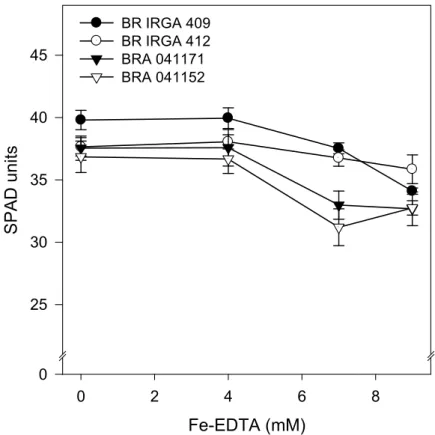
Chlorophyll eontent index
Excluding the genotype BR IRGA 412, which did not show significant
reduction in chlorophyll content, the increase in Fe%EDTA concentration over 7
mM resulted in lower chlorophyll content (Fig. 2). That genotype and BR IRGA
409 showed the highest chlorophyll content and lower sensitivity to the iron
treatments. These genotypes also reached the highest chlorophyll content when
exposed to 7 mM Fe%EDTA treatment, where the differences between genotypes
32 Table 1: Effects of iron treatments (Fe%EDTA) on dry weights of shoot and root, length of shoot and root and number of roots in rice plants.
Fe%EDTA
(mM)
BR IRGA
409
BR IRGA
412
BRA
041171
BRA
041152 Mean
Shoot dry
weight
0.019 0.395±0.06 0.411±0.08 0.453±0.12 0.193±0.07 0.363a
4 0.607±0.08 0.600±0.08 0.280±0.05 0.212±0.03 0.425a
(g plant%1) 7 0.460±0.05 0.384±0.1 0.392±0.13 0.177±0.03 0.353a
9 0.343±0.08 0.348±0.03 0.360±0.06 0.197±0.02 0.312a
Mean 0.451a 0.436a 0.371a 0.195b
Root dry weight 0.019 0.082±0.01 0.075±0.01 0.093±0.03 0.049±0.02 0.075a
(g plant%1) 4 0.081±0.01 0.089±0.01 0.037±0.00 0.037±0.01 0.061a
7 0.077±0.01 0.067±0.02 0.057±0.02 0.030±0.01 0.058a
9 0.063±0.02 0.059±0.00 0.054±0.01 0.037±0.01 0.053a
Mean 0.076a 0.072a 0.060ab 0.038b
Shoot lenght 0.019 60.4±4.3 61.0±3.2 53.7±0.7 44.2±3.4 54.8a
(Cm%1) 4 64.0±3.1 60.4±2.1 43.7±2.3 48.4±2.4 54.2a
7 61.8±2.1 52.3±6.2 50.9±4.6 45.7±3.5 52.7a
9 53.6±4.0 55.3±2.3 45.9±3.4 47.8±2.0 50.7a
Mean 60.0a 57.2a 48.6b 46.5b
Root lenght 0.019 30.7±2.5 26.8±1.2 27.1±2.7 24.0±1.6 27.2a
(Cm%1) 4 32.0±1.2 31.7±0.8 19.3±1.9 25.0±2.3 27.0a
7 32.7±1.0 31.4±2.9 25.2±2.1 26.6±2.4 28.9a
9 30.1±1.5 30.6±1.3 22.4±0.5 29.1±0.7 28.1a
Mean 31.4a 30.1a 23.5b 26.2b
Number of roots 0.019 31.9±2.0 30.6±3.7 32.6±4.1 22.9±2.8 29.5a
4 27.6±2.8 27.2±2.5 18.4±1.7 19.2±1.2 23.1b
7 23.3±1.6 19.4±1.9 22.8±5.2 15.4±1.4 20.2b
9 21.5±3.0 20.3±1.9 23.4±2.1 18.4±1.2 20.9b
Mean 26.1a 24.3a 24.3a 19.0b
The means of genotypes (columns) and Fe%EDTA doses (rows) are shown due to not significant interactions between genotypes and Fe%EDTA doses. Means followed by different letters are significant by Tukey’s test at 0.05 probability level. Means±SE, n=4.
Gas exehange parameters and photorespiration
The increase in Fe%EDTA doses in nutrient solution caused significant
changes in all gas exchange evaluated parameters (Fig. 3). However, the
genotypes differed only on net photosynthesis (A) and Ci/Ca ratio. Higher net
33 412 genotypes coincided with lower values of Ci/Ca ratio in these genotypes.
Exposure of plants to 4 mM Fe%EDTA decreased the Ci/Ca ratio in response to the
stomatal resistance; however, higher iron doses in nutrient solution brought
increases in this ratio, even under continuous reductions in gs (Fig. 3). The
photosynthetic CO2 assimilation rate increased with increasing PPFD (Fig. 4),
irrespective of iron treatments or genotypes. Under non%stressed conditions the
genotypes BR IRGA 409 and BRA 041171 showed similar responses, with light
saturation above 1000 µmol m−2 s−1 and lower light compensation point.
However, under iron excess was observed a marked change in the slope of the
light curve for photosynthesis near the compensation point, outstanding in
genotype BRA 041171 (Fig. 4). This genotype also showed the lowest
photosynthetic rate under high irradiances.
. ?) @
5 4 A B 8
/
"
5 4F D5 DF A5 AF
3 A56
3 A74
3 5A77E7
3 5A77F4
34
. ?) @
5 4 A B 8
"G
. ?) @
5 4 A B 8
?) ) & $ ) .4 .4 @ 5 4 A B ?) & $ ) .4 .4 @ 3 ? ) & $ ) .4 .4 @ 5 F 75 7F 45 4F 3 A56 3 A74 3 5A77E7 3 5A77F4 5H5 5HB 5HE 5H8 5H6 5H5 5H4 5HA 5HB 5H8
Figure 3: Changes in photosynthetic rate (A), stomatal conductance (gs), transpiration (E) and Ci/Ca ratio in four rice genotypes treated with Fe%EDTA in nutrient solution. Data are means±SE of four replicates.
The total electron flow (Jt) did not change with the Fe%EDTA treatment in
both genotypes (Tab. 2). However, the electron flow drive to carboxylative
capacity (Jc) was reduced mainly in the most sensitive genotype (BRA 041171).
This reduction was compensate with increasing in oxygenative electron flow (Jo),
35
? )&$ ).4 .7@
5 455 A55 B55 855 7555
?
)
&
$
)
.4
.7 @
5 75 45 D5
3 A56
3 5A77E7
Figure 4: Light response curve of net photosynthesis (A) of the rice genotypes BR IRGA 409 (circles) and BRA 041171 (inverted triangles) under control conditions (Fe%EDTA 0.019mM; open symbols) and under 7mM Fe%EDTA (closed symbols) in nutrient solution. Data are means±SE of four replicates. The data were fitted against exponential regression (Iqbal et al., 1997; p<0.001).
Chlorophyll a fluoreseenee parameters
The plants exposure to iron excess caused increases in basal fluorescence
(F0) and reduction in maximal photosystem II (PSII) quantum yield (Fv/Fm) of
dark%adapted samples (Fig. 5 and 6). The genotype BR IRGA 412 showed less
change in these parameters. At higher iron doses there were significant differences
in Fv/Fm values over the leaf surface of the genotypes BRA 041171 and BRA
041152 (Fig. 6), the iron toxicity effects were more severe on the extremities of
the leaf (Fig. 7).
Fe%EDTA treatments did not induce significant changes in photochemical
quenching coefficient based on a lake model (qL) neither in apparent electron
transport rate through PSII (ETR) of genotypes BR IRGA 409 and BR IRGA 412.
However, there was significant decrease in qL and ETR of genotypes BRA
36 Table 2: Effects of iron treatments (Fe%EDTA) on the total rate of electron transport through PSII (Jt ;µmol m−2 s−1), electron flow attributable to the carboxylation (Jc ;µmol m−2 s−1) and oxygenation (Jo ;µmol m−2 s−1) reactions of RuBP, ratio between these both factors (Jo/Jt), rate of CO2 production by photorespiration (Rl ;µmol m−2 s−1) and the ratio between Rl and net photosynthesis (Rl/A) in two genotypes of O. sativa.
Genotype Fe%EDTA Jt Jc Jo Jc/Jt Rl Rl/A
BR IRGA 409 0.019 mM 145.1±11.7 113.9±5.3
Aa
31.1±8.9 0.79±0.04 3.89±1.11 0.17±0.06Aa
7 mM 147.2±6.5 77.3±0.8Aa 69.9±6.4 0.53±0.02 8.74±0.80 1.01±0.16Ba
BRA 041171 0.019 mM 137.8±3.6 105.7±0.5
Ab
32.1±3.7 0.77±0.02 4.02±0.46 0.19±0.03Ab
7 mM 128.0±9.3 55.3±4.4Bb 72.7±5.8 0.43±0.02 9.09±0.73 3.80±0.74Aa
Analysis of varianee
Fe%EDTA (Fe) n.s. *** *** *** * ***
Genotype (G) n.s. ** n.s. n.s. n.s. **
Fe × G n.s. * n.s. n.s. n.s. **
Block n.s. n.s. n.s. n.s. n.s. n.s.
37
A) 6) A) 6) A) 6) A) 6)
5H576)
3 A56 3 A74 3 5A77E7 3 5A77F4
5 # G ) I J GA φ J φΙΙ φ 5H5 5H4 5HA 5HB 5H8 7H5 5H5 5H4 5HA 5HB 5H8 7H5 5H5 5H4 5HA 5HB 5H8 7H5 5H5 5H4 5HA 5HB 5H8 7H5 5H5 5H4 5HA 5HB 5H8 7H5 5H5 5H4 5HA 5HB 5H8 7H5 5H5 5H4 5HA 5HB 5H8 7H5
38
3 A56
5
5 F5 755 7F5
3 A74 3 5A77E7 3 5A77F4
#
G)
J
. ?) @
5 4 A B 8 75
5 75 45 D5
5 4 A B 8 75 5 4 A B 8 75 5 4 A B 8 75
5H5 5H4 5HA 5HB 5H8
5H7 5H4 5HD
5H5 5HD 5HB 5H6 7H4
Figure 6: Minimal fluorescence (F0), maximal PSII quantum yield (Fv/Fm), coefficient of photochemical quenching (qL), non%photochemical quenching (NPQ) and apparent electron transport rate (ETR) in four rice genotypes treated with Fe%EDTA in nutrient solution. The parameters were taken using the Imaging% PAM fluorometer. The leaf was divided in three areas of interest: left side, center and right side, depicted as the three bars to each Fe%EDTA concentration, respectively. Data are means±SE of four replicates.
Changes in qL over the leaf surface were observed only on the genotype
BRA 041152 at higher iron doses. ETR did not change through over the leaf.
The quantum yield of photochemical energy conversion in PSII (ϕΙΙ; or
39 041171 and BRA 041152 in response to iron excess under high PPFD (Tab. 3).
These reductions in ϕΙΙ come together with increases in quantum yield of regulated
non%photochemical energy loss in PSII (ϕNPQ); ϕNO remained practically
unchanged (Tab. 3). However, it was observed an increase in ϕNO values in the
genotype BRA 041171 in higher iron doses, and reduction in ϕNPQ was detected in
all genotypes under 9 mM Fe%EDTA treatment, but outstanding in the genotypes
BRA 041152 and BRA 041171 (Fig. 8).
The genotypes BR IRGA 409 and BRA 041171 did not shown differences
in ϕNPQ values over the light curve in imaging chlorophyll fluorescence
measurements (Fig. 9). Only in higher PPFD was observed significant decrease in
ϕII in the genotype BRA 041171, with considerable increase in ϕNO in both
genotypes. Reduction in qL was only detected at high PPFD. Under iron excess
and low PPFD the genotype BRA 041171 show higher qL values. This genotype
40
3 A56
5H576 ) A ) E ) 6 )
3 A74
#
G
)
3 5A77E7
3 5A77F4
"+K L
5H8
5HB
5HA
5H4
5H5
5H8
5HB
5HA
5H4
5H5
5H8
5HB
5HA
5H4
5H5
5H8
5HB
5HA
5H4
5H5