Vol-7, Special Issue3-April, 2016, pp1357-1364 http://www.bipublication.com
Research Article
A comparative study on the effects of clove oil, 2-phenoxy ethanol, and
MS-222 upon some enzymatic and hormonal activities in juvenile Caspian brown
trout (
Salmotruttacaspius
)
Masoumeh Bahr Kazemi*
Department of Fisheries,GhaemshahrBranch, Islamic Azad university,Ghaemshahr,Iran
*
Correspondence Author Email:m.bahrekazemi@qaemshahriau.ac.ir
ABSTRACT
In this study, the effects of threeanesthetics namelyclove oil(CO), 2-phenoxy ethanol(2-PE), and MS-222 were examined on blood enzymes including lactate dehydrogenase (LDH), creatine kinase (CK), alanine aminotransferase (ALT), aspartateaminotransferase (AST), protein, albumin, glucose, and cortisol levels in the Caspianbrowntrout (salmotruttacaspius). The biochemical composition of blood and cortisol levels were analyzed in fish anesthetized with CO (30 mg/l), 2-PE (0.3 ml/l) and MS-222 (100 mg/l) and acontrol group received no anesthetic agent. The long term stressing effect of 2-PEwas detected to belesser than those found for both MS-222 and CO. MS-222, however, revealedliver tissue damage in long run indicating that it would be inappropriate for aquaculturespecies. After all, since no stressful effect of MS-222 was noticed on the Caspianbrown trout in short term, it can be effectively usedin conditions when itbecomes important to reduce severe instantaneous stress such as surgery and broodstock spawning.
Key words: 2-phenoxy ethanol, Blood enzymes, Caspianbrown trout, clove oil, MS-222.
1. INTRODUCTION
Transportation of fish within and/or out of their natural environments is often associated with many problems. They struggle during capture and transport leading to large effects on their physiology. As a result, it often seems necessary to immobilize the fish before taking any simple operation. For this purpose, anesthesia accounts for a valuable tool in fisheries management (12). Common anesthetics in aquaculture include
tricainemethane sulfonate (MS-222),
benzocaine, Kuinaldine, Methomydate, clove oil extract, and 2-phenoxy ethanol (20).The use of anesthetics can cause complications for fish, which may not be desirable for both the culturists and farming purposes. Research has been conducted on stress-induced anestheticsin captive, transportable fish (16-20). Assessment of cardiovascular effects, blood pressure in dorsal aorta, cortisol response, and some blood
enzymes could provideindicators that
thesesubstancesmay be stressful (15). The use of
anesthetics in aquaculture procedures is
inevitable, though,appropriate anesthetics should be selectedassociated with lowest risk, injury, and stress. This, in particular, becomes more important in the case of species that are economically and/or environmentally influential
(12).The Caspian brown trout
(salmotruttacaspius) is a commercial species with high economic value. During the years 1936 to 1939,this species was fished between 410 to 620 tons per year,but the catch rate recorded in the past two decades does not exceed a few fish (24). For restocking as well as
food consumption purposes, Salmotruttacaspius
artificially propagated in a research center on
cold-water fish.In order to raise the
It is difficult to characterize direct stressful effects of anesthetics on captive and/or portable fish; however, the effect of anesthetic use on the blood cells and hormones (particularly cortisol) has been investigated in rainbow trout (Oncorhynchusmykiss) (7-19). The same effects on blood enzymes, blood glucose, and albumin has been studied in different fish species (4-20).The aim of this study is to compare stressful effects of threeanesthetics commonly used in aquaculture (clove oil, 2-phenoxy ethanol, and MS-222) on the Caspian trout in order to select an anesthetic withthe lowest risk to be used in aquaculture and restocking systems.
2. MATERIALS AND METHODS
Anesthesia tests on the Caspian trout using clove oil (CO), 2-phenoxy ethanol (2-PE), and MS-222 was conducted in a Fish Research Centre, Tonkabon, northern Iran. A total of 160 farmed Caspian trout (25.18 ± 4.01 g; 14.19 ± 1.62 cm;
1.0 year old)with equal genderswere
initiallyadopted to the experimental conditions. The fish were then randomly distributed in four fiberglass tanks (four treatments, one control group and three experimental treatments). All of the physicochemical conditions of water (such as temperature, oxygen content, pH, etc., Table 1) was maintained at optimal levels during the experiment.
Table 1: Physicochemical conditions of water throughout the experimental period
Parameter Level
Temperature ( ֯ ◌C) 12.6 ± 1
Dissolved oxygen (mg/l) 6.5 ± 0.5
EC(µs) 602 ± 9
pH 7.4 ± 0.4
To prepare the anesthetic solution, first the amounts of anesthetics were added to the water using a calibrated pipette according to Table 2; then the solution was stirred and homogenated completely (9). To conduct anesthesiatests, fish feedingwas stopped 24 h before the start of the experiment. After anesthesia, the fish were monitored for 24 h to ensure that no side effects of the anesthetics happen during this time (17).
Table 2:The doses of anesthetics(Ackerman et al., 2005) used in this study
Anesthetic Recommended dose
MS-222 100 mg/l
Clove oil (CO) 30 mg/l 2-phenoxy ethanol
(2-PE)
0.3 ml/l
Blood samples (three per treatment) were taken from the caudal peduncle by plastic syringes (2mm) in two steps: 10 minand 24 h after recovery of fish. Blood enzyme activities, albumin, and glucose were assayed by an
autoanalyzer (Technicon RA-1000).Serum
cortisol level was measured through
radioimmunoassay(RIA) by Gama Counter LKB.The enzymes creatinekinase (CK) and lactate dehydrogenase (LDH) wereestimated photometricallyat 340 nm (16-18). Albumin was assayed colorimetrically (5) using a kit, BCG (Bromo Corozel Green) to create a complex with albumin andproduce measurable color at
620-640 nm. The enzymesalanine
aminotransferase (ALT) and aspartate
aminotransferase (AST) were measuredby photometric method (Thomas, 1998). Glucose was estimated by spectrophotometrically at 500-540 nm (2).
The statistical design used in this study was
completely randomized design withthree
anesthetic treatments and a control group each with 3 replications. Data were analyzedby SPSS software (version 21) at a confidence level of 95 percent, and the diagrams were drawn by Microsoft Excel (version 2010). Differences between the data obtained from the treatments were determined by one-way ANOVA and the meanswerecompared with Duncan's test. Paired t-test was applied to compare thenumbers of blood sampling after 10 min and
24 h post-anesthesia.
3. RESULTS
Based on the results of this study, blood albumin in the fish from 10 min after anesthesia showed a significant reduction (p<0.05)only in 2-PE treatment (1.05±0.08mg/ml) compared to the control group (1.26±0.16 mg/ml). The amount of blood albumin in the fish24 h after
(p<0.05)only in the treatment MS-222 (1.46±0.04 mg/ml) than the control group. The other treatments did not reveal significant differencesin comparison with control group
(Table 3). In contrast, no significant
changeswere observedat the levels of this protein in samples from 24 h post-anesthesia compared to 10 min treatment(p<0.05)(Table 4).
Table 3: Comparison of significant differences in studied parameters among the treatments and control
Treatment Parameter Control 2-phenoxy ethanol (10min) Clove oil (10min) MS-222 (10min) 2-phenoxy ethanol(24h) Clove oil (24h) MS-222 (24h)
Albumin(mg/ml) 1.26±0.16 b 1.05±0.08a 1.25±0.073 b 1.24±0.15 b 1.25±0.17 B 1.24±0.04 B 1.46±0.04 C
CK (IU/l) 3960±618.33 b
449.11a
±
2150
316.44 b
±
3973.33 4083.33±856.52b 2003.33±515.98 A ±520.03B
4446.67 122.88
B
±
4030
LDH (IU/l) 2280±147.30bc 1910±364.28abc 1636.67±125.03a ±128.97abc
2113.33
1923.33±198.58ABC 1710±245.76AB 2450±259.39C
ALT (IU/l) 35.67±4.51b
1.53a ± 21.67 3.63b ± 31.00 5.51b ± 35.33 2.52B ± 35.67 4.59B ± 31.00 1.53C ± 51.67
AST (IU/l) 660±59.35bc 568±86.64ab 606.33±33.86abc 554±24.51a 675.66±51.29C 41.62
BC
±
658.33
828.33±59.94D
Glucose(mg/dl) 145.67±14.57a
6.43ab ± 170.33 9.87b ± 183.33 4.39ab ± 168 22.94A ± 149.67 15.69A ± 143.67 13.08C ± 230
Cortisol (ng/ml) 290.33±38.11a
66.56c ± 473.33 78.23bc ± 402.33 21.46a ± 290.33 54.37ABC ± 374 71.04C ± 469 41.52C ± 424.33 *
Different letters in each row indicate significant differencebetween treatments at P<0.05The amount of CKenzyme in the control (3960±618.33 IU/L) was not statistically different from those found in both 10 min after anesthesia with M-S-222
(4083.33±856.52 IU/L) and
CO(3973.33±316.44 IU/L); this enzyme activity, however,showed a significant decrease in 2-PEtreatment (2150±449.11 IU/L) as opposed to control (Table 3). The results of 24 h from anesthesia in all treatments were similar to those for 10 min after anesthesia. No significant differences were found in the levels of CK enzyme between samples from 10 minand 24 h after anesthesia in allthree treatments (Table
4).The amount of LDHin the
control(2280±147.30 IU/L) was not
significantly different fromthose in 10 min
after anesthesia with MS-222
(2113.33±128.97 IU/L) and
2-PH(1910.33±364.28 IU/L) treatments.
Nevertheless, the amount of this enzyme showed a significant decrease in samples
from 10 min after anesthesia with
CO(1636.67±125.03 IU/L) compared to the control. Serum levels of this enzyme in 24 h from the anesthesia treatment did not show a significant difference compared to control
(Table 3). In comparison,the fish displayed amarked increase in the level of LDH enzyme after 24 h of anesthesiawith MS-222 as opposed to those examinedafter 10 min anesthesia with MS-222 (Table 4).
The volume of ALT (35.67±4.51 IU/L) in
control fish showed no significant
differenceswith values in 10 min of anesthesia by each of MS-222 and CO,
while the activity of this enzyme
significantly decreased(21.67±1.53 IU/L) infish 10 min after anesthesia with 2-PE compared to control. This enzymeactivity significantlyrose in the samples treated with MS-222 (51.67±1.53 IU/L) following 24 hin
comparisonwith control,but the
enzymevalues fromCOand 2-PEtreatments were not significantly differentfrom control (Table 3). Additionally, 24 hafter anesthesia
with MS-222 and 2-PE, significant
elevations were recorded in the ALT levelscompared to thoseobserved in 10 min after anesthesia treatment. ALT activity in
CO treatment was notsignificantly
anesthesia with MS-222 (554±24.51 IU/L), which was significantly lower than the
control. In addition, the enzyme
activitymarkedly elevated (828.33±59.94 IU/L) at 24 h after anesthesia with MS-222 compared to the control, but the values for the fish in CO and 2-PE treatments did not
show significant differenceswith the control (Table 3). The amounts of this enzyme measured in the two samplingtimesfor all
three treatments displayed significant
increases in 24 h post-anesthesia as opposed to those estimated in 10 min after anesthesia treatments (Table 4).
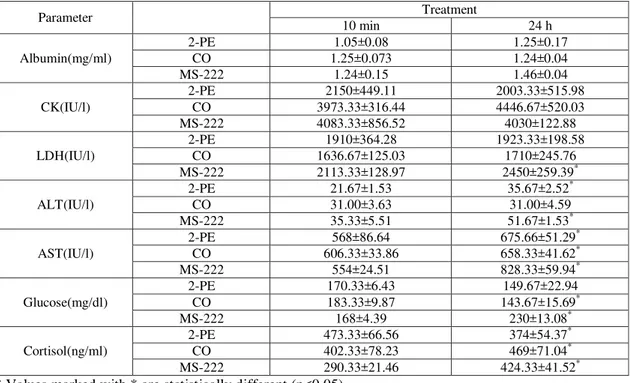
Table 4: Paired comparison among the measured parameters at different treatments of 10 min and 24 h post-anesthesia
Parameter Treatment
10 min
24 h
) mg/ml ( Albumin
2-PE 1.05±0.08 1.25±0.17
CO 1.25±0.073 1.24±0.04
MS-222
1.24±0.15 1.46±0.04
) IU/l ( CK
2-PE
2150±449.11 2003.33±515.98
CO
3973.33±316.44 4446.67±520.03
MS-222
4083.33±856.52 4030±122.88
) IU/l ( LDH
2-PE
1910±364.28 1923.33±198.58
CO
1636.67±125.03 1710±245.76
MS-222
2113.33±128.97 2450±259.39*
) IU/l ( ALT
2-PE
21.67±1.53 35.67±2.52*
CO
31.00±3.63 31.00±4.59
MS-222
35.33±5.51 51.67±1.53*
) IU/l ( AST
2-PE
568±86.64 675.66±51.29*
CO
606.33±33.86 658.33±41.62*
MS-222
554±24.51 828.33±59.94*
) mg/dl ( Glucose
2-PE
170.33±6.43 149.67±22.94
CO
183.33±9.87 143.67±15.69*
MS-222
168±4.39 230±13.08*
) ng/ml ( Cortisol
2-PE
473.33±66.56 374±54.37*
CO
402.33±78.23 469±71.04*
MS-222
290.33±21.46 424.33±41.52*
* Values marked with * are statistically different (p<0.05).
The results of serum glucose measurement showed an amount of 145.67±14.57 mg/dl in the control group. The increases in glucosein the fish from 10 min after anesthesia with MS-222 (168±4.39 mg/dl) and 2-PE (170.33±6.43 mg/dl) were not significant. Anesthesia with CO, on the other hand, resulted in significantly higher glucose level (183.33±9.87 mg/dl)after 10 minthan the control group. In samples from 24 h after anesthesia, only the anestheticMS-222 yielded a significant increase (230±13.08 mg/dl) in glucose levels compared to the control whereas in the samples anesthetized with CO and 2-PE, glucose levels returned to the baseline values (Table 3). Comparing the amounts of glucose from two sampling timesshowed that fish treated with MS-222 presented a significant increase in glucose level (230±13.08 mg/dl) following 24 h of anesthesia compared to 10
min post-anesthesia (168±4.39 mg/dl). In the CO treatment,a significant glucose reduction (143.67±15.69 mg/dl) was observed after 24 h of operation compared to 10 min after anesthesia (183.33±9.87 mg/dl). In the fish from 2-PE treatment, the average amount of glucose reducedafter 24 h of anesthesiain relation to the first stage of sampling (10 min after anesthesia), but the difference was not statistically significant (Table 4).
anesthesia with MS-222 (424.33±41.52ng/ml) and CO (469±71.04ng/ml) exhibited significant rises in the amount of cortisol compared to control. In the fish anesthetized by 2-PE, the amount of cortisol decreased (374±54.37ng/ml), but it was not significantly different from the control (p>0.05) (Table 3). Comparingthe two sampling times revealed that the amount of cortisol markedly rose in MS-222 and CO treatments, and significantly increased in 2-PE treatment after 24 hin comparison with hormone values obtainedfrom 10 min treatment after being contacted with three anesthetics examined (Table 4).
4. DISCUSSION
Any kind of general response to stress is
affected by two groups of
hormones:catecholamine and the hypothalamic-pituitary inter-renal (HPI) axis hormones, especially cortisol (Barton, 2002). The effect of a rise in cortisolis all-encompassing and often hazardous if prolonged leading to increased activity of enzymes, interruption in growth, reduced carbohydrate intake, increased glucose production from tissue protein, liver glycogen breakdown, changes in membrane permeability, increased ATPase activity, and increased
number of white blood cells.Following
prolonged exposure to chronic stressors, cortisol levels may return to normal levels (though stress still exists) (16). Stress may increase the
susceptibility to diseases, growth and
reproductive defects, all of which seem to be caused by cortisol secretion (10).The results of this study showed that at 10 min after anesthesia, the amount of albumin significantly decreased in the blood of fish treated with 2-PEcompared to the control group. Following 24 h of anesthesia, the amount of albumin significantly increasedonly in MS-222 treatment in comparison with the control fish. The elevated value of this protein after 24 h of anesthesia with MS-222 can be due to the albumin ability in transferring exotic chemicals. The decrease in the amount of albumin after 10 min anesthesia with 2-PE may be due to its effects on the liver and catabolism of albumin (2). Velisek and Svobodova (2004) observed a
significant albumin increase in rainbow trout (oncorhynchusmykiss) after 24 h exposure to 2-PE; such an effect was not observed in the Caspian trout. The study of Mavaddati and Habibian (2011) on the anesthetic effects of 2-PEand CO upon rainbow trout revealed no significant changesin the blood albumin after 15 min and 24 h of anesthesia. Our results showed that at 10 min after anesthesia, blood glucose levels in fish treated with COincreased significantly compared to the control and returned to the baseline valuesafter 24 h, which corresponds to the results of Velisek et al. (2011) in rainbow trout. At 24 hpost-anesthesia,the amount of glucose increased markedly only in MS-222treatmentas opposed to control. Velisek and Svobodova (2004b) found no changes in blood glucose levels in common
carp (cyprinuscarpio) anesthetized with 2-PE.
10 min and 24 h after anesthesia, the enzyme CK levels was significantly lower in fish anesthetized with 2-PEthan the control group. According to Henry (2001), a significant reduction in the amount of CK can be a result of
a sharp increase in basal metabolism.
Accordingly, it can be concluded that 2-PE greatly increased the metabolism of Caspian
trout at least 24 h after anesthesia.
Meanwhile,Congleton (2006) reported increased activity of CK in the blood of Chinook salmon (O. tshawytscha) exposed to acute doses of MS-222, which is completely contrary to the findings of this study. Evaluation of LDH at 10 min after anesthesia with COEshowed a significant declinecompared to the control. LDHlevels in the fish anesthetized with MS-222 markedly roseafter 24 h in relation to those following 10 min ofanesthesia. LDH serves as apotential indicator to determine the toxicity of the chemicals. Congleton (2006) noted elevated blood LDH activity in Chinook salmon (O.tshawytscha) exposed to acute doses of MS-222. Also,Velisek et al. (2004 a) detected no significant differencesin the blood LDH of rainbow trout treated with 2-PEandthose in control. However, Soltani et al. (2003),noticed a considerablereduction of this enzyme in the blood of common carp treated with CO, which is in accordance with that found after 10 min of anesthesia with the same material in the Caspian trout. Mavaddati and Habibian (2011) did not observe any significant changes inthe level of this enzyme in the blood of rainbow trout treated
with 2-PE, CO, and MS-222.
Additionally,Velisek et al. (2011) reported no significant changes in the level of this enzyme in the blood of rainbow trout treated with CO, 2-PE, and MS-222, all of which are against the results of present research.The results obtained from LDH activity in the examined sera might indicate non-toxicity and lack of injury from the tested anesthetics on the organs and internal body tissues of Caspian trout. Our results also showed that 10 min after anesthesia in fish treated with 2-PE, a significant decrease occurred in the amount of ALT enzymeas opposed to the control. Compared with the control, there was a significant increase in the
levels of this enzymeafter 24 h of anesthesia with MS-222. ALT enzyme activity in plasma rises as a result of damage to the liver membrane (11). According to Henry (2001), a significant reduction in ALT enzyme levels in the blood can increase dopamine release. It can, therefore, be concluded that the stress caused by 2-PE at 10 min after anesthesia should have led to the release of high dopamine levels rendering
reduced ALT vs. the control.However, the
significant increase in the activity of ALTafter 24 h of anesthesia with MS-222 could be explained through thepossibility of damagingthe liver tissue by this chemical. Velisek et al. (2004a) studiedthe enzyme activity in rainbow trout exposed to 2-PE and did not realize any significant difference, which disagrees with the results of this research. In accordance with the results of this study, Congleton (2006) reported a significant increase in serum ALT activity ofChinook salmon after exposure
toMS-222.Significant decrease and increase,
respectively, were recordedin the levels of the enzyme ASTafter 10 minand 24 h of anesthetizing the fish with MS-222. Increase in plasma AST activity is a result of mitochondria breakdownleading toinflation of liver tissue. In addition toan elevationwhenthe liver’smembrane is damaged, AST also rises withheart and muscle damages (11). Velisek et al. (2004a) observed a significant decrease in the level of AST activity in rainbow trout immediately after anesthesia with 2-PE. On the other hand, Congleton (2006) perceived a significant increase in serum activity of this enzyme in
Chinook salmon treated with MS-222
and dose of anesthetic used and other water physicochemical conditions. The results of current research suggest that the plant origin of an anesthetic cannot solelymake it a suitable choice for biological applications. Although the examined doses of the three anesthetics used caused no irreversible fish damages and mortalities, the results suggest that MS-222 rendered lesser stressful effects in short term than the other two anesthetics, hence, it can be used where the aim is to eliminate instantaneous stresses such as those induced by surgery and stripping. Nonetheless, where the fish is farmed for aquaculture purposes, the use of MS-222 is not recommended as this anesthetic revealed liver tissue damage through elevation ofALT andASTenzymes in Caspian trout juvenile aftera long-term (24 h)anesthesia.In contrast, 2-PE proved to have lesserstressful effects than MS-222 and CO inlong-term (24 h).
REFERENCES
1. Ackerman P. A., Morgan J. D., Iwama G. K.Anesthetics. CCAC guidelines on: The care and use of fish in research, teaching and testing. Canadian Council on Animal Care, Ottawa CA; 2005.
2. Barham D., Trinder P. An improved color reagent for the determination of blood glucose by the oxidase system.Analyst 1972; 97: 142-145.
3. Barton B. A Stress in Fishes: A Diversity of Responses with Particular Reference to Changes in Circulating Corticosteroids. Integ.and Comp. Biol 2002; 42: 517–525. 4. Congleton J. L. Short communication.
Stability of some commonly measured blood – chemistry varianles in juvenile salmonids exposed to a lethal dose of the anaesthetic
MS-222, Aquaculture Research 2006;
37:1146-1149.
5. Doumas B. T., Watson, W. A., Biggs, H. G. Albumin standards and the measurement of serum albumin with bromcresol green. ClinChimActa 1971; 31(1): 87-96.
6. Henry J. B. Clinical diagnosis and management by laboratory methods.20th Edition, W. B. Saunders, Philadelphia 2001. 7. Holloway A., Keene, J., Noakes D., Moccia
R. Effects of clove oil and MS 222 on blood
hormone profiles in rainbow trout
Oncorhynchusmykiss,Walbaum. Aquaculture Research 2004; 35: 1025-1030.
8. Mavadati A., Habibian R. Comparision of effect of clove oil and 2-phenoxy ethanol on
serom biochemical parameters in
Onchorhynchusmykiss. Word Journal of fish and marine sciences 2011; 3(4): 318-322. 9. MohammadiArani, d. Anesthetic effects of
clove (extract and essence) on sturgeon. MSc, Department of Natural Resources and
Marine Sciences, TarbiatModarres
University,2002 .
10. Pickering A. D. Rainbow trout husbandry:
management of the stress response.
Aquaculture 2003;100 (1-3): 125-139. 11. Porchas M., cordova l., enriquez r. Cortisol
and Glucose: Reliable indicators of fish
stress. Pan-American Journal of Aquatic
Sciences 2009; 4(2): 158-178.
12. Ross L. G., Ross B. Anaesthetic and sedative techniques for aquatic animals, Third Edition 2008; p. 222.
13. Small B. C. Anesthetic efficacy of metomidate and comparison of plasma
cortisol responses to
tricainemethanesulfonate. quinaldine and
clove oil anesthetized channel catfish Ictaluruspunctatus. Aquaculture 2003; 218: 177-185.
14. Soivio A., Nyholm K., Huhti M. Effects of anesthesia with MS 222,neutralised MS 222 and benzocaine on the blood constituents of
rainbow trout, Salmogairdneri. Journal of
Fish Biology 1977; 10: 91-101.
15. Soltani M., Ghaffari M., Khazraeinia P. C., Bokaei S. Anestheticeffects of Indian clove oil on hematological parameters. certain blood enzymes and tissue pathology of carp. Journal of Veterinary College of Tehran University. Volume 59, Number 3 2003; pp. 295-299.
16. Stein w. Creatine kinase (total activity). creatine kinase isoenzymes and variants. In.
Thomas L, (ed). Clinical laboratory
diagnostics. Frankfurt: TH-books
Verlagsgesellschaft 1998; pp 71-80.
17. Stoskopf M. K. Fish medicine. Chapter 6: Anesthesia and restraint. Academic press, USA; 1993.430 p.
18. Thomas. L .Clinical Laboratory
Diagnostics.1st ed. Frunkfurt, TH-books
Verlagsgesellschaft;1998.
(Salmotruttafario). Journal of animal and veterinary advances 2010; 9(14): 1925-1933. 20. Velisek J., Stara A., Li Z. H., Silovska S.,
Turek J. Comparison of the effects of four an aesthetics on blood biochemical profiles and oxidative stress biomarkers in rainbow trout. Aquaculture 2011; 310: 369-375. 21. Velisek J., Svobodova Z. Anaesthesia of
rainbow trout (Oncorhynchusmykiss) with
2-phenoxyethanol: Acute toxicity and
biochemical blood profile. ActaVeterinaria Brno 2004; 73: 379–384.
22. Velisek J., Svobodova Z. Anaesthesia of
common carp (Cyprinuscarpio L.) with
2-phenoxyethanol: acute toxicity and effects on biochemical blood profile. ActaVeterinaria Brno 2004; 73: 247–252.
23. Velisek J., Svobodova Z., Piackova V. Effects of 2-phenoxyethanol an aesthesia on haematological profile on common carp (cyprinuscarpio) and rainbow trout (Onchorhynchusmykiss).Acta vet brno 2007; 76: 487-492.
24. Vossoughi, G., Mostajeer, B., 2006. Fresh water fishes. Tehran University Press, Tehran, 123 pp.
25. Wagner G. N., Singer T. D., Mckinley R. S. The ability of clove oil and MS-222 to minimize handling stress in rainbow trout (OncorhynchusmykissWalbaum),