Article
Connecting Myokines and Metabolism
Rexford S. Ahima1, Hyeong-Kyu Park2
1
Division of Endocrinology, Diabetes and Metabolism, and the Institute for Diabetes, Obesity and Metabolism, Department of Medicine, Perelman School of Medicine at the University of Pennsylvania, Philadelphia, PA, USA; 2
Department of Internal Medicine, Soonchunhyang University College of Medicine, Seoul, Korea
Skeletal muscle is the largest organ of the body in non-obese individuals and is now considered to be an endocrine organ. Hor-mones (myokines) secreted by skeletal muscle mediate communications between muscle and liver, adipose tissue, brain, and oth-er organs. Myokines affect muscle mass and myofiboth-er switching, and have profound effects on glucose and lipid metabolism and inflammation, thus contributing to energy homeostasis and the pathogenesis of obesity, diabetes, and other diseases. In this re-view, we summarize recent findings on the biology of myokines and provide an assessment of their potential as therapeutic tar-gets.
Keywords: Muscle, skeletal; Myokine; Obesity; Diabetes; Exercise; Metabolism
INTRODUCTION
Lack of exercise and sedentary lifestyle have been linked to obesity, type 2 diabetes, cardiovascular diseases, cancer, osteo-porosis, and premature death [1-7]. Skeletal muscle is the most abundant tissue in non-obese adults, accounting for approxi-mately 40% of the body weight [8]. Skeletal muscle adapts to mechanical, neural and humoral stimuli, and plays critical roles in physical activity, energy expenditure, and glucose disposal [9,10]. Exercise and anabolic hormones, e.g., insulin, insullike growth factor 1, growth hormone and testosterone, in-crease skeletal muscle mass (Fig. 1) [11,12]. Conversely, phys-ical inactivity from aging or neuromuscular disorders, and chronic diseases, such as cancer, renal failure, respiratory fail-ure, infection, and some endocrine disorders, e.g., uncontrolled
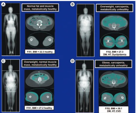
diabetes mellitus, hyperthyroidism and hypercortisolism, cause muscle atrophy or “sarcopenia” (Fig. 1) [13-17]. Sarcopenia has been linked to obesity, metabolic syndrome, and other dis-eases in aged populations, particularly in South Asia (Fig. 2) [18-21].
The concept that skeletal muscle secretes humoral factors that actively communicate with other organs was proposed many years ago [22-24]. Henningsen et al. [25] and Pedersen et al. [26] used the term “myokines” to describe cytokines and other peptides expressed and released by muscle cells. This re-view highlights the biological actions of myostatin and other myokines that regulate skeletal muscle mass and metabolism via autocrine, paracrine, and endocrine mechanisms.
Received: 25 March 2015, Revised: 22 June 2015, Accepted: 29 June 2015
Corresponding author: Rexford S. Ahima
Division of Endocrinology, Diabetes and Metabolism, Perelman School of Medicine at the University of Pennsylvania, 12-104 Smilow Translational Research Center, 3400 Civic Center Boulevard, Building 421, Philadelphia, PA 19104, USA
Tel: +1-215-573-1872, Fax: +1-215-898-5408,
E-mail: [email protected]
Copyright © 2015 Korean Endocrine Society
Fig. 1. Positive and negative regulators of skeletal muscle mass. Myokines are produced and secreted by skeletal muscle and act via au-tocrine, paracrine and endocrine mechanisms to regulate skeletal muscle mass and metabolism. IGF-1, insulin-like growth factor 1; IL, interleukin; BDNF, brain-derived neurotrophic factor; FGF-21, fibroblast growth factor 21; LIF, leukemia inhibitory factor.
Fig. 2. Magnetic resonance imaging scans comparing the distributions of abdominal and thigh fat and muscle in (A) lean, (B, C) over-weight, and (D) obese women. As in (B) and (D), increased visceral adiposity and sarcopenia are associated with diabetes mellitus (DM), hypertension (HT), dyslipidemia, and cardiovascular disease (CVD). BMI, body mass index.
A B
MYOSTATIN
Much attention has been focused on the biology of the
trans-forming growth factor β (TGF-β) superfamily of proteins since
the discovery of myostatin [27]. Myostatin, also known as growth differentiation factor 8, is expressed and secreted pre-dominantly by skeletal muscle and inhibits muscle growth. This function is conserved in many species, as evident by the hypermuscular phenotype resulting from inactivation of myo-statin gene in mice, sheep, cattle, and human [28-32]. During early postnatal development, myostatin inhibits muscle stem cell proliferation, differentiation, and protein synthesis [33]. Normally, the differentiation of skeletal muscle cells requires growth arrest followed by expression of muscle-specific genes. These processes are coordinated by activation of specific cy-clins, cyclin-dependent kinases (Cdk), Cdk inhibitors (CdkIs), and muscle regulatory factors. During the proliferation phase, myostatin up-regulates p21 (a CdkI), and decreases the levels of Cdk2 and Cdk4, leading to cell cycle arrest.
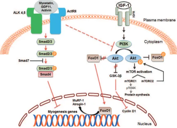
Myostatin inhibits satellite cell activation by down-regulat-ing the transcription factor Pax7, and also controls the myo-genic differentiation program through inhibition of myomyo-genic regulatory factors, such as Pax3, MyoD, and Myf5. Studies in-dicate that myostatin’s inhibitory effect on muscle differentia-tion in the postnatal period is mediated partly by perturbadifferentia-tion of Akt/mammalian target of rapamycin complex1 signaling [34-37]. In mature adult muscle fibers, the C-terminal dimer of myostatin binds to activin receptors II (ActRII), mainly ActRI-IB and to a lesser degree ActRIIA, which then recruits, phos-phorylates and activates activin receptor-like kinase (Alk) 4 and Alk5, leading to phosphorylation and activation of Smad2 and Smad3 [38,39]. Phosphorylated Smad2 and Smad3 form a heterodimeric complex with Smad4, which is translocated into the nucleus, and acts as a transcription factor to regulate gene expression. Myostatin signaling also leads to activation of Smad7 which functions as a negative feedback inhibitor [40, 41]. The activation of myostatin-Smad pathway inhibits the translation initiation complex and protein synthesis. Myostatin
suppresses Akt signaling and acts via forkhead box protein O1 transcription factors to promote protein breakdown through ac-tivation of the ubiquitin-proteasome system (Fig. 3). Myostatin also inhibits the autophagy-lysosome system [42,43].
Genetic or pharmacologic inhibition of myostatin, ActRIIB, Alk4/Alk5, or Smad2/3 results in skeletal muscle hypertrophy, associated with increased protein synthesis and reduced protein degradation [44]. Myostatin knockout (Mstn–/–) mice have in-creased skeletal muscle mass as well as reduced body fat [45]. Myostatin-null agouti lethal yellow or leptin deficient mice have drastically reduced body fat and glucose levels raising the possibility that blockade of myostatin signaling may be useful for treating obesity and diabetes [46]. Guo et al. [47] have shown that Mstn–/– mice have increased glucose utilization and insulin sensitivity. To determine whether these effects were due to a lack of myostatin signaling in muscle or adipose tis-sue, they compared the metabolic phenotypes of mice carrying a dominant negative ActRIIB receptor expressed in adipocytes or skeletal muscle. The absence of myostatin signaling in adi-pocytes did not affect body composition or glucose homeosta-sis, whereas inhibition of myostatin signaling in skeletal mus-cle recapitulated the phenotype of Mstn–/– mice, characterized by hypermuscularity, decreased body fat, and enhanced insulin sensitivity [47].
We studied the effects of pharmacological blockade of myo-statin and related peptides by treating mice on chow and high-fat diets with a soluble activin receptor type IIB (ActRIIB-Fc). ActRIIB-Fc treatment increased lean and skeletal muscle mass, grip strength, and contractile force, decreased body fat, and in-creased insulin sensitivity [48]. Mice lacking Akt1 or Akt2 have reduced muscle mass, grip strength and contractile force, consistent with a pivotal role of Akt signaling in promoting muscle growth [49,50]. Contrary to in vitro studies showing that Akt signaling is necessary for the ability of ActRIIB inhibi-tion to induce muscle hypertrophy, we found that Akt1 and Akt2 deficient mice responded similarly as wild type mice to ActRIIB-Fc in regard to increased muscle size, grip strength and contractile force, indicating these Akt isoforms are not es-sential for ActRIIB signaling [51].
ActRIIB-Fc has also been shown to decrease diet-induced obesity and improve glucose and lipid levels in mice [48]. Im-portantly, ActRIIB-Fc induced the browning of white adipose tissue (WAT), as shown by increased expression of the thermo-genic genes uncoupling protein 1 (UCP1) and peroxisomal
proliferator-activated receptor-γ coactivator 1α (PGC1α). Thus,
the anti-obesity effect of ActRIIB-Fc is partly by increasing
skeletal muscle mass as well as inducing thermogenesis in WAT [52]. Other studies have confirmed that deficiency of myostatin signaling in Mstn–/– mice promotes browning of WAT [53,54]. WAT of Mstn–/– mice displays features of brown adipose tissue, e.g., increased expression of including UCP1
and PGC1α, as well as expression of beige adipocyte markers,
e.g., Tmem26 and CD137. The enhanced browning of adipose tissue appears to be mediated by irisin (fibronectin type III do-main-containing 5, Fndc5), a myokine secreted from skeletal muscle in Mstn–/– mice. Myostatin deficiency stimulates AMPK expression and phosphorylation, which then activates
PGC1α and irisin and promotes the browning of adipose tissue
and thermogenesis [54]. Another study has shown that the re-duction of body fat in Mstn–/– mice is due to increased energy expenditure and leptin sensitivity [55]. The cross-talk of myo-kines and adipomyo-kines may provide novel therapeutic tools for treating obesity, diabetes, and diseases associated with muscle atrophy.
Does myostatin blockade have clinical potential? A double-blind, placebo-controlled study evaluated the safety, pharmaco-kinetics, and pharmacodynamics of a decoy ActRIIB receptor (ACE-031) in healthy postmenopausal women randomized to receive a single dose of ACE-031 (0.02 to 3 mg/kg subcutane-ous) or placebo. ACE-031 treatment had mild adverse events and produced significant increases of lean mass and thigh mus-cle volume at day 29 in those receiving 3 mg/kg. Moreover, ACE-031 treatment increased adiponectin by 51.3% and de-creased leptin by 27.7% demonstrating a favorable metabolic profile [56]. Androgen deprivation therapy for prostate cancer causes sarcopenia and increased body fat. An anti-myostatin peptibody (AMG 745/Mu-S) was evaluated in men undergoing androgen deprivation therapy for non-metastatic prostate can-cer [57]. The adverse events in AMG 745 versus placebo treat-ed groups were: diarrhea (13% vs. 9%), fatigue (13% vs. 4%), contusion (10% vs. 0%), and injection site bruising (6% vs. 4%). AMG 745 treatment increased the lean body mass and de-creased fat mass. These preliminary results provide support for further investigation into the safety profile and of therapeutic uses of myostatin blockade to reduce sarcopenia and improve metabolism.
with lean women [58,59]. The biological significance of these
findings, and whether myostatin and other TGF-β peptide su -perfamily can be targeted specifically for treatment of obesity and metabolic disorders requires further studies.
INTERLEUKIN 6
The cytokine interleukin 6 (IL-6) was named a myokine be-cause its levels increased in response to exercise and muscle contraction [60-62]. Evidence supporting the notion that is the source of IL-6 is based on transcriptional analysis of IL-6 mRNA levels during exercise, in situ hybridization and immu-nohistochemistry of IL-6, microdialysis of contracting skeletal muscle, and measurement of arteriovenous IL-6 concentrations and blood flow across an exercising leg [63]. Skeletal muscle adapts to exercise by altering glycogen content, increasing
β-oxidation of fatty acids, increasing intramyocellular triglyc -eride hydrolysis, and enhancing epinephrine-induced lipolysis [64]. Thus, the trained skeletal muscle uses fat as a substrate and is less dependent on glucose and muscle glycogen during exercise.
Epidemiological studies have found an inverse correlation of the amount of physical activity and plasma IL-6 concentration. The basal plasma levels of IL-6 are strongly associated with physical inactivity, obesity and metabolic syndrome [65-67]. Chronic exercise decreases the basal levels of IL-6, and the in-creases in plasma IL-6 and muscle IL-6 mRNA content during acute exercise are also blunted in response to endurance
train-ing [68]. IL-6 receptor (IL-6R) α is regulated opposite to IL-6, and the basal IL-6Rα mRNA content in muscle is increased
during endurance training, perhaps counteracting the reduction in IL-6 [69].
What are the biological roles of IL-6? Treatment of rat L6 myocytes with IL-6 increases basal glucose uptake via glucose transporter 4 translocation, as well as insulin-stimulated glu-cose uptake [70]. The in vitro effect of IL-6 on glucose uptake is mediated, at least partly, through AMP-activated protein ki-nase (AMPK) activation. Studies have also suggested that IL-6 may stimulate fatty acid oxidation via AMPK [71-73]. In rest-ing humans, acute administration of IL-6 infused to achieve physiological concentrations had no effect on either endoge-nous glucose production or glucose disposal [74]. In contrast, when IL-6 was infused to mimic the plasma of IL-6 observed during high-intensity exercise, the endogenous glucose produc-tion was markedly increased, suggesting that a muscle-liver crosstalk mediated via IL-6 may have a role in regulating
plas-ma glucose levels through endogenous glucose production dur-ing exercise [70]. In addition to its effects on glucose metabo-lism, infusion of IL-6 in healthy volunteers stimulates lipolysis in skeletal muscle without affecting adipose tissue [71,75]. IL-6 inhibits endotoxin-induced tumor necrosis factor (TNF) production in human monocytes, and infusion of IL-6 during exercise attenuates the ability of endotoxin to increase TNF levels in healthy individuals. These anti-inflammatory proper-ties of IL-6 are associated with induction of anti-inflammatory cytokines, e.g., IL-1 receptor agonist and IL-10 [76].
INTERLEUKIN 15
IL-15 was classified as an interleukin based on its 4-α-helical
secondary structure and its ability to mimic the functions of IL-2 [77]. The plasma membrane receptor for IL-15 was shown
to be composed of IL-2 receptor β (IL-2Rβ), the common gam
-ma chain (γc), and a specific IL-15 receptor α (IL-15Rα) chain [78,79]. Transcripts for IL-15 and IL-15Rα are widely ex -pressed, and skeletal muscle expresses IL15 and IL15RA mRNAs [77,78]. In vitro experiments in myogenic cells sug-gested that IL-15 was an anabolic factor for skeletal muscle; however, increasing IL-15 levels in vivo did not induce muscle hypertrophy [80-83]. Nonetheless, studies have revealed
differ-ent locomotor phenotypes of mice lacking IL-15Rα or IL-15 or IL-2Rβ [84,85]. Furthermore, single nucleotide polymorphisms
(SNPs) in the human IL15 and IL15RA genes have been associ-ated with different muscle phenotypes, responses to resistance training, and obesity [86-90].
We hypothesized that IL-15Rα has a role in determining the muscle phenotype in mice. We found that loss of IL-15Rα
leads to a remodeling of fast skeletal muscles to a more oxida-tive phenotype associated with increased spontaneous locomo-tor activity and exercise capacity, and resistance to fatigue [91]. The molecular signature of oxidative muscle phenotype from
IL-15Rα knockout mice included altered mitochondrial bio -genesis and calcium homeostasis. Consistent with our observa-tions in mice, we found a significant association between a SNP in exon 3 of the IL15RA gene and endurance in human athletes [91].
A recent paper by O’Connell and Pistilli [92] has shown
sponta-clear whether irisin is truly a myokine since human WAT is ca-pable of expressing FNDC5 and secreting irisin [100]. Some studies indicate that neither acute nor chronic exercise consis-tently increases expression FNDC5 and/or irisin in humans [101,102]. However, others have shown associations of plasma irisin and aging, obesity, physical activity, and metabolic out-comes [103,104]. These controversies surrounding the role of irisin may arise from different exercise regimens and assays for measuring irisin, suboptimal storage of tissue samples, as well as differences in the function of irisin in mouse versus human [101,104,105].
CONCLUSIONS
Myokines are proposed to play important roles in mediating the beneficial effects of skeletal muscle mass and exercise on health. Myokines have been implicated in the pathogenesis of obesity, substrate oxidation, lipid partitioning, insulin sensitivi-ty, and inflammation. While the list of putative myokines keeps growing, the specific physiological and pathological effects of these molecules are poorly understood. Important questions that need to be answered for a presumed myokine include whether skeletal muscle is the main or only source, how the lo-cal and systemic concentrations of the myokine are regulated, whether there are biological differences among species, and what specific signaling mechanisms mediate the biological ef-fects of the myokine in various organs. A better understanding of the actions of myokines may identify novel therapies for obesity, diabetes, cardiovascular diseases, cancer, and other diseases known to be improved by exercise.
CONFLICTS OF INTEREST
No potential conflict of interest relevant to this article was re-ported.
ACKNOWLEDGMENTS
We thank Dr. Lim Soo of the Seoul National University Col-lege of Medicine for providing MRI scans (Fig. 2). RSA is sup-ported by American Diabetes Association grant #7-13-BS-004, and National Institutes of Health grants R01-NS084965 and P01-DK049210.
neous activity was not different in muscle IL-15Rα deficient mice, unlike the whole body IL-15Rα knockout mouse [93]. Thus, muscle IL-15Rα has a role in altering contractile proper -ties and fatigue characteristics of skeletal muscles, but the
loco-motor behavior is likely to be controlled by IL-15Rα targets in
brain [84]. Further studies are needed to evaluate whether
IL-15Rα can be targeted specifically for obesity treatment by in -creasing energy expenditure and fatty acid oxidation.
IRISIN
Chronic exercise increases skeletal muscle mitochondrial
bio-genesis, which is regulated by PGC1α [94-96]. Bostrom et al.
[97] demonstrated that the inguinal subcutaneous WAT had
in-creased levels of UCP1 and Cidea in muscle-specific PGC1α
transgenic mice compared to wild-type mice. To address whether the browning of the subcutaneous WAT was due to a myokine, they cultured primary murine subcutaneous
adipo-cytes with conditioned media from PGC1α overexpressing
myocytes, and found that the conditioned media increased ex-pression of brown-fat specific genes in adipocytes. Using gene array and bioinformatic methods, Bostrom et al. [97] identified
FNDC5 as a gene target of PGC1α, and showed that FNDC5
expression was increased in muscle obtained from exercise-trained mice and humans. Primary subcutaneous adipocytes treated with recombinant-FNDC5 displayed an increased ex-pression of brown adipose genes, i.e., UCP1, Elovl3, Cox7a, and Otop1. Moreover, UCP1-positive cells treated with FNDC5 developed multi-loculated lipid droplets, increased mitochon-drial content and oxygen consumption, consistent with a ther-mogenic phenotype. Based on these results, the authors sur-mised that FNDC5 induced a beige phenotype of WAT in mice, and this effect was attenuated by a peroxisome
proliferator-ac-tivated receptor α antagonist treatment [97]. Further experi -ments revealed that the full-length FNDC5 was a transmem-brane protein, and the extracellular N-terminal portion of FNDC5 was secreted and was highly homologous between mouse and humans. This myokine was named “irisin” after the Greek messenger goddess Iris. Plasma irisin levels were shown to be increased in mice and humans after short-term exercise. Adenoviral expression of FNDC5 in liver increased plasma iri-sin levels which led to the browning of subcutaneous WAT, in-creased energy expenditure, and protection against obesity and insulin resistance [97].
un-REFERENCES
1. Irwin ML, Yasui Y, Ulrich CM, Bowen D, Rudolph RE,
Schwartz RS, et al. Effect of exercise on total and intra-ab-dominal body fat in postmenopausal women: a randomized controlled trial. JAMA 2003;289:323-30.
2. Irving BA, Davis CK, Brock DW, Weltman JY, Swift D,
Barrett EJ, et al. Effect of exercise training intensity on abdominal visceral fat and body composition. Med Sci Sports Exerc 2008;40:1863-72.
3. Church TS, Thomas DM, Tudor-Locke C, Katzmarzyk PT,
Earnest CP, Rodarte RQ, et al. Trends over 5 decades in U.S. occupation-related physical activity and their associa-tions with obesity. PLoS One 2011;6:e19657.
4. Tuomilehto J, Lindstrom J, Eriksson JG, Valle TT,
Ham-alainen H, Ilanne-Parikka P, et al. Prevention of type 2 dia-betes mellitus by changes in lifestyle among subjects with impaired glucose tolerance. N Engl J Med 2001;344:1343-50.
5. Manson JE, Hu FB, Rich-Edwards JW, Colditz GA,
Stamp-fer MJ, Willett WC, et al. A prospective study of walking as compared with vigorous exercise in the prevention of coro-nary heart disease in women. N Engl J Med 1999;341:650-8.
6. Nocon M, Hiemann T, Muller-Riemenschneider F, Thalau
F, Roll S, Willich SN. Association of physical activity with all-cause and cardiovascular mortality: a systematic review and meta-analysis. Eur J Cardiovasc Prev Rehabil 2008; 15:239-46.
7. Monninkhof EM, Elias SG, Vlems FA, van der Tweel I,
Schuit AJ, Voskuil DW, et al. Physical activity and breast cancer: a systematic review. Epidemiology 2007;18:137-57.
8. Janssen I, Heymsfield SB, Wang ZM, Ross R. Skeletal
muscle mass and distribution in 468 men and women aged 18-88 yr. J Appl Physiol (1985) 2000;89:81-8.
9. Friedrichsen M, Mortensen B, Pehmoller C, Birk JB,
Wojtaszewski JF. Exercise-induced AMPK activity in skel-etal muscle: role in glucose uptake and insulin sensitivity. Mol Cell Endocrinol 2013;366:204-14.
10. Turner N, Cooney GJ, Kraegen EW, Bruce CR. Fatty acid
metabolism, energy expenditure and insulin resistance in muscle. J Endocrinol 2014;220:T61-79.
11. Schiaffino S, Dyar KA, Ciciliot S, Blaauw B, Sandri M.
Mechanisms regulating skeletal muscle growth and atro-phy. FEBS J 2013;280:4294-314.
12. Kimball SR. Integration of signals generated by nutrients,
hormones, and exercise in skeletal muscle. Am J Clin Nutr 2014;99:237S-42S.
13. Egerman MA, Glass DJ. Signaling pathways controlling
skeletal muscle mass. Crit Rev Biochem Mol Biol 2014; 49:59-68.
14. Workeneh BT, Mitch WE. Review of muscle wasting
asso-ciated with chronic kidney disease. Am J Clin Nutr 2010; 91:1128S-32S.
15. Schakman O, Kalista S, Barbe C, Loumaye A, Thissen JP.
Glucocorticoid-induced skeletal muscle atrophy. Int J Bio-chem Cell Biol 2013;45:2163-72.
16. Bodine SC. Disuse-induced muscle wasting. Int J Biochem
Cell Biol 2013;45:2200-8.
17. Johns N, Stephens NA, Fearon KC. Muscle wasting in
can-cer. Int J Biochem Cell Biol 2013;45:2215-29.
18. Srikanthan P, Hevener AL, Karlamangla AS. Sarcopenia
exacerbates obesity-associated insulin resistance and dys-glycemia: findings from the National Health and Nutrition Examination Survey III. PLoS One 2010;5:e10805.
19. Lu CW, Yang KC, Chang HH, Lee LT, Chen CY, Huang
KC. Sarcopenic obesity is closely associated with meta-bolic syndrome. Obes Res Clin Pract 2013;7:e301-7.
20. Moon SS. Low skeletal muscle mass is associated with
in-sulin resistance, diabetes, and metabolic syndrome in the Korean population: the Korea National Health and Nutri-tion ExaminaNutri-tion Survey (KNHANES) 2009-2010. En-docr J 2014;61:61-70.
21. Han K, Park YM, Kwon HS, Ko SH, Lee SH, Yim HW, et
al. Sarcopenia as a determinant of blood pressure in older Koreans: findings from the Korea National Health and Nutrition Examination Surveys (KNHANES) 2008-2010. PLoS One 2014;9:e86902.
22. Goldstein MS. Humoral nature of the hypoglycemic factor
of muscular work. Diabetes 1961;10:232-4.
23. Pedersen BK, Steensberg A, Fischer C, Keller C, Keller P,
Plomgaard P, et al. Searching for the exercise factor: is IL-6 a candidate? J Muscle Res Cell Motil 2003;24:113-9.
24. Bortoluzzi S, Scannapieco P, Cestaro A, Danieli GA,
Schi-affino S. Computational reconstruction of the human skel-etal muscle secretome. Proteins 2006;62:776-92.
25. Henningsen J, Rigbolt KT, Blagoev B, Pedersen BK,
Kratchmarova I. Dynamics of the skeletal muscle secretome during myoblast differentiation. Mol Cell Proteomics 2010;9:2482-96.
26. Pedersen BK, Febbraio MA. Muscles, exercise and
Endocri-nol 2012;8:457-65.
27. McPherron AC, Lawler AM, Lee SJ. Regulation of
skele-tal muscle mass in mice by a new TGF-beta superfamily member. Nature 1997;387:83-90.
28. Szabo G, Dallmann G, Muller G, Patthy L, Soller M,
Var-ga L. A deletion in the myostatin gene causes the compact (Cmpt) hypermuscular mutation in mice. Mamm Genome 1998;9:671-2.
29. Clop A, Marcq F, Takeda H, Pirottin D, Tordoir X, Bibe B,
et al. A mutation creating a potential illegitimate microR-NA target site in the myostatin gene affects muscularity in sheep. Nat Genet 2006;38:813-8.
30. Grobet L, Martin LJ, Poncelet D, Pirottin D, Brouwers B,
Riquet J, et al. A deletion in the bovine myostatin gene causes the double-muscled phenotype in cattle. Nat Genet 1997;17:71-4.
31. Kambadur R, Sharma M, Smith TP, Bass JJ. Mutations in
myostatin (GDF8) in double-muscled Belgian Blue and Piedmontese cattle. Genome Res 1997;7:910-6.
32. Schuelke M, Wagner KR, Stolz LE, Hubner C, Riebel T,
Ko-men W, et al. Myostatin mutation associated with gross mus-cle hypertrophy in a child. N Engl J Med 2004;350:2682-8.
33. Rodgers BD, Garikipati DK. Clinical, agricultural, and
evolutionary biology of myostatin: a comparative review. Endocr Rev 2008;29:513-34.
34. Wullschleger S, Loewith R, Hall MN. TOR signaling in
growth and metabolism. Cell 2006;124:471-84.
35. Yang W, Zhang Y, Li Y, Wu Z, Zhu D. Myostatin induces
cyclin D1 degradation to cause cell cycle arrest through a phosphatidylinositol 3-kinase/AKT/GSK-3 beta pathway and is antagonized by insulin-like growth factor 1. J Biol Chem 2007;282:3799-808.
36. Amirouche A, Durieux AC, Banzet S, Koulmann N,
Bon-nefoy R, Mouret C, et al. Down-regulation of Akt/mam-malian target of rapamycin signaling pathway in response to myostatin overexpression in skeletal muscle. Endocri-nology 2009;150:286-94.
37. Trendelenburg AU, Meyer A, Rohner D, Boyle J,
Hatakeya-ma S, Glass DJ. Myostatin reduces Akt/TORC1/p70S6K signaling, inhibiting myoblast differentiation and myotube size. Am J Physiol Cell Physiol 2009;296:C1258-70.
38. Derynck R, Zhang Y, Feng XH. Smads: transcriptional
ac-tivators of TGF-beta responses. Cell 1998;95:737-40.
39. Lee SJ, McPherron AC. Regulation of myostatin activity and
muscle growth. Proc Natl Acad Sci U S A 2001;98:9306-11.
40. Zhu X, Topouzis S, Liang LF, Stotish RL. Myostatin
sig-naling through Smad2, Smad3 and Smad4 is regulated by the inhibitory Smad7 by a negative feedback mechanism. Cytokine 2004;26:262-72.
41. Forbes D, Jackman M, Bishop A, Thomas M, Kambadur R,
Sharma M. Myostatin auto-regulates its expression by feedback loop through Smad7 dependent mechanism. J Cell Physiol 2006;206:264-72.
42. Sandri M, Sandri C, Gilbert A, Skurk C, Calabria E, Picard
A, et al. Foxo transcription factors induce the atrophy-re-lated ubiquitin ligase atrogin-1 and cause skeletal muscle atrophy. Cell 2004;117:399-412.
43. Stitt TN, Drujan D, Clarke BA, Panaro F, Timofeyva Y,
Kline WO, et al. The IGF-1/PI3K/Akt pathway prevents ex-pression of muscle atrophy-induced ubiquitin ligases by in-hibiting FOXO transcription factors. Mol Cell 2004;14:395-403.
44. Rodriguez J, Vernus B, Chelh I, Cassar-Malek I, Gabillard
JC, Hadj Sassi A, et al. Myostatin and the skeletal muscle atrophy and hypertrophy signaling pathways. Cell Mol Life Sci 2014;71:4361-71.
45. Lin J, Arnold HB, Della-Fera MA, Azain MJ, Hartzell DL,
Baile CA. Myostatin knockout in mice increases myogene-sis and decreases adipogenemyogene-sis. Biochem Biophys Res Com-mun 2002;291:701-6.
46. McPherron AC, Lee SJ. Suppression of body fat
accumula-tion in myostatin-deficient mice. J Clin Invest 2002;109:595-601.
47. Guo T, Jou W, Chanturiya T, Portas J, Gavrilova O,
McPher-ron AC. Myostatin inhibition in muscle, but not adipose tis-sue, decreases fat mass and improves insulin sensitivity. PLoS One 2009;4:e4937.
48. Akpan I, Goncalves MD, Dhir R, Yin X, Pistilli EE,
Bogda-novich S, et al. The effects of a soluble activin type IIB re-ceptor on obesity and insulin sensitivity. Int J Obes (Lond) 2009;33:1265-73.
49. Chen WS, Xu PZ, Gottlob K, Chen ML, Sokol K,
Shiya-nova T, et al. Growth retardation and increased apoptosis in mice with homozygous disruption of the Akt1 gene. Genes Dev 2001;15:2203-8.
50. Garofalo RS, Orena SJ, Rafidi K, Torchia AJ, Stock JL,
Hildebrandt AL, et al. Severe diabetes, age-dependent loss of adipose tissue, and mild growth deficiency in mice lacking Akt2/PKB beta. J Clin Invest 2003;112:197-208.
51. Goncalves MD, Pistilli EE, Balduzzi A, Birnbaum MJ,
inhibition. PLoS One 2010;5:e12707.
52. Koncarevic A, Kajimura S, Cornwall-Brady M, Andreucci
A, Pullen A, Sako D, et al. A novel therapeutic approach to treating obesity through modulation of TGFbeta signaling. Endocrinology 2012;153:3133-46.
53. Zhang C, McFarlane C, Lokireddy S, Masuda S, Ge X,
Gluckman PD, et al. Inhibition of myostatin protects against diet-induced obesity by enhancing fatty acid oxidation and promoting a brown adipose phenotype in mice. Diabetolo-gia 2012;55:183-93.
54. Shan T, Liang X, Bi P, Kuang S. Myostatin knockout drives
browning of white adipose tissue through activating the AMPK-PGC1alpha-Fndc5 pathway in muscle. FASEB J 2013;27:1981-9.
55. Choi SJ, Yablonka-Reuveni Z, Kaiyala KJ, Ogimoto K,
Schwartz MW, Wisse BE. Increased energy expenditure and leptin sensitivity account for low fat mass in myostatin-deficient mice. Am J Physiol Endocrinol Metab 2011;300: E1031-7.
56. Attie KM, Borgstein NG, Yang Y, Condon CH, Wilson
DM, Pearsall AE, et al. A single ascending-dose study of muscle regulator ACE-031 in healthy volunteers. Muscle Nerve 2013;47:416-23.
57. Padhi D, Higano CS, Shore ND, Sieber P, Rasmussen E,
Smith MR. Pharmacological inhibition of myostatin and changes in lean body mass and lower extremity muscle size in patients receiving androgen deprivation therapy for pros-tate cancer. J Clin Endocrinol Metab 2014;99:E1967-75.
58. Hittel DS, Berggren JR, Shearer J, Boyle K, Houmard JA.
Increased secretion and expression of myostatin in skeletal muscle from extremely obese women. Diabetes 2009;58: 30-8.
59. Allen DL, Hittel DS, McPherron AC. Expression and
function of myostatin in obesity, diabetes, and exercise ad-aptation. Med Sci Sports Exerc 2011;43:1828-35.
60. Bartoccioni E, Michaelis D, Hohlfeld R. Constitutive and
cytokine-induced production of interleukin-6 by human myoblasts. Immunol Lett 1994;42:135-8.
61. Ostrowski K, Rohde T, Zacho M, Asp S, Pedersen BK.
Evidence that interleukin-6 is produced in human skeletal muscle during prolonged running. J Physiol 1998;508(Pt 3):949-53.
62. Keller C, Steensberg A, Pilegaard H, Osada T, Saltin B,
Pedersen BK, et al. Transcriptional activation of the IL-6 gene in human contracting skeletal muscle: influence of muscle glycogen content. FASEB J 2001;15:2748-50.
63. Pedersen BK, Febbraio MA. Muscle as an endocrine
or-gan: focus on muscle-derived interleukin-6. Physiol Rev 2008;88:1379-406.
64. Egan B, Zierath JR. Exercise metabolism and the
molecu-lar regulation of skeletal muscle adaptation. Cell Metab 2013;17:162-84.
65. Colbert LH, Visser M, Simonsick EM, Tracy RP, Newman
AB, Kritchevsky SB, et al. Physical activity, exercise, and inflammatory markers in older adults: findings from the Health, Aging and Body Composition Study. J Am Geriatr Soc 2004;52:1098-104.
66. Platat C, Wagner A, Klumpp T, Schweitzer B, Simon C.
Re-lationships of physical activity with metabolic syndrome features and low-grade inflammation in adolescents. Diabe-tologia 2006;49:2078-85.
67. Hamer M, Sabia S, Batty GD, Shipley MJ, Tabak AG,
Singh-Manoux A, et al. Physical activity and inflammatory markers over 10 years: follow-up in men and women from the Whitehall II cohort study. Circulation 2012;126:928-33.
68. Fischer CP, Plomgaard P, Hansen AK, Pilegaard H, Saltin B,
Pedersen BK. Endurance training reduces the contraction-induced interleukin-6 mRNA expression in human skeletal muscle. Am J Physiol Endocrinol Metab 2004;287:E1189-94.
69. Keller C, Steensberg A, Hansen AK, Fischer CP, Plomgaard
P, Pedersen BK. Effect of exercise, training, and glycogen availability on IL-6 receptor expression in human skeletal muscle. J Appl Physiol (1985) 2005;99:2075-9.
70. Carey AL, Steinberg GR, Macaulay SL, Thomas WG,
Holmes AG, Ramm G, et al. Interleukin-6 increases insulin-stimulated glucose disposal in humans and glucose uptake and fatty acid oxidation in vitro via AMP-activated protein kinase. Diabetes 2006;55:2688-97.
71. van Hall G, Steensberg A, Sacchetti M, Fischer C, Keller C,
Schjerling P, et al. Interleukin-6 stimulates lipolysis and fat oxidation in humans. J Clin Endocrinol Metab 2003;88:3005-10.
72. Bruce CR, Dyck DJ. Cytokine regulation of skeletal
mus-cle fatty acid metabolism: effect of interleukin-6 and tu-mor necrosis factor-alpha. Am J Physiol Endocrinol Metab 2004;287:E616-21.
73. Al-Khalili L, Bouzakri K, Glund S, Lonnqvist F, Koistinen
74. Steensberg A, Fischer CP, Sacchetti M, Keller C, Osada T,
Schjerling P, et al. Acute interleukin-6 administration does not impair muscle glucose uptake or whole-body glucose disposal in healthy humans. J Physiol 2003;548(Pt 2):631-8.
75. Wolsk E, Mygind H, Grondahl TS, Pedersen BK, van Hall G.
IL-6 selectively stimulates fat metabolism in human skeletal muscle. Am J Physiol Endocrinol Metab 2010;299:E832-40.
76. Steensberg A, Fischer CP, Keller C, Moller K, Pedersen
BK. IL-6 enhances plasma IL-1ra, IL-10, and cortisol in hu-mans. Am J Physiol Endocrinol Metab 2003;285:E433-7.
77. Grabstein KH, Eisenman J, Shanebeck K, Rauch C,
Srini-vasan S, Fung V, et al. Cloning of a T cell growth factor that interacts with the beta chain of the interleukin-2 re-ceptor. Science 1994;264:965-8.
78. Giri JG, Kumaki S, Ahdieh M, Friend DJ, Loomis A,
Shanebeck K, et al. Identification and cloning of a novel IL-15 binding protein that is structurally related to the al-pha chain of the IL-2 receptor. EMBO J 1995;14:3654-63.
79. Giri JG, Ahdieh M, Eisenman J, Shanebeck K, Grabstein K,
Kumaki S, et al. Utilization of the beta and gamma chains of the IL-2 receptor by the novel cytokine IL-15. EMBO J 1994;13:2822-30.
80. Quinn LS, Haugk KL, Grabstein KH. Interleukin-15: a
novel anabolic cytokine for skeletal muscle. Endocrinolo-gy 1995;136:3669-72.
81. Quinn LS, Anderson BG, Drivdahl RH, Alvarez B, Argiles
JM. Overexpression of interleukin-15 induces skeletal muscle hypertrophy in vitro: implications for treatment of muscle wasting disorders. Exp Cell Res 2002;280:55-63.
82. Furmanczyk PS, Quinn LS. Interleukin-15 increases
myo-sin accretion in human skeletal myogenic cultures. Cell Biol Int 2003;27:845-51.
83. Pistilli EE, Alway SE. Systemic elevation of interleukin-15
in vivo promotes apoptosis in skeletal muscles of young
adult and aged rats. Biochem Biophys Res Commun 2008; 373:20-4.
84. He Y, Wu X, Khan RS, Kastin AJ, Cornelissen-Guillaume
GG, Hsuchou H, et al. IL-15 receptor deletion results in circadian changes of locomotor and metabolic activity. J Mol Neurosci 2010;41:315-21.
85. Wu X, He Y, Hsuchou H, Kastin AJ, Rood JC, Pan W.
Es-sential role of interleukin-15 receptor in normal anxiety behavior. Brain Behav Immun 2010;24:1340-6.
86. Pistilli EE, Devaney JM, Gordish-Dressman H, Bradbury
MK, Seip RL, Thompson PD, et al. Interleuk15 and
in-terleukin-15R alpha SNPs and associations with muscle, bone, and predictors of the metabolic syndrome. Cytokine 2008;43:45-53.
87. Riechman SE, Balasekaran G, Roth SM, Ferrell RE.
Asso-ciation of interleukin-15 protein and interleukin-15 recep-tor genetic variation with resistance exercise training re-sponses. J Appl Physiol (1985) 2004;97:2214-9.
88. Di Renzo L, Bigioni M, Bottini FG, Del Gobbo V,
Prem-rov MG, Cianci R, et al. Normal Weight Obese syndrome: role of single nucleotide polymorphism of IL-1 5Ralpha and MTHFR 677C-->T genes in the relationship between
body composition and resting metabolic rate. Eur Rev Med Pharmacol Sci 2006;10:235-45.
89. Nielsen AR, Hojman P, Erikstrup C, Fischer CP, Plomgaard
P, Mounier R, et al. Association between interleukin-15 and obesity: interleukin-15 as a potential regulator of fat mass. J Clin Endocrinol Metab 2008;93:4486-93.
90. Di Renzo L, Gloria-Bottini F, Saccucci P, Bigioni M,
Abenavoli L, Gasbarrini G, et al. Role of interleukin-15 re-ceptor alpha polymorphisms in normal weight obese syn-drome. Int J Immunopathol Pharmacol 2009;22:105-13.
91. Pistilli EE, Bogdanovich S, Garton F, Yang N, Gulbin JP,
Conner JD, et al. Loss of IL-15 receptor alpha alters the en-durance, fatigability, and metabolic characteristics of mouse fast skeletal muscles. J Clin Invest 2011;121:3120-32.
92. O’Connell GC, Pistilli EE. Interleukin-15 directly
stimu-lates pro-oxidative gene expression in skeletal muscle
in-vitro via a mechanism that requires interleukin-15 receptor
alpha. Biochem Biophys Res Commun 2015;458:614-9.
93. O’Connell G, Guo G, Stricker J, Quinn LS, Ma A, Pistilli
EE. Muscle-specific deletion of exons 2 and 3 of the IL-15RA gene in mice: effects on contractile properties of fast and slow muscles. J Appl Physiol (1985) 2015;118:437-48.
94. Baar K, Wende AR, Jones TE, Marison M, Nolte LA,
Chen M, et al. Adaptations of skeletal muscle to exercise: rapid increase in the transcriptional coactivator PGC-1. FASEB J 2002;16:1879-86.
95. Kelly DP, Scarpulla RC. Transcriptional regulatory
cir-cuits controlling mitochondrial biogenesis and function. Genes Dev 2004;18:357-68.
96. Handschin C, Spiegelman BM. Peroxisome
proliferator-ac-tivated receptor gamma coactivator 1 coactivators, energy homeostasis, and metabolism. Endocr Rev 2006;27:728-35.
97. Bostrom P, Wu J, Jedrychowski MP, Korde A, Ye L, Lo
brown-fat-like development of white fat and thermogene-sis. Nature 2012;481:463-8.
98. Raschke S, Elsen M, Gassenhuber H, Sommerfeld M,
Schwahn U, Brockmann B, et al. Evidence against a bene-ficial effect of irisin in humans. PLoS One 2013;8:e73680.
99. Erickson HP. Irisin and FNDC5 in retrospect: an exercise
hormone or a transmembrane receptor? Adipocyte 2013;2: 289-93.
100. Moreno-Navarrete JM, Ortega F, Serrano M, Guerra E,
Par-do G, Tinahones F, et al. Irisin is expressed and produced by human muscle and adipose tissue in association with obesi-ty and insulin resistance. J Clin Endocrinol Metab 2013;98: E769-78.
101. Hecksteden A, Wegmann M, Steffen A, Kraushaar J,
Morsch A, Ruppenthal S, et al. Irisin and exercise training in humans: results from a randomized controlled training trial. BMC Med 2013;11:235.
102. Norheim F, Langleite TM, Hjorth M, Holen T, Kielland A,
Stadheim HK, et al. The effects of acute and chronic exer-cise on PGC-1alpha, irisin and browning of subcutaneous adipose tissue in humans. FEBS J 2014;281:739-49.
103. Huh JY, Panagiotou G, Mougios V, Brinkoetter M,
Vamvi-ni MT, Schneider BE, et al. FNDC5 and irisin in humans: I. predictors of circulating concentrations in serum and plasma and II. mRNA expression and circulating concen-trations in response to weight loss and exercise. Metabo-lism 2012;61:1725-38.
104. Huh JY, Siopi A, Mougios V, Park KH, Mantzoros CS.
Iri-sin in response to exercise in humans with and without met-abolic syndrome. J Clin Endocrinol Metab 2015;100:E453-7.
105. Albrecht E, Norheim F, Thiede B, Holen T, Ohashi T,