NAD
-Glycohydrolase Promotes Intracellular
Survival of Group A
Streptococcus
Onkar Sharma¤, Maghnus O’Seaghdha, Jorge J. Velarde, Michael R. Wessels*
Division of Infectious Diseases, Boston Children’s Hospital, and Department of Pediatrics, Harvard Medical School, Boston, Massachusetts, United States of America
¤ Current address: Affinivax, Inc., Cambridge, Massachusetts, United States of America *michael.wessels@childrens.harvard.edu
Abstract
A global increase in invasive infections due to group AStreptococcus(S.pyogenesor GAS) has been observed since the 1980s, associated with emergence of a clonal group of strains of the M1T1 serotype. Among other virulence attributes, the M1T1 clone secretes NAD+-glycohydrolase (NADase). When GAS binds to epithelial cellsin vitro, NADase is translocated into the cytosol in a process mediated by streptolysin O (SLO), and expression of these two toxins is associated with enhanced GAS intracellular survival. Because SLO is required for NADase translocation, it has been difficult to distinguish pathogenic effects of NADase from those of SLO. To resolve the effects of the two proteins, we made use of anthrax toxin as an alternative means to deliver NADase to host cells, independently of SLO. We developed a novel method for purification of enzymatically active NADase fused to an amino-terminal fragment of anthrax toxin lethal factor (LFn-NADase) that exploits the avid, reversible binding of NADase to its endogenous inhibitor. LFn-NADase was translo-cated across a synthetic lipid bilayerin vitroin the presence of anthrax toxin protective anti-gen in a pH-dependent manner. Exposure of human oropharyngeal keratinocytes to LFn-NADase in the presence of protective antigen resulted in cytosolic delivery of LFn-NADase activ-ity, inhibition of protein synthesis, and cell death, whereas a similar construct of an enzymat-ically inactive point mutant had no effect. Anthrax toxin-mediated delivery of NADase in an amount comparable to that observed duringin vitroinfection with live GAS rescued the defective intracellular survival of NADase-deficient GAS and increased the survival of SLO-deficient GAS. Confocal microscopy demonstrated that delivery of LFn-NADase prevented intracellular trafficking of NADase-deficient GAS to lysosomes. We conclude that NADase mediates cytotoxicity and promotes intracellular survival of GAS in host cells.
Author Summary
Invasive infections due to group AStreptococcus(S.pyogenesor GAS) have become more frequent since the 1980s due, in part, to the emergence and global spread of closely related strains of the M1T1 serotype. A feature of this clonal group is the production of a secreted enzyme, NAD+-glycohydrolase (NADase), which has been suggested to contribute to GAS OPEN ACCESS
Citation:Sharma O, O’Seaghdha M, Velarde JJ, Wessels MR (2016) NAD+-Glycohydrolase Promotes Intracellular Survival of Group AStreptococcus. PLoS Pathog 12(3): e1005468. doi:10.1371/journal. ppat.1005468
Editor:Carlos Javier Orihuela, The University of Alabama at Birmingham, UNITED STATES
Received:January 11, 2016
Accepted:February 2, 2016
Published:March 3, 2016
Copyright:© 2016 Sharma et al. This is an open access article distributed under the terms of the
Creative Commons Attribution License, which permits unrestricted use, distribution, and reproduction in any medium, provided the original author and source are credited.
Data Availability Statement:All relevant data are within the paper and its Supporting Information files.
Funding:This work was supported by NIH grants AI070926 and AI29952 to MRW. The funders had no role in study design, data collection and analysis, decision to publish, or preparation of the manuscript.
virulence by intoxication of host cells. For NADase to exert its toxic effects, it must be translocated into the host cell by a second GAS protein, streptolysin O (SLO). SLO is a pore-forming toxin that damages cell membranes in addition to its role in translocating NADase. In order to distinguish effects of NADase on host cell biology from those of SLO, we used components of anthrax toxin to deliver NADase to human throat epithelial cells, independently of SLO. Introduction of NADase into GAS-infected cells increased the intracellular survival of GAS lacking NADase or SLO, and the increase in bacterial survival correlated with inhibition of intracellular trafficking of GAS to lysosomes that mediate bacterial killing. The results support an important role for NADase in enhancing GAS sur-vival in human epithelial cells, a phenomenon that may contribute to GAS colonization and disease.
Introduction
Since the 1980’s, there has been a sustained, worldwide increase in the incidence of severe, invasive infections due to group AStreptococcus(Streptococcus pyogenesor GAS), particularly necrotizing fasciitis and streptococcal toxic shock syndrome [1–3]. The reasons for the emer-gence of invasive GAS disease are incompletely understood; however, a partial explanation may be the global dissemination of a clonal group of strains of the M1T1 serotype. The invasive M1T1 strains harbor bacteriophage-associated genes encoding such virulence factors as the pyrogenic exotoxin SpeA and the secreted DNase Sda1 (also called SdaD2), both of which have been associated with GAS pathogenicity in model systems. In addition, these strains secrete NAD+-glycohydrolase (NADase), a property that generally was not present among M1 strains isolated prior to 1988 [4–6]. NADase is encoded bynga, which is located in an operon together withifs, encoding an intracellular inhibitor that dissociates from NADase upon NADase secre-tion, andsloencoding the cholesterol-dependent cytolysin/hemolysin, streptolysin O (SLO) [4,7–9]. Genomic analyses of multiple M1 isolates from the past century indicate that the inva-sive M1T1 strain acquired a 36-kb chromosomal region that includes thengaandslogenes prior to emergence of this strain in the 1980s [10–12]. The association of NADase activity with contemporary invasive M1T1 isolates has suggested that production of the enzyme might con-tribute to virulence.
Physical association of NADase with hemolytic activity in GAS culture supernatants led to early misidentification of NADase and SLO as a single protein, although subsequent studies clearly separated the two [13–15]. A new paradigm for the interaction of NADase and SLO was proposed by Maddenet al., who found that NADase could be translocated into the cytosol of epithelial cellsin vitroafter its secretion from GAS bound to the cell surface [16]. Translocation required the concomitant expression of SLO, which suggested a model in which NADase associ-ates with SLO on the epithelial cell surface and is transferred across the cell membrane in a pro-cess dependent on SLO. These and subsequent studies provided evidence that SLO-mediated delivery of NADase augmented the cytotoxic effect of SLO and induced epithelial cell apoptosis [16,17]. NADase-deficient mutants were found to have reduced virulence in mice compared to wild type GAS, supporting a role of the enzyme in pathogenesis of invasive infection [18,19].
to study the role of each toxin in enhancing intracellular survival of GAS. These studies revealed that NADase-deficient GAS are more efficiently killed after internalization by kerati-nocytes compared to SLO+NADase+GAS [21]. The increased survival of NADase-producing strains is associated with failure of GAS-containing vacuoles to fuse with lysosomes to form an acidic, bactericidal compartment [21]. While these observations have suggested a role for NADase in GAS pathogenesis, prior studies have been limited in their capacity to distinguish effects of NADase from those of SLO, since SLO is itself a cytotoxin and is required to deliver NADase to host cells. The goal of the present investigation was to distinguish effects of NADase from those of SLO during interaction of GAS with human epithelial cells. To this end, we developed a system that delivers NADase to the cytosol of host cells independently of SLO. Uti-lizing the anthrax toxin platform to deliver enzymatically active or inactive forms of recombi-nant NADase to cells infected with various GAS strains, we have obtained direct evidence that the catalytic activity of NADase is a critical effector of GAS intracellular trafficking and intra-cellular survival.
Anthrax toxin, the major virulence factor ofBacillus anthracis, is an A-B type toxin com-posed of the catalytic moieties, lethal factor (LF) and edema factor (EF), and the receptor bind-ing/pore forming protective antigen (PA; MW 83 kDa). Upon release by the bacteria, PA83
binds to its cellular receptors and is cleaved by cell surface furin to a 63 kDa form (PA63),
which then self-assembles to form a heptameric or octameric prepore [22–24]. The prepore binds the enzymatic LF and/or EF moieties to form complexes that are subsequently endocy-tosed [25,26]. The low pH of the endosome causes the PA prepore to undergo a conformational change into the pore form, which inserts into the endosomal membrane and translocates the catalytic LF and EF moieties into the cytoplasm [27–29].
The intrinsic activity of the anthrax toxin system for intracellular delivery of its catalytic components can be harnessed to translocate heterologous proteins into the cytosol of its target cells. Fusing the non-catalytic N-terminal PA-binding domain of LF (LFn, residues 1 to 263)) [30] to any of a variety of unrelated“cargo”proteins enables them to undergo PA-dependent translocation to the cytosol. Examples include a cytotoxic T lymphocyte epitope fromListeria monocytogenes, the gp120 portion of the HIV-1 envelope protein, and the activity domains of
Pseudomonasexotoxin, diphtheria toxin, or shiga toxin [31–35]. In the current study, we fused LFn to NADase or its variants and utilized the anthrax toxin platform to deliver enzymatically active or inactive forms of the enzyme to human oropharyngeal keratinocytes independently of SLO. Results ofin vitroinfection experiments utilizing this system provide direct evidence that the enzymatic activity of NADase is a critical effector of GAS intracellular trafficking and survival.
Results
A novel expression and purification protocol yields enzymatically active
NADase alone and fused to anthrax toxin lethal factor N-terminal domain
(LFn)
In order to produce sufficient quantities of NADase free of IFS, we developed a novel scheme for expression and purification. Because initial experiments indicated low expression levels of NADase and its fusion constructs, thengaandifsgene sequences were codon-opti-mized for expression inE.coli. We then exploited the high-affinity binding of IFS to NADase to purify native and variant forms of the enzyme and various fusion constructs using His6
-tagged IFS (S1 Fig). In the first step, we purified the NADase-IFS-His6complex, which bound
to a Ni-charged resin. His6-tagged IFS was then released from untagged NADase by denaturing
the two proteins with guanidinium chloride. A second round of affinity chromatography was used to separate His6-tagged IFS, which was retained by the Ni column, from untagged
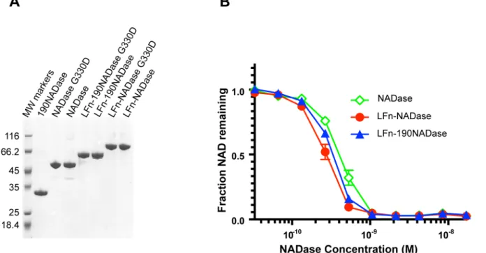
NADase in the flow through fraction. NADase was then refolded slowly by removal of guanidi-nium chloride by dialysis. High protein purity was achieved by Q column purification of pro-teins after the first Ni column affinity purification, and then again after renaturation of IFS-free NADase constructs. Each of the purified recombinant proteins migrated predominantly as a single band of the expected molecular size on SDS-PAGE (Fig 1A).
In addition to native NADase, two variant forms were expressed and purified, both as indi-vidual proteins and as fusions to LFn. Variant 190NADase lacks the N-terminal 190 amino acids required for SLO-dependent translocation of NADase [37]; NADaseG330D harbors a point mutation that almost completely abrogates NAD-glycohydrolase activity [6,38,39]. Since the protocol involved protein denaturation and renaturation, we confirmed that the purified LFn-NADase, LFn-190NADase, and NADase proteins retained similar levels of NAD-glycohy-drolase activity (Fig 1B). The Kcatvalue for LFn-NADase was estimated at 4200 reactions/sec,
which compares favorably with published estimates of 3700 and 8000 reactions/sec, deter-mined for purified NADase using a highly sensitive HPLC-based assay [36,38].
LFn-Fig 1. Purification and biochemical characterization of recombinant proteins.(A) Coomassie-stained SDS-PAGE of purified recombinant proteins. Proteins LFn-NADase, LFn-190NADase, NADase, and 190NADase were released from IFS and purified using a novel protocol in which the proteins were denatured, purified, and then refolded (Materials and Methods). (B) Recombinant LFn-NADase, LFn-190NADase, and NADase proteins had similar NADase activities after renaturation.
NADaseG330D and NADaseG330D lacked detectable catalytic activity. However, both LFn-NADaseG330D and LFn-NADaseG330D were able to compete with NADase for binding of IFS after renaturation (S2 Fig). We also analyzed the secondary structure of purified recombinant NADaseG330D by circular dichroism spectroscopy and found nearly identical results as those for purified recombinant (and enzymatically active) NADase (S2 Fig). Together, these analyses provide evidence that renaturation of the enzymatically inactive variants LFn-NADaseG330D and NADaseG330D restored the native conformations of the purified proteins.
LFn-NADase is translocated across PA pores in planar lipid bilayers
We tested the ability of LFn-NADase and its variants to interact with and translocate across PA pores in planar bilayersin vitroas measured by ion conductance. Occlusion of pores in DPhPC bilayers was monitored for 60 sec following addition of each recombinant protein (final concentration 1μg/ml) to theciscompartment. All of the constructs tested (LFn,
LFn-NADase, LFn-190LFn-NADase, LFn-NADaseG330D, and LFn-190NADaseG330D) blocked con-ductance rapidly (within 20 sec) and almost completely (Fig 2A). Subsequently, translocation was initiated by addition of KOH to thetranscompartment to increase the pH to ~7.5. Trans-location of free LFn and LFn-190NADase, as measured by return of ion conductance, was rapid (within ~80 sec) and essentially complete (~80–90%) (Fig 2B). LFn-NADase took longer (240 sec) to achieve comparable translocation. LFn-NADaseG330D and LFn-190NADa-seG330D constructs were less efficiently translocated, with about ~60% translocation achieved in 240 sec. Interestingly, addition of IFS to theciscompartment (final concentration 6μg/ml)
before addition of KOH to thetranscompartment prevented translocation of LFn-NADase
(Fig 2B). To test whether the binding of NADase to IFS prevents NADase unfolding, which is
necessary for translocation, we used differential scanning fluorimetry to measure the melting temperature of NADase, IFS, and NADase-IFS complex. The melting temperatures were deter-mined to be 43°C, 60°C and 76°C, respectively (S3 Fig). Thus, the tight binding of IFS increases the Tmof NADase by more than 30°C, presumably preventing its unfolding, a required step for
the translocation of LFn-NADase across PA pores.
Delivery of NADase to the cytosol of human oropharyngeal
keratinocytes independently of SLO
Having determined that LFn-NADase could be translocated through PA pores in an artificial membranein vitro, we investigated whether PA pores could mediate delivery of LFn-NADase into human oropharyngeal keratinocytes. We reasoned that NAD+-glycohydrolase activity of LFn-NADase would deplete cellular energy stores resulting in inhibition of protein synthesis. Accordingly, NADase and its variants were tested for PA-mediated translocation into OKP7 cells by measuring inhibition of cellular protein synthesis. In the presence of PA, LFn-NADase and LFn-190NADase were efficiently translocated, with half-maximal inhibition of protein synthesis observed at a LFn-NADase concentration of ~1 nM in the cell culture medium (Fig 2C). LFn-190NADase gave an almost identical result, a finding that implies the N-terminal domain of NADase involved in SLO-mediated translocation is dispensable for delivery of the enzyme by the anthrax toxin system. In the absence of PA, no LFn-NADase translocation was observed (S4 Fig), and, as expected, NADase and 190NADase also did not translocate. Translo-cation of the enzymatically inactive forms of NADase, NADaseG330D and
LFn-190NADaseG330D, did not inhibit cellular protein synthesis, even at concentrations up to 1,000 times that required for inhibition by LFn-NADase (Fig 2C).
LFn-190NADase for cytotoxicity in the presence of PA F427H, a mutant form of PA, which forms pores that lack the ability to mediate translocation. The F427H mutation completely blocked LFn-NADase translocation (Fig 2C), implying PA-mediated translocation is dependent on interaction with the Phe clamp and occurs through the central pore.
Fig 2. Anthrax toxin-mediated translocation of NADase.(A) Kinetics of occlusion of PA pores in planar bilayers by LFn-NADase, LFn-190NADase, LFnNADase G330D, and LFn-190NADase G330D. Interaction with PA channels was monitored by the decrease in conductance, which is plotted as fraction of open PA channels. (B) Translocation of LFn-NADase, LFn-190NADase, LFn-NADase G330D and LFn-190NADase G330D proteins across PA channels in planar bilayers. After complete occlusion of PA channels, KOH was added to thetranscompartment raising the pH from 5.5 to 7.5, triggering translocation, which is visualized by an increase in conductance and is plotted as fraction of open PA channels. (C) Inhibition of protein synthesis by PA-mediated translocation of LFn-NADase or variants in OKP7 cells. Protein synthesis was assayed by incorporation of tritiated leucine and is plotted as the fraction of activity in OKP7 cells in the absence of recombinant protein.
NADase-mediated cytotoxicity is due to NAD
+-glycohydrolase activity
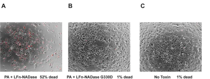
It has been suggested that introduction of NADase into host cells exerts cytotoxic effects that are independent of NAD+-glycohydrolase activity of the protein [38]. The anthrax toxin delivery system enabled us to test this hypothesis in the absence of other GAS virulence fac-tors. We found that exposure of OKP7 keratinocytes to LFn-NADase in the presence of PA resulted in rounding, pyknosis, and uptake of propidium iodide indicating loss of cell viabil-ity (Fig 3). Treatment with LFn-NADase resulted in 52% cell death as assessed by propidium iodide staining. In addition, treatment with LFn-NADase caused significant cell loss when compared to untreated cells, presumably due to cells becoming non-adherent upon loss of viability. In contrast, identical exposure to enzymatically inactive LFn-NADaseG330D in the presence of PA caused no cytotoxicity compared to untreated cells (1% cell death for each condition). These results provide direct evidence that the cytotoxic effects of NADase are due solely to its enzymatic activity.
PA-mediated translocation delivers LFn-NADase in an amount
comparable to that associated with exposure of cells to live GAS
Previous studies on the effects of NADase on epithelial cells have utilized model systems in which cells are exposed to live GASin vitro[17,21]. In order to compare effects of NADase delivered by the anthrax toxin system with those associated with exposure to live GAS, we mea-sured intracellular NAD-glycohydrolase activity under both conditions. Our goal was to deter-mine the concentration of LFn-NADase to be added to OKP7 cells so that the subsequent PA-mediated delivery would result in an intracellular NADase activity comparable to that achieved by exposure to live GAS in prior studies. We found that addition of LFn-NADase to a concen-tration of 10 nM achieved a level of NADase activity in the cytosol of the keratinocytes that corresponded to approximately 50% of that associated with infection by NADase-producing GAS strain 188 at an MOI of 10 (Fig 4,S5 Fig).
Fig 3. Cytotoxic effects of NADase depend on enzymatic activity.(A) Propidium iodide uptake (red) indicating cell membrane damage is superimposed on bright field images of OKP7 cells exposed to LFn-NADase or (B) enzymatically inactive LFn-NADase G330D, both in the presence of anthrax toxin PA, or (C) untreated control cells.
Enzymatically active NADase rescues the defective intracellular survival
of NADase-deficient GAS
Infection of OKP7 cells with GAS is associated with survival of 10 to 15% of intracellular bacte-ria, whereas fewer than 1% of GAS deficient in NADase activity survive intracellularly for 24 hours [21]. We reasoned that anthrax toxin-mediated delivery of exogenous NADase might rescue intracellular GAS that did not produce enzymatically active NADase. We found that addition of 10 nM LFn-NADase to OKP7 cells in the presence of 20 nM PA increased the intra-cellular survival of GAS strain 188 G330D, which expresses enzymatically inactive NADase, by 14-fold, from 0.35% at 24 hours to 5% (Fig 5A). Thus, addition of exogenous NADase restored intracellular GAS survival to an extent roughly commensurate with the amount of NADase activity delivered to the cytosol of the host cell, i.e., approximately 50% of the activity associated with infection of the cell by the parent strain, 188. Addition of 1 nM LFn-NADase had a lesser and not statistically significant effect, increasing survival at 24 h of 188 G330D 2.5-fold to 0.9%. The ability of exogenously delivered NADase to restore intracellular GAS survival was dependent on the catalytic activity of the protein: addition of LFn-NADaseG330D had no effect on the 24-hour survival of GAS within OKP7 cells, even at 100 nM (Fig 5B).
The process of SLO-dependent translocation of NADase across the eukaryotic cell mem-brane requires a 190-aa domain in the amino terminus of the NADase protein, a part of the molecule that is dispensable for enzymatic activity [37]. As suggested by the protein synthesis inhibition assays (Fig 2C), we found that 190NADase could function in lieu of
Fig 4. Intracellular NADase activity during infection by GAS compared with that delivered by LFn-NADase.Cells were exposed to GAS strain 188 or to anthrax toxin PA and LFn-NADase for 2 h, after which cytosolic NADase activity was measured. Data represent the fraction of NAD+substrate depleted by the cytosolic fraction of treated cells. Experiments were performed 3 times, and results are shown as mean ±SEM.
NADase to increase the intracellular survival of 188 G330D, albeit slightly less efficiently than LFn-NADase (7-fold increase in survival versus 14-fold for LFn-NADase,Fig 5C). These results imply that the catalytic domain of NADase plays a dominant role in the intracellular survival of GAS. However, the small but reproducible improvement in survival imparted by LFn-NADase compared to LFn-190NADase suggests that the N-terminal translocation domain has an as-yet-unidentified function in enhancing intracellular survival.
Fig 5. Effect of anthrax toxin-mediated delivery of LFn-NADase or variants on the intracellular survival of GAS deficient in enzymatically active NADase (188 G330D) or SLO (188 SLO-).Survival of intracellular GAS at the indicated time points is plotted as percent of CFU recovered at 2 hours of infection. (A) Dose-dependent increase in survival of 188 G330D in the presence of LFn-NADase. (B) Increase in survival of 188 G330D in the presence of NADase but not in the presence of enzymatically inactive NADase G330D. (C) Increase in survival of 188 G330D in the presence of either LFn-NADase or LFn-190LFn-NADase, which lacks the N-terminal translocation domain. (D) Increase in survival of 188 SLO-in the presence of LFn-NADase. Values represent mean±SEM of at least 3 independent experiments.*,P<0.05;**,P<0.01;****,P<0.0001.
Intracellular delivery of NADase enhances survival of SLO-deficient
GAS
The anthrax toxin delivery system allowed us to evaluate the contribution of NADase to GAS intracellular survival in the absence of SLO. Because SLO is ordinarily required to translocate NADase during GAS infection, it has not been possible previously to assess the role of NADase on GAS intracellular survival independently of SLO. To address the discrete contribution of each toxin, we added LFn-NADase and PA during infection of OKP7 cells with 188 SLO-and assessed the effect on intracellular survival (Fig 5D). Delivery of LFn-NADase increased intra-cellular survival of 188 SLO- by ~8 fold, from 0.25% to 2.0%. Thus, delivery of NADase pro-longs the survival of both 188 G330D and 188 SLO-strains, results that imply both SLO and NADase are required for maximum resistance to intracellular killing. The fact that SLO-inde-pendent delivery of NADase partially corrects the survival defect of an SLO-deficient strain indicates that the reduced survival of SLO- GAS is due in part to the absence of NADase deliv-ery, but also that SLO possesses NADase-independent activities that contribute to the intracel-lular survival of GAS. Thus, the synergistic action of SLO and NADase mediates optimal intracellular survival.
NADase prevents trafficking of intracellular GAS to lysosomes for
efficient killing
Previous studies have implicated SLO and NADase in GAS resistance to killing by epithelial cells. After internalization, SLO-deficient mutants are contained within endosomes or autop-hagosomes that fuse with lysosomes, an event associated with acidification of the GAS-contain-ing vacuole and efficient bacterial killGAS-contain-ing [21,41]. The anthrax toxin system allowed us to assess directly the ability of NADase to interfere with fusion of the GAS-containing compartment with lysosomes. We found that delivery of exogenous LFn-NADase to the cytosol of GAS-infected OKP7 cells reduced the co-localization of 188 G330D with the lysosomal marker LAMP-1 (Lysosomal–Associated Membrane Protein 1) by 4-fold, from 41% to 10% at 6 h of infection (P<0.001,Fig 6). Delivery of LFn-NADase also inhibited trafficking of 188 SLO-to a
LAMP-1-positive compartment, reducing co-localization with LAMP-1 from 86% to 51% (P<0.05). These findings correlate with the effects of LFn-NADase on the intracellular survival
of 188 G330D and 188 SLO-and provide direct evidence that NADase contributes to GAS intracellular survival by interfering with lysosomal fusion to the GAS-containing vacuole.
Because the effect of LFn-NADase on endosomal trafficking was evident within the first few hours of infection, it seemed likely that the survival of intracellular GAS was largely determined during this time period. We found that a delay of only 2 hours in the addition of LFn-NADase to cells infected with 188 G330D largely abrogated the 24-hour survival benefit of LFn-NADase compared with that conferred by addition of the toxin at the time of initial infection (Fig 7). This result is consistent with the finding that, in the absence of NADase expression, GAS are trafficked to a degradative compartment by lysosomal fusion as early as 1 hour after infection (Fig 6).
Discussion
Fig 6. Effect of anthrax toxin-mediated delivery of LFn-NADase on intracellular trafficking of GAS.LFn-NADase inhibits trafficking of GAS deficient in expression of enzymatically active NADase (188 G330D) or SLO (188 SLO-) to lysosomes. (Left panels) Data points indicate percent co-localization of intracellular GAS with the lysosomal protein LAMP-1 as assessed by confocal microscopy at 1, 3, or 6 hours of infection of OKP7 cells in the absence or presence of LFn-NADase. Values represent mean±SEM of at least 3 independent experiments. (Right panels) Representative confocal microscopy images at 6 hours of infection. Intracellular GAS are labeled green (Alexa-488), extracellular GAS are labeled blue (Alexa-660) and green, and LAMP-1 is labeled red (Alexa-568). Percent co-localization of GAS with LAMP-1 is indicated. Scale bar, 10μm. (A) Infection with GAS strain 188; (B) Infection with GAS strain 188 G330D alone or with PA-mediated delivery of LFn-NADase; (C) Infection with GAS strain 188 SLO- alone or with PA-mediated delivery of LFn-NADase.*,
P<0.05;***,P<0.001.
and in enhancing intracellular survival of GAS internalized by epithelial cells [17,38]. However, these functions of NADase during GAS infection have been inferred almost entirely from com-parisons with mutants that lacked NADase or produced an enzymatically defective protein. The requirement of SLO for translocation of NADase has made it difficult to analyze the bio-logical effects of NADase separately from those of SLO, which is required for NADase delivery, but which also has intrinsic cytotoxicity due to its pore-forming activity. An additional level of experimental complexity arises from the tightly bound endogenous inhibitor of NADase, IFS, whose co-expression is required for NADase production, but which must be removed to restore enzymatic activity.
In the current study, the anthrax toxin system provided a tractable platform to deliver enzy-matically active, highly purified, IFS-free NADase or variant forms to the cytosol of human oropharyngeal keratinocytes. This system permitted direct investigation of the function of NADase in the cell biology of GAS infection, independent of the effects of SLO. We found that SLO-independent cytosolic delivery of LFn-NADase inhibited protein synthesis in oropharyn-geal keratinocytes in a dose-dependent manner (Fig 2C). Nearly identical inhibition was observed upon delivery of LFn-190NADase, which lacks the N-terminal domain of NADase required for SLO-mediated translocation, but preserves the catalytic domain. By contrast, LFn-NADase G330D, an enzymatically inactive variant, had no inhibitory effect, even at high doses. Consistent with these results, sufficient doses of NADase delivered by the anthrax toxin system resulted in cytotoxicity and cell death that was dependent on the catalytic activity of the protein (Fig 3). These results support the view that the intrinsic cytotoxic activity of NADase on eukaryotic cells depends on the enzymatic activity of the toxin. Depletion of cellular NAD+and ATP is expected to have a broad range of inhibitory effects on cellular functions. It remains possible that the synergistic toxicity of NADase with SLO also involves a second, non-enzy-matic mechanism, as suggested by Chandrasekaranet al, although the molecular basis for such an effect has not been determined [38].
Previous studies found that SLO was required for prolonged GAS intracellular survival in keratinocytes [21,41]. Shortly after bacterial internalization, GAS production of SLO results in damage to the endosomal membrane, which exposes the bacteria to the cytosol where they become ubiquitinated. Ubiquitin is a signal for targeting intra-cytosolic bacteria to
Fig 7. Intracellular survival of GAS 188 G330D following anthrax toxin-mediated delivery of LFn-NADase at time 0h or at 2h of infection.Values represent mean±SEM of at least 3 independent experiments.*,P<0.05.
autophagosome-like compartments [21,42]. Fusion of lysosomes with these compartments leads to their maturation into degradative autolysosomes and efficient bacterial killing. Autop-hagosomes containing NADase-deficient GAS appear to follow this pathway; however, the step of lysosomal fusion is impaired for autophagosomes containing NADase-producing GAS, and this impairment is associated with enhanced intracellular survival [21]. The anthrax toxin sys-tem allowed us to study directly whether NADase prevents lysosomal fusion with GAS-con-taining vacuoles in infected cells. We found that cytosolic delivery of NADase inhibited the co-localization of GAS 188 G330D (expressing an enzymatically inactive NADase) with the lyso-somal marker LAMP-1 (Fig 6). Inhibition of lysosomal fusion was associated with a 14-fold increase in intracellular survival to a level approaching that of the NADase-producing parent strain. Delivery of enzymatically inactive NADase G330D had no effect on GAS intracellular survival, supporting the essential role of enzymatic activity in enhancing intracellular survival. In similar experiments, we tested the effect of NADase delivery on the intracellular survival of the SLO-deficient GAS strain 188 SLO-. Supplying exogenous NADase partially rescued sur-vival of 188 SLO-, a result that implies that SLO contributes to GAS intracellular sursur-vival in part through delivery of NADase, but also through function(s), such as pore-formation, inde-pendent of NADase translocation. These data are consistent with the observation that a GAS strain producing a non-pore-forming SLO that is competent for NADase translocation (SLO Y2552A) was defective for intracellular survival [21].
Results of these experiments provide the most direct evidence to date on the contribution of NADase to the cell biology of GAS infection. Use of the anthrax toxin delivery system isolated the effects of NADase from those of SLO and defined an unambiguous role for NADase in cytotoxicity for host epithelial cells and in enhancing GAS intracellular survival. Both functions were dependent on cytosolic delivery of NADase and on the enzymatic activity of the toxin to degrade NAD+. Together, these findings provide a plausible molecular basis for the association of NADase expression with GAS virulence.
Materials and Methods
Cell culture
The OKP7/bmi1/TERT (OKP7) keratinocytes used in this study are immortalized normal human soft palate keratinocytes [43,44]. These cells were a gift of James Rheinwald and were provided through the Harvard Skin Disease Research Center. OKP7 cells were cultured in kera-tinocyte serum-free medium (KSFM, Gibco/Invitrogen) as described previously [41].
Bacterial strains and culture conditions
GAS strain 188 and its mutant derivatives were used in this study. GAS strain 188 is an isogenic unencapsulated mutant of the M type 3 necrotizing fasciitis isolate 950771 [45]. Use of an unencapsulated mutant allowed efficient internalization of GAS by human cellsin vitro
because the hyaluronic acid capsule inhibits GAS internalization.Escherichia coliXL1-Blue was used as a host for molecular cloning (NEB) and was grown in Luria-Bertani (LB) medium (Novagen). GAS was grown in L3 medium as described with two modifications: the final CaCl2
concentration was 0.015% and type 1-S bovine hyaluronidase was omitted [46].
Cloning and mutagenesis
Generation of LFn-NADase-IFS constructs. The LFn-NADase-IFS-encoding construct was
generate the LFn-NADase-IFS-encoding DNA fragment, incorporating a BamHI restriction site inserted between the LFn-encoding sequence andnga-ifs. This product was subsequently cloned into pET43.1a vector (Invitrogen, Grand Island, NY) between the NdeI/XhoI restriction sites such that the in-frame fusion construct generated a His6-tag at the C-terminus of IFS.
Protein expression from the LFn-NADase-IFS-encoding construct, named MRW001, was insufficient for downstream studies. To improve expression, a DNA fragment encoding NADase G330D-IFS (enzymatically inactive NADase and IFS) was codon-optimized for expression inE.coliand synthesized by GENEWIZ, Inc (South Plainfield, NJ 07080). Codon-optimizednga-ifswas generated by OuikChange site-directed mutagenesis (Agilent Technolo-gies) of the codon-optimized NADase G330D-IFS-encoding construct. These two constructs served as templates for PCR to generate DNA constructs encoding NADase, NADase G330D, 190NADase (aa 190–451), and 190NADase G330D using appropriate primers. Each of these PCR products was cloned between the BamHI/XhoI restriction sites in MRW001, in place of
nga-ifs(S1 Fig).
Generation of NADase constructs. Codon-optimized DNA fragments encoding NADase,
NADase G330D, and 190NADase were amplified by PCR using appropriate primers and cloned into the NdeI/XhoI restriction sites of pET43.1a to incorporate a C-terminal His6-tag
on the IFS protein.
Generation of IFS and LFn constructs. In the first step, DNA fragments encoding an N-ter-minally His6-tagged Sumo protein, codon-optimized IFS, and LFn were amplified by PCR in
separate reactions [47,48]. These PCR products served as template for overlap PCR to generate the His6-Sumo-IFS and His6-Sumo-LFn-encoding constructs, which were subsequently cloned
into the NdeI/XhoI restriction sites in the pET43.1a vector.
Expression and purification of recombinant proteins
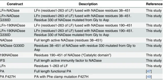
Recombinant proteins used in this study are described inTable 1.
LFn-NADase, LFn-190NADase, NADase and their variants were expressed in BL21(DE3) cells (Invitrogen) using IPTG induction. Proteins were initially purified using Ni-charged metal affinity chromatography. Each partially purified protein preparation was loaded onto a High Performance Q column (GE) in buffer A (20 mM Tris, pH 7.5), washed with buffer A, and eluted with a gradient of 0 to 1 M NaCl in the same buffer. The proteins were then
Table 1. Recombinant proteins used in this study.
Construct Description Reference
LFn-NADase LFn (residues1-263 of LF) fused with NADase residues 38–451 This study LFn-NADase
G330D
LFn (residues1-263 of LF) fused with NADase residues 38–451. Residue 330 of NADase mutated from Gly to Asp
This study
LFn-190NADase LFn (residues1-263 of LF) fused with NADase residues 190–451 This study LFn-190NADase
G330D
LFn (residues1-263 of LF) fused with NADase residues 190–451. Residue 330 of NADase mutated from Gly to Asp
This study
NADase Full length active NADase (residues 38–451) This study NADase G330D Residues 38–451 of NADase with residue 330 mutated from Gly to
Asp
This study
190NADase Residues 190–451 of NADase (“Catalytic domain”) This study
IFS Full length active immunity factor to NADase This study
LFn Residues 1–263 of LF This study
PA Full length functional PA [47]
PA F427H PA with Phe clamp mutation F427H [49]
denatured in 6 M guanidinium chloride, pH 8.0, and the His-tagged IFS was removed from untagged LFn-NADase proteins by Ni-charged metal affinity chromatography. The IFS-free proteins were renatured by dialysis into buffer A containing 350 mM NaCl and 5 mM DTT. The renatured proteins were subsequently dialyzed in buffer A containing 5 mM DTT. Finally, the proteins were subjected to another round of Q column purification. Protein solutions were filter sterilized and stored at -80°C.
LFn and IFS fused with N-terminally His6-tagged Sumo protein were overexpressed
using IPTG in BL21(DE3) cells (Invitrogen). The proteins were initially purified using Ni-charged metal affinity chromatography. Sumo was removed by cleavage with Sumo protease, and the reaction was monitored by SDS-PAGE. N-terminally His6-tagged Sumo and Sumo
protease were removed from the now untagged protein of interest using Ni-charged metal affinity chromatography. DTT (5 mM) was addedto the final protein eluate for NADase constructs.
Recombinant wild type PA and PA F427H were overexpressed in the periplasm ofE.coli
BL21 (DE3), purified by anion-exchange chromatography, and converted to the prepore form of PA using a protocol published elsewhere [50].
Circular dichroism (CD) spectroscopy
NADase and NADaseG330D were dialyzed against 10 mM sodium phosphate, 0.5 mM DTT, pH 8.0, and introduced at a concentration of 3.55μM (determined by A280measurements) into
a stoppered 0.1 cm quartz cuvette. Equal concentration of the two proteins was confirmed by SDS-PAGE and Coomassie staining. CD spectra were measured in a JASCO J-815 Spectropo-larimeter at 20°C from 185–260 nm in 0.5 nm steps with a 1 nm bandwidth. Five scans were averaged and smoothed, a background buffer-only spectrum was subtracted, and the data for the two protein species were plotted and overlayed to assess similarity.
Differential scanning fluorimetry
Differential scanning fluorimetry was used to calculate the melting temperature of NADase, IFS, or NADase-IFS complex. A 10μM solution of each protein was prepared in PBS
contain-ing 5X SYPRO Orange (Sigma), and the solution was dispensed in wells of a 96-well PCR plate. The plate was subjected to a temperature scan from 10 to 93°C at a rate of 1°C min−1in an ABI Prism 7300 real time PCR instrument (Applied Biosystems/Invitrogen) using an excitation wavelength of 492 nm; fluorescence emission was recorded at 610 nm. Fluorescence emission of SYPRO Orange in aqueous solution increases upon binding to hydrophobic regions of pro-teins exposed by temperature-induced protein unfolding. The peak of the curve of the first derivative of the measured fluorescence intensity, plotted as a function of temperature, repre-sents the melting temperature of the protein.
Determination of NADase activity of recombinant proteins
Assessment of IFS binding ability of enzymatically inactive variants of
LFn-NADase constructs
Thirty-five nM NADase was added to 17.5 nM, 35 nM and 70 nM of LFn-NADase G330D, LFn-190NADase G330D and NADase G330D in a 96-well plate. Seventy nM IFS, sufficient to completely inhibit enzymatic activity of 35 nM NADase, was then added to the wells. To this mixture, 0.67 mM NAD+was added and the reaction incubated for a period of 1 h at 37°C. The reaction was then terminated by the addition of 2 M NaOH and the fluorescence of uncleaved NAD+was allowed to develop for 1 h at which point the plates were read in a fluorimeter with excitation/emission wavelengths of 355nm/560nm. Samples without NADase served as con-trols. The results were expressed as percentage inhibition of NADase activity. Complete cleav-age of NAD was labeled as 0% inhibition of NADase activity and no cleavcleav-age was labeled as 100% inhibition of NADase activity.
Measurement of intracellular NADase activity
OKP7 cells were grown in 6-well dishes at 37°C in 5% CO2to approximately 70% confluence
(~2x10⁵cells/well). Cells were washed and incubated in KSFM containing GAS at a multiplicity of infection (MOI) of 10 unless otherwise indicated or supplemented with 20 nM PA and LFn-NADase at 10−8, 10−9, or 10−10M for 2 h. A control lacking PA protein was also included. Fif-teen min prior to harvesting cells, clindamycin (10μg/ml) was added to prevent NADase
pro-duction by GAS during sample processing. For intracellular NADase measurements, cells were washed, trypsinized, and permeabilized by incubation in PBS containing saponin (0.005% w/v) and protease inhibitors for 20 min at 37°C. Cells were removed by centrifugation for 2 min at maximum speed on a bench-top centrifuge and the supernatant containing cytosolic material was passed through a 0.2μm filter. This filtrate, the cytosolic fraction, was kept on ice until
NADase measurement. NADase activity was determined as previously described [17]. Experi-ments were performed three times. Intracellular activity was represented as the percentage NAD+substrate depletion.
Planar lipid bilayer experiments
Planar phospholipid bilayer experiments were performed in a Warner Instruments Planar Lipid Bilayer Workstation (BC 525D, Hamden, CT). Planar bilayers were formed by painting a 35 mM solution of 1,2-diphytanoyl-sn-glycerol-3-phosphocholine (DPhPC) in n-decane (Avanti Polar Lipids, Alabaster, AL) on a 200μm aperture of a Delrin cup in a Lucite chamber.
The aperture separated two compartments, each containing one ml of 100 mM KCl, 1 mM eth-ylenediaminetetraacetic acid (EDTA), and 10 mM each of sodium oxalate, potassium phos-phate, and 2-(N-morpholino)ethanesulfonic acid (MES), pH 5.5. Both compartments were stirred continuously.
Upon formation of a bilayer membrane, up to 5μg PA prepore (25 pM) was added to thecis
compartment in the presence of a constant voltage of +20 mV with respect to thetrans com-partment. After incorporation of PA pores as monitored by conductance across the membrane, theciscompartment was perfused to remove any free PA. Once the current had stabilized, 1μg
Protein synthesis inhibition assay
OKP7 cells were plated in a 96-well plate at a density of 104cells/well approximately 40 h prior to the protein synthesis inhibition assay. PA (20 nM) and LFn-NADase diluted in KSFM were added to the plates. The plates were then incubated at 37°C for 24 h, after which toxin-contain-ing medium was removed and was replaced with L-Leucine-deficient F-12 medium supple-mented with L-[4, 5-3H] Leucine (Perkin Elmer). The plates were incubated for 1 h at 37°C. Next, the plates were washed with ice-cold PBS, liquid scintillation cocktail was added, and incorporation of radioactivity in the cells was measured in a scintillation counter. Results were normalized and expressed as a fraction of the radioactivity incorporated in OKP7 cells that were not treated with toxin.
Intracellular survival assay
OKP7 cells were infected at an MOI of 10 with GAS that had been grown to exponential phase (A600nm~0.25) and washed twice in KSFM. When appropriate, PA (20 nM) and LFn-NADase
(1 nM or 0.1 nM) were added to the cells at the time of infection. Infected cell monolayers were treated with 20μg/ml penicillin G and 200μg/ml gentamicin for 45 minutes beginning 1 h 15
min post-infection. At 2 h post-infection, viable intracellular bacteria were quantified as described previously [51]. To determine intracellular survival at later time points, infected monolayers were washed at 2 h post-infection and fresh medium containing penicillin G (1μg/
ml), but not PA or LFn-NADase, was added. Infected monolayers were incubated for 4 h or 24 h post-infection, at which times the total intracellular CFU were determined as above.
Confocal microscopy
OKP7 cells were cultured on coverslips in 24-well plates. Cells were infected with GAS as described above except that antibiotics were omitted to prevent cellular uptake of non-viable bacteria. Instead, extracellular bacteria were removed by extensively washing the cells with PBS at 2 h post infection, after which the cells were incubated in fresh KSFM. Infected cells were processed 1 h, 3 h, or 6 h post-infection. At each of these time points, monolayers were washed three times with PBS and extracellular GAS were stained with Alexa Fluor 660-conjugated anti-GAS IgG at 4°C for 15 min in the dark. Excess unbound antibody was removed by wash-ing with PBS. Subsequently, cells were fixed and permeabilized by incubation in ice-cold meth-anol at−20°C for 5 min. Cells were then washed three times with PBS and incubated at room temperature for 1 h with mouse anti-LAMP-1 IgG. After three washes in PBS, cells were incu-bated for 1 h with goat anti-mouse Alexa Fluor 568-conjugated IgG and with Alexa Fluor 488-conjugated anti-GAS IgG at room temperature in the dark for 1 h. Slides were mounted using Prolong Gold (Molecular Probes) and stored at room temperature in the dark for 16–24 h prior to imaging. Confocal microscopy was performed at the Harvard Digestive Diseases Center core facility as previously described [51]. Images were acquired and analyzed using Sli-debook 5 and SliSli-debook 6 (Intelligent Imaging Innovations, Denver, CO). For quantification, co-localization of intracellular bacteria with LAMP-1 marker was determined from three inde-pendent experiments. Images were evaluated by an observer who was blind to the experimental conditions. At least 100 intracellular bacteria were scored for each experiment.
Cytotoxicity assays
served as a negative control. Cells were then washed with PBS and incubated with 500μl of
PBS containing 1μg/ml propidium iodide for a period of 30 min at room temperature in the
dark. Cells were then visualized under a Nikon Eclipse TS100 fluorescence microscope with a standard TRITC filter set (Ex 535/50, Em 610/75, DM 565) and images were acquired. For easy visualization, images showing dead cells stained with propidium iodide were colored red and merged with bright-field images showing the total number of cells (ImageJ software).
Statistical analysis
Significance of differences between experimental groups was assessed by Student’s t-test. P val-ues of less than 0.05 were considered statistically significant.
Supporting Information
S1 Fig. Schematic diagrams of expression constructs for recombinant proteins prepared in this study.
(PDF)
S2 Fig. Evidence that enzymatically inactive variants of NADase retain their native confor-mations after purification.(A) Ability of enzymatically inactive variants of LFn-NADase to compete for binding of NADase to IFS. Plots demonstrate dose-dependent inhibition of NADase activity. Reactions contained 35 nM NADase, 70 nM IFS, and the indicated concen-tration of inactive variant protein. (B) Circular dichroism spectra of NADase and NADa-seG330D after purification and renaturation. The nearly identical spectra suggest similar secondary structures.
(PDF)
S3 Fig. Increase in NADase melting temperature associated with binding of IFS.Plots
rep-resent melting curves of NADase, IFS, and NADase-IFS complex as determined by differential scanning fluorimetry. The peak of the curve of the first derivative of the measured fluorescence intensity, plotted as a function of temperature, represents the melting temperature of the pro-tein.
(PDF)
S4 Fig. Dependence on PA of LFn-NADase translocation into OKP7 keratinocytes.Data
represent inhibition of protein synthesis by PA-mediated translocation of LFn-NADase in OKP7 cells. Protein synthesis was assayed by incorporation of tritiated leucine and is plotted as the fraction of activity in OKP7 cells in the absence of recombinant protein. Inhibition of pro-tein synthesis by LFn-NADase or LFn-190NADase did not occur in the absence of PA, which is required for LFn-mediated translocation.
(PDF)
S5 Fig. Intracellular delivery of NADase varies with multiplicity of infection.NADase activ-ity was measured in the cytosolic fraction of OKP7 cell lysates after 2 hours exposure of OKP7 cells to GAS strain 188 at the indicated multiplicity of infection. Values represent mean±SEM from three independent experiments.
(PDF)
Acknowledgments
Robin Ross, Ben Seiler, Erica Gardner, and the Biomolecule Production Core of the New England Regional Center of Excellence for Biodefense and Emerging Infectious Diseases for help with the production of recombinant proteins used in this study.
Author Contributions
Conceived and designed the experiments: OS MO JJV MRW. Performed the experiments: OS MO JJV. Analyzed the data: OS MO JJV. Contributed reagents/materials/analysis tools: OS MO JJV. Wrote the paper: OS MO JJV MRW.
References
1. Aziz RK, Kotb M (2008) Rise and persistence of global M1T1 clone of Streptococcus pyogenes. Emerg-ing Infect Dis 14: 1511–1517. doi:10.3201/eid1410.071660PMID:18826812
2. Cleary PP, Kaplan EL, Handley JP, Wlazlo A, Kim MH, et al. (1992) Clonal basis for resurgence of seri-ousStreptococcus pyogenesdisease in the 1980s. Lancet 339: 518–521. PMID:1346879
3. Musser JM, Kapur V, Szeto JXP, Swanson D, Martin D (1995) Genetic diversity and relationships amongStreptococcus pyogenesstrains expressing serotype M1 protein: recent intercontinental spread of a subclone causing episodes of invasive disease. Infect Immun 63: 994–1003. PMID:
7868273
4. Stevens DL, Salmi DB, McIndoo ER, Bryant AE (2000) Molecular epidemiology of nga and NAD glyco-hydrolase/ADP-ribosyltransferase activity amongStreptococcus pyogenescausing streptococcal toxic shock syndrome. J Infect Dis 182: 1117–1128. PMID:10979908
5. Sumby P, Porcella SF, Madrigal AG, Barbian KD, Virtaneva K, et al. (2005) Evolutionary origin and emergence of a highly successful clone of serotype M1 group a Streptococcus involved multiple hori-zontal gene transfer events. J Infect Dis 192: 771–782. PMID:16088826
6. Tatsuno I, Sawai J, Okamoto A, Matsumoto M, Minami M, et al. (2007) Characterization of the NAD-gly-cohydrolase in streptococcal strains. Microbiology 153: 4253–4260. PMID:18048938
7. Ajdic D, McShan WM, Savic DJ, Gerlach D, Ferretti JJ (2000) The NAD-glycohydrolase (nga) gene of
Streptococcus pyogenes. FEMS Microbiol Lett 191: 235–241. PMID:11024269
8. Kimoto H, Fujii Y, Hirano S, Yokota Y, Taketo A (2006) Genetic and biochemical properties of strepto-coccal NAD-glycohydrolase inhibitor. J Biol Chem 281: 9181–9189. PMID:16380378
9. Meehl MA, Pinkner JS, Anderson PJ, Hultgren SJ, Caparon MG (2005) A Novel Endogenous Inhibitor of the Secreted Streptococcal NAD-Glycohydrolase. PLoS Pathog 1: e35. PMID:16333395
10. Maamary PG, Ben Zakour NL, Cole JN, Hollands A, Aziz RK, et al. (2012) Tracing the evolutionary his-tory of the pandemic group A streptococcal M1T1 clone. FASEB J 26: 4675–4684. doi: 10.1096/fj.12-212142PMID:22878963
11. Nasser W, Beres SB, Olsen RJ, Dean MA, Rice KA, et al. (2014) Evolutionary pathway to increased vir-ulence and epidemic group A Streptococcus disease derived from 3,615 genome sequences. Proc Natl Acad Sci U S A 111: E1768–1776. doi:10.1073/pnas.1403138111PMID:24733896
12. Sumby P, Barbian KD, Gardner DJ, Whitney AR, Welty DM, et al. (2005) Extracellular deoxyribonucle-ase made by group A Streptococcus assists pathogenesis by enhancing evasion of the innate immune response. Proc Natl Acad Sci U S A 102: 1679–1684. PMID:15668390
13. Carlson AS, Kellner A, Bernheimer AW, Freeman EB (1957) A streptococcal enzyme that acts specifi-cally upon diphosphopyridine nucleotide: characterization of the enzyme and its separation from strep-tolysin O. J Exp Med 106: 15–26. PMID:13439111
14. Fehrenbach FJ (1971) Identity of streptolysin-O and NAD-glycohydrolase (EC 3.2.2.5). Z Naturforsch B 26: 1336–1338. PMID:4401888
15. Shany S, Grushoff PS, Bernheimer AW (1973) Physical separation of streptococcal nicotinamide ade-nine dinucleotide glycohydrolase from streptolysin O. Infect Immun 7: 731–734. PMID:4357989 16. Madden JC, Ruiz N, Caparon M (2001) Cytolysin-mediated translocation (CMT): a functional equivalent
of type III secretion in gram-positive bacteria. Cell 104: 143–152. PMID:11163247
17. Bricker AL, Cywes C, Ashbaugh CD, Wessels MR (2002) NAD+-glycohydrolase acts as an intracellular toxin to enhance the extracellular survival of group A streptococci. Mol Microbiol 44: 257–269. PMID:
11967084
19. Tatsuno I, Isaka M, Minami M, Hasegawa T (2010) NADase as a target molecule of in vivo suppression of the toxicity in the invasive M-1 group A Streptococcal isolates. BMC microbiology 10: 144. doi:10. 1186/1471-2180-10-144PMID:20470439
20. Michos A, Gryllos I, Hakansson A, Srivastava A, Kokkotou E, et al. (2006) Enhancement of streptolysin O activity and intrinsic cytotoxic effects of the group A streptococcal toxin, NAD-glycohydrolase. J Biol Chem 281: 8216–8223. PMID:16431917
21. O'Seaghdha M, Wessels MR (2013) Streptolysin O and its Co-Toxin NAD-glycohydrolase Protect Group A Streptococcus from Xenophagic Killing. PLoS pathog 9: e1003394. doi:10.1371/journal.ppat. 1003394PMID:23762025
22. Feld GK, Thoren KL, Kintzer AF, Sterling HJ, Tang II, et al. (2010) Structural basis for the unfolding of anthrax lethal factor by protective antigen oligomers. Nat Struct Mol Biol 17: 1383–1390. doi:10.1038/ nsmb.1923PMID:21037566
23. Kintzer AF, Thoren KL, Sterling HJ, Dong KC, Feld GK, et al. (2009) The protective antigen component of anthrax toxin forms functional octameric complexes. J Mol Biol 392: 614–629. doi:10.1016/j.jmb. 2009.07.037PMID:19627991
24. Young JA, Collier RJ (2007) Anthrax toxin: receptor binding, internalization, pore formation, and translo-cation. Annual review of biochemistry 76: 243–265. PMID:17335404
25. Abrami L, Liu S, Cosson P, Leppla SH, van der Goot FG (2003) Anthrax toxin triggers endocytosis of its receptor via a lipid raft-mediated clathrin-dependent process. J Cell Biol 160: 321–328. PMID:
12551953
26. Boll W, Ehrlich M, Collier RJ, Kirchhausen T (2004) Effects of dynamin inactivation on pathways of anthrax toxin uptake. Eur J Cell Biol 83: 281–288. PMID:15511085
27. Blaustein RO, Koehler TM, Collier RJ, Finkelstein A (1989) Anthrax toxin: channel-forming activity of protective antigen in planar phospholipid bilayers. Proc Natl Acad Sci U S A 86: 2209–2213. PMID:
2467303
28. Koehler TM, Collier RJ (1991) Anthrax toxin protective antigen: low-pH-induced hydrophobicity and channel formation in liposomes. Mol Microbiol 5: 1501–1506. PMID:1787799
29. Wesche J, Elliott JL, Falnes PO, Olsnes S, Collier RJ (1998) Characterization of membrane transloca-tion by anthrax protective antigen. Biochemistry 37: 15737–15746. PMID:9843379
30. Quinn CP, Singh Y, Klimpel KR, Leppla SH (1991) Functional mapping of anthrax toxin lethal factor by in-frame insertion mutagenesis. J Biol Chem 266: 20124–20130. PMID:1939073
31. Arora N, Leppla SH (1993) Residues 1–254 of anthrax toxin lethal factor are sufficient to cause cellular uptake of fused polypeptides. J Biol Chem 268: 3334–3341. PMID:8429009
32. Arora N, Leppla SH (1994) Fusions of anthrax toxin lethal factor with shiga toxin and diphtheria toxin enzymatic domains are toxic to mammalian cells. Infect Immun 62: 4955–4961. PMID:7927776 33. Ballard JD, Collier RJ, Starnbach MN (1996) Anthrax toxin-mediated delivery of a cytotoxic T-cell
epi-tope in vivo. Proc Natl Acad Sci U S A 93: 12531–12534. PMID:8901616
34. Goletz TJ, Klimpel KR, Arora N, Leppla SH, Keith JM, et al. (1997) Targeting HIV proteins to the major histocompatibility complex class I processing pathway with a novel gp120-anthrax toxin fusion protein. Proc Natl Acad Sci U S A 94: 12059–12064. PMID:9342362
35. Milne JC, Blanke SR, Hanna PC, Collier RJ (1995) Protective antigen-binding domain of anthrax lethal factor mediates translocation of a heterologous protein fused to its amino- or carboxy-terminus. Mol Microbiol 15: 661–666. PMID:7783638
36. Ghosh J, Anderson PJ, Chandrasekaran S, Caparon MG (2010) Characterization of Streptococcus pyogenes beta-NAD+ glycohydrolase: re-evaluation of enzymatic properties associated with pathogen-esis. J Biol Chem 285: 5683–5694. doi:10.1074/jbc.M109.070300PMID:20018886
37. Ghosh J, Caparon MG (2006) Specificity of Streptococcus pyogenes NAD(+) glycohydrolase in cytoly-sin-mediated translocation. Mol Microbiol 62: 1203–1214. PMID:17042787
38. Chandrasekaran S, Ghosh J, Port GC, Koh EI, Caparon MG (2013) Analysis of polymorphic residues reveals distinct enzymatic and cytotoxic activities of the Streptococcus pyogenes NAD+ glycohydro-lase. J Biol Chem 288: 20064–20075. doi:10.1074/jbc.M113.481556PMID:23689507
39. Riddle DJ, Bessen DE, Caparon MG (2010) Variation in Streptococcus pyogenes NAD+ glycohydro-lase is associated with tissue tropism. J Bacteriol 192: 3735–3746. doi:10.1128/JB.00234-10PMID:
20494994
40. Krantz BA, Melnyk RA, Zhang S, Juris SJ, Lacy DB, et al. (2005) A phenylalanine clamp catalyzes pro-tein translocation through the anthrax toxin pore. Science 309: 777–781. PMID:16051798
42. Nakagawa I, Amano A, Mizushima N, Yamamoto A, Yamaguchi H, et al. (2004) Autophagy defends cells against invading group A Streptococcus. Science 306: 1037–1040. PMID:15528445
43. Crowe DL, Hu L, Gudas LJ, Rheinwald JG (1991) Variable expression of retinoic acid receptor (RARß) mRNA in human oral and epidermal keratinocytes; relation to keratin 19 expression and keratinization potential. Differentiation 48: 199–208. PMID:1725165
44. Dickson MA, Hahn WC, Ino Y, Ronfard V, Wu JY, et al. (2000) Human keratinocytes that express hTERT and also bypass a p16INK4a-enforced mechanism that limits life span become immortal yet retain normal growth and differentiation characteristics. Mol Cell Biol 20: 1436–1447. PMID:10648628 45. Ashbaugh CD, Warren HB, Carey VJ, Wessels MR (1998) Molecular analysis of the role of the group A
streptococcal cysteine protease, hyaluronic acid capsule, and M protein in a murine model of human invasive soft-tissue infection. J Clin Invest 102: 550–560. PMID:9691092
46. Hill JE, Wannamaker LW (1981) Identification of a lysin associated with a bacteriophage (A25) virulent for group A streptococci. J Bacteriol 145: 696–703. PMID:7007344
47. Christensen KA, Krantz BA, Melnyk RA, Collier RJ (2005) Interaction of the 20 kDa and 63 kDa frag-ments of anthrax protective antigen: kinetics and thermodynamics. Biochemistry 44: 1047–1053. PMID:15654761
48. Sharma O, Collier RJ (2014) Polylysine-Mediated Translocation of the Diphtheria Toxin Catalytic Domain through the Anthrax Protective Antigen Pore. Biochemistry 53: 6934–6940. doi:10.1021/ bi500985vPMID:25317832
49. Sun J, Lang AE, Aktories K, Collier RJ (2008) Phenylalanine-427 of anthrax protective antigen func-tions in both pore formation and protein translocation. Proc Natl Acad Sci U S A 105: 4346–4351. doi:
10.1073/pnas.0800701105PMID:18334631
50. Miller CJ, Elliott JL, Collier RJ (1999) Anthrax protective antigen: prepore-to-pore conversion. Biochem-istry 38: 10432–10441. PMID:10441138
51. Logsdon LK, Hakansson AP, Cortes G, Wessels MR (2011) Streptolysin O inhibits clathrin-dependent internalization of group A Streptococcus. mBio 2: e00332–00310. doi:10.1128/mBio.00332-10PMID: