(3
9
-Indolyl)-1-(Aromatic) Methane Analog through
Induction of the Orphan Nuclear Receptor, NR4A2 (Nurr1)
Cedar H. A. Boakye1., Ravi Doddapaneni1., Punit P. Shah1
, Apurva R. Patel1, Chandraiah Godugu1, Stephen Safe2, Santosh K. Katiyar3, Mandip Singh1*
1College of Pharmacy and Pharmaceutical Sciences, Florida A&M University, Tallahassee, Florida, United States of America,2Department of Veterinary Physiology and Pharmacology, Texas A&M University, College Station, Texas, United States of America,3Department of Dermatology, University of Alabama at Birmingham, Birmingham, Alabama, United States of America
Abstract
Background:The objective of this study was to demonstrate the anti-skin cancer and chemopreventive potential of 1,1-bis(39-indolyl)-1-(p-chlorophenyl methane) (DIM-D) using an in vitro model.
Methods:In vitro cell cytotoxicity and viability assays were carried out in A431 human epidermoid carcinoma cell line and normal human epidermal keratinocytes (NHEK) respectively by crystal violet staining. Apoptosis induction in A431 cells (DIM-D treated) and NHEK cells pretreated with DIM-D (2 hr) prior to UVB irradiation, were assessed. The accumulation of reactive oxygen species (ROS) in DIM-D pretreated NHEK cells (2 hr) prior to UVB exposure was also determined. Immunocytochemistry and western blot analysis was performed to determine cleaved caspase 3 and DNA damage markers in DIM-D treated A431 cells and in DIM-D pretreated NHEK cells prior to UVB irradiation.
Results:The IC50 values of DIM-D were 68.767.3, 48.3610.1 and 11.563.1mM whilst for Epigallocatechin gallate (EGCG) were 419.168.3, 186.165.2 and 56.763.1mM for 24, 48 and 72 hr treatments respectively. DIM-D exhibited a significantly (p,0.05) greater induction of DNA fragmentation in A431 cells compared to EGCG with percent cell death of 38.9. In addition, DIM-D induced higher expression in A431 cells compared to EGCG of cleaved caspase 3 (3.0-fold vs. 2.4-fold changes), Nurr1 (2.7-fold vs. 1.7-fold changes) and NFkB (1.3-fold vs. 1.1-fold changes). DIM-D also exhibited chemopreventive activity in UVB-irradiated NHEK cells by significantly (p,0.05) reducing UVB-induced ROS formation and apoptosis compared to EGCG. Additionally, DIM-D induced expression of Nurr1 but reduced expression of 8-OHdG significantly in UVB-irradiated NHEK cells compared to EGCG and UV only.
Conclusion:Our results suggest that DIM-D exhibits Nurr1-dependent transactivation in the induction of apoptosis in A431 cells and it protects NHEK cells against UVB-induced ROS formation and DNA damage.
Citation:Boakye CHA, Doddapaneni R, Shah PP, Patel AR, Godugu C, et al. (2013) Chemoprevention of Skin Cancer with 1,1-Bis (39-Indolyl)-1-(Aromatic) Methane Analog through Induction of the Orphan Nuclear Receptor, NR4A2 (Nurr1). PLoS ONE 8(8): e69519. doi:10.1371/journal.pone.0069519
Editor:Rajvir Dahiya, UCSF/VA Medical Center, United States of America ReceivedMay 8, 2013;AcceptedJune 11, 2013;PublishedAugust 7, 2013
Copyright:ß2013 Boakye et al. This is an open-access article distributed under the terms of the Creative Commons Attribution License, which permits unrestricted use, distribution, and reproduction in any medium, provided the original author and source are credited.
Funding:This work was supported by National Center on Minority Health and Health Disparities grant P20 MD 006738-01 and SC-1 grant 5SC1CA161676-03 from National Institutes of Health. The funders had no role in study design, data collection and analysis, decision to publish, or preparation of the manuscript.
Competing Interests:The authors have declared that no competing interests exist. * E-mail: mandip.sachdeva@gmail.com
.These authors contributed equally to this work.
Introduction
Skin cancer incidence has been increasing and over 2 million new cases are diagnosed each year in the United States [1]. It has been estimated that one in five Caucasian Americans will develop skin cancer at least once in the course of his/her lifetime [2]. Melanoma is the most severe form of skin cancer and accounts for 5% of all skin cancer cases in America, is responsible for most skin
cancer deaths [3][4], with an impact estimated at$2.36 billion in
2010 [5]. The increasing incidence of skin cancer is expected to continue as the population ages, greater amounts of UV radiation reach the surface of the earth due to depletion of the ozone layer, and continuous use of sun tanning devices [6][7][8].
Many phytochemicals and synthetic analogs have the ability to reverse and/or decrease the onset and progression of skin carcinogenesis and angiogenesis [16][17]. These phytochemicals are primarily polyphenols, which include but are not limited to silymarin, epigallocatechin 3-gallate (EGCG), curcumin, myrice-tin, quercetin and hesperitin. EGCG is an abundant polyphenol present in green tea extract and is a potent antioxidant flavonoid that has chemopreventive potential [18]. EGCG can induce cell cycle arrest and apoptosis in hepatoma cells by inducing p53 and Fas/FasL apoptotic pathway respectively [19]. The cytotoxicity of EGCG in vitro requires relatively high concentrations [20] that are not readily achieved in the serum and both oral and topical formulations of EGCG exhibit minimal protection against photoaging and UV-induced inflammatory responses in the skin [21][22][23].
3,39-Diindolylmethane (DIM) (Fig. S1) is a natural product derived from indole-3-carbinol (I3C) which is present in crucifer-ous vegetables such as brussels sprouts, broccoli and cauliflower. DIM has generated much interest in cancer research because of its low toxicity and cytotoxic effects on cancer cells in vitro and inhibition of tumor growth in vivo [24]. For example, DIM induced expression of cell cycle inhibitors such as p21 and p27 and downregulated-cyclin proteins including cyclin D1 and also decreased expression of survival and antiapoptotic proteins including survivin, bcl-2, bax and induced poly (ADP-Ribose) polymerase (PARP) cleavage, mitochondrial cytochrome c release and procaspase cleavage [25][26][27]. A series of novel synthetic
1,1-bis(39-indolyl)-1-(p-substituted phenyl) methane analogs
(C-DIMs), are also potent anticancer agents [25][28][29] and their activities are structure-dependent. The t-butylphenyl and p-biphenyl derivatives activate peroxisome proliferator-activated
receptor c (PPARc) whereas the unsubstituted phenyl and
p-methoxyphenyl analogs activate the orphan receptor NR4A1 (Nurr77/TR3) [30]. Studies in our laboratory have reported a synergistic effect between 1,1-bis(39-indolyl)-1-(p-biphenyl) meth-ane (DIM-C-pPhC6H5) and Docetaxel in non-small cell lung cancer cells through enhanced induction of cleaved PARP, bax and N-cadherin and inhibition of phospho-Akt, cyclin D1, survivin, NF-kB, Mcl-1 and phospho JNK2 [29][31].
A member of the nerve growth factor I-B Nurr1 (NR4A2) is another NR4A receptor, which has been implicated, in various hormonal, physiological and pathophysiological processes includ-ing cardiovascular, neurological and metabolic diseases, inflam-mation and oncogenesis. Nurr1 plays a role in brain function and hence has been linked to Alzheimer’s disease, Schizophrenia and Parkinson’s disease [32]. Nurr1 is highly expressed in Panc1 and Pan28 pancreatic and some human bladder cancer cell lines [28][33]. In this study, we illustrate the significance of Nurr1 in the chemopreventive potential of 1,1-bis(39-indolyl)-1-(p-chlorophenyl methane) (DIM-D) in skin cancer using an in vitro UVB induced skin cancer model. DIM-D has shown to induce Nurr1-dependent transactivation and results of our study suggest a possible role for DIM-D/Nurr1 in chemoprevention of skin cancer.
Materials and Methods
1. Materials
p-Substituted C-DIM analogs C-pPhCl; DIM-D), (DIM-C-pPhCN; DIM-B) and (DIM-C-pPhBr; DIM-C) were synthe-sized as described [36]. EGCG was purchased from Selleck Chemicals (Houston, TX, USA). A431 human epidermoid carcinoma cell line and normal human epidermal keratinocytes (NHEK) were acquired from Invitrogen (Grand Island, NY, USA). Phosphate buffered saline (PBS) was purchased from Invitrogen.
The NR4A2 (Nurr 1) antibody was obtained from Santa Cruz Biotechnology, Inc. (Santa Cruz, CA). The antibodies directed against 8-hydroxy deguanosine (8-OHdG), CCAAT/-enhancer-binding protein homologous protein (CHOP), nuclear factor
kappa-light-chain-enhancer of activated B cells (NF-kB) and
cleaved caspase 3 were also obtained from Santa Cruz Biotech-nology, Inc. (Santa Cruz, CA).
2. Cell lines and cell cultures
A431 cells were maintained in Dulbecco’s modified Eagle’s medium (DMEM; Sigma Aldrich, St Louis, MO) nutrient mixture supplemented with 10% fetal bovine serum (FBS) from Invitrogen (Grand Island, NY) and antibiotic-antimycotic mixture comprising penicillin (5000 U/mL), streptomycin (0.1 mg/mL), and neomy-cin (0.2 mg/mL) from Sigma Aldrich (St Louis, MO, USA) at
37uC in the presence of 5% CO2 and 95% relative humidity.
NHEK cells were maintained in Epilife medium (Invitrogen, Grand Island, NY, USA) and were sustained by either epidermal growth supplement (EDGS) or human keratinocyte growth supplement (HKGS) from Invitrogen (Grand Island, NY) and antibiotic-antimycotic mixture comprising penicillin (5000 U/mL), streptomycin (0.1 mg/mL), and neomycin (0.2 mg/mL) from
Sigma Aldrich (St Louis, MO) at 37uC. Both cells lines were sub
cultured when approximately 80–90% confluent with 0.25% trypsin-EDTA (Invitrogen; Grand Island, NY). The cells were
cultivated on 1.12 cm2 0.4mm pore polycarbonate membrane
inserts in 12 mm612-transwell permeable support plates
(Corning, NY).
3.In vitroCell Cytotoxicity and Viability Assays
Cell cytotoxicity and cell viability assays were carried out in A431 and NHEK cells respectively using the conventional crystal violet staining assay. The A431 and NHEK cells were both seeded
in 96-well plates (104cells/well) and incubated overnight in 5%
CO2at 37uC. The A431 cells were treated with the solvent control
(DMSO) and various concentrations of B, C and DIM-D and EGCG, whilst NHEK cells were exposed to UVB radiation
(150 J/m2) 2 hr following treatment and then incubated for 24 hr
in growth media. The A431 cells in drug and media mixture were incubated for 24, 48 and 72 hr. The viable cells were fixed with glutaraldehyde and stained with crystal violet for 15 min at room temperature. The crystal violet was washed; the residue solubilized with sodium hydrogen phosphate and the absorbance was measured with a spectrophotometer at wavelength of 462 nm. Viability of NHEK cells treated with C-DIMs or EGCG were expressed as percentages of the absorbance of control cells, which were regarded as 100% viable and IC50 values were determined by the following formula, [(50– lowest kill)/(highest kill – lowest kill) * (highest conc. – lowest conc.)]+lowest conc.
4. TUNEL Assay
A431 cells were treated with 34.4mM DIM-D (50 percent of
IC50 value) and incubated for 24 hr. Cells were then washed with PBS buffer and fixed in 10% formaldehyde on microscopic sides.
The ApoTag Red In Situ Apoptosis detection kitH (Millipore,
Billerica, MA) was used for the detection of apoptosis in accordance with the manufacturer’s protocol. Briefly, the fixed
cells were incubated in 20mg/mL proteinase K solution for
15 min at room temp, followed by incubation with equilibration buffer for 10 sec. The cells were washed in PBS and incubated
with TdT enzyme at 37uC for 1 hr in a humidified chamber for
incorporation of conjugated nucleotides at the 39-OH ends of
counterstained with DAPI. The microscopic images of the fixed cells on the slides were visualized with an Olympus BX40 light microscope equipped with a computer-controlled digital camera (DP71, Olympus Center Valley, PA, USA). DNA fragmentation was indicated by Rhodamine positive staining (red). Untreated cells were maintained as control.
5. Acridine orange/ethidium bromide staining
Morphological changes in NHEK cell nuclei following exposure to UVB radiation was determined to assess the protective effects of DIM-D and EGCG in the exposed cells. Cells were seeded in
96-well plate (104 cells/well) and treated for 2 hr with different
concentrations of solutions of DIM-D and EGCG in DMSO prior to UVB radiation exposure at dose of 150 mJ for 30 sec. Cells were then incubated in growth media for 24 hr and staining was carried out with acridine orange/ethidium bromide (AO/EB) as described [34] and analyzed by fluorescence microscopy. Briefly, the differential uptake of the two dyes was used to ascertain cells viability. After incubation for 24 hr, cells were washed with PBS (2X) and stained with a mixture of acridine orange and ethidium bromide.
6. Determination of intracellular reactive oxygen species (ROS)
The fluorescent dye, 29,79-dichlorofluorescein diacetate (DCF-DA) was used to assess the accumulation of ROS in NHEK cells after UVB exposure. Cells were seeded in a 24-well plate at a
density of 0.056106cells/well, treated with different
concentra-tions of DIM-D and EGCG in DMSO for 2 hr, aspirated a thin film of PBS was added and cells were then exposed to UVB radiation dose of 150 mJ for 30 sec. Cells were then incubated for
24 hr in growth media only. DCF-DA solution (10mM) was added
to the cells (0.056106/mL) and the mixture incubated at 37uC for 1 hr in the dark. Cells were washed with PBS (2X) and the fluorescence intensity of the cells was determined by fluorescence microscopy.
7. Immunocytochemical analysis
Both A431 and NHEK cells were treated with DIM-D and
EGCG (34.4mM and 210.0mM respectively) and incubated for
24 hr. Cells were then fixed in 10% formaldehyde and cytospun into microscopic slides. The analysis was carried out following the
protocol specified in the SignalStainTMIHC kit (Cell Signaling,
Beverly, MA). Fixed cells were hydrated with varying concentra-tions of alcohol and PBS (3X), incubated with the primary antibodies against cleaved caspase 3, Nurr1 and 8-OHdG
overnight at 4uC and detected by HRP-conjugated secondary
antibody. Cells were stained with Nova Red stain and counter-stained with hematoxylin. Microscopic analysis of the fixed cells was carried out with an Olympus BX40 light microscope equipped with computer-controlled digital camera (DP71, Olympus Center Valley, PA, USA).
8. Western blot analysis
Proteins were collected from both A431 and NHEK cells as described [33]. Briefly, after 24 hr treatment with DIM-D
(17.2mM) and EGCG (104.8mM), cells were treated with RIPA
buffer (50 nM Tris-HCl, pH 8.0, with 150 mM sodium chloride, 1.0% Igepal CA-630 (NP-40), 0.5% sodium deoxychlorate, and 0.1% sodium dodecyl sulfate) with protease inhibitor (500 mM phenylmethylsulfonyl fluoride). Protein concentrations were de-termined according to BCA protein assay reagent protocol (PIERCE, Rockford, IL) and the standard plot was generated by
using bovine serum albumin, 50mg of supernatant protein from
the control group and all the different treatment samples were
denatured by boiling at 100uC for 5 min in SDS sample buffer and
were subsequently electrophoresed in 10% SDS-PAGE gel, transferred into nitrocellulose membranes, blocked with 5% skim milk in tris-buffered saline with tween 20 (10 mM tris-HCl (pH 7.6), 150 mM NaCl, and 0.5% Tween), and probed with
antibodies against Nurr1 (1:400), NFkB (1:500) and cleaved
caspase 3 (1:1000) for A431 cells. Proteins were detected with HRP conjugated secondary antibodies using SuperSignal West pico chemiluminescent solution (PIERCE, Rockford, IL). Quan-tification and analysis of the results were carried out with Image J software (v1.33u, NIH, USA). Results were expressed as percent-age ratios of protein expression tob-actin (set to 100%).
9. Statistical analysis
Results are expressed as the mean 6 S.D for at least three
replicates and comparison between multiple groups was estab-lished by a one-way analysis of variance (ANOVA) and between
two groups by student’s t test analysis. A p value ,0.05 was
considered significant.
Results
1. Anticancer activity of DIM analogues
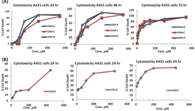
1.1 Effect of DIM analogues on A431 skin cancer cells. A431 cells were treated with three different DIM analogues, which differ structurally at the p-phenyl position (Fig. S1). A431 cells were treated with DIM-B for 24, 48 and 72 hr
and IC50 values for cytotoxicity were 56.8610.7, 30.866.6 and
7.261.2mM respectively (Fig. 1A). The IC50 values for DIM-C
after treatment for 24, 48 and 72 hr were 78.3616, 50.2612.2
and 7.261.5mM respectively and the corresponding IC50 values
for DIM-D were 68.7627.3, 48.3610.1and 11.563.1mM after
treatment for 24, 48 and 72 hr. In contrast, IC50 values for
EGCG after treatment for 24, 48 and 72 hr were 419.168.3,
186.165.2 and 56.763.1mM respectively (Fig. 1B), which were
significantly higher than observed for C-DIMs (Table 1). All three DIM derivatives showed more potency than EGCG with little variations between their potencies. DIM-D was however selected for further studies as a model drug because it exhibited more photostability than DIM-B and DIM-C.
1.2 Apoptotic activity of DIM-D against A431 cells. The effects of DIM-D on apoptosis in A431 cells were examined by the TUNEL method using flow cytometry. A431 cells treated with
DIM-D (34.4mM) for 24 hr significantly induced cell death
(38.9%) compared to DMSO control (Fig. 2A). We also investigated the effects of DIM-D and EGCG on DNA fragmentation of A431 cells using TUNEL method by microscopic analysis (Fig. 2B). In control cells treated with PBS, minimal DNA fragmentation was observed whereas DIM-D significantly in-creased DNA fragmentation as evidenced by inin-creased red
fluorescence. Similarly, EGCG (104.8mM) also increased DNA
fragmentation but comparatively less when compared with DIM-D (Fig. 2B).
1.3 Effect of DIM-D on apoptotic proteins and Nurr1. A431 cells were treated with 34.4mM DIM-D and
104.8mM EGCG for 24 hr and whole cell lysates were analyzed
by western blots (Fig. 3A). DIM-D significantly induced expression of cleaved (activated) caspase-3 and Nurr1 proteins and similar results were observed for EGCG. Quantitation of these results (Fig. 3B) showed that DIM-D was more potent that EGCG as an inducer of cleaved caspase-3 (3.0 fold vs 2.4 fold) and Nurr1 (2.7
fold vs 1.1 fold). Expression of cleaved caspase-3 and Nurr1 was
significantly induced by DIM-D and EGCG (p,0.05). DIM-D
(34.4mM) and EGCG (104.8mM) also induced CHOP expression
as indicated by immunostaining of A431 cells after treatment for 24 hr (Fig. 3C). Quantitation of these results showed significant increase of CHOP positively stained cells (79%) in DIM-D treated cells as compared to 67% of cells positively stained after EGCG treatment. These results suggest that the anti-skin carcinogenic potency of DIM-D is greater than EGCG under identical conditions.
2. Chemopreventive activity of DIM-D on normal human epidermal keratinocyte (NHEK) cells
2.1 Cytotoxic effect of DIM-D on NHEK cells. In order to investigate the effects of DIM-D against normal skin cells, NHEK cells were treated with various concentrations of DIM-D (15 to
140mM) for 24 hr and results indicated that DIM-D was
minimally toxic to NHEK cells (Fig. 4A). At the maximum
concentration of 140mM, percentage cell death was 56.261.3
whilst at the IC50 concentration (68.767.3mM) observed in A431
cells, NHEK cell death was reduced to 48.762.1%. Hence,
concentration of DIM-D 68.767.3mM was employed for all
subsequent investigations. On the other hand, exposure of the pretreated NHEK cells to UV radiation, reduced cell viability further. This was however not significant with increase in percent
cell death to 67.562.6 and 65.963.1 for 140mM and 68.7mM
respectively (Fig. 4A).
2.2 Anti-oxidant potential and apoptotic activity of DIM-D on NHEK cells. The antioxidant activity of DIM-D and protection against UV-induced ROS was also investigated in
NHEK cells treated with 34.4mM DIM-D and 209.6mM EGCG
for 24 hr. The UVB-induced ROS levels in NHEK cells pretreated with DIM-D were significantly reduced compared to EGCG; this demonstrates that DIM-D has more potent anti-oxidant activity than EGCG (Fig. 4B). In addition, the ability of DIM-D to scavenge hydroxyl (-OH) radicals in a cell-free in vitro system was utilized to confirm the enhanced anti-oxidant potential of DIM-D compared to EGCG. The results in Figure 4C revealed
that at concentrations of 34.4mM to 280mM, DIM-D was able to
quench approximately 30% to 90% of the hydroxyl radicals. Next, apoptotic activity was analyzed in NHEK cells, which
were pretreated with EGCG (17.2mM and 68.7mM) and DIM-D
(8.6 and 34.4mM) prior to UVB exposure. For efficient
comparison between EGCG and DIM-D with respect to protection against apoptosis, the IC50 and one-fourth the IC50 concentrations of DIM-D were employed for EGCG pretreatment of cells whilst half of the respective concentrations were employed for DIM-D pretreatment. The acridine orange/ethidium bromide double staining was utilized for the apoptotic assay because the acridine orange dye has ability to stain the nuclei of viable cells green whilst the ethidium dye, an intercalating agent permeated apoptotic or necrotic cells to stain their nuclei orange. We Figure 1. Cytotoxicity profile of DIM analogues and EGCG at different concentrations in A431 cells.(A) Cytotoxicity profile (Plot of % cell death vs concentration of drug at A) 24 hr, B) 48 hr and C) 72 hr where, DIM-B = pPhCN, DIM-C = pPhBr and DIM-D = DIM-C-pPhCl). Data represent mean6SD. (B) Cytotoxicity profile (Plot of % cell death vs concentration of drug) at A) 24 hr, B) 48 hr, C) 72 hr for EGCG in A431 cells.
doi:10.1371/journal.pone.0069519.g001
Table 1.IC50 values of DIM-B pPhCN), DIM-C (DIM-C-pPhBr) and DIM-D (DIM-C-pPhCl) and ECGC at 24, 48 and 72 hr. Data represent mean6SD (n = 3).
Time (hr) IC50 valuesmM
DIM-B DIM-C DIM-D
Epigallocatechin gallate (EGCG)
24 56.8610.7 78.3616.0 68.767.3 419.168.3
48 30.866.6 50.2612.2 48.3610.1 186.165.2
72 7.261.2 7.261.5 11.563.1 56.763.1
demonstrated that in cells, which were treated with DIM-D, there was a significant reduction apoptotic cell death (red staining) compared to cells treated with EGCG (Fig. 4D). No apoptotic cell death was observed in the control cells, which were treated with either PBS or vehicle only. DIM-D treatment caused significant decrease in induction of apoptosis by UVB irradiation in NHEK cells. EGCG, however, showed minimal protection of the cells against apoptosis. The cells pretreated with EGCG prior to UVB irradiation were markedly stained red by ethidium bromide. Overall, these findings suggest that although there was reduction
in cell viability in DIM-D+UV treated cells compared to DIM-D
only treated cells (as shown in fig. 4A); DIM-D treatment relatively prevents the induction of apoptosis by UVB radiation and hence enhances cell viability more significantly than EGCG.
2.3 Effect of DIM-D on UVB-induced-oxidative stress. To further investigate whether DIM-D inhibits UV radiation-induced oxidative stress and inflammation through enhancement of Nurr1 in UV-exposed skin cells, we examined
the expression of Nurr1 and 8-OHdG in NHEK cells (Fig. 5).
After UVB irradiation, cells were treated with DIM-D (34.4mM)
and EGCG (209.6mM) for 24 hr and formation of 8-OHdG was
determined by immunostaining (Fig. 5A). UVB significantly increased 8-OHdG but both DIM-D and EGCG significantly decreased the UVB-induced response thus confirming the antioxidant activity of both compounds. Their relative potency was evident by immunostaining. Only 3.6% of cells were positively stained in DIM-D treatment as compared to EGCG treated cells (20% cells).
Immunocytochemical analysis revealed that, the Nurr1 expres-sion was significantly stronger as compared to EGCG and UV only (Fig. 5A). Quantitation of these results showed that approximately 83% of cells were positively stained for Nurr1 in DIM-D treated cells as compared to EGCG treated cells (66%). These results clearly demonstrated that DIM-D was more potent that EGCG as an inducer of Nurr1. Further these results were confirmed by western blot analysis. For this purpose, NHEK cells Figure 2. Determination of apoptosis in A431 cells after treatment with DIM-D and EGCG by TUNEL assay.(A) Detection of apoptotic cells by the TUNEL method using flow cytometric analysis. A431 cells were stained for apoptotic cells with FITC by the TUNEL method before and after 24 hr treatment. (i) Dot plot depicting FSC versus SSC profile of cells after treatment. (ii) Dot plot depicting profile of FITC fluorescence versus FSC for cells after treatment. Most of the TUNEL-stained apoptotic cells are slightly smaller than unstained live cells. (iii) Histogram depicting cells before treatment in the gate R1. Very few apoptotic cells are detected. (iv) Histogram depicting cells after treatment with DIM-D (DIM-C-pPhCl) in the gate R1. Apoptotic cells (brightly stained by TUNEL) are detected. (B) Detection of apoptotic cells by the TUNEL method using microscopic analysis. A431 cells were stained for apoptotic cells with Rhodamine by the TUNEL method for 24-hr treatment. Cells were counterstained with Hoechst dye (DAPI).
were treated with DIM-D (34.4mM) and EGCG (209.6mM) for 24 hr and whole cell lysates were analyzed (Fig. 5B). NHEK cells treated with DIM-D showed Nurr1 expression was increased significantly more in DIM-D treated cells (1.8 fold) as compared to EGCG (1.4 fold) (Fig. 5C).
Discussion
Poor clinical outcome of the current treatment avenues has prompted the need for the development of new therapeutic strategies for treatment of skin cancer. There is an urgent need to identify molecular targets using dietary compounds, which have both therapeutic as well as chemopreventive activity. Nurr1 is an orphan nuclear receptor and preliminary reports suggest a role for Nurr1 in rheumatoid arthritis and cancer through modulation of apoptosis. Further it has also been demonstrated that DIM analogues activates the orphan nuclear receptor Nurr1 and inhibits bladder cancer growth [28]. However there are no reports on the chemopreventive potential of DIM-D activated Nurr1 in skin cancer. Therefore, in the first part of present study we focused
on anticancer or therapeutic activity of DIM-D using A431 skin cancer cells. In the second part of our study we investigated the chemopreventive activity of DIM-D through Nurr1 mediated signaling using NHEK.
In first part of our study, we assessed the cytotoxic effect of D on A431 skin cells. It is important to point out that DIM-D showed cytotoxic effect on these cells and its cytotoxic effect was greater than EGCG. Similar to EGCG, previous studies have demonstrated that DIM inhibits growth of cancer cells derived from tumors of the prostate, breast, colon, cervix and pancreas [34]. The role of apoptosis was further investigated by determining expression of apoptotic protein such as cleaved caspase-3 in A431 cells. The western blot analysis showed significant increase in expression of caspase-3 following DIM-D treatment in A431 cells. We also studied DNA fragmentation in A431 cells after DIM-D treatment because DNA fragmentation is a hallmark of apoptosis, which commits cells to die. DNA fragmentation was highly induced by DIM-D compared to EGCG, thus confirming that apoptosis is an important pathway associated with the anticancer activity of these compounds. This was well correlated with our Figure 3. Determination of protein expression levels by western blot and immunocytochemistry.(A) Expression of cleaved caspase 3, Nurr1 and NFkB in comparison to the controlb-actin in Dim-D and EGCG treated A431 cells by western blot. Untreated cells were maintained as control. (B) Western blot analysis of expression of different proteins increased significantly in DIM-D treated cells. (C) ICC peroxidase staining of A431 cells treated with DIM-D and EGCG, respectively for the pro-apoptotic protein, CHOP. Brown staining was considered positive result. Untreated cells were maintained as control. Data are calculated from triplicate experiments and presented as mean, and error bars refer to SD, *P,0.05, **P,0.01 compared with control.
previous study of enhancement of anticancer activity by a DIM compound in human non-small cell lung cancer cells [29]. Previous studies have shown that DIM-D activates endoplasmic reticulum stress in pancreatic and ovarian cancer cells [35][36]. DIM-D induced expression of endoplasmic reticulum stress protein GRP78 through enhanced expression of CHOP and this was accompanied by inhibition of tumor growth [37]. Similarly, our immunocytochemical studies showed that DIM-D increased the expression of CHOP in A431 cells after treatment for 24 hr. These results demonstrate that DIM-D exerts its anti-cancer effects through targeting multiple molecular targets associated with cell survival and apoptosis.
Overexpression of Nurr1 decreases inflammatory mediators, scavenger receptor expression and lowers LDL accumulation in macrophages [38][39]. In our study, expression of cleaved caspase-3 was increased in A431 cells after DIM-D treatment.
The repression of inflammatory markers including NFkB provide
protection to normal cells from the damaging effects of UVB irradiation [40] whilst on the other hand, their stimulation in cancer cells can induce stress and subsequently, apoptosis. This is in complete concordance with our study where we have shown the
pronounced upregulation of NFkB in A431 cancer cells treated
with DIM-D and to a lesser extent, in EGCG treated cells, all
compared to control. NFkB regulates the expression of genes
involved in many processes that play a key role in the development and progression of cancer such as proliferation, migration and apoptosis.
In second part of our study, we assessed the chemopreventive effect of DIM-D in NHEK. For this purpose, cells were exposed to UVB with and without treatment with DIM-D to examine the cytotoxic effect. It is important to point out that DIM-D relatively did not show cytotoxic effect on normal cells but after exposing these cells to UVB, the viability of the cells were reduced further. In spite of this observation, the acridine orange/ethidium bromide double staining revealed the relative cytoprotective effect of DIM-D in the NHEK cells compared to EGCG. Hence, though
percentage cell death increased with DIM-D+ UV treatment,
there was protection to an extent against induction of apoptosis in these cells. Another explanation of this observation is the fact that this effect of DIM-D on UVB-irradiated cells protects the photodamaged cells from further proliferation, which may be mutated or malignant. Thus, it shows the chemopreventive activity of DIM-D. Excessive exposure of the skin to solar UV- radiation is Figure 4. Effect of cytotoxicity, antioxidant potential and apoptosis in NHEK cells treated by DIM-D for 24 hr.(A) Line graph of cytotoxicity profile (Plot of % cell death vs concentration of drug) of NHEK cells treated with DIM-D (DIM-C-pPhCl) at various concentrations with and without UVB radiation. Data represent mean6SD. (B) Production of Reactive oxygen species (ROS) with DIM-D and EGCG treatment prior to UVB exposure. Each value is expressed as the mean6standard deviation. (C) Hydroxyl radical scavenging in an in vitro cell-free system treatment by DIM-D. The data is expressed in terms of percentage of control in which the cell-free system was not treated with DIM-DIM-D. (D) Apoptosis determination in NHEK cells treated with DIM-D (DIM-C-pPhCl) and EGCG at different concentrations.
one of the major etiologic factors for the development of skin cancer.
Hence, after exposing NHEK cells to UVB radiation, we investigated the antioxidant potential of DIM-D. Here we also used EGCG because of its known anticarcinogenic and antioxi-dant activities [41]. The present study demonstrates that DIM-D and EGCG reduced ROS levels in UVB irradiated NHEK cells. The induction of oxidative stress also overwhelms the antioxidant defense ability of the cutaneous system and leads to the onset of several disease states including skin cancer or photocarcinogenesis and photoaging [42]. It was reported that dietary grape seed proanthocyanidins also inhibit UVB-induced photocarcinogenesis in mice by reducing the levels of UVB-induced oxidative stress [43]. In our study, the hydroxyl radical scavenging activity of DIM-D in an in vitro cell-free system was pronounced suggesting that the DIM-D will protect normal skin against UVB induced oxidative stress, which leads to photocarcinogenesis. Our results were consistent with Wiseman et al [44] who demonstrated that green tea extract effectively scavenged superoxide free radicals, hydroxyl radicals and prevented Cu-mediated LDL oxidation.
The anti-oxidant effect of DIM-D was however superior to EGCG because treatment with DIM-D scavenged more hydroxyl radicals as compared with EGCG.
To investigate the possible involvement of apoptosis in the chemopreventive effects of DIM-D, we examined apoptosis in UVB irradiated NHEK cells after DIM-D treatment. Our results demonstrated that in UVB irradiated NHEK cells; apoptosis was low as compared to EGCG showing its chemopreventive activity was superior to that of EGCG. EGCG has been reported to have anticancer activity in various cancers and demonstrated that EGCG inhibited the growth of squamous carcinoma cell’s via S and G(2)/M phase arrest [45]. Although the molecular mecha-nisms of DIM-D action are not yet clearly understood, it appears to have potential as a therapeutic agent. Recently Katiyar et al [46] also demonstrated that treatment of NHEK with silymarin inhibits UVB-induced apoptosis of keratinocytes, and in this process UVB-induced DNA damage was significantly reduced or repaired after silymarin treatment.
Nurr1 is known to play important role in regulating inflamma-tory disease. The inflammainflamma-tory response contributes to the Figure 5. Expression of Nurr1 and 8-OHdG in NHEK cells by immunocytochemistry and further confirmation by western blot.(A) Immunocytochemical peroxidase staining of Nurr1 and 8-OHdG in NHEK cells treated with DIM-D and EGCG. Presence of brown stain indicated positive staining for the primary antibody. (B)&(C) Western blot analysis of expression of Nurr1 in comparison to the controlb-actin in Dim-D and EGCG treated NHEK cells. Untreated cells were maintained as control. Data are calculated from triplicate experiments and presented as mean6SD, *P,0.05, **P,0.01 compared with control.
pathogenesis of the sunburn reaction, photocarcinogenesis, pho-toaging of the skin and UV-induced immune suppression [47]. Nurr1 is induced by atherogenic stimuli in macrophages and smooth muscle cells and is found in atherosclerotic plaques [42]. In the current study, western blot and immunocytochemical analysis showed that in UVB irradiated NHEK cells, expression of Nurr1 was significantly induced in DIM-D and EGCG treated cells but expression was higher in DIM-D treated cells. According to Bensinger&Tontonoz, examination of isolated microglia and astrocytes demonstrated that Nurr1 potently influences inflamma-tory gene expression in these cells [48]. Measurement of the levels of 8-OHdG is used as a biomarker of oxidative stress [49]. The molecular marker 8-OHdG expression was decreased after DIM-D treatment, showing its superior activity of reducing UVB-induced oxidative stress.
Nurr1 mediated signaling by DIM-D decreased UVB-induced skin damage and may play vital role in complex pathways of cell survival and apoptosis of cancer cells. Previous studies suggested that DIM-D activates Nurr1 in human bladder cancer cells and inhibits bladder tumor growth [28][3–4]. Therefore, it is expected that Nurr1 mediated signaling by DIM-D would inhibit initiation and promotion stages of UVB-induced photocarcinogenesis in skin. Like silymarin, DIM-D may act as chemotherapeutic agents by sensitizing tumors. In addition to its chemopreventive effects, silymarin exhibits antitumor activity against human tumors (e.g., prostate and ovary) in rodents [50]. Likewise, our study also suggests that DIM-D shows anticancer and chemopreventive activities and more importantly this activity was superior to well known natural anticancer compound EGCG. Therefore, the mechanism involved in chemoprevention of DIM-D through
Nurr1 mediated signaling is novel and in future, will be confirmed further by using Nurr1 deficient mice. The overall mechanism of DIM-D suggested in this manuscript is the fact that DIM-D has potential to induce inflammation, oxidative stress and apoptosis
which is evident in the induction of NFkB, CHOP and cleaved
caspase 3, in the skin cancer cells to result in death of the cancer cells through the induction and mediation by the orphan nuclear receptor, Nurr1. Our study also suggests that DIM-D is a potential novel agent for developing chemopreventive strategies against UVB induced skin cancer because of its significant reduction in DNA damage and mutation and apoptosis of cells after UV irradiation.
In summary, data from this study suggest that DIM-D may be efficacious in treating skin cancer by acting as a potent and specific stimulator of the Nurr1-mediated apoptosis in skin cancer cells. Further studies are required to evaluate the chemopreventive effects of DIM-D on skin cancer development using in vivo animal models.
Supporting Information
Figure S1 Structure of DIM analogues (DIM-C, DIM-B and DIM-D).
(JPG)
Author Contributions
Conceived and designed the experiments: PS AP CG. Performed the experiments: CB RD CG AP PS. Analyzed the data: RD PS AP CG. Contributed reagents/materials/analysis tools: MS SS SK. Wrote the paper: CB RD SS SK MS AP.
References
1. (2009) American Cancer Association: Cancer facts & Figures. Available: http:// www.cancer.org/research/cancerfactsfigures/cancerfactsfigures/cancer-facts-figures-2009. Accessed 2013 Jan 5.
2. Robinson JK (2005) Sun exposure, sun protection, and vitamin D. JAMA 294: 1541–1543.
3. (2012) Cancer facts and figures. Available: http://www.cancer.org/acs/groups/ content/@epidemiologysurveilance/documents/document/acspc. Accessed 2013 Jan 5.
4. (2012) American Cancer Association: Cancer facts and figures. Available: http://www.cancer.org/research/cancerfactsfigures/cancerfactsfigures/cancer-facts-figures-2012. Accessed 2013 Jan 5.
5. (2011) National Cancer Institute: Skin cancer facts. Available: http://www. cancer.gov/cancertopics/types/skin. Accessed 2013 Jan 5.
6. Miller DL, Weinstock MA (1994) Nonmelanoma skin cancer in the United States: incidence. J Am Acad Dermatol 30: 774–778.
7. Urbach F (1991) Incidence of nonmelanoma skin cancer. Dermatol Clin 9: 751– 755.
8. Johnson TM, Dolan OM, Hamilton TA, Lu MC, Swanson NA, et al. (1998) Clinical and histologic trends of melanoma. J Am Acad Dermatol 38: 681–686. 9. Wright TI, Spencer JM, Flowers FP (2006) Chemoprevention of nonmelanoma
skin cancer. J Am Acad Dermatol 54: 933–946; quiz 947–950.
10. Ouhtit A, Ananthaswamy HN (2001) A Model for UV-Induction of Skin Cancer. J Biomed Biotechnol 1: 5–6.
11. Scharffetter-Kochanek K, Wlaschek M, Brenneisen P, Schauen M, Blaudschun R, et al. (1997) UV-induced reactive oxygen species in photocarcinogenesis and photoaging. Biol Chem 378: 1247–1257.
12. Rittie L, Fisher GJ (2002) UV-light-induced signal cascades and skin aging. Ageing Res Rev 1: 705–720.
13. Cimino F, Cristani M, Saija A, Bonina FP, Virgili F (2007) Protective effects of a red orange extract on UVB-induced damage in human keratinocytes. Biofactors 30: 129–138.
14. Thiele JJ, Schroeter C, Hsieh SN, Podda M, Packer L (2001) The antioxidant network of the stratum corneum. Curr Probl Dermatol 29: 26–42.
15. Sander CS, Chang H, Salzmann S, Muller CS, Ekanayake-Mudiyanselage S, et al. (2002) Photoaging is associated with protein oxidation in human skin in vivo. J Invest Dermatol 118: 618–625.
16. Stagos D, Amoutzias GD, Matakos A, Spyrou A, Tsatsakis AM, et al. (2012) Chemoprevention of liver cancer by plant polyphenols. Food Chem Toxicol 50: 2155–2170.
17. Glauert HP, Calfee-Mason K, Stemm DN, Tharappel JC, Spear BT (2010) Dietary antioxidants in the prevention of hepatocarcinogenesis: a review. Mol Nutr Food Res 54: 875–896.
18. Hou Z, Lambert JD, Chin KV, Yang CS (2004) Effects of tea polyphenols on signal transduction pathways related to cancer chemoprevention. Mutat Res 555: 3–19.
19. Kuo PL, Lin CC (2003) Green tea constituent (2)-epigallocatechin-3-gallate inhibits Hep G2 cell proliferation and induces apoptosis through p53-dependent and Fas-mediated pathways. J Biomed Sci 10: 219–227.
20. Yang CS, Maliakal P, Meng X (2002) Inhibition of carcinogenesis by tea. Annu Rev Pharmacol Toxicol 42: 25–54.
21. Chow HH, Cai Y, Alberts DS, Hakim I, Dorr R, et al. (2001) Phase I pharmacokinetic study of tea polyphenols following single-dose administration of epigallocatechin gallate and polyphenon E. Cancer Epidemiol Biomarkers Prev 10: 53–58.
22. Chiu AE, Chan JL, Kern DG, Kohler S, Rehmus WE, et al. (2005) Double-blinded, placebo-controlled trial of green tea extracts in the clinical and histologic appearance of photoaging skin. Dermatol Surg 31: 855–860; discussion 860.
23. Elmets CA, Singh D, Tubesing K, Matsui M, Katiyar S, et al. (2001) Cutaneous photoprotection from ultraviolet injury by green tea polyphenols. J Am Acad Dermatol 44: 425–432.
24. Hong C, Firestone GL, Bjeldanes LF (2002) Bcl-2 family-mediated apoptotic effects of 3,39-diindolylmethane (DIM) in human breast cancer cells. Biochem Pharmacol 63: 1085–1097.
25. Patel AR, Spencer SD, Chougule MB, Safe S, Singh M (2012) Pharmacokinetic evaluation and in vitro-in vivo correlation (IVIVC) of novel methylene-substituted 3,39diindolylmethane (DIM). Eur J Pharm Sci 46: 8–16. 26. Vanderlaag K, Samudio I, Burghardt R, Barhoumi R, Safe S (2006) Inhibition
of breast cancer cell growth and induction of cell death by 1,1-bis(39 -indolyl)methane (DIM) and 5,59-dibromoDIM. Cancer Lett 236: 198–212. 27. Rahimi M, Huang KL, Tang CK (2010) 3,39-Diindolylmethane (DIM) inhibits
the growth and invasion of drug-resistant human cancer cells expressing EGFR mutants. Cancer Lett 295: 59–68.
28. Inamoto T, Papineni S, Chintharlapalli S, Cho SD, Safe S, et al. (2008) 1,1-Bis(39-indolyl)-1-(p-chlorophenyl)methane activates the orphan nuclear receptor Nurr1 and inhibits bladder cancer growth. Mol Cancer Ther 7: 3825–3833. 29. Ichite N, Chougule MB, Jackson T, Fulzele SV, Safe S, et al. (2009)
30. Lei P, Abdelrahim M, Cho SD, Liu X, Safe S (2008) Structure-dependent activation of endoplasmic reticulum stress-mediated apoptosis in pancreatic cancer by 1,1-bis(39-indoly)-1-(p-substituted phenyl)methanes. Mol Cancer Ther 7: 3363–3372.
31. Ichite N, Chougule M, Patel AR, Jackson T, Safe S, et al. (2010) Inhalation delivery of a novel diindolylmethane derivative for the treatment of lung cancer. Mol Cancer Ther 9: 3003–3014.
32. Shi Y (2007) Orphan nuclear receptors in drug discovery. Drug Discov Today 12: 440–445.
33. Li X, Lee SO, Safe S (2012) Structure-dependent activation of NR4A2 (Nurr1) by 1,1-bis(39-indolyl)-1-(aromatic)methane analogs in pancreatic cancer cells. Biochem Pharmacol 83: 1445–1455.
34. Banerjee S, Wang Z, Kong D, Sarkar FH (2009) 3,39-Diindolylmethane enhances chemosensitivity of multiple chemotherapeutic agents in pancreatic cancer. Cancer Res 69: 5592–5600.
35. Lei P, Abdelrahim M, Safe S (2006) 1,1-Bis(39-indolyl)-1-(p-substituted phenyl)methanes inhibit ovarian cancer cell growth through peroxisome proliferator-activated receptor-dependent and independent pathways. Mol Cancer Ther 5: 2324–2336.
36. Chintharlapalli S, Papineni S, Safe S (2006) 1,1-Bis(39-indolyl)-1-(p-substituted phenyl)methanes inhibit colon cancer cell and tumor growth through PPARgamma-dependent and PPARgamma-independent pathways. Mol Cancer Ther 5: 1362–1370.
37. Abdelrahim M, Newman K, Vanderlaag K, Samudio I, Safe S (2006) 3,39 -diindolylmethane (DIM) and its derivatives induce apoptosis in pancreatic cancer cells through endoplasmic reticulum stress-dependent upregulation of DR5. Carcinogenesis 27: 717–728.
38. Huang W, Glass CK (2010) Nuclear receptors and inflammation control: molecular mechanisms and pathophysiological relevance. Arterioscler Thromb Vasc Biol 30: 1542–1549.
39. Bonta PI, van Tiel CM, Vos M, Pols TW, van Thienen JV, et al. (2006) Nuclear receptors Nur77, Nurr1, and NOR-1 expressed in atherosclerotic lesion
macrophages reduce lipid loading and inflammatory responses. Arterioscler Thromb Vasc Biol 26: 2288–2294.
40. Ishida T, Sakaguchi I (2007) Protection of human keratinocytes from UVB-induced inflammation using root extract of Lithospermum erythrorhizon. Biol Pharm Bull 30: 928–934.
41. Du GJ, Zhang Z, Wen XD, Yu C, Calway T, et al. (2012) Epigallocatechin Gallate (EGCG) is the most effective cancer chemopreventive polyphenol in green tea. Nutrients 4: 1679–1691.
42. Katiyar SK (2007) UV-induced immune suppression and photocarcinogenesis: chemoprevention by dietary botanical agents. Cancer Lett 255: 1–11. 43. Aziz MH, Reagan-Shaw S, Wu J, Longley BJ, Ahmad N (2005)
Chemopre-vention of skin cancer by grape constituent resveratrol: relevance to human disease? FASEB J 19: 1193–1195.
44. Thiagarajan G, Chandani S, Sundari CS, Rao SH, Kulkarni AV, et al. (2001) Antioxidant properties of green and black tea, and their potential ability to retard the progression of eye lens cataract. Exp Eye Res 73: 393–401. 45. Liu X, Zhang DY, Zhang W, Zhao X, Yuan C, et al. (2011) The effect of green
tea extract and EGCG on the signaling network in squamous cell carcinoma. Nutr Cancer 63: 466–475.
46. Vaid M, Prasad R, Singh T, Elmets CA, Xu H, et al. (2013) Silymarin inhibits ultraviolet radiation-induced immune suppression through DNA repair-dependent activation of dendritic cells and stimulation of effector T cells. Biochem Pharmacol 85: 1066–1076.
47. Davies MR, Harding CJ, Raines S, Tolley K, Parker AE, et al. (2005) Nurr1 dependent regulation of pro-inflammatory mediators in immortalised synovial fibroblasts. J Inflamm (Lond) 2: 15.
48. Bensinger SJ, Tontonoz P (2009) A Nurr1 pathway for neuroprotection. Cell 137: 26–28.
49. Valavanidis A, Vlachogianni T, Fiotakis C (2009) 8-hydroxy-29-deoxyguanosine (8-OHdG): A critical biomarker of oxidative stress and carcinogenesis. J Environ Sci Health C Environ Carcinog Ecotoxicol Rev 27: 120–139.