Recebido em 10.10.2001. / Received in October, 10t hof 2001.
Aprovado pelo Conselho Consultivo e aceito para publicação em 25.11.2002. / Approved by the Consultive Council and accepted for publication in November, 25thof 2002. * Trabalho realizado no Instituto Oswaldo Cruz. / Work done at "Instituto Oswaldo Cruz".
1Pesquisador titular do Departamento de Endemias Samuel Pessoa - ENSPE / FIOCRUZ; Doutor em Paleoparasitologia pela FIOCRUZ. / Titular Researcher, Dept. of Endemic Diseases Samuel Pessoa - ENSPE/FIOCRUZ; Ph.D Paleoparasitology from FIOCRUZ.
2Professor adjunto da Universidade Federal Fluminense; Doutor em Parasitologia pela FIOCRUZ. / Adjunct Professor, "Universidade Federal Fluminense"; Ph.D. in Parasitology from FIOCRUZ.
3Professora da UERJ; Dermatologista; Chefe do setor de Dermatopatologia da Universidade do Estado do Rio de Janeiro. / Professor, UERJ; Dermatologist; Head of the Dermatopathology Dept., "Universidade do Estado do Rio de Janeiro".
4Doutor em Biologia Parasitaria pela FIOCRUZ; Professor adjunto da Universidade Federal Fluminense. / Ph.D. in Parasitic Biology, from FIOCRUZ; Adjunct Professor, "Universidade Federal Fluminense".
©2003by Anais Brasileiros de Dermatologia
Ocorrência de onicomicose em pacientes atendidos em
consultórios dermatológicos da cidade do Rio de
Janeiro, Brasil
*
Occurrence of onychomycosis among patients attended in
dermatology offices in the city of Rio de Janeiro, Brazil
*
Adauto José Gonçalves de Araújo
1Otilio Machado P. Bastos
2Maria Auxiliadora Jeunon Souza
3Jeferson Carvalhaes de Oliveira
4Resumo:FUNDAMENTOS- A infecção por dermatófitos afeta aproximadamente 40% da população mundial, representa 30% de todas as infecções micóticas cutâneas, e a onicomicose por dermatófito é a mais freqüente, representando entre 18 e 40% de todas as onico-patias. A onicomicose é causada primariamente por dermatófitos, Candida spp. e outros fungos não dermatófitos. Entre os agentes fúngicos, os dermatófitos, particularmente Trichophyton rubrum, são os mais comuns desses patógenos. Atualmente, Candida spp. pode invadir a unha distal e proximal. As onicomicoses provocadas por fungos não dermatófitos vêm aumentando em sua prevalên-cia, e, pela similaridade clínica com a onicomicose por dermatófito, é necessário o diagnóstico laboratorial para sua diferenciação. OBJETIVOS- O objetivo do presente trabalho é avaliar a ocorrência de onicomicose na cidade do Rio de Janeiro.
MÉTODOS- Foram avaliados 2.920 pacientes entre janeiro de 1998 e dezembro de 1999, dos quais 1.416 apresentaram anormalidades
nas unhas.
RESULTADOS- A confirmação micológica de onicomicose foi possível em 565 dos 2.920 pacientes, e a prevalência estimada pontual na
cidade do Rio de Janeiro é 19,34%. Os homens foram responsáveis por 34,16% das onicomicoses, e as mulheres por 65,84%. A dis-tribuição por agente etiológico dos 224 pacientes com onicomicose que apresentaram cultura positiva foi: dermatófitos 64,7%,
Candida spp. 30,1% e outros não dermatófitos 5,2%.
CONCLUSÃO- A microbiota fúngica é completamente trocada no mundo periodicamente em sua composição quantitativa e qualitativa,
sendo afetada por vários fatores ambientais. Assim, o exame periódico da composição desta microbiota vem a ser vantajoso e de importância epidemiológica e terapêutica.
Palavras-chave: arthrodermataceae; Candida; epidemiologia; onicomicose; Trichophyton.
Summary:BACKGROUND- Infection by dermatophytes affects 40% of the world population and represents 30% of all mycotic cuta-neous infections, onychomycosis being the most frequent disease of the nails, counting for 18 to 40% of all onychopathies. Onychomycosis is caused primarily by dermatophytes, Candida spp. and other non-dermatophyte fungi. Among the etiologic agents the most common are dermatophytes and especially Trichophyton rubrum. Nowadays Candida spp. can invade the distal and prox-imal nails. Onychomycosis due to non-dermatophyte fungi has been increasing in prevalence and in view of the clinical similari-ty with onychomycosis caused by dermatophytes, a laboratorial diagnosis is necessary for its differentiation.
OBJETIVES- The objective of the present work is to evaluarte the occurrence of onychomycosisin the city of Rio de Janeiro. METHODS- Out of 2920 patients attended between January 1998 and December 1999, 1416 presented abnormalities in their nails. RESULTS- Mycological confirmation of onychomycosis was performed in 565 patients and the estimated prevalence in the city of Rio
de Janeiro is 19.34%. Women were more affected than men, with a ratio of almost 2:1. The distribution according to etiological agent of 224 patients with onychomycosis and who presented positive culture was: dermatophytes 64.7%; Candida spp.30.1%; and non-dermatophytes 5.2%.
CONCLUSION- The fungal flora changes completely periodically in the world in terms of its quantitative and qualitative composition as it is affected by several environmental factors. Consequently, a periodic exam of the composition of the microflora can be inter-esting and of epidemiological and therapeutic importance.
INTRODUÇÃO
A infecção por dermatófitos afeta aproximadamente 40% da população mundial e representa 30% de todas as infecções micóticas cutâneas, sendo as mais comuns as que afetam a pele e as mucosas.1,2As onicomicoses são as mais
freqüentes das doenças das unhas, representando de 18 a 40% de todas as onicopatias. A onicomicose do hálux fre-qüentemente está associada com tinea interdigitaleou tinea pedis plantar.3
A prevalência da onicomicose na Inglaterra situa-se entre três e 8%.4Em alguns países, como a Irlanda, 15 a
20% das pessoas, entre 40 e 60 anos de idade, sofrem da doença, sendo a prevalência maior nos idosos.5
A importância da onicomicose reside no aumento de sua prevalência. Nos Estados Unidos da América do Norte, é estimada entre dois e 13%,3,6 e aproximadamente uma
entre cinco pessoas de 40 a 60 anos de idade apresenta oni-comicose, os indivíduos idosos apresentando essa infec-ção.3,6,8 em percentual que varia de 15 a 20%,3,6,7 podendo
atingir até 50%.
É uma infecção que deve ser considerada importan-te pelo dano que acarreta. A qualidade de vida dos doenimportan-tes é prejudicada, a auto-estima pode ser reduzida, a capacida-de funcional por vezes é afetada capacida-de maneira a interferir nas atividades rotineiras. A onicomicose pode agravar outras afecções clínicas, especialmente no indivíduo idoso; tal como as amputações de membros inferiores nos portadores de diabetes mellitus correlacionadas à onicomicose. Os
pacientes diabéticos apresentam prevalência mais alta de onicomicose do que os não diabéticos. A prevalência de onicomicose na unha do pé em pessoas diabéticas em Ontário e Massachusetts é estimada em 32,3% e 34,9%, res-pectivamente.3 A incidência de infecções secundárias por
outros microorganimos é mais alta nos pacientes diabéticos com onicomicose do que em diabéticos que não apresentam tal afecção.9A onicomicose crônica, portanto, pode
aumen-tar os custos dos cuidados com a saúde. O intertrigo inter-digital mostra forte associação com erisipela ou celulite da perna.10 Outras causas que contribuem para o aumento do
número de infectados por dermatófitos são: a epidemia da Aids, o uso incrementado de corticosteróides e antibióticos, e a utilização de drogas imunodepressoras em pacientes transplantados.11Outro elemento de fomento das
dermatofi-toses é o aumento de sua freqüência em academias de con-dicionamento físico e a maior participação da população das cidades em atividades esportivas comunitárias.12
Existem diferenças geográficas na epidemiologia e etiologia das onicomicoses, especialmente na freqüência de cada grupo de fungos responsável pela infecção. T. rubrum
é a espécie mais freqüente na Inglaterra, Alemanha, no Canadá, nos EUA e Índia, enquanto a alta prevalência de
Candida spp.na unha tem sido relatada na Bélgica, Arábia
Saudita e Espanha. Em outras regiões tropicais do mundo, a etiologia da onicomicoses também pode ser atribuída ao
Scytalidium spp., fungo filamentoso não dermatófito.6
INTRODUCTION
Infection by dermatophytes affects approximately 40% of the world population and represents 30% of all cutaneous mycotic infections, the most common ones being those that affect the skin and the mucous membranes.1,2
Onychomycoses are the most frequent of nail diseases, rep-resenting 18 to 40% of all onychopathies. Onychomycosis of the hallux is frequently associated with tinea
interdigital-isor tinea plantar.3
The prevalence of onychomycosis in England is between 3 and 8% .4In some countries, such as Ireland, 15
to 20% of the population in the 40 to 60 years of age bra-cket suffer from the disease, with greater predominance among the elderly.5
The importance of the study of onychomycosis lies in the increase of its prevalence. In the United States of America, it is estimated at between 2% and 13%,3,6 and
approximately one in five people from 40 to 60 years of age present onychomycosis, again the more elderly individuals presenting this infection3,6,8 in a percentile that varies from
15% to 20%,3,6,7 and can reach as much as 50%.
It is an infection that should be considered important because of the damage it causes. The patients' quality of life is impaired, self-esteem may be reduced, and functional capacity at times is affected to such an extent that it interferes in routine activities. Onychomycosis can worsen other clinical afflic-tions, especially in the elderly individual; as in the case of
dia-betes mellitus patients requiring amputations of the inferior members because of conditions related to onychomycosis. Diabetic patients present a greater predisposition to ony-chomycosis than non-diabetics. The prevalence of onychomy-cosis in the toenails of diabetic individuals in Ontario and Massachusetts is estimated at 32.3% and 34.9%, respectively.3
The incidence of secondary infections of other microorganisms is higher in diabetic patients with onychomycosis than in dia-betics that do not present this disease.9Chronic
onychomyco-sis, therefore, can increase the costs of health care. Interdigital intertrigo shows a strong association with erysipela or celluli-tis of the leg.10 Other reasons that contribute to the increasing
number of individuals infected by dermatophytes are: the Aids epidemic; the increased use of corticosteroids and antibiotics; and the use of immunodepressant drugs in organ transplant patients.11Another element in the fomentation of
dermatophy-toses is the increase of its frequency in academies of physical conditioning and the greater participation of the population of cities in community sporting activities.12
There are geographical differences in the epidemiolo-gy and etioloepidemiolo-gy of onychomycosis, especially in the frequen-cy found for each group of fungi responsible for the infection.
T. rubrumis the most frequent species in England, Germany, Canada, the USA and India, while the high prevalence of
Candida spp. in the nail has been reported in Belgium, Saudi Arabia and Spain. In other tropical areas of the world, the etiology of onychomycosis can also be attributed to
Variations between reports can be due to the distinct groups studied and their respective geographical climatic differences, as probable determinants for the occurrence of
Scytalidium sppin particular.6 Sampling with variations in
other parameters, such as age, life habits, sex, location and clinical type, for instance, can also generate divergent results in studies on the subject. From the clinical point of view, a pathogenic fungus is considered to be that in which its elimination from the organism via antifungal therapy is followed by clinical cure of the nail.13
The quantitative and qualitative composition of fun-gal microbiota undergoes periodic alterations in the world, as a function of environmental factors, such as urban deve-lopment, industrialization, geographical location and cli-matic conditions, for example temperature and duration of exposure to ultraviolet radiation.4 Such that, a periodic
examination of the composition of the microbiota becomes necessary and of epidemic importance.1The determination
of etiology, in some cases, should follow criteria such as those for Aspergillus and Scopulariopsis, that are only accepted as agents if the direct exam is positive, despite the obtaining of two successive isolations in culture.1
Authors of the area of Lodz (Poland) studying 2,282 patients, isolated: T. rubrumin the toenail of 8.9% and the fin-gernail of 2.7%; T. mentagrophytes var. mentagrophytes in the toenail of 4.5%; and T. mentagrophytes var. interdigitalein the toenail of 2.2%. In this work, the dermatophytes were isolated from the toenail in 17.9% and from the fingernail in 4.3%.1
Dermatophytes have been isolated not only in the dust of the patients' residences, but also from the floor of public toilets and swimming pools.14
To prevent dermatophytosis it is important to deter-mine the minimum period for penetration of the dermato-phyte into the corneum stratum. Increased temperature and humidity are important environmental factors in the growth of the fungus. A humidity of 90% and a temperature of 35°C enable the penetration of the fungus in four days. Humidity is more important than temperature as an environmental factor of penetration. Interdigital humidity between the fourth and the fifth toe is approximately 98%.14Even with
less than 90% humidity, it is possible for the dermatophyte to penetrate the corneum stratum within a few days.
The study of the frequency of dermatophytosis in the city of Porto Alegre over a period of 13 years, has showed that the development of dermatophytosis is influenced by the age of the patient, genetic factors, bioclimatic condi-tions, promiscuity, perspiration, demographic movement or migration, care of domestic animals and contact with swimming pool decks and closed areas.15
Factors that can contribute to the increased preva-lence onychomycosis in the elderly population are reduc-tion in the rate of growth of the ungual covering and a greater possibility of traumas.16
The prevalence of onychomycosis in Ontario, Canada is 11.7% in men and 6.5% in women. The high
Variações entre os relatos podem ser devidas a dis -tintos grupos geográficos estudados e respectivas diferen-ças climáticas, como prováveis determinantes para ocorrên-cia de Scytalidium spp em particular.6 Amostragens com
variações de outros parâmetros, como idade, hábitos de vida, sexo, localização, tipo clínico, por exemplo, também podem gerar resultados divergentes nos estudos sobre o assunto. Do ponto de vista clínico, considera-se fungo pato-gênico aquele que, uma vez removido do organismo com terapia antifúngica, permite a cura clínica da unha.13
A microbiota fúngica é alterada no mundo periodi-camente em sua composição quantitativa e qualitativa, em função de fatores ambientais, como o desenvolvimento urbano, a industrialização, a localização geográfica e condi-ções climáticas, tais como temperatura e tempo de exposi-ção à radiaexposi-ção ultravioleta.4Assim, o exame periódico da
composição da microbiota torna-se vantajoso e de impor-tância epidemiológica.1 A determinação da etiologia, em
alguns casos, deve obedecer a critérios como os existentes para Aspergilluse Scopulariopsis, que só são aceitos como
agentes se o exame direto for positivo, além da obtenção de dois isolamentos sucessivos em cultura.1
Autores da região de Lodz, Polônia, estudando 2.282 pacientes, isolaram T. rubrumna unha do pé em 8,9% deles
e na unha da mão em 2,7%; T. mentagrophytes var. menta-grophytesna unha do pé em 4,5%, T. mentagrophytes var. interdigitalena unha do pé em 2,2%. Neste trabalho, os
der-matófitos foram isolados da unha do pé em 17,9% e da unha da mão em 4,3%.1
Os dermatófitos têm sido isolados não só do pó de casa de pacientes residentes, mas também do piso de banheiros públicos e piscinas.14
Para prevenir a dermatofitose é importante determi-nar o período mínimo de penetração do dermatófito no estrato córneo. O aumento da temperatura e o da umidade são importantes fatores ambientais para o crescimento do fungo. A umidade de 90% e a temperatura de 35°C possibi-litam a penetração do fungo em quatro dias. A umidade é mais importante do que a temperatura como fator ambiental de penetração. A umidade interdigital entre o quarto e o quinto dedo do pé é de aproximadamente 98%.14 Com
menos de 90% de umidade, é possível ao dermatófito pene-trar o estrato córneo em poucos dias.
O estudo da freqüência da dermatofitose, na cidade de Porto Alegre em 13 anos, mostrou que é influenciada pela faixa etária, fatores genéticos, condições bioclimáticas, promiscuidade, transpiração, movimento ou migração humana, contato com animais domésticos e contato com chão de piscina e áreas fechadas.15
Os fatores que podem contribuir com o aumento de prevalência de onicomicose na população de terceira idade são a redução da taxa de crescimento da lâmina ungueal e o aumento da possibilidade de traumas.16
pre-valência no primeiro grupo em relação ao segundo prova-velmente resulta de maior probabilidade de trauma e gran-de oclusão por uso gran-de calçados. Diferença gran-de taxas gran-de pro-gesterona e esteróides entre os sexos poderia também expli-car esse fato, pois essas taxas causam inibição do cresci-mento dos dermatófitos.16
A proporção de pacientes com onicomicose foi maior na unha do pé,4,16em vários trabalhos, e os fatores
res-ponsabilizados por essa diferença foram: o crescimento mais lento e a maior probabilidade de trauma nos pés. A unha do hálux foi mais afetada devido à predisposição de trauma nessa região. A onicomicose da mão é mais diag-nosticada do que a infecção do pé por chamar mais a aten-ção médica, mas certamente é menos freqüente.16
Apenas 40% das unhas com aparência anormal tinham evidência micológica de onicomicose. Muitos dos pacientes com onicomicose relatados por Gupta não tinham ciência de seu diagnóstico.16
A onicomicose é doença infecciosa, estando os membros da mesma família sob maior risco de contraí-la de parceiros ou irmãos afetados.3Altos níveis de contaminação
em locais de banhos comunitários não podem ser reduzidos, a menos que os indivíduos afetados sejam tratados de forma bem-sucedida.3
A onicomicose pode exercer impacto adverso importante na qualidade de vida dos indivíduos afetados, causando redução da auto-estima e possivelmente afetando o potencial de trabalho.1,14As unhas, como componente da
extremidade dos dedos, são parte integrante da estrutura sensorial da mão. A perda da margem livre das unhas pode reduzir drasticamente a capacidade sensorial dos dedos, com conseqüente limitação da destreza manual. A onico-micose do pé pode causar dor e desconforto, tornando difí-cil permanecer em pé, andar e praticar esportes. A infecção pode também resultar em prejuízo significativo para a saúde geral, a aparência física e o desempenho social. A onicomicose pode ter conseqüências psicológicas impor-tantes, incluindo-se constrangimento constante, depressão, ansiedade, preocupação com aparência e receio de situa-ções íntimas.7
Ficam objetivadas a ocorrência, etiologia e epide-miologia da onicomicose por dermatófito.
PACIENTES E MÉTODOS
A amostra populacional foi constituída por doentes atendidos em consultórios dermatológicos da cidade do Rio de Janeiro, Brasil, no período de janeiro de 1998 a dezem-bro de 1999. Pacientes de todas as idades e com vários pro-blemas dermatológicos eram encaminhados ao laboratório para esclarecimento diagnóstico. As unhas de mãos e pés eram classificadas em normais e anormais, sendo coletado material subungueal para exame micológico apenas das unhas afetadas. A classificação das onicomicoses foi basea-da nos quatros tipos clínicos específicos de alterações pre-sentes, segundo Roberts:17
prevalence in the first group in relation to the second proba-bly was the result of a greater probability of trauma and greater occlusion in the type of shoes used. The different levels of progesterone and steroids between sexes could also explain this fact because these levels can inhibit the growth of dermatophytes.16
The proportion of patients with onychomycosis was higher in the case of toenails,4,16in several studies, and the
fac-tors considered responsible for this difference were: the slower growth and greater probability of trauma in the feet. The nail of the hallux was most affected, due to the likelihood of trauma in this location. Onychomycosis of the hand is diagnosed more often than infections of the foot even though it is certainly less frequent, because it attracts more medical attention.16
Only 40% of the nails with abnormal appearance presented mycological evidence of onychomycosis. Many of the patients with onychomycosis reported by Gupta were not aware of the condition diagnosed.16
Onychomycosis is an infectious disease, rendering other members of a family under greater risk of contagion from affected partners or siblings.3High levels of
contami-nation in places such as communal showers cannot be reduced unless affected individuals are treated in an effi-cient and successful manner.3
Onychomycosis can cause an important adverse impact on the quality of the affected individual's life, leading to reduced self-esteem and possibly affecting work poten-tial.1,14The nails, as components of the extremities of the
fin-gers, are integral parts of the sensorial structure of the hand. Loss of the outer edge of the nails can reduce the sensorial capacity of the fingers drastically, with consequent limita-tion of manual dexterity. While onychomycosis of the foot can cause pain and discomfort, making it difficult to prac-tice sports, walk or even remain standing. The infection can also result in significant damage to general health, physical appearance and social performance. Onychomycosis can have important psychological consequences, including con-stant embarrassment, depression, anxiety, concern over appearance and fear of intimate situations.7
The objective of this work was to focus on the occur-rence, etiology and epidemiology of onychomycosis caused by dermatophytes.
PATIENTS AND METHODS
The population sample constituted patients attended at dermatology clinics in the city of Rio de Janeiro, Brazil, from January 1998 to December 1999. Patients of all ages and with various dermatological problems were referred to the laboratory for diagnostic clarification. The nails of hands and feet were classified as normal or abnormal. Only subungual material from affected nails was collected for mycological exam. Classification of the onychomycoses was based on the four specific clinical types of alteration presented, according to the method of Roberts:17
most common type of onychomycosis. Dermatophytes pre-dominate in this clinical type with occasional involvement of nondermatophytes.
2. White superficial onychomycosis is a superficial infection of the nail caused primarily by T. mentagrophytes and, sometimes, by the mold Acremonium sp. This type has recently been receiving more attention in view of its greater occurrence in patients with Aids.
3. Proximal subungual infection, frequently affecting the fingernails, it results primarily from an infection by
Candida. In patients with Aids, it can be caused by a der-matophyte.
4. Total dystrophic onychomycosis, characterized by the total destruction of the nail plate (covering), is fre-quently the result of a dermatophytic infection.
A clinical sample of the nail was submitted to direct mycological exam, after clarification with 20% NaOHand culture in Sabouraud's agar and Mycosel medium.
Confirmation of dermatophytosis was based on the etiological agent's growth in the culture medium. In the case of Candida, diagnosis was based on direct exam for the presence of hyphas, pseudo-hyphas and blastoconidia. When a nondermatophytic fungus was isolated in addition to the hypha in the direct exam and growth in culture, a sec-ond sample was collected for confirmation of the mycologi-cal exam.
Statistical analysis was performed in the sample population of 2,920 patients attended at dermatology cli-nics in the city of Rio de Janeiro. After tabulation, calcula-tions were made for verification of prevalence, distribution, regression, confidence limits and test of hypotheses (Table1).
RESULTS
Of the total of 2,920 examined patients, 1,107 were men and 1,813 were women. The mean age was 39.05 years. Besides the patients of unknown age, another was excluded because there was no reference to either the loca-tion of the lesion or age. Patients with tinea pedis were included in the research. Of the 2,920 patients, 1,416 (48.49%) presented nails with abnormal aspects, of these 358 were men and 1,058 were women; the mean age was 46.14 ± 0.39 (least mean square error) (Table 1).
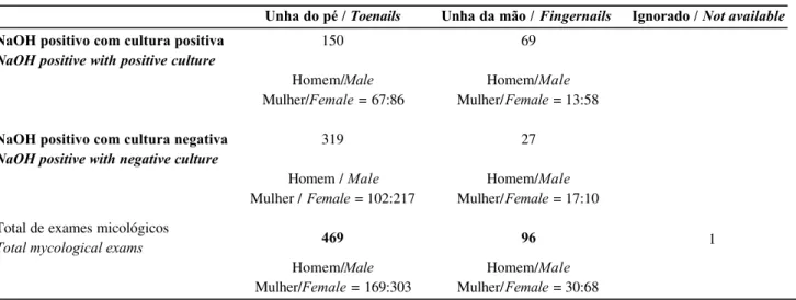
Out of the total patients (2,920) Onychomycosis was present in 19.35%. Mycological exam was performed in 1,416 patients and there was mycological confirmation of onychomycosis in 565 (39.90%), of which 193 were men and 372 were women, with a mean age of 44.88 ± 0.69 (least mean square error). Of these, the culture was only positive in 224 (7.50% of the total patients, 15.82% with ungual abnormality and 39.65% with positive direct exam). Furthermore, of these 224 patients, there was were involve-ment of the toenails in 153, distributed as follows: der-matophytic, 64.7%; Candida spp, 30.1%; and nondermato-phytic, 5.2% (Table 2). Negative direct exams and positive
1. Onicomicose subungueal distal e lateral é o tipo mais comum de onicomicose. Os dermatófitos predomi-nam nesse tipo clínico, com envolvimento ocasional por não dermatófito.
2. Onicomicose superficial branca é uma infecção superficial da unha causada primariamente por T. menta-grophytes e, algumas vezes, pelo mofo Acremonium sp.
Esse tipo tem recebido, recentemente, maior atenção por causa da maior ocorrência em paciente com Aids.
3. Infecção subungueal proximal, freqüentemente afetando as unhas dos dedos das mãos, é primariamente resultado de infecção por Candida. No paciente portador de
Aids, pode ser causada por dermatófito.
4. Onicomicose distrófica total, caracterizada pela total destruição da placa (lâmina) ungueal, é freqüentemen-te resultado de infecção dermatofítica.
A amostra clínica da unha foi submetida ao exame micológico direto, após clarificação com NaOHa 20%, e a cultura em meio de Sabouraud e Mycosel.
A confirmação da dermatofitose foi baseada no cres-cimento do agente etiológico no meio de cultura. No caso da espécie Candida, o diagnóstico era baseado no exame direto
pela presença de hifas, pseudo-hifas e blastoconídios. Quando era isolado fungo não dermatófito, além da hifa no exame direto e do crescimento em cultura, uma segunda cole-ta era realizada para confirmação do exame micológico.
A pesquisa estatística foi executada por meio de uma amostra de 2.920 pacientes dos consultórios de derma-tologia da cidade do Rio de Janeiro; após tabulação foram feitos os cálculos para verificação de prevalência, distribui-ção, regressão, intervalo de confiança e teste de hipóteses (Tabela 1).
RESULTADOS
Do total de 2.920 pacientes examinados, 1.107 eram homens e 1.813 mulheres, com média de 39,05 anos. Além dos pacientes com idade ignorada, um foi excluído por não haver a referência da localização da lesão e idade. Pacientes portadores de tinea pedisforam incluídos na
pes-quisa. Dos 2.920 pacientes, 1.416 (48,49%) apresentavam unhas com aspecto anormal, sendo 358 homens e 1.058 mulheres, média de 46,14 ± 0,39 (erro médio quadrático) anos (Tabela 1).
Do total de pacientes (2.920), a onicomicose esteve presente em 19,35%. Dos 1.416 pacientes em que se reali-zou a coleta para exame micológico, houve confirmação micológica de onicomicose em 565 (39,90%), dos quais 193 eram homens e 372 mulheres, com idade média de 44,88 ± 0,69 (erro médio quadrático). Desses, a cultura só foi positiva em 224 (7,50% do total de pacientes, 15,82% com anormalidade ungueal e 39,65% com exame direto positivo). Desses 224 pacientes, a unha do pé estava com-prometida em 153, assim distribuídos: dermatófito 64,7%,
Candida spp30,1% e não dermatófito 5,2% (Tabela 2). O
não foram incluídos como onicomicose.
A projeção da prevalência de onicomicose na cidade do Rio de Janeiro tem a estimativa pontual de 19,34%; se estimada por intervalo, a prevalência situa-se entre 18,61% e 20,07%, com 95% de confiança.
A prevalência de onicomicose apresenta histograma simétrico em relação à média de idades. Apenas nos der-matófitos, a proporção de homens com onicomicose nos pés por dermatófito foi maior do que a das mulheres (Tabela 2). A onicomicose nas unhas das mãos estava pre-sente em 96 pacientes (6,8%), sendo 30 homens e 68 mulheres, do grupo de 1.416 pacientes com anormalidades ungueais (Tabela 3). Alguns com onicomicose nas mãos tinham lesão concomitante nas unhas dos pés. A relação de onicomicose em pés e mãos foi aproximadamente 5:1 (Tabela 3).
A localização mais freqüente da onicomicose foi no hálux direito. Nos 565 pacientes com onicomicose, a forma clínica subungueal distal da classificação de Roberts foi a mais freqüente. Os dermatófitos mais comumente associa-dos com a onicomicose foram Trichophyton rubrum e T. mentagrophytes.
A detecção de fungos não dermatófitos, leveduras não Candida spp. e bactérias foi 5,2% (Tabela 2), o que
chama atenção para esses microorganismos emergentes como causadores de onicomicoses.
cultures for Candida were not included as onychomycosis. The projection of the prevalence of onychomycosis in the city of Rio de Janeiro was estimated at 19.34%; if estimated by interval, the prevalence was between 18.61% and 20.07%, with 95% confidence.
The prevalence of onychomycosis presents a sym-metrical histogram in relation to the mean ages. Only in the dermatophytes was the proportion of men with onychomy-cosis in the feet due to dermatophytes greater than that of women (Table 2). Onychomycosis in the fingernails was present in 96 patients (6.8%), of these 30 were men and 68 were women out of the group of 1,416 patients with ungual abnormalities (Table 3). Some with onychomycosis in the hands had concomitant lesions in their toenails. The ratio of feet to hands in onychomycosis was approximately 5:1 (Table 3).
The most frequent location of onychomycosis was in the right hallux. In the 565 patients with onychomycosis, the most frequent clinical form was distal subungual according to Roberts' classification. The dermatophytes more com-monly associated with onychomycosis were Trichophyton
rubrumand T. mentagrophytes.
In the detection of nondermatophytic fungi, yeasts other than Candida spp. and bacteria were 5.2% (Table 2), which calls attention to these emerging microorganisms as etiological agents of onychomycosis.
Tabela 1: Distribuição do número de pacientes por idade com aparência anormal das unhas, segundo idade, sexo e presença de onicomicose / Table 1: Distribution of the patients as a whole, patients with abnormal
nails and patients with onychomycosis, according to age, sex and percentage.
Grupo de pessoas Total sample
Pacientes com unha anormal Patients with abnormal nails
Pacientes com onicomicose Patients with onychomycosis
Idade Homem Mulher Total
Age Male Female Total
Homem Mulher Total %
Male Female Total %
Homem Mulher Total %
Male Female Total %
0 – 10 11 – 20 21 – 30 31 – 40 41 – 50 51 – 60 61 – 70 71 – 80 Ž 81 Ignorado* Not available* Total/Total Total válido Total valid 102 132 173 260 184 130 84 39 3 31 1,138 1,107 84 156 286 342 365 304 179 85 12 34 1,847 1,813 6 19 41 84 71 62 47 25 3 4 362 358 6 42 113 207 248 219 148 66 9 2 1,060 1,058 1 2 6 1 154 291 319 281 195 9 1 1 2 6 1,422 1,416 6.45 21.18 33.55 48.34 58.11 64.75 74.14 73.39 80 9.23 47.64 48.49 1 5 2 1 4 4 4 3 3 8 2 6 1 3 2 3 196 193 2 13 37 71 82 78 51 34 4 2 374 372 3 1 8 5 8 115 125 116 7 7 4 7 6 5 570 565 1.62 6.25 12.64 19.10 22.77 26.73 29.28 37.90 40 7.69 19.10 19.35 186 288 459 602 549 434 263 124 1 5 6 5 2,985 2,920
DISCUSSION
In this study, the occurrence of onychomycosis among 2,920 patients in the city of Rio de Janeiro was 19.35%. The advantage of this research is that all the patients were examined by dermatologists and confirmation of onychomycosis was made through mycological exam and not merely based on the completion of a questionnaire for its identification. Also considered were the clinical history and risk factors, such as age, sex, physical activity,
occupa-DISCUSSÃO
Nesse estudo, a ocorrência de onicomicose em 2.920 pacientes foi de 19,35%, para a cidade do Rio de Janeiro. A vantagem dessa pesquisa é que todos os pacien-tes foram examinados pelos dermatologistas, e a confirma-ção da onicomicose fez-se por meio do exame micológico, não se baseando só no preenchimento de questionário para sua identificação. Foram também considerados a história clínica e fatores de risco, como idade, sexo, atividade
físi-Tabela 2: Agentes etiológicos associados com onicomicose de mãos e pés de amostras de 1.416 pacientes com alteração ungueal. / Table 2: Etiological agents associated with onychomycosis of the hands and feet in a
sample of 1,416 patients with ungual alterations.
Número de organismos / Number of organisms
Tipo de organismo / Type of organism
Dermatófito / Dermatófito
Trichophyton rubrum Trichophyton rubrum
T. mentagrophytes var. mentagrophytes T. Maletagrophytes var. Maletagrophytes
T. mentagrophytes var. interdigitale T. Maletagrophytes var. interdigitale
T. tonsurans T. tonsurans
Microsporum canis Microsporum canis
Epidermophyton floccosum Epidermophyton floccosum
Candida spp. / Candida spp.
Não dermatófito e outros microorganismos
Non-dermatophyte and other microorgan-isms
Scytalidium dimidiatum Scytalidium dimidiatum
Curvularia sp. Curvularia sp.
Trichosporon beigelii Trichosporon beigelii
Nocardia sp. Nocardia sp.
Total
Unha do pé / Toenails Unha da mão / Fingernails Ignorado / Not available
99 (64.7%) Homem/Male Mulher/Female= 56:43
65
25
5
1
0
3
46 (30.1%)
Homem/Male Mulher/Female= 9:37
8 (5.2%)
Homem/Male Mulher/Female= 2:6
4
1
3
0
153 (100%)
4 (5.6%) Homem/Male Mulher/Female= 2:2
2
1
0
0
1
0
63 (88.8%)
Homem/Male Mulher/Female= 9:54
4 (5.6%)
Homem/Male Mulher/Female= 2:2
1
0
2
1
71 (100%)
Tabela 3: Número de exames micológicos positivos de 565 amostras de unhas. / Table 3: Number of positive mycological exams out of 565 nail samples.
NaOH positivo com cultura positiva
NaOH positive with positive culture
NaOH positivo com cultura negativa
NaOH positive with negative culture
Total de exames micológicos Total mycological exams
Unha do pé / Toenails Unha da mão / Fingernails Ignorado / Not available
150
Homem/Male Mulher/Female= 67:86
319
Homem / Male Mulher / Female= 102:217
469
Homem/Male Mulher/Female= 169:303
69
Homem/Male Mulher/Female= 13:58
27
Homem/Male Mulher/Female= 17:10
96
Homem/Male Mulher/Female= 30:68
1
ca, ocupação e presença de outras doenças facilitadoras (diabetes, doença cardiovascular e psoríase). O estudo baseou-se na chegada aleatória dos pacientes ao consultório dos dermatologistas.
O encontro de onicomicose observada no estudo é diferente da prevalência de 8,4% (n = 800), 8,7% (n = 1.038), 9,1% (n = 2.001) e 2.4% (n = 1.000) relatadas na literatura.2,18-20 Definiu-se onicomicose por dermatófito
quando, na cultura, um dos fungos desse grupo era isolado. Nessa amostra, a taxa positiva foi de 103/2.920 = 3,53%. A explicação dessa diferença talvez esteja ligada ao uso de medicação, ao clima tropical e à maior tendência ao cresci-mento de outros microorganismos saprófitas. Estudos em que o dermatologista tinha examinado o paciente e houve confirmação micológica exibiram prevalências altas em relação aos valores de 2,7% e 2,8%, obtidos usando o méto-do de questionário.16,18
O principal parâmetro de associação decorreu do diagnóstico de dermatofitose e da faixa etária correspon-dente. De fato, a onicomicose esteve presente em apenas 194 (12,6%) de 1.535 pacientes com idade inferior a 40 anos. Na população acima de 40 anos (1.385 pacientes), acima de 50 anos (836 pacientes), acima de 60 anos (402 pacientes) e acima de 70 anos (139 pacientes), a ocorrência de onicomicose foi de 26,73, 29,28, 37,90 e 40,00%, res-pectivamente. Os fatores que podem contribuir para o cres-cimento da prevalência de onicomicose na população mais idosa incluem a taxa reduzida de crescimento da unha e o aumento de trauma em relação ao grupo mais jovem, ofere-cendo ao organismo maior oportunidade de causar doença no leito ungueal e de invadir a lâmina ungueal.
Detectou-se onicomicose em 17,43% dos homens e 20,52% das mulheres que fizeram parte do estudo, sendo significante, no nível de 5%, a hipótese de que as proporções são diferentes. Assim, as mulheres apresentam maior risco
tion and the presence of other facilitative diseases (diabetes, cardiovascular disease and psoriasis). The study was based on the random arrival of patients at dermatological clinics.
The findings on onychomycosis observed in this study differ from the prevalence of 8.4% (n = 800), 8.7% (n = 1,038), 9.1% (n = 2,001) and 2.4% (n = 1,000) reported in the literature.2,18-20 Onychomycosis has been defined as being
caused by a dermatophyte when, in the culture, one of the fungi of that group was isolated. In this sampling, the positive rate was 103/2,920 (3.53%). The explanation of this differ-ence may be linked to the use of medication, to the tropical cli-mate and the greater tendency for growth of other saprophyt-ic msaprophyt-icroorganisms. Studies in whsaprophyt-ich the dermatologist had examined the patient and there was a mycological confirma-tion presented high prevalence in relaconfirma-tion to the values of 2.7% and 2.8%, obtained using the questionnaire method.16,18
The main parameter of association arose from the diagnosis of dermatophytosis and corresponding age group. In fact, onychomycosis was present in only 194 (12.6%) of the 1,535 patients less than 40 years of age. In the population > 40 years of age (1,385 patients), > 50 years (836 patients), > 60 years (402 patients) and > 70 years (139 patients), the occurrence of onychomycosis was 26.73, 29.28, 37.90 and 40.00%, respectively. Factors that can contribute to the increased prevalence of onychomyco-sis in the more senior population include the reduced rate of nail growth and greater frequency of trauma in relation to the younger age groups. These offer the organism a more propitious opportunity for causing disease in the nail bed and plate.
prevalence of women in the group can be the result of trau-mas, as much because of manual activities as for the use of high-heeled shoes, which facilitate such injury. The pres-ence of progesterone can inhibit the growth of dermato-phytes. Candida spp. was more frequently found in finger-nails and in women; this observation has also been report-ed by other researchers.21,22 The present study did not
estab-lish a significant relationship between concomitant disease (diabetes, psoriasis and vascular disease) and the frequen-cy of tinea unguium, due probably to the low number of per-sons in this sample with these diseases.
In the study by Gupta et al., the prevalence of ony-chomycosis in patients with diabetes type I was 13%. Subungual onychomycosis in distal and lateral forms was the most common.23
The prevalence of onychomycosis in the toenails of diabetic patients in Ontario and Massachusets is estimated at 32.3% and 34.9%, respectively.23In this work, due to the
small number of individuals with diabetes, that correlation was not made.
The relationship of patients with onychomycosis in the toenail against those with onychomycosis in the hands was 5:1. In the research done by Roberts, Charif, Elewski and Gupta, this ratio was 6.1:1, 23:1 and 10.1:1, respectively. The factors that explain why the nails of the feet present a higher rate of onychomycosis compared to that of the hands, are as follows: slow growth (fingernail: 3 to 4mm / month vs. toenail: 1.5 to 2mm/month)16,25and the greater exposure of the toenail
to trauma. The nail of the hallux is involved most often, because it is the most liable to trauma. Finally, the patients are much more concerned over their fingernails and hence there is a greater likelihood of seeking medical assistance.
CONCLUSION
Of the 1,416 patients with clinically abnormal nails, onychomycosis was confirmed in 39.9%. Many patients with abnormal nails were not aware that they might have onychomycosis nor that it is considered a curable infection. This suggests that there is a need to clarify patients and to provide information regarding onychomycosis for the ge-neral population and particularly regarding when a myco-logical exam is indicated.
Onychomycosis is not only an aesthetic problem, it can cause harmful effects to the individual and health risks, particularly among the elderly. Furthermore, the conse-quences of its spread throughout a community can be cost-lier than treating all the infected individuals. q
de onicomicose do que os homens. A prevalência no grupo pode ser resultado de maior trauma, tanto por atividades manuais como pelo uso de calçados com salto alto, o que facilita a agressão. A presença de progesterona pode inibir o crescimento dos dermatófitos. Candida spp. foi mais
fre-qüente nas unhas das mãos e em mulheres, e essa observa-ção é mostrada, também, por outros investigadores.21,22 O
presente estudo não estabeleceu relação significante entre doenças concomitantes (diabetes, psoríase e doença vascu-lar) e a freqüência de tinea unguium, devido provavelmente
ao pequeno número de pessoas com essas doenças.
No estudo de Gupta et al., a prevalência de
onicomi-cose em pacientes com diabetes tipo I foi de 13%. A onico-micose subungueal nas formas distal e lateral foi a mais comum.23
A prevalência de onicomicose na unha do pé em pes -soas diabéticas em Ontário e Massachusets é estimada em 32,3% e 34,9%, respectivamente.23 Neste trabalho, devido
ao pequeno número de pessoas portadoras de diabetes, essa correlação não foi feita.
A relação de pacientes com onicomicose na unha do pé e daqueles com onicomicose nas mãos foi de 5:1. Nos trabalhos de Roberts, de Charif e Elewski e de Gupta, a relação entre onicomicose em pés e mãos foi de 6,1:1, de 23:1 e de 10,1:1, respectivamente. Fatores que levam as unhas dos pés a apresentarem alta taxa de onicomicose, comparada à das mãos, são: crescimento lento (unha da mão: 3 - 4mm/ mês vs unha do pé: 1,5 - 2mm/mês)16,25e
maior exposição da unha do pé ao trauma. A unha do hálux foi a mais comprometida, porque é mais predisposta ao trauma. Finalmente, a unha da mão desperta maior atenção do paciente, direcionando busca de médico.
CONCLUSÃO
Dos 1.416 pacientes com unha anormal clinicamen-te, a onicomicose foi confirmada em 39,9%. Muitos pacien-tes com unhas anormais não estavam cienpacien-tes de que pode-riam ter onicomicose nem de que se trata de infecção tratá-vel, sugerindo que há oportunidade de educação do pacien-te e incremento do conhecimento da onicomicose na popu-lação geral quando se indica exame micológico.
A onicomicose não é apenas um problema estético, mas pode exercer efeitos prejudiciais ao indivíduo, acarre-tar risco particular à saúde do idoso, e as conseqüências de sua disseminação na comunidade podem ser mais dispen-diosas do que tratar todos os indivíduos infectados. q
AGRADECIMENTO
Os autores agradecem ao professor Paulo Afonso Lopes pelo estudo estatístico.
ACKNOWLEDGEMENT
REFERÊNCIAS / REFERENCES
1. Kaszuba A, Seneczko F, Lipowczan G et al. Fungal flora in human skin and skin appendages infections in the region of Lódz, Poland. Mycoses 1998 May; 41(5-6): 249-253.
2. Evans EGV. Causative pathogens in onychomycosis and the possibility of treatment resistance: A review. J Am Acad Dermatol 1998 May; 38(5): S32-S56.
3. Gupta AK, Konnikov N, MacDonald P et al. Prevalence and epidemiology of toenail onychomycosis in diabetic subjects: a multicentre survey. Br J dermatol 1998 Oct; 139(4): 665-671. 4. Roberts DT. Prevalence of dermatophyte onychomycosis in the United Kingdom: results of na omnibus survey. Br J Dermatol 1992 Feb; 126 Suppl. 39: 23-7.
5. Jaffe R. Onychomycosis: recognition, diagnosis, and manage-ment. Arch Fam Med 1998; 7 (6): 587-92.
6. Vélez A, Linares MJ, Fenández-Roldán JC, Casal M. Study of onychomycosis in Cordoba, Spain: prevailing fungi and pattern of infection. Mycopathologia 1997; 137(1): 1-8.
7. Lubeck DP, Patrick DL, McNulty P et al. Quality of life of per-sons with onychomycosis. Qual Life Res 1993 Oct; 2 (5): 341-8. 8. Scher RK. Onychomycosis: therapeutic update. J Am Acd Dermatol 1999 Jun; 40 (6 Pt 2): S21 - 6. Review.
9. Boyko WL, Doyle JJ, Seonyoung R, Gause D. Poster presenta-tion, 4th Annual International Meeting of the International Society for Pharmacoeconomics and Outcomes Research, May 23-26, 1999, Arlington, Virginia, USA.
10. Dupuy A, Benchikhi H, Roujeau J-C et al. Risk factors for erysipelas of the leg (cellulits): case - control study. BMJ. 1999 Jun 12; 318 (7198): 1591-4.
11. Levy LA. Epidemiology of onychomycosis in special - risk popu-lations. J Am Podiatr Med Assoc 1997 Dec; 87 (12): 546-50. Review. 12. Evans EGV, Sigurgeirsson B. Double-blind, randomised study of continous terbinafine compored with intermited itraconazole intreatment of toenail onychomicosis. Br Med J 1999, 318: 1031-5. 13. Maraki S, Tselentis Y. Dermatophytoses in Crete, Greece, between 1992 and 1996. Mycoses 1998 Mar; 41(3-4): 175-178. 14. Ninomiya J, Ide M, Ito Y, Takiuchi I. Experimental penetra-tion of Trichophyton mentagrophytes into human stratum corneum. Mycpathologia 1998; 141(3): 153-157.
15. Mezzari A. Frequency of dermatophytes in the metropolitan area of Porto Alegre, RS, Brazil. Rev Inst Med Trop São Paulo 1998 Mar; 40(2): 71-76.
16. Gupta AK, Jain HC, Lynde CW et al. Prevalence and
epi-demiology of unsuspected onychomycosis in patient visiting der-matologists' offices in Ontario, Canada - a multicenter of 2001 patients.Int J Dermatol 1997 Oct; 36(10): 783-787.
17. Roberts DT, Evans EGV, Allen R. Fungal nail disease. London, England: Gower Medical Publishing; 1990. p. 86. 18. Heikkila H, Stubb S. The prevalence of onychomycosis in Finland. Br J Dermatol 1995; 133: 699-703.
19. Charif MA, Elewski BE. Prevalence Of onychomycosis in the United States: results of a population based survey. Abstract Form 27. In: International Summit on Cutaneous Antifungal Therapy Programme, May 26-28,1996. Vancourver:1996.
20. Perea S, Ramos MJ, Garau M, Gonzalez A, Noriega AR, del Palacio A. Prevalence and risk factors of tinea unguium and tinea pedis in the general population in spain. J Clin Microbiol. 2000 Sep; 38 (9): 3226-30.
21. Daniel CR. Nonfungal infections. In: Scher RK< Daniel CR, eds. Nails: therapy, Diagnosis, Surgery. Toronto: W. B. Saunders Company, 1990: 120-126.
22. Greer DL. Evolving role of nondermatophytes in onychomy-cosis. Int J Dermatol 1995; 34: 521-524.
23. Gupta AK, Konnikov N, MacDonald P et al. Prevalence and epidemiology of toenail onychomycosis in diabetic subjects: a multicentre survey. Br J Dermatol 1998; 139: 665-671.
24. Reis CMS, Gaspar PA, Gaspar NK, Santos MG. Avaliação das condições sócio-econômicas na composição da flora dermatofítica do Distrito Federal. An Bras Dermatol 1992, 67 (4): 151-154. 25. Gayoso CW, Mendonça IRSM, Azulay RD. O estudo das unhas. Novos aspectos após o exame de 1.100 pacientes. An Bras Dermatol 1992, 67 (4): 169-181.
26. Montenegro FF, Roselino AMF, Mazzocato TS, Maffei CML. Aspectos clínicos e micológicos de pele sã e lesada em transplan-tados renais. An Bras Dermatol 1997, 72 (3): 231-6.
ENDEREÇO PARA CORRESPONDÊNCIA: / MAILINGADDRESS: Jeferson Carvalhaes de Oliveira
Rua Carvalho Alvim N. 201, ap. 101. Tijuca Rio de Janeiro RJ 20510-100
Tel/Fax: (21) 2569-9777