Márcio Bruno Figueiredo Amaral
EFICÁCIA DO LASER CIRÚRGICO DE DIODO NO
TRATAMENTO DA HIPERPLASIA FIBROSA
INFLAMATÓRIA
Universidade Federal de Minas Gerais Faculdade de Odontologia
Belo Horizonte – MG
Márcio Bruno Figueiredo Amaral
EFICÁCIA DO LASER CIRÚRGICO DE DIODO NO
TRATAMENTO DA HIPERPLASIA FIBROSA
INFLAMATÓRIA
Tese apresentada ao Colegiado do Programa de Pós-Graduação da Faculdade de Odontologia da Universidade Federal de Minas Gerais, como requisito parcial para obtenção do grau de doutor em Odontologia - área de concentração em Estomatologia.
Orientador: Prof. Dr. Ricardo Alves Mesquita
Universidade Federal de Minas Gerais Faculdade de Odontologia
Belo Horizonte – MG
FICHA CATALOGRÁFICA A485e
2014 T
Amaral, Márcio Bruno Figueiredo
Eficácia do laser cirúrgico de diodo no tratamento da hiperplasia fibrosa inflamatória / Márcio Bruno Figueiredo
Amaral. – 2014. 117f. : il.
Orientador: Ricardo Alves de Mesquita
Tese (Doutorado) – Universidade Federal de Minas Gerais, Faculdade de Odontologia.
1. Lasers semicondutores. 2. Hiperplasia. 3. Cirurgia bucal.
I. Mesquita, Ricardo Alves de. II. Universidade Federal de Minas Gerais. Faculdade de Odontologia. III. Título.
A Deus que me deu saúde, a minha esposa e família que entenderam os meus momentos de ausência possibilitando a realização de mais este trabalho.
AGRADECIMENTOS
Primeiramente a Deus, por me guiar, dar sabedoria e discernimento
durante minhas escolhas até este momento.
A minha esposa Paulinha, pelo carinho, amor e principalmente por
entender e meus vários momentos de ausência!
Aos meus pais Geraldo e Elziron, que sempre fizeram de tudo para que
eu chegasse até este momento.
Ao meu irmão, Juliano, pela amizade e companheirismo durante toda a
minha vida.
Ao meu orientador Professor Doutor Ricardo Alves Mesquita, pelos
ensinamentos e dedicação para que mais este trabalho fosse realizado.
Agradeço pela parceria profissional que deu e continuará a dar bons frutos.
Ao Professor João Batista de Freitas, pela amizade e ensinamentos
durante minha vida pessoal e profissional.
Aos professores Belini Freire-Maia e Vasco Araújo, pela amizade e
ensinamentos durante minha vida profissional.
Às acadêmicas Juliana Ávila e Larissa Correia, que não mediram
esforços em auxiliar na pesquisa durante a aplicação do laser.
A humildade exprime, ao contrário, uma das raras certezas de que
estou certo: a de que ninguém é superior a ninguém.
LISTA DE SÍMBOLOS E SIGLAS
® Marca registrada
100x Cem vezes
AsGaAl Arseneto de Gálio e Alumínio
CNPq Conselho Nacional para o Desenvolvimento Científico e Tecnológico
CO2 Dióxido de Carbono
COEP Comitê de Ética em Pesquisa
CONSORT Consolidated Standards of Reporting Trials Er–YAG Érbio–Ítrio–Alumínio–Garnet
FFH Focal Fibrous Hyperplasia
FH Fibrous Hyperplasia
FHID Fibrous Hyperplasia Induced by Denture
HE Hematoxilina e Eosina
HFI Hiperplasia Fibrosa Inflamatória
KTP Potassium-titanium-phosphorous
Mg Miligrama
mW Miliwatts
Nd:YAG Neodimio-Ítrio-Aluminio-Garnet
nm Nanômetro
PTR Prótese Total Removível
SPSS Statistical Package for Social Sciences
UFMG Universidade Federal de Minas Gerais
VAS Visual Analog Scale
W Watt
RESUMO
O objetivo deste trabalho foi avaliar a eficácia do laser cirúrgico de diodo no tratamento da hiperplasia fibrosa inflamatória comparada com a técnica convencional utilizando o bisturi em um ensaio clínico randomizado. Trinta e oito pacientes com hiperplasia fibrosa inflamatória foram avaliados. No grupo controle os pacientes foram submetidos ao tratamento com bisturi, e no grupo de estudo ao tratamento com laser cirúrgico de diodo. Os pacientes do grupo de estudo foram tratados utilizando um laser de diodo em um comprimento de onda de 808nm em modo contínuo, com uma média de potência de 2.96W. Uma escala visual numérica foi aplicada para avaliar a dor pós-operatória, as alterações funcionais durante a fala, a mastigação e a satisfação do paciente em relação ao tratamento. Adicionalmente, o tipo de anestesia, a necessidade de medicação analgésica pós-operatória, a presença de sangramento, edema e o tempo de cirurgia também foram avaliadas. A relação entre os grupos tratados com relação à dor pós-operatória e alterações funcionais, foi avaliada através do teste U de Mann-Whitney. A relação entre os grupos e o tempo de cirurgia foi avaliada pelo teste t student devido à distribuição normal dos dados. As variáveis categóricas foram avaliadas pelo teste qui-quadrado ou pelo teste exato de Fisher. Trinta e quatro pacientes foram analisados em conformidade com os critérios de inclusão e exclusão. O tempo de cirurgia foi significativamente menor no grupo de estudo (p= 0.04). A necessidade de medicação analgésica no período pós-operatório foi significativamente maior no grupo controle (p= 0.01). A cura clínica das feridas pós-operatórias foi significativamente maior no grupo de estudo (p= 0.01). O laser cirúrgico de diodo provou ter efetivo desempenho no manejo da hiperplasia fibrosa inflamatória com mínimo sangramento, menos invasivo evitando o uso suturas, e diminuindo o tempo da cirurgia. Assim, o laser de diodo demonstrou ser menos invasivo quando comparado com a cirurgia com bisturi. Ao contrário, a cicatrização da ferida operatória provou ser mais rápida com o uso do bisturi quando comparado com o laser cirúrgico de diodo.
ABSTRACT
The objective of this study was investigated the efficacy of diode laser surgery in the treatment of inflammatory fibrous hyperplasia compared with conventional technique using a scalpel in randomized clinical trial. Thirty eight patients with inflammatory fibrous hyperplasia were evaluated. In the control group, patients were submitted to scalpel surgery; in the study group, patients were submitted to treatment with diode laser surgery. The patients of the study group were treated using an 808nm diode laser in a continuous mode, with a mean of potency of 2.96W. A numeric visual analogue scale was applied to assess the post-operative pain, functional alterations during speech and chewing and patient satisfaction with the treatment modalities. Additionally, type of anesthesia, needed of postoperative analgesic medicine, bleeding, post-operative edema and time of the surgery were assessed. The relationship between groups with post-operative pain and functional alterations was assessed by the U Mann-Whitney test. The relationship among groups with time of the surgery was assessed by student t test due to normal distribution of the data. Categorical variables were assessed by Chi-Square test or Fisher exact test. Thirty-four patients were analyzed in accordance with inclusion and exclusion criteria. The time of surgery was significantly minor in the study group (p= 0.04). The need of analgesic medicine in the post-operative period was significantly higher in the control group (p= 0.01). However, clinical healing of the post-operative wounds was significantly higher in the study group (p= 0.01). The diode laser surgery proved to be well-tolerated by patients reducing the needed of analgesic medicine in the postoperative period when compared with scalpel surgery. Furthermore, the diode laser surgery proved to be effective in the management of fibrous hyperplasia with minimal bleeding, less invasive avoiding sutures, and decreasing the time of surgery. Thus, diode laser demonstrated less invasiveness when compared with scalpel surgery. By contrast, wound healing proved to be faster with scalpel surgery when compared with diode laser surgery.
1. INTRODUÇÃO 10
2. SÍNTESE BIBLIOGRÁFICA 12
2.1. Hiperplasia Fibrosa Inflamatória 12
2.2 Tratamento da Hiperplasia Fibrosa Inflamatória 14
2.3. Lasers de Alta Intensidade 15
2.3.1. Laser de Diodo 16
3. JUSTIFICATIVA 17
4. OBJETIVOS 18
4.1. Objetivo geral 18
4.2. Objetivos específicos 18
5. HIPÓTESE 18
6. METODOLOGIA 19
6.1 Desenho do estudo 19
6.2 Universo 21
6.3 Critérios de inclusão e exclusão 22
6.4 Plano amostral 22
6.5 Análise estatística dos dados 23
7. RESULTADOS 24
7.1 Artigo 1 25
7.2 Artigo 2 44
8. CONSIDERAÇÕES FINAIS 68
9. CONCLUSÕES 68
10. REFERÊNCIAS 69
1. INTRODUÇÃO
A população idosa tem aumentado comparada com outras faixas etárias
em muitos países, incluindo no Brasil. A população acima dos 60 anos de idade
deve passar de 14,9 milhões (7,4% do total), em 2013, para 58,4 milhões
(26,7% do total), em 2060 no Brasil. No período, a expectativa média de vida
do brasileiro deve aumentar dos atuais 75 anos para 81 anos (IBGE, 2013),
representando um aumento significativo na população idosa. Desta forma, o
bem estar e a qualidade de vida desta população torna-se um desafio para
profissionais da área da saúde. Assim, é essencial o conhecimento não só
fisiológico, mas também das alterações patológicas e principalmente formas de
tratamento que possibilite um melhor conforto para este grupo etário (Freitas et
al., 2008).
Países em desenvolvimento como o Brasil, onde desigualdades sociais
são encontradas, um grande número de idosos são desdentados, requerendo o
uso de próteses totais removíveis (PTR). As PTR podem melhorar a qualidade
de vida restabelecendo a função do sistema estomatognático; entretanto,
podem causar lesões na mucosa bucal. Entre as alterações patológicas
comumente encontradas em associação ao uso de PTR mal adaptadas está a
hiperplasia fibrosa inflamatória (HFI) (Coelho, 2004, Firoozmand et al., 2005).
A HFI representa um aumento do número de tecido da mucosa bucal em
resposta a uma irritação crônica de baixa intensidade, cujo o importante fator
causal é uma prótese total removível mal adaptada (Firoozmand et al., 2005).
Clinicamente a HFI varia em relação à coloração, consistência,
localização e tempo de evolução. É uma doença comum que afeta
vida, representando 16,7% das doenças relacionadas ao uso de próteses
removíveis (Cutright, 1971; Budtz-Jorgensen, 1981; Pinto-Coelho and Zucoloto,
2000; Coelho et al., 2004).
O tratamento mais comumente realizado para a HFI é a remoção
cirúrgica com auxílio do bisturi associado à retirada do fator irritante (Monteiro
et al., 2012). Em casos de lesões extensas, a não coaptação dos bordos da
ferida cirúrgica é necessária para não causar a perda de profundidade do
vestíbulo bucal e dificuldades de adaptação de uma nova prótese dentária.
Desta forma, a cicatrização da ferida acontece por segunda intenção
(Niccoli-Filho et al., 1999; Monteiro et al., 2012).
Lasers de alta intensidade tem se tornado mais uma ferramenta no
tratamento de lesões bucais. Vários tipos de lasers tem sido descritos para o
uso nos tecidos moles da cavidade oral incluindo potássio-titânio-fósforo (KTP),
neodimio-ítrio-aluminio-garnet (Nd:YAG), dióxido de carbono (CO2) e lasers de
diodo com semi-condutores. Os benefícios do laser de alta intensidade para o
tratamento das lesões da cavidade oral têm sido reportados na literatura, entre
eles podemos citar: incisão precisa, hemostasia eficiente evitando o uso de
suturas, possibilidade de diminuição do tempo operatório e redução bacteriana
na ferida operatória (Romanos e Nentwig, 1999).
Considerando os benefícios do laser, este trabalho teve como relevância
avaliar outra forma de tratamento, utilizando o laser cirúrgico de diodo, no
tratamento da HFI comparada com a forma clássica de tratamento com a
2. SÍNTESE BIBLIOGRÁFICA
2.1 Hiperplasia Fibrosa Inflamatória (HFI)
A HFI é uma lesão reacional de tecido conjuntivo fibroso em resposta a
irritação local ou por traumatismo crônico de baixa intensidade, normalmente
por próteses mal adaptadas ou eventualmente por hábitos parafuncionais. A
HFI é considerada um processo proliferativo não-neoplásico. Lesões
proliferativas não neoplásicas correspondem a crescimentos teciduais,
geralmente, em resposta a um estímulo crônico de longa duração ( Tamarit-Borràs et al., 2005; Firoozmand et al., 2005).
HFI induzida por prótese tem sido designada por outros sinônimos:
epúlide fissurada, hiperplasia fibrosa induzida por prótese, hiperplasia de
irritação por prótese, hiperplasia papilar inflamatória, hiperplasia gengival
inflamatória e hiperplasia por dentadura (Budtz-Jorgensen, 1981; Bezzon et al.,
1994; Jin et al., 2010).
Esta lesão caracteriza-se por um crescimento, limitado, de consistência
firme que pode ser ulcerado ou não, e desenvolve-se lentamente cessando-se
após a remoção do agente causal. Adicionalmente, sua coloração varia de
eritematosa a pálida e assintomática na maioria dos casos. Entretanto o
paciente pode queixar-se de dor se a área estiver ulcerada ou sobreposta por
infecção fúngica por Cândida sp. Os aspectos clínicos variam de uma lesão
inflamada e ulcerada a uma lesão fibrosa. A borda da prótese total ou parcial
frequentemente adapta-se convenientemente aos lóbulos da lesão
(Firoozmand et al., 2005).
Ao exame histopatológico observa-se hiperplasia de tecido conjuntivo
de intensidade variável. Menos frequentemente, leucócitos polimorfonucleares
também podem ser observados. A camada epitelial é geralmente
hiperparaceratinizada e demonstra hiperplasia (Tamarit-Borràs et al., 2005; Firoozmand et al., 2005).
O tecido conjuntivo fibroso varia de acordo com o estágio de
desenvolvimento da lesão, onde, em muitos casos, a mesma lesão pode
apresentar diferentes achados histopatológicos. Tecido conjuntivo maduro com
grupos de feixes de colágeno e muita proliferação vascular podem ser
observados em lesões recentemente formadas e expostas a trauma contínuo.
Em áreas distantes da irritação direta ou aquelas que têm sido expostas a
trauma por um longo período, tecido conjuntivo maduro pode ser encontrado
onde fibras colágenas são predominantes e o número de células e vasos
sanguíneos é reduzido. Em áreas de ulceração focal onde há irritação ativa, a
camada de epitélio pode estar menos desenvolvida ou ausente (Firoozmand et
al., 2005).
O diagnóstico clínico da HFI é realizado através das características
clínicas. Entretanto, há a necessidade de ocasionalmente realizar exames
radiográficos para determinar se o tecido ósseo está envolvido. O diagnóstico é
baseado na observação clínica da hiperplasia e confirmação histopatológica da
hiperplasia fibrosa inflamatória (Firoozmand et al., 2005).
O tratamento da HIF é realizado através da remoção cirúrgica.
Entretanto, a remoção cirúrgica é ocasionalmente desnecessária e deve ser
considerada somente quando não há contra-indicações sistêmicas. A remoção
pode ser feita utilizando um bisturi, eletrocautério, crioterapia, e recentemente a
2.2 Tratamento da Hiperplasia Fibrosa Inflamatória
Frequentemente, o tratamento da HFI é a remoção cirúrgica com o uso
do bisturi. Entretanto, esta técnica está significativamente associada à
diminuição da profundidade do sulco vestibular e algumas vezes a perda do
vestíbulo oral. Este problema pode ser reduzido através da realização da
vestibuloplastia com aprofundamento do vestíbulo e não união das bordas
cirúrgicas. Entretanto, a não sutura da ferida cirúrgica pode dificultar a
hemostasia, principalmente em pacientes com discrasias sanguíneas ou em
uso de anticoagulantes e anti-agregantes plaquetários (Monteiro et al., 2012).
Além disso, a utilização da técnica convencional para tratamento de
lesões dos tecidos moles está mais associada à dor e desconforto durante a
fala, mastigação e alimentação no período pós-operatório quando comparado
com outras técnicas de tratamento, entre elas o uso do eletrocautério e os
sistemas de lasers cirúrgicos (Haytac & Ozcelik, 2006).
A utilização do eletrocautério é uma opção no tratamento da HFI,
entretanto produz uma úlcera por lesão termal importante. Além disso,
prejudica a avaliação histológica completa da peça cirúrgica devido à necrose
tecidual extensa das margens da lesão (Tamarit-Borrás, 2005).
Os lasers cirúrgicos ou de alta intensidade tem demonstrado ser uma
ferramenta útil no tratamento das lesões de tecidos moles da região oral e
maxilofacial entre elas pode-se citar: a HFI, a mucocele, o papiloma, o
2.3 Lasers de alta intensidade
A palavra LASER é o acrônimo Light Amplification by Stimulated
Emission of Radiation (Amplificação da Luz por Emissão Estimulada de
Radiação). Ao contrário de outras fontes de luz, lasers emitem radiação
eletromagnética colimada, monocromática e coerente. Estas características
proporcionam aos lasers várias e únicas aplicações. Os mais comuns lasers
cirúrgicos emitem comprimentos de onda na parte infravermelho do espectro
de luz, sendo eles: laser de neodimio:ítrio–alumínio–garnet (Nd–YAG, =1,064
nm), laser de érbio–ítrio–alumínio–garnet (Er–YAG, =2.94 m), e laser de
CO2 ( =10.6 e 9.6 m). Dentro da parte visível do espectro eletromagnético,
lasers de argônio emitem luz entre 458 e 515 nm, e lasers excimer estão
localizados na parte ultravioleta do espectro (100 to 400 nm). Lasers de diodo
emitem comprimentos de onda de = 810 e 980 nm (Deppe & Horch, 2007).
Para se determinar se o laser é adequado para incisão, vaporização ou
coagulação devem-se levar em consideração os seguintes parâmetros: 1)
comprimento de onda, 2) fluência da energia, 3) características ópticas do
tecido e 4) como o laser é aplicado. Em modo contínuo o laser proporciona
uma constante e estável deposição de energia. Sistemas de laser pulsado, ao
contrário, proporcionam explosões de energia no tecido. Lasers dentro do
comprimento de onda ultravioleta (100 a 380 nm) são capazes de ionizar os
tecidos, processo este conhecido como dissorção fotoquímica. Lasers com
maiores comprimentos de onda, especialmente dentro do comprimento de
onda infravermelho (700 a 10.000 nm), causam significante aquecimento
tecidual. A maioria dos lasers cirúrgicos está inserido neste comprimento de
lasers é rapidamente convertida em energia termal, causando desnaturação de
proteínas, decomposição tecidual, micro-explosões de células ricas em água e
carbonização (Deppe & Horch, 2007; Strauss & Fallon, 2004).
2.3.1 Laser de Diodo
O laser de diodo é um semicondutor que utiliza elementos no estado sólido (ex:
gálio, arsênio, alumínio, índio) para transformar energia elétrica em energia
luminosa. A energia luminosa dos lasers de diodo é rapidamente absorvida
pelos tecidos moles e pobremente absorvida pelos tecidos duros em um
comprimento de onda entre 805 a 980 nm (Kravitz and Kusnoto, 2008).
Lasers de diodo são portáteis, compactos, menos onerosos em relação
a outros sistemas de lasers e tem se mostrado eficiente e confiável para uso
em cirurgia oral e maxilofacial. Dependendo do tipo de lesão, o laser de diodo
pode ser utilizado em modo contínuo ou modo pulsado. A energia é depositada
no tecido por contato ou não através de uma fibra óptica. No comprimento de
onda de 980 nm a penetração óptica é menor que no laser de Nd: YAG (1064
nm), sendo útil no tratamento de lesões superficiais (Romanos and Nentwig,
1999; Strauss and Fallon, 2004).
Quando utilizado em modo de contato, com um comprimento de onda
contínuo e em baixa potência os lasers cirúrgicos de diodo são uma ferramenta
útil para excisão, cauterização e redução bacteriana dos tecidos moles (Bader,
2000). Além disso, tem demonstrado efeito biomodulador residual quando
aplicado no modo desfocado, proporcionando melhor reparo tecidual (Romanos
& Nentwig, 1999). Pode ser utilizado para remoção de lesões orais sem a
al., 2009). A incisão marginal de lesões usando o laser de diodo é mais precisa
comparado com outros sistemas de lasers, incluindo o laser de CO2 e Nd:YAG
(Romanos & Nentwig, 1999).
3. JUSTIFICATIVA
O tratamento da HFI é frequentemente realizado através da remoção cirúrgica
com bisturi. Entretanto, este tratamento pode levar a perda de profundidade de
vestíbulo, nos casos associados ao rebordo alveolar, devido à aproximação
das bordas da ferida, ou deixar uma ferida cirúrgica cruenta provocando
dificuldade para a hemostasia, dor, dificuldade para mastigar e dificuldade de
fala. Além disso, a necessidade de remoção de sutura no pós-operatório pode
proporcionar dor e sangramento de intensidade leve a moderada. O laser de
diodo possui propriedades específicas entre elas: incisão precisa, hemostasia
imediata, cauterização das terminações nervosas periféricas podendo ser
utilizado sob anestesia tópica, menor tempo para cicatrização devido ao efeito
biomodulador e redução bacteriana. Desta forma, justificou-se avaliar a eficácia
do laser cirúrgico de diodo no tratamento na HFI comparada com a técnica
convencional de remoção utilizando o bisturi através um estudo de ensaio
4. OBJETIVOS
4.1 Objetivo geral
Avaliar a eficácia do laser cirúrgico de diodo no tratamento da HFI
comparada com a técnica convencional de remoção cirúrgica utilizando o
bisturi.
4.2 Objetivos específicos
- Avaliou-se a eficácia do laser cirúrgico de diodo no tratamento da HFI quanto
ao tempo de cicatrização total da ferida operatória;
- Avaliar a eficácia do laser cirúrgico de diodo no tratamento da HFI
comparando com técnica convencional de remoção com bisturi quanto:
- a dor pós-operatória;
- ao edema;
- alterações funcionais durante a alimentação e/ou mastigação;
- as alterações funcionais durante a fala;
- quanto ao tempo cirúrgico em minutos dos procedimentos;
- Avaliou-se a necessidade ou não de anestesia infiltrativa nos pacientes
tratados pelo laser cirúrgico de diodo;
- Avaliou-se histologicamente o dano tecidual provocado pelo laser cirúrgico de
diodo no tecido da HFI;
- Avaliou-se a satisfação do paciente ao término do tratamento.
5. HIPÓTESE
O laser cirúrgico de diodo, quando comparado com a técnica de remoção com
6. METODOLOGIA
6.1 Desenho do estudo
Foi realizado um ensaio clínico randomizado no qual todos os pacientes
com diagnóstico clínico de HFI associado à prótese removível ou não, foram
submetidos a tratamento cirúrgico. No grupo controle as lesões foram
removidas pela técnica convencional utilizando uma lâmina de bisturi número
15 após anestesia infiltrativa (Cloridrato de lidocaína 2% com epinefrina
1:100.000; Alphacaine 100, DFL Indústria e Comércio S.A., Rio de Janeiro,
Brasil). Suturas interrompidas ou contínuas foram utilizadas dependendo da
localização e tamanho das lesões para controle da hemostasia. No grupo
estudo, os pacientes foram submetidos à remoção das lesões utilizando o laser
cirúrgico de diodo (AsGaAl – Arseneto de Gálio e Alumínio) com os seguintes
parâmetros: 1) comprimento de onda de 808 nm, 2) potência média de 2.96 W
(variação 2.0 a 3.5 W), 3) em modo contínuo por contato, e 4) fibra óptica de
600 µm. Inicialmente somente anestesia tópica (Emla® AstraZeneca do Brasil
LTDA, São Paulo,Brasil ou Benzocaína 20%, DFL Indústria e Comércio S.A.,
Rio de Janeiro, Brasil) foi utilizada. Em caso de qualquer incômodo por parte do
paciente, a anestesia infiltrativa foi realizada. Ambos os grupos foram
acompanhados por 8 semanas até a completa cicatrização da ferida cirúrgica.
As duas modalidades de tratamento foram avaliadas através de: 1)
tempo de cicatrização total da ferida cirúrgica, acompanhando o paciente por
um período de oito semanas; 2) avaliação do edema no momento do 7° dia
pós-operatório por questionamento; 3) avaliação da dor no momento do 1° e 7°
dia pós-operatório; 4) avaliação das funções de mastigação durante a
cirúrgico; 6) satisfação do paciente em relação às formas de tratamento. O
tempo de cicatrização das lesões foi avaliado através da medida da ferida
cirúrgica em milímetros e semanalmente até o momento de cicatrização
completa da ferida cirúrgica. A dor pós-operatória e funções foram avaliadas
através da escala análoga visual numérica de 0 a 10. Em relação à dor
pós-operatória a marcação 0 significa “sem dor” e 10 significa “pior dor imaginável”
(Mannion et al, 2007). Em relação às funções a marcação 0 significa “sem
desconforto” e 10 significa “desconforto extremo” (Haytac and Ozcelik, 2006). O
mesmo pesquisador registrou os escores no 1° e 7° dia pós-operatórios. O
tempo operatório foi registrado em cronometro digital levando em consideração
somente o ato cirúrgico em si, desconsiderando o processo de anestesia tópica
ou infiltrativa. A satisfação do paciente foi avaliada em escala análoga visual
numérica de 0 a 10, quando 0 significa “totalmente insatisfeito” e 10 significa
“totalmente satisfeito” (Peñarrocha et al, 2007).
Todos os pacientes foram orientados a tomar a mesma medicação
analgésica contendo paracetamol (750mg Tylenol®, de 6 em 6 horas, Johnson
& Johnson do Brasil Indústria e Comércio de Produtos para Saúde Ltda, São
Paulo, Brasil) caso fosse necessário. A necessidade de uso de analgésico foi
avaliada entre os grupos controle e estudo.
Os dados foram coletados e anotados em ficha clínica específica para
cada paciente (Anexo 2).
Cada paciente incluído na pesquisa foi locado no grupo estudo ou
controle através da técnica de amostragem aleatória simples.
Todos os espécimes cirúrgicos foram encaminhados ao laboratório de
No grupo intervenção o dano tecidual causado pelo laser cirúrgico foi avaliado
histologicamente. Os espécimes foram fixados em formol 10% tamponado e
emblocados em parafina. Os cortes de 4 µm foram corados com hematoxilina e
eosina (HE). A desnaturação termal histológica foi avaliada no espécime até o
final da desnaturação termal visível em aumento óptico final de 100x (Angiero
et al., 2012). Alterações morfológicas no epitélio de revestimento e na lâmina
própria foram avaliadas.
6.2 Universo
Foram estudados indivíduos independente de sexo e idade, com diagnóstico
clínico HFI, encaminhados à clínica de Patologia, Estomatologia e Radiologia
da Faculdade de Odontologia da UFMG. Este projeto foi aprovado pelo comitê
de ética em Pesquisa (COEP) da Universidade Federal de Minas Gerais
(CAAE: 23083713.1.0000.5149) (Anexo 1).
Os pacientes foram esclarecidos da não maleficência e da beneficência
do tratamento e puderam negar a participação na pesquisa, porém continuaram
a receber algum tipo de tratamento. Aqueles que interessaram em participar da
pesquisa leram e assinaram o Termo de Consentimento Livre e Esclarecido
(ANEXO 3).
Ao final da pesquisa, os resultados foram divulgados à comunidade
científica independente de serem inéditos ou que contrariem as hipóteses
propostas no estudo. A confidencialidade da identidade dos participantes foi
resguardada.
Foram incluídos no estudo todos os indivíduos com diagnóstico clínico de HFI.
Para inclusão dos indivíduos as lesões apresentaram as seguintes
características clínicas: 1) lesão hiperplásica localizada em qualquer parte da
mucosa bucal associada diretamente a um trauma específico de prótese parcial
ou total removível mal adaptada, hábito de mordedura ou hábito de sucção, 2)
lesão de coloração e sintomatologia variável e 3) consistência variável.
Pacientes em uso de próteses removíveis suspenderam o uso por 15 dias
antes do tratamento.
Pacientes em uso contínuo de medicação analgésica ou antiinflamatória,
pacientes imunodeprimidos ou imunossuprimidos, diabéticos e hipertensos
descontrolados foram excluídos do estudo.
Todos os casos encaminhados com diagnóstico diferente de HFI, ou que
não se encaixaram nos critérios descritos, foram submetidos à biópsia
incisional ou excisional para confirmação diagnóstica e tratados
adequadamente.
Os pacientes que necessitaram de confecção de nova prótese removível
foram encaminhados adequadamente.
6.4 Plano Amostral
Foi realizado um ensaio clínico com todos os pacientes encaminhados à
Clínica de Patologia, Estomatologia e Radiologia em um período de 10 meses
após aprovação do comitê de ética (Anexo 1), onde todos os indivíduos foram
submetidos ao tratamento proposto, sendo 50% dos casos tratados com o laser
cirúrgico de diodo comparado com 50% dos casos tratados pela técnica
O tamanho da amostra e a definição do erro do tipo I e do erro do tipo II
foram baseados na literatura de acordo com os estudos de Haytac and Ozcelik
(2006) e El-Kholey (2013), e devido à escassez de trabalhos com o desenho de
estudo proposto associando o tratamento de HFI com o laser cirúrgico de
diodo.
6.5 Análise estatística dos dados
Os dados foram submetidos à análise estatística utilizando o software SPSS
(versão 17.0, Chicago, IL, USA). As amostras foram submetidas a testes de
normalidade (Shapiro-Wilk). Em amostras com distribuição normal foram
aplicados o teste-t de Student. Em amostras com distribuição não-normal foi
aplicado o teste Mann-Whitney. Para as variáveis categóricas o teste
qui-quadrado ou teste exato de Fisher foram aplicados. Análise de sobrevida em
relação ao tempo de cicatrização e os grupos foi avaliado pelo método de
Kaplan-Meier. Significância estatística foi alcançada quando os valores de p ≤
7. RESULTADOS
Os resultados foram escritos em língua inglesa na forma de dois artigos.
7.1 Artigo 1
Submetido ao periódico Journal of Cranio-Maxillofacial Surgery (qualis – A2/
Fator de impacto 1.5) e aguardando resposta dos revisores.
Ms. Ref. No.: JCMS-D-14-00443
Title: Diode laser surgery in the treatment of fibrous
hyperplasia: a prospective study
Journal of Cranio-Maxillofacial Surgery
Dear Márcio,
Your submission "Diode laser surgery in the treatment of
fibrous hyperplasia: a pilot study" has been assigned
manuscript number JCMS-D-14-00443. To track the status of
your paper, please do the following:
1. Go to this URL: http://ees.elsevier.com/jcms/
2. Enter your login details
3. Click [Author Login]
This takes you to the Author Main Menu.
4. Click [Submissions Being Processed]
Thank you for submitting your work to Journal of
Cranio-Maxillofacial Surgery.
Kind regards,
Jörg Wiltfang, MD, DMD, PhD
Editor-in-Chief
Title: Diode laser surgery in the treatment of fibrous hyperplasia: a
prospective study
Running title: Diode laser in the treatment of fibrous hyperplasia
Abstract
Fibrous hyperplasia is frequently treated by surgical incision with a scalpel
associated with retirement of chronic trauma. However, hemostasis of the
surgical wound is especially difficult for patients with hemorrhagic disorders or
those undergoing antithrombotic therapy without the suturing of the wound
borders. High-power lasers have been applied as a useful tool in the
management of soft tissue lesions. Therefore, the present study aimed to
present a prospective study of fibrous hyperplasia treated using a high-power
diode laser. Fifteen patients with fibrous hyperplasia were enrolled in this study.
Laser irradiation was performed using an 808 nm diode laser with an optical
fiber of 600 µm, at a potency of 2.0W to 3.5 W (average 2.96 W), in a
continuous-wave mode. The treatment performance of fibrous hyperplasia using
a high-power diode laser was determined by evaluating the pain, postoperative
functional alterations, edema, secondary infection, bleeding, and satisfaction of
the patients after treatment. Diode laser surgery proved to be effective and
presented a good performance in the treatment of fibrous hyperplasia.
Randomized clinical trials may be performed to compare diode laser and other
laser systems with conventional surgery and electrosurgery in the management
of fibrous hyperplasia and other oral lesions.
Introduction
Hyperplasia is an increase in the number of cells in any portion of human
tissues,including the oral cavity tissues. Fibrous hyperplasia (FH) is caused by a
low-intensity chronic trauma, often provoked by ill-fitting dentures or by
parafunctional habits,and is represented by an increase in fibroblast cells and
collagen fibers (Canger et al., 2009). FH is a frequent oral mucosal disease that
affects 5% to 16.7% of the population (Cobert et al., 1994; Coelho et al., 2004).
FH first appears as a limited-size growth, with a fibrous to flaccid
consistency, and an erythematous to pale color lesion that may be ulcerated.
FH presents a slow growth that ceases with the removal of the traumatic agent.
In the majority of cases, as FH is painless, the patient may not realize its
existence. However, the patient may complain of pain if the area is ulcerated or
has an associated infection caused by a fungus, such as Candida ssp.
Moreover, the flange of the complete or partial denture often fits conveniently
into the folds of the lesion (Firoozmand et al., 2005; Freitas et al., 2008).
FH is frequently treated by surgical incision with a scalpel associated with
retirement of chronic trauma. Scalpel techniques are associated with a loss of
sulcus depth and/or with the full elimination of the vestibule in cases induced by
ill-fitting dentures (Keng and Loh, 1992). Also, hemostasis of the surgical wound
is especially difficult for patients with hemorrhagic disorders or those
undergoing antithrombotic therapy without the suturing of the wound borders
(Keng and Loh, 1992; Niccoli-Filho et al., 1999; Monteiro et al., 2012).
Electrocautery has been applied in the management of oral tissues and
provides enhanced hemostasis by sealing blood vessels before cutting.
healing is delayed by extensive thermal damage when compared to scalpel
surgery (Liboon et al., 1997).
High-power lasers have been applied as a useful tool in the management
of soft tissue lesions. Surgical lasers have been usedto treat oral lesions,
including: 1) potassium-titanium-phosphorous (KTP), 2)
neodymium-yttrium-aluminium-gamet (Nd:YAG), 3) carbon dioxide (CO2), and 4) diode lasers with
semiconductors (Romanos and Nentwig, 1999; Angiero et al., 2012).
High-power diode lasers, as compared toother high-High-power lasers,are more portable,
compact, and cost effective. Diode lasers have wavelengths of between 805
and 980nm that can be used in continuous or pulsed mode, according to the
clinical recommendation,using an optical fiber with or without contact (Jackson
and Lauto, 2002).
High-power diode lasers can be applied in the management of oral
tissues due to high absorption by water and hemoglobin, thus providing positive
results in periodontal surgery, tissue alteration related to orthodontic treatments,
and oral lesions (Romanos and Nentwig, 1999; Elanchezhiyan et al., 2013;
El-Kholey, 2014). Considering that diode laser surgery may well produce a solid
performance in thetreatment of oral diseases, including FH, and that prior
literature is based on case reports (Niccoli-Filho et al., 1999; Monteiro et al.,
2012), the current study aimed to present a pilot study of FH managed using a
high-power diode laser.
Patients and methods
This study was approved by the ethics committee and informed written consent
recruited from the Oral Medicine Clinic of the Universidade Federal de Minas
Gerais (UFMG), Belo Horizonte, in a period of 10 months. The selection of
cases included patients who presented FH induced either by dentures or by
parafunctional habits. Patients with limited-size growths, with flaccid to a fibrous
consistency, that were sessil or pedicle,with an erythematous to pale color,and
that were associated with dentures or parafuctional habits were enrolled in this
study. Dentures were removed 2 weeks before the surgical procedures to
eliminate inflammation and/or chronic pain. Patients currently using
anti-inflammatory or analgesic medications as well as patients with non-controlled
deseases were excluded.
Surgical procedures
Topical anesthesia (Emla ® AstraZeneca do Brasil LTDA, São Paulo, Brazil or
Benzocaine 20%, DFL Indústria e Comércio S. A., Rio de Janeiro, Brazil)was
applied to all patients. Infiltrative anesthesia with 2% lidocaine and adrenaline at
1:100.000 (DFL Indústria e Comércio S. A., Rio de Janeiro, Brazil) was applied
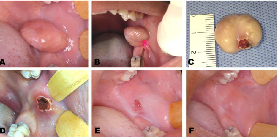
if the patient complained of any kind of pain. Slight traction of the lesion using
mosquito forceps was performed to facilitate the application of the diode laser
incision. Sutures were not performed. The surgical specimens were fixed in
10% buffer formalin and sent for histopathological analysis.
Laser parameters
Laser irradiation was performed using an 808 nm diode laser (Thera Lase
Surgery, DMC LTDA, São Carlos, Brazil), with an optical fiber of 600 µm, at a
Post-surgical evaluations
The treatment performance of FH with high-power diode laser was determined
by evaluating pain, postoperative functional alterations, edema, secondary
infection, bleeding, and patient satisfaction with the treatment. The patients
were asked to separately rate the degree of pain and postoperative functional
alterations, which included discomfort during eating and speech,on a 10cm
horizontal visual analog scale (VAS) by placing a vertical mark to assess the
position between the two endpoints (Mannion et al., 2007). The left endpoint of
the pain scale was designated as “no pain, and the right endpoint was marked
as unbearable pain.” The end-points of the scales for the degree of discomfort
during eating and speech were marked as no discomfort on the left side and
unbearable discomfort on the right side. The patients were asked to mark the
position between the two endpoints that best described their personal
perception of the degree of pain and discomfort during eating and speech that
they had experienced on postoperative days 1 and 7. The hatch mark placed by
the patient was measured to the nearest centimeter; the scores for the degree
of pain and functional complications were between 0 and 10. A single operator
recorded these scores on postoperative days 1 and 7. After completion, all
recordings were analyzed. All patients were instructed to use the same
analgesic medicine containing paracetamol, if needed to alleviate the pain, and
were subsequently analyzed. In addition, the patients were asked whether or
not an edema was present. Secondary infection was investigated by the
presence or absence of local exudation and fever. Bleeding was investigated by
of the post-operative wounds until they had been completely healed. Patient
treatment satisfaction was evaluated after post-surgical wounds had been
completely epithelized. A visual analogue scale (VAS) was applied to verify the
satisfaction degree: 0 = totally unsatisfied and 10 = totally satisfied with the
treatment (Peñarrocha et al., 2007).
Post-surgical care
All patients underwent special oral hygiene care, especially as regards hot,
hard, and acidic foods, during the post-surgical laser period. Patients were
instructed not to ingest any form of analgesic during the post surgical period,
except in case of unbearable pain.
Results
The clinical profiles of patients and data concerning FH lesions treated with
diode laser surgery are presented in Table 1.
Patient ages ranged from 12 to 76 years (mean 56.13 ± 17.55 years).
The study sample consisted of 12 females (80%) and 3 males (20%). Ten
(66.67%) patients presented a clinical diagnosis of FH induced by denture
(FHID), while five (33.33%) presented a diagnosis of focal FH (FFH). The size
of the lesions ranged from 5 to 90 mm (Mean: 33 mm). Six (40%) patients with
FHID presented a lesion on the superior vestibule, three (20%) patients
presented the lesion on the lower vestibule, and one (6.67%) presented the
lesion on the floor of the mouth. Two (13.33%) patients with FFH presented
lesions on the buccal mucosa, two (13.33%) on the lower lip, and one (6.67%)
three (20%) needed only topic anesthesia to remove the lesions. All lesions
were removed without the need for complementary sutures to control the
bleeding during and after diode laser surgery. No edema was reported by
eleven (74.34%) patients, while four (26.66%) reported edema in the
operative period. The patients classified the intensity of pain on the first
post-operative day as no pain in 53.33%, mild in 27.67%, moderate in 13.33% and
severe in 6.67%. On day 7, the patients classified the edema as no pain in
66.67%, mild in 26.66%, moderate in 6.66%; no patients reported unbearable
pain. Regarding analgesic medication in the post-operative period, twelve
(73.33%) patients reported no need for use, while three (26.67%) took an
analgesic due to moderate or severe pain. Considering functional alterations
during the chewing reported by the patients on the first post operative day, no
discomfort was reported in 66.66% of the cases, mild discomfort in 13.34%,
moderate in 13.34%, and severe in 6.65%. On day 7, no discomfort was
reportedin 60% of the patients, mild discomfortin 26.66%, severe in 13.33%,
and moderate in 6.66%. Considering functional alterations during speech
reported by the patients on the first post operative day, no discomfort was
reported in 46.67%, mild discomfort in 33.33% and moderate in 20%. In the
same category, on day 7, no discomfort was reported in 66.67% of the patients,
as compared to mild discomfort in 20% and moderate discomfort in13.33%. No
patient reported unbearable discomfort. All patients were totally satisfied with
the treatment. No persistent bleeding or infections could be observed. All
patients presented a clinical healing of the surgical wounds in a period that
ranged from 3 to 5 weeks (mean 3.5 weeks). Regarding patients with a
after the clinical healing of the surgical wounds, which left a minimal mucosa
scar (Figure 1).
In all specimens, microscopic analysis showed oral mucosal fragments
with hyperplastic stratified squamous epithelium, propria lamina of the densely
collagenized connective tissue, and chronic inflammatory cells. A band of
coagulation necrosis was present in the lower border of the specimens
(opposite to the epithelium) (Figure 2).
Discussion
The current study aimed to verify the performance of diode laser surgery on the
treatment of FH. The main observations included: 1) diode laser surgery proved
to be effective on the treatment of FH, 2) diode laser surgery shows low
postoperative pain and no complications, 3) diode laser surgery shows low
discomfort during chewing and speech in the postoperative period, 4) all
patients were satisfied with the applied treatment,and 5) there was minimal
thermal damage in the treated specimens.
Laser surgery treatment has been used as an adjuvant or substitute to
conventional therapies due to several advantages, including cutting, ablation or
vaporization, hemostasis, bacterial reduction, and surgical procedures without
infiltrative anesthesia (Romanos and Nentwig, 1999; Kara, 2008; Aras et al.,
2010).
Diode laser is considered a good cutting device for oral tissues
(Romanos and Nentwig, 1999). However, more tissue damage occurs than with
the use of a scalpel (Jin et al., 2010), but this damage is not prejudicial to the
least 5 mm (Angiero et al., 2012). In the current study, the tissue damage was
minimal, and only one lesion was measured at 5 mm in diameter. In addition,
the laser characteristics and settings, such as power output, wavelength,
emission modalities, type of optical fiber, and affinity with target tissues can
control the width and severity of the thermal damage caused to the tissue
(Angiero et al., 2012). By contrast, diode laser surgery provides a thermal
damage zone of less than 1 mm, which allows for surgical precision and
excellent hemostasis within a dry operative field (Coleton, 2004; Strauss and
Fallon, 2004). Hemostasis caused by laser surgery is due to the increase in
platelet activation at the end point of the wound, which leads to the sealing of
the blood vessels (Mordon et al., 2002).
Rapid wound healing with diode laser surgery has been described in
prior literature (Elanchezhiyan et al., 2013). This benefit is related to the photobiomodulation phenomenon, which works at cellular levels by promoting
faster healing with a toxin reduction through the accceleration of lymphatic flow,
thereby helping to reduce pain, enhance repair, and induce regeneration
(Bornstein, 2004; Elanchezhiyan et al., 2013). The current results demonstrated
that each of the 15 cases of FH treated with diode laser surgery with specific
parameters presented a clinical healing without complications in an average
time of 3.5 weeks.
Considering surgical procedures without infiltrative anesthesia, Fornaini
et al. (2007) reported the surgical management of oral tissues with only topical
anesthesia using diode and Nd:YAG laser systems. However, Aras et al. (2010)
demonstrated that, when compared with diode lasers, a lower quantity of
study, twelve (80%) patients needed infiltrative anesthesia, while three (20%)
needed only topic anesthesia to remove the lesions.
Management of oral tissues causes post-operative pain and functional
discomfort during chewing and speech (Aras et al., 2010). Benefits of laser
surgery systems, as compared to the use of scalpels, in decreasing
post-operative pain and functional complications have been demonstrated in
randomized clinical trials reported in prior literature (Haytac and Ozcelik, 2006).
These benefits may be explained due to the protein coagulum that is formed on
the wound surface, thereby acting as a biological dressing, sealing the end of
sensory nerves, as well producing photobiomodulation (Elanchezhiyan et al.,
2013). Moreover, less edemas have been reported when using laser surgery
systems, which is related to the sealing of the lymphatic vessels (Cernavin et
al., 1994; Coleton, 2004). In this prospective case series study, the
post-operative follow-up was uneventful with no edema in 74.34% of the patients.
The majority of patients (73.33%) reported no need to take analgesic
medication in the post-operative period. Additionally, the majority of patients
reported no or mild pain on post-operative day 1 (80%) and day 7 (93.33%).
Considering functional alterations during chewing, the majority of patients
reported no or mild discomfort on post-operative day 1 (80%) and day 7
(86.66%). Regarding functional alterations during speech, the majority of
patients reported no or mild discomfort on post-operative day 1 (80%) and day 7
(86.67%).
Therefore, diode laser surgery proved to be effective and presented a
satisfactory performance in the treatment of FH. The use of diode laser surgery
decreasing surgery time and maintaining the vestibule fundus in patients with
FHID. However, randomized clinical trials may be performed to compare diode
laser and other laser systems with conventional surgery and electrosurgery in
the management of FH and other oral lesions.
Funding
Conselho Nacional de Desenvolvimento Científico e Tecnológico (CNPq,
#309209/2010-2, #472045/2011-3)
Conflict of interest
None declared
Ethical Approval
Approved by the Ethics Committee of Universidade Federal de Minas Gerais,
under protocol number 23083713.1.0000.5149.
Acknowledgments
The authors would like to thank the Conselho Nacional de Desenvolvimento
References
1- Canger EM, Celenk P, Kayipmaz S: Denture-related hyperplasia: A clinical
study of a turkish population group. Braz Dent J 20:243 248, 2009.
2- Corbet EF, Holmgren CJ, Philipsen HP: Oral mucosal lesions in
65-year-old Hong Kong Chinese. Community Dent Oral Epidemiol 22:392 395,
1994.
3- Coelho CM, Souza YT, Daré AM: Denture-related oral mucosal lesions in
a Brazilian school of dentistry. J Oral Rehabil 31:135 139, 2004.
4- Firoozmand LM, Almeida JD, Cabral LA: Study of denture-induced fibrous
hyperplasia cases diagnosed from 1979 to 2001. Quintessence Int 36:825
829, 2005.
5- Freitas JB, Gomez RS, De Abreu, MH, Ferreira e Ferreira, E: Relationship
between the use of full dentures and mucosal alterations among elderly
Brazilians. J Oral Rehabilit 35:370 374, 2008.
6- Keng SB, Loh HS: The treatment of epulis fissuratum of the oral cavity by
CO 2 laser surgery. J Clin Laser Med Surg 10:303 306, 1992.
7- Niccoli-Filho W, Neves ACC, Penna LAP, Seradairian PI, Riva R: Removal
of epulis fissuratum associated to vestibuloplasty with carbon dioxide
laser. Lasers Med Sci 14:203 206, 1999.
8- Monteiro LS, Mouzinho J, Azevedo A, Câmara MI, Martins MA, La Fuente
JM: Treatment of epulis fissuratum with carbon dioxide laser in a patient
with antithrombotic medication.Braz Dent J 23:77 81, 2012.
9- Liboon J, Funkhouser W, Terris DJ: A comparison of mucosal incisions
made by scalpel, CO2 laser, electrocautery, and constant-voltage
10- Romanos G, Nentwig GH: Diode laser (980 nm) in oral and maxillofacial
surgical procedures: clinical observations based on clinical applications. J
Clin Laser Med Surg 17:193 197, 1999.
11- Angiero F,Parma L, Crippa R, Benedicenti S: Diode laser (808 nm) applied
to oral soft tissue lesions: a retrospective study to assess histopathological
diagnosis and evaluate physical damage. Lasers Med Sci 27:383 388,
2012.
12- Jackson SD, Lauto A: Diode-pumped fiber lasers: a new clinical tool?
Lasers Surg Med30:184 190, 2002.
13- Elanchezhiyan S,Renukadevi R,Vennila K: Comparison of diode
laser-assisted surgery and conventional surgery in the management of
hereditary ankyloglossia in siblings: a case report with scientific
review.Lasers Med Sci 28:7 12, 2013.
14- El-Kholey KE: Efficacy and safety of a diode laser in second-stage
implantsurgery: a comparative study. Int J Oral Maxillofac Surg 43:633
638, 2014.
15- Mannion AF, Balagué F, Pellisé F, Cedraschi C: Pain measurement in
patients with low back pain. Nature Clin Pract Rheumatol 3:610 618, 2007.
16- Peñarrocha M, Carrillo C, Boronat A, Martí E: Level of satisfaction in
patients with maxillary full-arch fixed prostheses: zygomatic versus
conventional implants. Int J Oral Maxillofac Implants 22:769 773, 2007
17- Kara, C: Evaluation of patient perceptions of frenectomy: a comparison of
Nd:YAG laser and conventional techniques. Photomed Laser Surg 26:147
18- Aras MH, Göregen M, Güngörmüş M, Akgül HM: Comparison of diode
laser and Er:YAG lasers in the treatment of ankyloglossia. Photomed
Laser Surg28:173 177, 2010.
19- Jin JY, Lee SH, Yoon HJ: A comparative study of wound healing following
incision with a scalpel, diode laser or Er,Cr:YSGG laser in guinea pig oral
mucosa: A histological and immunohistochemical analysis. Acta Odontol
Scand 68:232 238, 2010.
20- Coleton, S: Lasers in surgical periodontics and oral medicine. Dent Clin N
Am 48:937 962, 2004.
21- Strauss RA, Fallon SD: Lasers in contemporary oral and maxillofacial
surgery. Dent Clin N Am 48:861 868, 2004.
22- Mordon S, Begu S, Buys B, et al: Study of platelet behavior in vivo after
endothelial stimulation with laser irradiation using fluorescence in trivital
videomicsocopy and PEG-ylated liposome staining. Microvasc 64:316
325, 2002.
23- Bornstein E: Near infra red dental diode lasers. Scientific and
photobiologic principles and applications. Dent Today 23:102 108, 2004.
24- Fornaini C, Rocca JP, Bertrand MF, Merigo E, Nammour S, Vescovi P:
Nd:YAG and diode laser in the surgicalmanagement of soft tissues related
to orthodontic treatment. Photomed Laser Surg 25:381 392, 2007.
25- Haytac MC, Ozcelik O: Evaluation of patient perceptions after frenectomy
operations: a comparison of carbon dioxide laser and scalpel techniques.
J. Periodontol 77:1815 1819, 2006.
26- Cernavin I, Pugatpchew A, De Boer N, Tyas MJ. Laser applications in
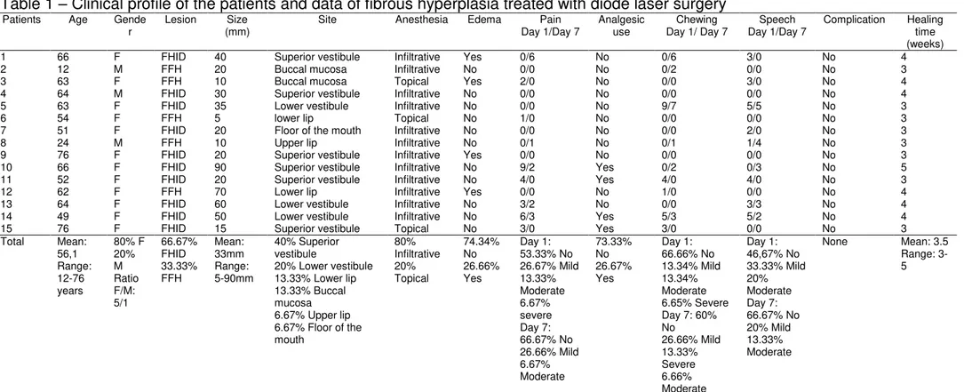
Table 1 – Clinical profile of the patients and data of fibrous hyperplasia treated with diode laser surgery Patients Age Gende
r Lesion (mm) Size Site Anesthesia Edema Day 1/Day 7 Pain Analgesic use Day 1/ Day 7 Chewing Day 1/Day 7 Speech Complication Healing time (weeks) 1 66 F FHID 40 Superior vestibule Infiltrative Yes 0/6 No 0/6 3/0 No 4 2 12 M FFH 20 Buccal mucosa Infiltrative No 0/0 No 0/2 0/0 No 3 3 63 F FFH 10 Buccal mucosa Topical Yes 2/0 No 0/0 3/0 No 4 4 64 M FHID 30 Superior vestibule Infiltrative No 0/0 No 0/0 0/0 No 4 5 63 F FHID 35 Lower vestibule Infiltrative No 0/0 No 9/7 5/5 No 3 6 54 F FFH 5 lower lip Topical No 1/0 No 0/0 0/0 No 3 7 51 F FHID 20 Floor of the mouth Infiltrative No 0/0 No 0/0 2/0 No 3 8 24 M FFH 10 Upper lip Infiltrative No 0/1 No 0/1 1/4 No 3 9 76 F FHID 20 Superior vestibule Infiltrative Yes 0/0 No 0/0 0/0 No 3 10 66 F FHID 90 Superior vestibule Infiltrative No 9/2 Yes 0/2 0/3 No 5 11 52 F FHID 20 Superior vestibule Infiltrative No 4/0 Yes 4/0 4/0 No 3 12 62 F FFH 70 Lower lip Infiltrative Yes 0/0 No 1/0 0/0 No 4 13 64 F FHID 60 Lower vestibule Infiltrative No 3/2 No 0/0 3/3 No 4 14 49 F FHID 50 Lower vestibule Infiltrative No 6/3 Yes 5/3 5/2 No 4 15 76 F FHID 15 Superior vestibule Topical No 3/0 Yes 3/0 0/0 No 3 Total Mean:
56,1 Range: 12-76 years 80% F 20% M Ratio F/M: 5/1 66.67% FHID 33.33% FFH Mean: 33mm Range: 5-90mm 40% Superior vestibule
20% Lower vestibule 13.33% Lower lip 13.33% Buccal mucosa 6.67% Upper lip 6.67% Floor of the mouth 80% Infiltrative 20% Topical 74.34% No 26.66% Yes Day 1: 53.33% No 26.67% Mild 13.33% Moderate 6.67% severe Day 7: 66.67% No 26.66% Mild 6.67% Moderate 73.33% No 26.67% Yes Day 1: 66.66% No 13.34% Mild 13.34% Moderate 6.65% Severe Day 7: 60% No 26.66% Mild 13.33% Severe 6.66% Moderate Day 1: 46,67% No 33.33% Mild 20% Moderate Day 7: 66.67% No 20% Mild 13.33% Moderate
None Mean: 3.5 Range: 3-5
Figures and Legends
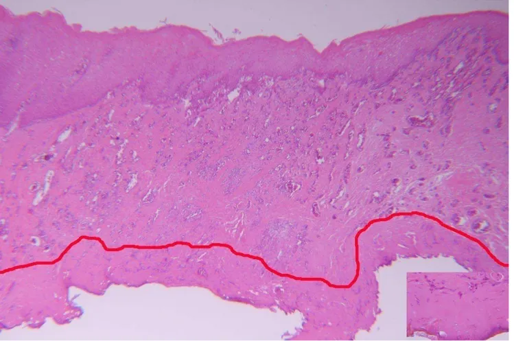
Figure 2- Fragment of fibrous hyperplasia submitted to diode laser surgery. An oral mucosal fragment with hyperplastic stratified squamous epithelium and lamina propria can be observed, represented by densely collagenized connective tissue and chronic inflammatory cells. A band of coagulation necrosis is present on the lower border of the specimens, opposite to the epithelium and outlined by a red line (Haematoxylin and eosin, 25X magnification original).
7.2 Artigo 2
Artigo submetido ao periódico International Journal of Oral and Maxillofacial
Surgery (qualis – A1/ Fator de impacto 1.5) em 19 de maio de 2014 e
encontra-se aceito para publicação..
Journal title: International Journal of Oral & Maxillofacial Surgery
Corresponding author: Prof. Márcio Bruno Amaral
Article title: Diode laser surgery versus scalpel surgery in the treatment of
fibrous hyperplasia: a randomized clinical trial
Manuscript number: IJOMS-D-14-00408R2
Dear Prof. Amaral,
I am pleased to confirm that your manuscript has been accepted and sent to the
Technical Editor for editing.
Once we have the final edited version of your paper a second confirmation
acceptance letter will be sent to you directly.
Yours sincerely
Jacqui Merrison
Title: Diode laser surgery versus scalpel surgery in the treatment of
fibrous hyperplasia: a randomized clinical trial
Running title: Diode laser versus scalpel surgery in treatment of fibrous
hyperplasia
Abstract
Fibrous hyperplasia (FH) is treated by surgical incision using a scalpel, together
with the removal of chronic trauma. However, scalpel techniques do not provide
the haemostasis that is necessary when dealing with high vascular tissues.
Diode laser surgery can be used in the management of oral tissues due to its
high absorption by water and hemoglobin, and has provided good results in
periodontal surgery and oral lesions. The aim of this present study was to
compare the effects of diode laser surgery to those of the conventional
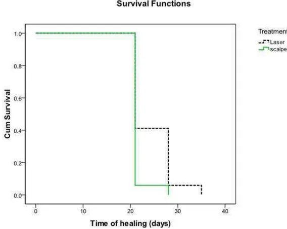
technique in patients with fibrous hyperplasia. A randomized clinical trial was
performed in which surgical and postoperative evaluations were analyzed. On
comparison of the laser- treated (study group) patients to those treated with a
scalpel (control group), significant differences were observed in the duration of
surgery and the use of analgesic medications. Over a 3-week period, clinical
healing of the postoperative wound was significantly faster in the control group
as compared to the study group. In conclusion, diode laser surgery proved to be
more effective and less invasive when compared to scalpel surgery in the
management of fibrous hyperplasia. However, wound healing proved to be
Introduction
Fibrous hyperplasia is a chronic low-grade irritation occurring as a
consequence of ill-fitting dentures. Fibrous hyperplasia is frequently the result of
the resorption of the alveolar ridge in such a way that the denture moves further
into the vestibular mucosa, leading to fibrous hyperplasia that proliferates over
the flange. Additionally, parafunctional habits can also induce a focal fibrous
hyperplasia.1
Fibrous hyperplasia is a frequent oral lesion that affects 5% to 16.7% of
the population. 2, 3 Its commonly appears as a small-sized, painless lesion with
fibrous to flaccid consistency that is pale to erythematous in color and that can
be found in any part of the oral cavity.4,5 Fibrous hyperplasia occurs as a result
of chronic irritation to grow, and when the source of trauma is removed, the
lesion commonly decreases in size or regresses.4
Conventionally, fibrous hyperplasia is treated by surgical incision using a
scalpel, together with the removal of chronic traumatic factors. Scalpels have
been used for many years due to their case of use, accuracy, and minimal
damage to the surrounding tissues. However, haemostasis of the surgical
wound can be difficult, especially for patients with haemorrhagic disorders or
those on anti-thrombotic therapy, without a suturing of the wound borders. 6,7-9
In the management of fibrous hyperplasia, haemostatic problems can be
controlled by the use of electrocautery, which provides enhanced hemostasis by
sealing blood vessels before cutting. However, cutting performance can be
reduced by muscle fasciculation, while wound healing is delayed by extensive
thermal damage when compared with scalpel surgery.6,10 It is important to
vestibule can appear in cases induced by ill-fitting dentures when suturing is
performed on the wound borders.7
Surgical laser systems have been applied in the treatment of oral lesions.
The main types are: 1) potassium-titanium-phosphorous (KTP), 2)
neodymium-yttrium-aluminum-gamet (Nd:YAG), 3) carbon dioxide (CO2), and 4) diode
lasers with semiconductors.11,12 Diode laser systems are portable, compact, and
cost effective when compared with other high-power lasers. Diode lasers have a
wavelength of between 805 and 980nm, which can be used in continuous or
pulsed mode, depending on the clinical requirement, using an optical fiber with
or without contact.13 Diode laser surgery is often used in the management of
oral tissues due to high absorption by water and haemoglobin, and has
provided sound results in periodontal surgery, tissue alteration related to
orthodontic treatment, and oral lesions.11,14-16
Considering that (1) diode laser surgery appears to be a good option for
the treatment of oral diseases, including fibrous hyperplasia; (2) only a few
clinical studies have been published;8,9 and (3) the conventional treatment using
scalpels can lead to clinical complications, the aim of the present study was to
compare the effects of diode laser surgery with conventional techniques using
scalpels.
Patients and methods
Patients and study design
Patients were recruited consecutively from the oral medicine clinic of the study
university in Belo Horizonte, Brazil, from February to October 2013. The cases
parafunctional habits. Sample size was calculated based on dependent
variables (postoperative pain and postoperative functional alterations) and
analyzed considering a 95% confidence interval. The parameters used to
perform the sample size calculation were identified from studies with a similar
design published in the literature. 16,17 Limited-sized lesions with flaccid to
fibrous consistency, sessil or had a pedicle, that were pale to erythematous in
color, and that were associated with dentures or parafunctional habits, were
included in this study. Dentures were removed 2 weeks prior to treatment to
eliminate inflammation and/or chronic pain. Patients currently using
anti-inflammatory or analgesic medications were excluded. The lesions were
measured in their largest diameter with a millimeter rule. A randomized clinical
trial was carried out and patients were divided into two groups. A
computer-generated list of random numbers was used to allocate subjects to the groups,
considering a randomization ratio of 1:1 (Microsoft Office Excel Software, 2007).
This study was approved by the Human Research Ethics Committee (protocol
number 23083713.1.0000.5149) and was conducted in accordance with the
Consolidated Standards of Reporting Trials (CONSORT) statement.18 Patients
signed a statement of informed consent prior to inclusion in this study.
Surgical procedures
The control group (conventional technique using a scalpel) consisted of 19
patients. For these patients, after infiltrative anesthesia with 2% lidocaine and
adrenaline at 1:100.000 (DFL Indústria e Comércio S. A., Rio de Janeiro, Brazil)
was applied. Following this, the fibrous hyperplaisa was removed through