Antibody-Independent Control of
c
-Herpesvirus
Latency via B Cell Induction of Anti-Viral
T Cell Responses
Kelly B. McClellan1, Shivaprakash Gangappa2, Samuel H. Speck3, Herbert W. Virgin IV1*
1Department of Pathology and Immunology, Washington University School of Medicine, Saint Louis, Missouri, United States of America,2Emory Transplant Center, Emory University School of Medicine, Atlanta, Georgia, United States of America,3Division of Microbiology and Immunology, Yerkes Regional Primate Research Center, Emory University, Atlanta, Georgia, United States of America
B cells can use antibody-dependent mechanisms to control latent viral infections. It is unknown whether this represents the sole function of B cells during chronic viral infection. We report here that hen egg lysozyme (HEL)-specific B cells can contribute to the control of murinec-herpesvirus 68 (cHV68) latency without producing anti-viral antibody. HEL-specific B cells normalized defects in T cell numbers and proliferation observed in B cell/mice during the early phase ofcHV68 latency. HEL-specific B cells also reversed defects in CD8 and CD4 T cell cytokine production observed in B cell/mice, generating CD8 and CD4 T cells necessary for control of latency. Furthermore, HEL-specific B cells were able to present virally encoded antigen to CD8 T cells. Therefore, B cells have antibody independent functions, including antigen presentation, that are important for control ofc-herpesvirus latency. Exploitation of this property of B cells may allow enhanced vaccine responses to chronic virus infection.
Citation: McClellan KB, Gangappa S, Speck SH, Virgin HW IV (2006) Antibody-independent control ofc-herpesvirus latency via B cell induction of anti-viral T cell responses. PLoS Pathog 2(6): e58. DOI: 10.1371/journal.ppat.0020058
Introduction
c-Herpesviruses such as Epstein Barr virus (EBV), Kaposi’s sarcoma herpesvirus (KSHV), and murine c–herpesvirus 68 (cHV68) latently infect lymphocytes and other cells as part of a strategy for maintaining life-long infection. Latent in-fection represents a balance between the virus and the host to which immunity makes an essential contribution.c -herpesvi-rus latency and replication of vi-herpesvi-rus that has reactivated from latently infected cells contribute toc-herpesvirus-associated diseases [1–7]. The stability of this balance between virus and host is demonstrated by the observation in mice that a latency ‘‘set point’’ exists such that the same number of cells are latently infected regardless of the dose or route of infection [8], and in humans by the observation that individuals have a stable level of EBV latency over years [9]. Despite the stability ofc-herpesvirus latency, the balance between virus and host is delicate sincec-herpesvirus-induced disease is most often seen in immunocompromised hosts. In addition, deletion of individual host [7,10,11] or viral [12–14] genes disrupts this balance with consequent inefficient infection or development of disease.
To understand the stable but delicate balance between the host andc-herpesviruses present during life-long infection, it is necessary to define mechanisms of immunity responsible for holding the virus at bay. To define these mechanisms many groups have studied infection of mice with cHV68, which provides a relevant small animal model for c -herpesvirus infection and immunity. After clearance of acute infection,cHV68 latently infects macrophages, B cells, and dendritic cells [8,15–18].cHV68 infection is associated with development of B cell malignancies, vasculitis, and athero-sclerosis [2,7,19,20].
Immunity controls latentcHV68 infection by limiting the number of cells carrying viral genome during latency
[10,21,22] and by regulating the efficiency with which these cells reactivate from latency when explanted [10,11,23,24]. In addition, the immune system regulates persistent viral replication, which is detected as the presence of preformed infectious virus in tissues after clearance of the acute infection [7,10,11,17,23]. Persistent cHV68 replication is distinct from replication occurring during acute infection (acute replication) since thecHV68 v-cyclin and v-Bcl-2 genes are required for persistent but not acute replication [12–14]. Persistent replication is observed in normal mice, and is more prominent in immunocompromised mice such as those lacking B cells or interferon-c (IFNc) [7,10,14,17,23]. It is likely that persistent replication involves virus that has reactivated from latently infected cells since the v-cyclin and v-Bcl-2 genes are required for both efficient reactivation from latency and for persistent replication [12,13]. Persistent replication may contribute to latency via infection of new cells that enter the latent pool [25,26].
There are two forms ofcHV68 latency that are distinguish-able experimentally [8,10–12]. The early form of latency is measurable 16 d after infection when acute infection has
Editor:Grant McFadden, Robarts Research Institute, Canada
ReceivedFebruary 1, 2006;AcceptedApril 28, 2006;PublishedJune 23, 2006 DOI:10.1371/journal.ppat.0020058
Copyright:Ó2006 McClellan et al. This is an open-access article distributed under
the terms of the Creative Commons Attribution License, which permits unrestricted use, distribution, and reproduction in any medium, provided the original author and source are credited.
Abbreviations:cHV68, murinec-herpesvirus 68; APC, antigen presenting cell; BCR, B cell receptor; EBV, Epstein-Barr virus; ELISA, enzyme-linked immunosorbent assay; HEL, hen egg lysozyme; IFNc, interferon-c; KSHV, Kaposi’s sarcoma associated virus; Vb4þTCR, variable region geneb4 containing T cell receptor
been cleared. At this time most cells carrying latent viral genome reactivate when cultured ex vivo [11]. The late form of latency, typically measured at 42 d after infection, is characterized by inefficient reactivation ex vivo with 10% or less of genome bearing cells reactivating when explanted [10,11]. Latency is typically measured in the spleen as a lymphoid site and the peritoneum as a body cavity site. Analysis of these sites is of interest since both EBV and KSHV establish latency in lymphoid sites and KSHV causes body cavity based lymphomas [3–5]. Both the early and late forms of latency in splenocytes and peritoneal cells are observed regardless of the route (intraperitoneal or intranasal) or dose of viral inoculation [8].
Several components of the immune system contribute to the control of latent and persistentcHV68 infection. These include CD8 T cells [10,21,22], CD4 T cells [21,27–29], perforin [10], granzymes [30], caspase 3 [30], IFNc
[6,7,10,24,29], IFNab [23], and antibody and B cells [11,25,26]. Of these, the role of B cells is of particular interest because c-herpesviruses including EBV, KSHV, and cHV68 establish latency in B cells. In addition to serving as a site of latent infection, B cells regulate latency in non-B cells [11]. B cell/mice exhibit increased frequencies of reactivating and viral genome bearing cells compared to B6 mice [11], and show increased persistent replication in lung and aorta [6,7,14,31]. Thus, removal of one latent reservoir, B cells, increases the level of latency in non-B cells, and diminishes control of persistent replication. Lack of specific anti-viral antibody likely explains some of the abnormalities in latency and persistence seen in B cell/ mice. Passive transfer of antibody significantly decreases the frequency of latently infected cells in B cell/or T cell depleted CD28/mice [25,26]. However, it is not known whether the important role of B cells in control of cHV68 latency and persistent replication is explained by production of anti-viral antibody or other antigen-specific B cell receptor (BCR)-dependent activities of B cells.
To identify B cell activities during infection that are independent of antibody production and expression of a virus antigen-specific BCR we bred a mouse that contained B cells but cannot mount an antigen specific anti-viral B cell
functions during chroniccHV68 infection that are at least in part explained by B cell-dependent induction of CD8 and CD4 T cell responses.
Results
HEL-Specific B Cells Decrease the Frequency of
Splenocytes That Reactivate from Latency and Decrease the Efficiency of Reactivation from Latency
B cell/mice have higher numbers of cells carrying latent
cHV68 than wild type mice [11]. We wanted to determine whether the effects of B cells on latent cHV68 infection require production of anti-viral antibody and engagement of a virus antigen-specific BCR. One approach to this question would be to study the effects of adoptively transferred B cells oncHV68 infection of B cell/mice. However, adoptively transferred B cells do not persist in this setting [32,33], necessitating a genetic approach to engrafting virus antigen non-specific mice onto a B cell/background. We therefore bred the HEL-specific IgM/IgD BCR bearing the IgMaallotype from MD4 mice [34] onto thelMT B cell/background [35] (Figure 1, HELMET mice). HELMET mice contained IgMaand CD19 double positive B cells in spleen (Figure 1) and lymph nodes (unpublished data) while B6 mice and B cell/mice did not.
To determine if HEL-specific B cells can control the late form of latency in a lymphoid site we examined splenic latency in B6, HELMET, and B cell/mice (Figure 2). We confirmed a prior report [11] that the frequency of splenocytes from B cell/mice that reactivate from latency ex vivo is significantly elevated compared to the frequency observed in splenocytes from B6 mice (p¼0.003, Figure 2A). The frequency of splenocytes from HELMET mice that reactivated ex vivo was indistinguishable from the frequency observed in splenocytes from B6 mice and was significantly lower than that observed in splenocytes from B cell/mice (p ¼0.0001). Therefore, the presence of HEL-specific B cells
Figure 1.HELMET Mice Contain Transgenic B Cells
Splenocytes from naı¨ve B6, HELMET, and B cell/mice were stained with antibodies against CD19 and IgMa. Numbers represent percentages of cells in the indicated quadrants.
DOI: 10.1371/journal.ppat.0020058.g001
normalized defects in the control of splenic latency observed in B cell/mice.
We next determined whether effects of HEL-specific B cells on splenic latency were due to alterations in the frequency of viral genome bearing cells or in the efficiency of reactivation of explanted latently infected cells (defined as the frequency of cells reactivating ex vivo divided by the frequency of cells bearing viral genome multiplied by 100). The frequencies of genome positive splenocytes in B cell/, HELMET, and B6 mice were similar (Figure 2B, right panel). About 15% of viral genome positive splenocytes from B cell/mice reactivated ex vivo compared to about 2% in B6 mice and 3% in HELMET mice (Figures 2A and 2B, right panels). Therefore the effects of HEL-specific B cells on splenic latency were due to changes in the efficiency of ex vivo reactivation rather than the frequency of cells bearing viral genome.
HEL-Specific B Cells Decrease the Frequency of Peritoneal Cells That Reactivate from Latency and That Carry Latent cHV68 Genome
To determine whether viral antigen non-specific B cells can restore normal regulation of latency in a body cavity site we compared the late form of latency in B6, HELMET, and B cell/ mice. Consistent with previous results [11], the frequency of reactivating cells in B cell/mice was 200-fold higher than in B6 mice (Figure 2A, left panel,p¼0.008). HEL-specific B cells were able to decrease reactivation from latency in peritoneal cells since the frequency of reactivating cells was 20-fold less in HELMET mice in comparison to B
cell/ mice (p ¼ 0.0053). However, the frequency of reactivating peritoneal cells in HELMET mice was 11-fold higher than that in peritoneal cells from B6 mice (Figure 2A, left panel, p ¼ 0.05). These data demonstrate that HEL-specific B cells limit reactivation from latency in peritoneal cells, but do not fully compensate for the presence of normal B cells.
We next determined whether the decreased frequency of peritoneal cells that reactivate from latency in HELMET mice compared to B cell/ mice was due to a decrease in the frequency of latently infected cells by measuring the efficiency of reactivation. Consistent with published results [11], the frequency of viral genome-positive cells was 10-fold higher in peritoneal cells from B cell/mice than in B6 mice (Figure 2B, left panel, p ¼ 0.01). Thus, roughly 100% of latently infected peritoneal cells from B cell/ mice reactivated from latency when explanted while only 6% of latently infected peritoneal cells from B6 mice reactivated (compare Figures 2A and 2B). HEL-specific B cells decreased the frequency of cells carrying latent viral genome to levels similar to those observed for B6 mice (p¼0.36). However, about 81% of viral genome bearing peritoneal cells from HELMET mice reactivated when explanted. Thus the effect of HEL-specific B cells on latency in peritoneal cells is largely via decreasing the frequency of cells carrying latent viral genome.
The Effects of HEL-Specific B Cells oncHV68 Latency Are Not Due to Cross-Reactivity between HEL andcHV68
The data provided above show that the presence of HEL-specific B cells has significant effects oncHV68 latency in two different sites. To determine whether these effects were virus-specific antibody independent we needed to determine whether anti-cHV68 antibody is generated in either B cell/ or HELMET mice. To address this issue we used enzyme-linked immunosorbent assay (ELISA), viral neutralization assays, and passive transfer experiments (Figure 3).
As expected, anti-viral antibody was found by ELISA in serum from infected B6 mice but not in serum from infected B cell/or HELMET mice (Figure 3A, left panel). Antibody specific to HEL was detected in mock infected and cHV68 infected HELMET mice (unpublished data). Similarly, serum from cHV68 infected B6 mice neutralized cHV68, while serum from infected B cell/ or HELMET mice did not (Figure 3A, right panel). Thus anti-HEL antibodies do not cross-react withcHV68 in ELISA or neutralization assays and infected B cell/and HELMET mice fail to make detectable antibody tocHV68.
To rule out the possibility that ELISA and neutralization assays failed to detect physiologically relevant anti-cHV68 antibody in the serum of HELMET mice and that HEL specific antibody in the serum of HELMET mice might alter
cHV68 latency, we compared the effect of passively trans-ferred serum from infected HELMET or B6 mice on latency in B cell/ mice. We and others previously demonstrated that the passive transfer of anti-viral antibody can decrease the number of latently infected cells [25] and decrease the recrudescence of lytic virus [26] in latently infected mice by blocking lytic viral replication [25]. The transfer of control serum from either B6 or HELMET mice had no effect on latency in B cell/mice (compare Figures 2A and 3B). As previously published [25], passively transferred immune Figure 2.Virus Non-Specific B Cells Control Latent Infection
(A) Ex vivo reactivation from latency in peritoneal cells (left) and splenocytes (right) from B6, HELMET, and B cell/mice 42 d after infection. Dotted lines indicate the 63.2% Poisson distribution line used to calculate the frequency of cells reactivating virus.
serum fromcHV68 infected B6 mice decreased the frequency of peritoneal cells reactivating from latency more than 100-fold compared to the transfer of control serum from mock infected B6 mice (Figure 3B, p¼ 0.05), and of splenocytes more than 4-fold (Figure 3B, p¼ 0.01). In contrast, serum from infected HELMET mice had no effect on latency (Figure 3B).
Together these data show that there is noc HV68-reactive-antibody in infected HELMET mice and that HEL-specific antibody in serum of HELMET mice did not alter cHV68 latency. We conclude that the control of latency observed in HELMET mice (Figures 2A and 2B) is dependent on the presence of HEL-specific B cells rather than antibody or a virus antigen-specific BCR.
Effects of HEL-Specific B Cells on Latency Are Not Explained by Decreases in AcutecHV68 Replication
The above data showed that B cells that do not produce
anti-viral antibody or express a virus antigen-specific BCR can nevertheless have significant effects on latent cHV68 infection. To define the mechanism responsible for this effect we first determined whether effects of HEL-specific B cells on acutecHV68 replication might explain decreases incHV68 latency detected in HELMET mice. We therefore measured
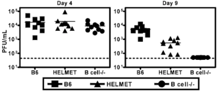
cHV68 replication in B6, HELMET, and B cell/mice 4 and 9 d after infection. 4 d after infection, viral titers were similar in B6, HELMET, and B cell/mice (p.0.2, Figure 4). On day 9,cHV68 titers were more than 100-fold greater in B6 mice (p
, 0.0001) than in B cell/mice (Figure 4), reflecting the importance of B cells for maintainingcHV68 replication [36]. The presence of HEL-specific B cells led to increasedcHV68 replication compared to B cell/mice (p,0.0001) 9 d after infection, but did not fully normalize splenic titers (p ,
0.0001). These data show that decreased latency in HELMET mice compared to B cell/ mice cannot be explained by decreasedcHV68 replication in HELMET mice.
HEL-Specific B Cells Correct Abnormalities in Splenocyte Numbers Observed in B cell/Mice
Based on studies in other systems comparing responses to infection in the presence and absence of B cells [33,37–40], we reasoned that increases in T cell responses in the presence of HEL-specific B cells might contribute to control of cHV68 latency. We therefore determined whether HEL-specific B cells restore abnormalities in splenic cellularity that have been reported in B cell/mice including reduced numbers of splenocytes, CD8 T cells, and CD4 T cells compared to B6 mice [41]. We confirmed that mock infected or naı¨ve B cell/ had decreased numbers of CD4 T cells compared to B6 mice [41] (Figure 5). In contrast to B cell/mice, we found that the absolute number of splenocytes, B cells, CD4 T cells, and CD8 T cells was comparable between in naı¨ve and/or mock-infected B6 and HELMET mice (Figure 5). Thus, the presence of HEL-specific B cells effectively corrected deficiencies in T cell numbers found in uninfected B cell/mice.
We next evaluated the T and B cell response to cHV68 infection in HELMET mice compared to B6 and B cell/ mice by quantifying the spleen cell populations 16 and 42 d after infection. Consistent with previous data [36], we found thatcHV68 infection of B6 increased total splenocytes (p¼ 0.03), CD4 T cells (p¼0.03), CD8 T cells (p,0.0001), and B cells (p ¼ 0.02) compared to uninfected mice 16 d after Figure 3.Control ofcHV68 Latency by HEL-Specific B Cells Is Not due to
Production of Cross-Reactive Antibody
(A) (Left) ELISA for cHV68-specific antibodies in serum from mock infected and virus infected B6, HELMET, and B cell/mice. Data are representative of two independent experiments. (Right) Sera from mock infected and cHV68 infected B6, HELMET, and B cell/mice were incubated withcHV68 and tested for their ability to neutralizecHV68 infection. Percent neutralization was normalized to results obtained using serum from mock-infected mice of the same genotype.
(B) Beginning at day 16 post-infection, B cell/mice were treated with serum from mock infected orcHV68 infected B6 and HELMET mice. Ex vivo reactivation from latency was assessed 42 d after infection. DOI: 10.1371/journal.ppat.0020058.g003
Figure 4.HEL-Specific B Cells Restore Acute PhasecHV68 Replication to the B Cell/Background
infection. B cell/ mice did not show increased total splenocytes (p¼0.37) or CD4 T cells (p . 0.05). Although there were slight increases in CD8 T cells (p¼0.005) in B cell/ mice, the number was significantly reduced in comparison to the number of CD8 T cells in B6 mice 16 d after infection (p¼ 0.0036). At 16 d after infection the presence of HEL-specific B cells normalized total splenocyte numbers (p¼0.78) and CD4 (p¼0.91), and CD8 T cell numbers (p¼0.81) to levels observed in B6 mice (Figure 5). Taking these data together, HEL-specific B cells restored defects in cell numbers observed in infected B cell/ mice 16 d after infection. We did not identify all of the types of cells present in the spleens of the different mice used here, and thus it is possible that the presence of B cells alters the number of cells other than CD4 and CD8 T cells. The B cell response to infection observed in B6 mice was not fully restored by the presence of HEL-specific B cells (Figure 5). In addition, HEL-HEL-specific B cells did not restore CD4 or CD8 responses to the level seen in B6 mice 42 d after infection. Thus, HEL-specific B cells can normalize splenic lymphocyte numbers in uninfected mice early after infection withcHV68, but HEL-specific B cells do not replace all normal B cell functions.
HEL-Specific B Cells Are Activated during EarlycHV68 Latency
The normalization of T cell proliferative responses early after infection in the presence of HEL-specific B cells raised the question of whether HEL-specific B cells are activated in response to infection. To address this we used flow cytometry to determine the percentage of activated B cells in B6 and HELMET mice 16 d after infection (Figure 6). Compared to mock-infected controls, we found that the percentage of B
cells that expressed CD69 increased 2-fold in both B6 (p¼ 0.01) and HELMET mice (p¼0.002) (Figure 6A) compared to mock infected mice. Further histological examination anal-ysis showed that germinal center-like structures formed in HELMET and B6 mice after cHV68 infection (unpublished data). Thus, HEL-specific B cells can be activated during
cHV68 infection independent of a virus antigen-specific BCR or antiviral antibody.
We questioned whether this activation might be explained by direct infection of HEL-specific B cells by cHV68. We therefore determined whether HEL specific B cells carry latent cHV68 16 d after infection. We found that approx-imately 1/150 CD19þ cells in HELMET mice carried viral genome compared to 1/700 total splenocytes (Figure 6B). We entertained the possibility that our purification procedure non-specifically enriched for viral genome bearing cells, but found that,1:10,000 CD8 T cells purified in parallel, carried viral genome (unpublished data). As an additional control we determined the frequency of viral genome bearing cells in CD19-depleted splenocytes. We found that only 1:4,400 CD19-depleted splenocytes carried viral genome. By multi-plying these frequencies times the numbers of cells per spleen from Figure 5, we determined that HEL-specific B cells account for the majority of latently infected splenocytes in HELMET mice (3.33105viral genome bearing B compared to 3.43104viral genome bearing non-B cells). These findings in HELMET mice agree with previously reported data in B6 mice showing that B cells carry the majority of cHV68 genome 16 d after infection [17,18,42]. Interestingly, the frequency of viral genome bearing HEL-specific B cells was 33-fold lower than the frequency of HEL-specific B cells expressing CD69 (compare Figures 6A and 6B), indicating that direct virus infection cannot fully explain activation of HEL-specific B cells duringcHV68 infection.
Virus Non-Specific B Cells Rescue T Cell Defects in B Cell/Mice
Taken together the experiments above indicate that HEL-specific B cells play an important role in generating a full CD4 and CD8 T cell proliferative response to cHV68 infection. Since CD4 and CD8 T cells are important for the control of cHV68 latency [10,21,22,27], we postulated that HEL-specific B cells could control latentcHV68 infection by promoting protective CD4 and CD8 T cell responses. To address this postulate, we further characterized T cell expansion and activation in B6, HELMET, and B cell/mice after infection.
The CD8 T cell response tocHV68 is heavily biased toward T cells whose receptors are encoded by the Vb4 gene, and expansion of Vb4þCD8 T cells is dependent on the presence of B cells [43–46]. We used flow cytometry to evaluate the Vb4þ
T cell receptor (TCR)þCD8 T cell expansion at 21 d after infection, a time when it has been consistently observed in wild-type mice [47] (Figure 7). Flow-cytometry revealed a 10-fold expansion of Vb4þ
TCRþCD8 T cells in B6 (p¼0.0053) and HELMET mice (p ¼ 0.016) relative to mock-infected controls (Figure 7A). As expected, we did not detect the Vb4þ
TCRþCD8 T cell expansion aftercHV68 infection in B cell/mice. Thus, the B cell-dependent induction of Vb4þ CD8 T cell responses tocHV68 does not required anti-viral antibody or a virus antigen-specific BCR.
To further examine T cell activation, we used flow Figure 5.HEL-Specific B Cells Normalize Some but Not All Abnormalities
in Splenic Cellularity Observed in B Cell/Mice
Splenocytes from pooled naı¨ve or mock infected andcHV68 infected B6, HELMET, and B cell/mice were counted and stained with antibodies against CD8, CD4, and CD19. The splenocyte numbers were the same in naı¨ve and mock-infected mice, and data from mock infected and naı¨ve mice were pooled. Numbers represent number of cells/spleen3106. The data are pooled from two to six independent experiments evaluating three to five mice per experiment.
cytometry to assess the number of CD8 and CD4 T cells that produce IFNc. Chronic activation of CD8 T cells has been reported during long term cHV68 infection [48–50]. We measured the number of T cells that produce IFNc16 d after infection based on studies showing that day 16 falls within the peak of T cell activation duringcHV68 infection [50] and that IFNcis important for the control ofcHV68 latency [10,24]. Compared to mock infection,cHV68 at day 16 induced a 10-fold expansion of CD8 T cells that produced IFNcin B6 (p¼ 0.0027) and HELMET mice (p¼ 0.01) while no significant expansion was observed in B cell/ mice (Figure 7B). Additionally,cHV68 infection induced a significant expan-sion of CD8 T cells that expressed CD69 in B6 and HELMET mice while no significant expansion was found in B cell/ mice (unpublished data). Similar studies of CD4 T cells revealed a 6-fold expansion of IFNcproducing CD4 T cells in B6 mice (p¼0.002) and in HELMET mice (p¼0.006), but not in B cell/mice (Figure 7C). Together these data show that the presence of HEL-specific B cells allows the induction of an activated population of CD8 and CD4 T cells incHV68 infected mice.
HELMET Mice Use Both CD4 T and CD8 T Cell-Dependent Mechanisms to LimitcHV68 Latent Infection
We questioned whether the observed differences between HELMET and B cell/mice in T cell numbers and activation were functionally relevant to the control of latency. We reasoned that activated T cells present in HELMET mice and wild-type mice but not B cell/mice might contribute to the control of latent cHV68 infection observed in B6 and HELMET mice. To investigate this, we determined the effect of T cell depletion on latency in B cell/and HELMET mice (Figure 8).
Treatment with isotype control had some effect on levels of peritoneal cell latency in both B cell/and HELMET mice (compare Figures 2A and 8A). We therefore considered the levels of latency observed in isotype control antibody treated mice as the proper control for treatment with T cell depleting antibodies. CD4-depletion of B cell/mice had a small effect on latency in peritoneal cells relative to isotype-control treated mice (Figure 8A), increasing the frequency of reactivating peritoneal cells 3-fold (p , 0.0001) in both HELMET mice and B cell/ mice. CD4-depletion had no effect on the frequency of viral genome bearing peritoneal Figure 6.HEL-Specific B Cells Are Activated duringcHV68 Infection and Are Infected bycHV68
(A) Splenocytes from mock infected andcHV68 infected B6 and HELMET mice were stained with antibodies specific for CD19 and CD69 16 d after infection. Shown are the percentages of splenic CD19þ
cells that express CD69 (left). Shown are representative dot plots (right).
(B) Frequency splenic HEL-specific B cells that carry viral genome 16 d post infection (left). Shown is a representative histogram of the purity of the B cell population after purification (right). These data are representative of two independent experiments using five mice per experiment.
cells in HELMET mice or in B cell/mice compared to isotype control treated mice. Thus, CD4-depletion increased the efficiency with which peritoneal cells reactivate from latency.
We observed that CD4-depletion had no effect on latency in splenocytes from B cell/mice (Figure 8A and 8B). However, we observed a five-fold increase in reactivating HELMET splenocytes upon CD4-depletion (p,0.0001, Figure 8A). CD4-depletion also increased the frequency of splenocytes carrying viral genome more than 6-fold in HELMET mice (p,0.0001, Figure 8B). Therefore, the increase in reactivation was partially due to an increase in the frequency of viral
genome-bearing splenocytes. We interpreted these data to mean that CD4 T cells are important for the control of latent
cHV68 infection in HELMET mice.
As previously shown [29], CD8 T cell depletion of B cell/ mice had no effect on the frequency of peritoneal cells that reactivated ex vivo or the frequency of viral genome-bearing peritoneal cells compared to mice treated with isotype-control antibodies (Figures 8A and 8B). Depletion of CD8 T cells from HELMET mice resulted in a 15-fold increase in the frequency peritoneal cells that reactivated from latency compared to isotype-control treated groups (p , 0.0001, Figure 8A). This increase in reactivation was largely due to an increase in the frequency of cells harboring viral genome because 12-fold more peritoneal cells carried viral genome in CD8 T cell-depleted HELMET mice compared to isotype control treated HELMET mice (p,0.0001, compare Figures 8A and 8B). Thus, CD8 T cells are important for controlling latency in peritoneal cells from HELMET mice.
Similar to the results obtained from peritoneal cells, CD8-depletion of B cell/mice had no effect on the frequency of splenocytes reactivating ex vivo or the frequency of viral genome-bearing splenocytes compared to isotype-control treated mice (Figures 8A and 8B). Administration of isotype control antibodies had no effect on splenic latency (compare Figures 2A and 8A). However, the frequency of splenocytes reactivating ex vivo in CD8 T cell depleted-HELMET mice was conservatively estimated to be at least 3-fold greater than in isotype control treated HELMET mice (p,0.0001, Figure 8A). Furthermore, CD8-depletion led to a 5-fold increase in the frequency of splenocytes carrying viral genome compared to isotype control treated HELMET mice (p,0.0001 Figure 8B). Together these data indicate that CD4 and CD8 T cell dependent mechanisms control cHV68 latency in HELMET mice, but not in B cell/mice.
Virus Non-Specific B Cells Present Virally Encoded Antigen to CD8 T Cells
We hypothesized that antigen presentation by B cells could provide one explanation for the observed differences in CD8 T cell activation and function between HELMET and B cell/ mice. To test this hypothesis, antigen presentation assays were performed using the response of T cell receptor transgenic T cells specific for the SIINFEKL epitope of ovalbumin (OVA, OT1 T cells, Figure 9). Purified splenic B cells from HELMET mice were infected in vitro with either
cHV68.OVA orcHV68.LacZ and subsequently cultured with CFSE labeled OT1 CD8 T cells. We quantitatively measured the ability of infected B cells to induce antigen-specific OT1 T cell proliferation.cHV68.OVA infected B cells were able to induce about 10-fold more OT1 T cell proliferation than
cHV68.LacZ infected B cells (p ¼ 0.03, Figure 9A). We additionally found that antigen presentation was dependent on the number of antigen presenting cells (APCs) (unpub-lished data). To determine whether virus replication was required for antigen presentation, we incubated B cells with UV-inactivatedcHV68.OVA. Incubation with UV-inactivated
cHV68.OVA did not induce OT1 proliferation beyond levels observed withcHV68.LacZ infection (Figure 9A). Therefore, effective antigen presentation required virus replication.
Although the fractionated B cell population was .98% pure across multiple experiments (Figure 9B) and we depleted CD11b/cþ cells prior to B cell purification, we wanted to exclude the possibility that the,2% contaminat-Figure 7.Virus Non-Specific B Cells Restore CD8 T Cell Defects Found in
B Cell/Mice
Splenocytes from mock infected andcHV68 infected B6, HELMET, and B cell/mice at day 21 (A) or day 16 (B and C) were counted and stained with antibodies specific for CD8 and TCR Vb4 (A) or CD8, CD4, and IFNc
(B and C). Shown are the numbers of CD8 T cells/spleen that are TCR Vb4þ
(A) and the numbers of CD8 and CD4 T cells/spleen that produce IFNcupon restimulation with PMA/ionomycin (B and C). These data were pooled from two to three independent experiments using three to five mice per experiment.
ing cells (,13104cells) accounted for the observed antigen presentation. To address this issue, antigen presentation assays were performed using purified CD11b/cþcells in place of B cells. While purified CD11b/cþcells were able to elicit OT1 proliferation at higher numbers (unpublished data),,1
3 104 purified CD11b/cþ cells were unable to induce significant OT1 proliferation (Figure 9B), indicating that contaminating CD11b/cþcells in purified B cell preparations did not explain antigen presentation by infected B cells. Taking these data together, we conclude that B cells present virally encoded antigen to CD8 T cells.
Discussion
We show here that B cells can have a significant role in the control of chronicc-herpesvirus infection even when they do not produce antiviral antibody and do not express a virus antigen-specific B cell receptor. The presence of virus-antigen non-specific B cells normalized defective CD4 and CD8 T cell responses observed in B cell/mice, generating T cells sufficient for the control of latent infection. Further-more, B cell activation duringc-herpesvirus infection occurs independent of virus antigen-specific BCR expression, a phenomenon that is not explained by latent infection of proliferating B cells. Identification of physiologically
impor-tant effects of virus antigen non-specific B cells on virus infection suggests that exploitation of this property of B cells may allow enhanced vaccine responses to chronic virus infection. Identification of molecular mechanisms responsi-ble for these antibody- and BCR-independent effects on virus infection, and for B cell activation during virus infection in the absence of cognate antigen, may provide fundamental insights into B cell biology and immune physiology.
Viral Antigen Non-Specific B Cells Can Control Chronic cHV68 Infection
Since B cell/ mice lack B cells as well as antiviral antibody, it was unclear whether phenotypes observed in these mice such as increased cHV68 latency and increased persistentcHV68 replication are due to the lack of B cells, the lack of antiviral antibody, or both [7,11,14,31]. Previous reports found that a significant portion of the polyclonal antibody response that occurs aftercHV68 infection is not specific to cHV68 [25,50,51], suggesting that virus antigen non-specific B cell responses are induced bycHV68 infection. The physiologic importance of these responses was not clear, but provided an impetus for development of the HELMET mouse model for analysis of virus antigen non-specific B cells duringcHV68 infection.
We conclude here that B cells can control both the number Figure 8.CD8 T Cells Induced by the Presence of HEL-Specific B Cells Are Essential for Control ofcHV68 Latency
(A) Effects of CD8- and CD4-depletion on the frequency of cells reactivating ex vivo from latency in peritoneal cells (left two graphs) and splenocytes (right two graphs) 42 d after infection in HELMET and B cell/mice.
(B) Effects of CD8- and CD4-depletion on the frequency of cells carrying viral genome in peritoneal cells and splenocytes 42 d after infection. These data were pooled from two to three independent experiments using three to five mice per experiment.
of latently infected cells and the efficiency with which these cells reactivate from latency without producing antibody or being activated by a virus-antigen specific BCR. The lack of a complete cellular and molecular definition ofcHV68 latency led us to measure latency using the simple parameters of the number and reactivation phenotype of the total latent cell pool. Thus, these studies demonstrate a fundamentally important function of antigen non-specific B cells on the overall level of latency, but do not address the role of B cells in the regulation of latency in individual cell compartments or at the molecular level.
Our conclusion that virus non-specific B cells can control latentcHV68 infection rests in part on our data showing that HEL-specific antibody does not bind cHV68 specifically as measured by neutralization assays or ELISA. However, since not all physiologically important activities of antibody are recapitulated in in vitro assays [52–54], we also performed passive transfer experiments to test for the presence of serum components that would controlcHV68 infection. Despite the capacity of immune serum from normal mice to control infection in B cell/mice, serum from HELMET mice had no effect on latent infection. These data conclusively show that production of virus-specific antibody is not required in order
for B cells to have profound effects on chronic herpesvirus infection.
Antibody-Dependent Effects of B Cells oncHV68 Infection While we show here that not all effects of B cells on chronic
cHV68 infection require antiviral antibody production, previously published data and data presented here make it clear that antigen specific B cell responses play an important role in control ofcHV68 infection and the normal immune response to cHV68 infection. Passive transfer of polyclonal antibody or antibody specific for lytic cycle viral antigens significantly decreases acute and latent cHV68 infection [25,26,55]. Furthermore, passive transfer of immune serum prior to infection can significantly limit latent infection [56]. Antibody specific forcHV68 can neutralize virus infectivity (this study and [25,50]). Together these data convincingly show that specific antibody can limitcHV68 infection.
In addition to studies directly showing that antibody specific forcHV68 can controlcHV68 infection, data from this study shows that HEL-specific B cells fail to fully complement all abnormalities observed in cHV68 infected B cell/mice. For example, the presence of HEL-specific B cells failed to reconstitute abnormal splenic T cell responses in B cell/mice observed 42 d aftercHV68 infection (Figure Figure 9.Virus Non-Specific B Cells PresentcHV68-Encoded Ag to CD8 T Cells
(A) Antigen presentation by HEL-specific B cells infected withcHV68.OVA,cHV68.LacZ, or UV-inactivatedcHV68.OVA. Purified B cells were incubated with CFSE labeled OT1 T cells to determine their ability to present antigen. Antigen presentation was measured by the ability of APCs to induce proliferation in OT1 T cells, as shown by dilution of CFSE after cell division. Shown are representative dot plots from flow cytometry experiments with percentages noted in the quadrants. Plots were gated on TCR Va2þ
cells. (B) Contamination of purified B cells with CD11b/cþ
cells cannot account antigen presentation (left panel). Purity of fractionated CD19þ
cells (right panel). These data are representative of two to six independent experiments using three to five mice per experiment.
important during cHV68 infection both by provision of immune antibody and via roles of normal B cell differ-entiation in viral infection.
Mechanisms by Which HEL-Specific B Cells Contribute to Control of Latent Infection
There are multiple explanations for how HEL-specific B cells might control latency, but we focused our attention on the possibility that the mechanism of control of latency in HELMET mice involved B cell dependent T cell responses. Other groups have found that CD8 T cell responses to several pathogens including lymphocytic choriomeningitis virus [39] andListeria monocytogenes[38] are abnormal in B cell/mice. We therefore addressed two points: (i) whether T cell responses tocHV68 in B cell/mice are abnormal during chroniccHV68 infection, and if so (ii) whether HEL-specific B cells are able to normalize T cell responses. We confirmed prior reports that the expansion of Vb4þCD8 T cells induced by cHV68 infection is compromised in B cell/ mice [43,44,46,57]. While the physiologic role of these Vb4þ
CD8 T cells has not been demonstrated [58], their expansion provides one measure of CD8 T cell proliferative responses tocHV68 infection. Abnormal Vb4 biased CD8 responses observed in B cell/mice were restored by the presence of HEL-specific B cells. Several investigators have observed chronic activation of T cells duringcHV68 infection [48,49]. We found that both CD4 and CD8 T cells were activated incHV68 infected B6 and HELMET mice, but not in B cell/mice.
These results, together with the demonstration that CD4 and CD8 T cells are critical for control of latent cHV68 infection [10,21,22,27], led us to question whether the correlation between T cell expansion and activation and control ofcHV68 latency in HELMET mice was physiologi-cally meaningful. Similar to previous reports [29], CD8 T cell depletion had no effect on latent infection in B cell/mice. However, CD4 T cell depletion slightly increased the frequency of reactivating peritoneal cells in B cell/mice. Depletion of either T cell subset increased latency in HELMET mice. We interpreted these data to mean that CD4 and CD8 T cells were functionally relevant to the control of cHV68 latency in HELMET mice. Thus, the presence of HEL-specific B cells resulted in expansion and activation of CD4 and CD8 T cells that are required for control ofcHV68 infection.
Since B cells are the major reservoir ofcHV68 latency in the spleen of normal mice [18,42,58], the latency observed in B cell/mice resides in non-B cells. HEL-specific B cells decreased splenic latency overall, despite being themselves latently infected (Figures 2A and 2B). Thus, the presence of virus antigen non-specific B cells is associated with significant decreases in splenic latency in non-B cells. Our data do not directly address whether B cell-dependent T cells control latency in B cells. Additionally, while we showed that HEL-specific B cell control viral latency in splenocytes and peritoneal cells, our data do not address whether this effect
Several groups have shown that antigen non-specific B cells can take up and present LCMV-derived peptides [60] and HEL-derived peptides [61] to CD4 T cells. In addition,cHV68 infected B cells can present a ligand to CD8 T cells bearing Vb4 with consequent activation of the Vb4þ
T cells [46,57]. These data, together with the demonstration that HEL-specific B cells can be infected by cHV68, suggest the possibility that HEL-specific B cells may function as antigen presenting cells. Our data further support this hypothesis by demonstrating that HEL-specific B cells can present virally encoded antigen to CD8 T cells.
An alternative mechanism by which HEL-specific B cells might contribute to normalized T cell responses is via restoration of abnormalities in splenic architecture and chemokine environment observed in B cell/ mice [41]. Intact splenic architecture is critical for the control of LCMV [62]. In this regard it is important to note that HEL-specific B cells restored some of the abnormalities in splenic cellularity observed in uninfected and infected B cell/mice. cHV68 infection of LTa/mice, which lack lymph nodes and have a disrupted splenic architecture [63], results in the eventual control of latent infection [64]. These data imply that, although intact splenic architecture is required for the control of some viral infections, it might not be an absolute requirement for controllingcHV68 latency. Data presented here do not rule out the possibility that intact splenic architecture in HELMET mice is important for T cell-mediated control ofcHV68 latency.
Activation of B Cells duringc-Herpesvirus Infection During EBV infection, viral genes expressed in latently infected B lymphocytes [4] drive B cell expansion. In addition to proliferating, EBV transformed B cells express surface phenotypes consistent with those seen during antigen activation [65]. Studies of B cell activation during cHV68 infection have been limited to date. A component of the expansion of B cells in vivo duringcHV68 is dependent on CD4 T cells [66]. However, exposure of splenocytes tocHV68 infection in culture results in a CD4 T cell independent expansion and activation of B cells to express CD69 [66,67]. The requirement for virus infection and BCR specificity in those studies was not established. However, data presented here argues that B cells can be activated in vivo independent of either antibody or acHV68-specific BCR and that this is not explained by latent virus infection as might have been predicted based on studies with EBV.
critical to maintaining B cell responses, possibly through retaining and sequestering antigen.
Implications of these Findings for Understanding of Immune Control of Chronic Viral Infection
It is not clear how general our finding that antigen non-specific B cells can influence anti-viral responses will be. It is possible, for example, that the tropism ofcHV68 for B cells during both acute and chronic infection results in a fundamental role for antigen-non-specific B cells during this infection that will not be present in infections by other pathogens. However, several other studies have found a lack of correlation between anti-viral antibody effects and B cell effects on viral infection [39,68,69]. Together with studies presented here, these data suggest that virus antigen non-specific B cells may play a generally important role in the control of virus infection.
Materials and Methods
Mice, infections, cells, and tissue harvests. HELMET mice were
bred and maintained at Washington University School of Medicine, St. Louis, Missouri, United States, in accordance with all federal and University policies. B6 mice were purchased from The Jackson Laboratory, Bar Harbor, Maine, United States. Unless otherwise stated, mice were age and sex matched and infected at 7–12 wk of age with 13106plaque forming units (PFU) ofcHV68 by intraperitoneal injection (IP) in 500 ll complete DMEM [13]. Peritoneal cells, splenocytes, and lymph node cells were harvested and processed as described from groups of five mice per experiment in two to six independent experiments [36]. Crossing MD4 transgenic mice [34] on to the B6.129S2-Igh-6tm1Cgn (also referred to as lMT, B cell/)
background generated HELMET mice and B cell/ littermate controls used in these studies. The presence of HEL-specific B cells was confirmed by flow cytometric analysis of peripheral blood using allotype-specific antibodies to IgMa (DS-1, Pharmingen, San Jose, California, United States).
Viruses, tissue culture, neutralization, and plaque assays.cHV68 clone WUMS (ATCC VR1465) was passaged, grown and titered in NIH 3T12 cells or BALB/c or C57Bl/6 (B6) murine embryonic fibroblasts (MEFs) as described [13]. cHV68.OVA and cHV68.LacZ were generated and handled as previously described [28]. Neutralization and plaque assays were performed as previously described [25].
Latency assays.The frequency of cells reactivating from latency ex vivo was determined using a limiting dilution reactivation assay as previously described [11]. Briefly, we performed serial 2-fold dilutions of harvested cells (24 wells per dilution starting at 13105cells per well for splenocytes and 43104cells per well for peritoneal cells). These cells were plated onto permissive mouse embryonic fibroblast monolayers for 21 d and then scored for cytopathic effect due to reactivating virus [11]. Preformed virus in tissues was detected by plating parallel cell samples that had been subjected to mechanical disruption using a procedure that does not significantly inactivate preformed virus [36]. Samples reported here did not contain detectable persistently replicating virus. The frequency of cells harboring thecHV68 genome was determined in serial dilutions of latently infected cells by a limiting dilution nested PCR assay that amplifiescHV68 gene 50 or gene 72 sequences with single copy sensitivity [11]. Ten PCR reactions were analyzed for each of six-cell dilutions per sample per experiment. There were no false-positive reactions in assays reported here, and all assays demonstrated approximately one-copy sensitivity for plasmid DNA, with control reactions containing 10, 1, or 0.1 copies of plasmid DNA positive in 87%, 37%, and 4% of all reactions.
In vivo depletion of lymphocyte subsets and lymphocyte purifica-tion.Monoclonal antibodies (mAbs) specific to CD8b, H35, [21] were used to deplete latently infected mice of CD8 T cells. Beginning 28 d aftercHV68 infection, 1 mg of lymphocyte-depleting antibody or an isotype-matched control antibody, SFR3-DR5, IgG2b, ATCC HB-151, [70] was administered to each mouse by IP injection. mAb treatment was repeated every fourth day. The efficacy of depletion was monitored using flow cytometric analysis of splenocytes using anti-mouse CD8a. The efficacy of depletion was.95% in B cell/mice
and.90% in HELMET mice. CD19-postive B cells and CD8þ T cells were fractionated to .99% purity via magnetic beads (Miltenyi
Biotec, Auburn, California, United States) according to the manu-facturer’s directions for limiting dilution PCR assays.
For antigen presentation assays, APCs were isolated as previously described [71]. First, cells expressing CD11b and CD11c were isolated simultaneously using CD11b and CD11c microbeads (CD11b/c subset) (Miltenyi). CD19þ
cells were subsequently isolated from the CD11b/c-depleted flow through. More than 84% of cells within the CD11b/c subset expressed CD11b, CD11c, or both CD11b and CD11c. In this population, about 10% of cells expressed both CD11b and CD19 and 6% expressed CD19 alone. Cells in the CD19þ
subset were .98% CD19þ
. We did not detect any CD11cþ
cells in the CD19þ fraction, however 4% of the CD19þ
cells were also CD11bþ
. CD8 T cells were isolated from OT1 or OT1/RAG mice using magnetic beads according to the manufacturer’s instructions (Miltenyi). CD8 T cells were then stained with CFSE using the Cell Tracee CFSE proliferation kit according to the manufacturer’s instructions (Molecular Probes, Eugene, Oregon, United States).
Serum harvest, passive transfer, and ELISA.Serum was collected
from B6, HELMET, and B cell/mice 42 d aftercHV68 (immune) or mock-virus stock (control) inoculation. Passive transfers were performed as previously described [25]. ELISAs were performed as previously described using paraformaldehyde fixed viral antigen and goat anti-mouse IgG (HþL) (Jackson ImmunoResearch, West Grove, Pennsylvania, United States) [25].
Antibodies and flow cytometry. The following antibodies and
isotype-matched controls were purchased from BD Pharmingen (San Jose, California, United States): anti-CD4-FITC (RM4-5), anti-CD19-APC (1D3), anti-CD69-PE (H1.2F3), and unlabeled/purified anti-CD40 (HM40-3). Anti-CD8-TC (CT-CD8a) and anti-Vb4-PE (CTVB4), anti-Va2-APC (B20.1), and isotype-matched controls were purchased from Caltag (Burlingame, California, United States). Single-cell suspen-sions were incubated with Fc-block (2.4G2) and then stained for cell surface markers in 96-well round-bottom plates prior to analysis using a FACSCalibur cytometer BD Biosciences Pharmingen, and FCS Express, version 2 (De Novo software, Toronto, Ontario, Canada). For intracellular staining, cells were activated with 20ng/ml PMA plus1lM ionomycin for 4–6 h in the presence of GolgiPlug from the Cytofix/ Cytoperm kit from BD Pharmingen. After staining the cells for surface markers, cells were fixed and permeabilized using the Cytofix/ Cytoperm kit according to the manufacturer’s instructions. Next, the cells were stained with antibodies specific to IFNc, washed and resuspended in FACS buffer prior to analysis.
Antigen presentation assays. CD19þ
and CD11b/cþ
subsets were incubated with eithercHV68.OVA orcHV68.LacZ [72] at a multi-plicity of infection (MOI) of 10 for 1 h (positive and negative controls using 1nM SIINFEKL and media alone were also performed in each experiment). APCs were then washed and incubated with 2.53105 CFSE-labeled OT1 T cells in U-bottom 96-well plates with 200ll of complete RPMI media with 1:3,500 dilution of anti-CD40 mAb. 4 d later, flow cytometry was performed to determine the percentage of proliferating T cells.
Statistical methods.All data were analyzed using GraphPad Prism
software (GraphPad Software, San Diego, California, United States). Frequencies were obtained from the cell number at which 63% of the wells scored positive for either reactivating virus or the presence of viral genome based on Poisson distribution. Data were subjected to nonlinear regression analysis to obtain single-cell frequency for each limiting dilution analysis. The curves were statistically analyzed by unpairedttest. All differences not specifically stated to be significant were insignificant (p.0.05). Acute virus titer data were analyzed with the nonparametric Mann-Whitney test. Data from experiments analyzed by flow cytometry were subjected to unpairedttests. Except where noted, data shown are the mean of two to six independent experiments6SEM.
Acknowledgments
We would like to acknowledge the contributions of Darren and Alan Kraemelmeyer and Jennifer Tobias to the breeding of HELMET mice.
Author contributions. KBM, SG, SHS, and HWV conceived and
designed the experiments. KBM and SG performed the experiments. KBM, SG, and HWV analyzed the data. KBM and HWV wrote the paper.
Funding. HWV was supported by NIH grant HL60090 and
CA74730. SHS was supported by NIH grant RO1 CA74730. SG was supported by T32AI07163. KBM was supported by training grant AI07163-27.
Competing interests.The authors have declared that no competing
5. Moore PS, Chang Y (2001) Kaposi’s Sarcoma-associated herpesvirus. In: Knipe DM, Howley P, editors. Fields virology. Philadelphia: Lippincott Williams & Wilkins. pp. 2803–2833.
6. Dal Canto AJ, Swanson PE, O’Guin AK, Speck SH, Virgin HW (2001) IFN-gamma action in the media of the great elastic arteries, a novel immunoprivileged site. J Clin Invest 107: R15–R22.
7. Weck KE, Dal Canto AJ, Gould JD, O’Guin AK, Roth KA, et al. (1997) Murine gammaherpesvirus 68 causes severe large vessel arteritis in mice lacking interferon-gamma responsiveness: A new model for virus induced vascular disease. Nat Med 3: 1346–1353.
8. Tibbetts SA, Loh J, Van Berkel V, McClellan JS, Jacoby MA, et al. (2003) Establishment and maintenance of gammaherpesvirus latency are inde-pendent of infective dose and route of infection. J Virol 77: 7696–7701. 9. Khan G, Miyashita EM, Yang B, Babcock GJ, Thorley-Lawson DA (1996) Is
EBV persistence in vivo a model for B cell homeostasis? Immunity 5: 173– 179.
10. Tibbetts SA, Van Dyk L, Speck SH, Virgin HW (2002) Immune control of the number and reactivation phenotype of cells latently infected with a gamma-herpesvirus. J Virol 76: 7125–7132.
11. Weck KE, Kim SS, Virgin HW, Speck SH (1999) B cells regulate murine gammaherpesvirus 68 latency. J Virol 73: 4651–4661.
12. Gangappa S, Van Dyk LF, Jewett TJ, Speck SH, Virgin HW (2002) Identification of the in vivo role of a viral bcl-2. J Exp Med 195: 931–940. 13. Van Dyk LF, Virgin HW, Speck SH (2000) The murine gammaherpesvirus
68 v-cyclin is a critical regulator of reactivation from latency. J Virol 74: 7451–7461.
14. Van Dyk LF, Virgin HW, Speck SH (2003) Maintenance of gammaherpes-virus latency requires viral cyclin in the absence of B lymphocytes. J Virol 77: 5118–5126.
15. Weck KE, Kim SS, Virgin HW, Speck SH (1999) Macrophages are the major reservoir of latent murine gammaherpesvirus 68 in peritoneal cells. J Virol 73: 3273–3283.
16. Sunil-Chandra NP, Efstathiou S, Nash AA (1992) Murine gammaherpesvirus 68 establishes a latent infection in mouse B lymphocytes in vivo. J Gen Virol 73: 3275–3279.
17. Flano E, Kim IJ, Moore J, Woodland DL, Blackman MA (2003) Differential gamma-herpesvirus distribution in distinct anatomical locations and cell subsets during persistent infection in mice. J Immunol 170: 3828–3834. 18. Willer DO, Speck SH (2003) Long-term latent murine Gammaherpesvirus
68 infection is preferentially found within the surface immunoglobulin D-negative subset of splenic B cells in vivo. J Virol 77: 8310–8321. 19. Tarakanova VL, Suarez FS, Tibbetts SA, Jacoby M, Weck KE, et al. (2005)
Murine gammaherpesvirus 68 infection induces lymphoproliferative disease and lymphoma in BALBb2 microglobulin deficient mice. J Virol 79: 14668–14679.
20. Alber DG, Powell KL, Vallance P, Goodwin DA, Grahame-Clarke C (2000) Herpesvirus infection accelerates atherosclerosis in the apolipoprotein E-deficient mouse. Circulation 102: 779–785.
21. McClellan JS, Tibbetts SA, Gangappa S, Brett KA, Virgin HW (2004) Critical role of CD4 T cells in an antibody-independent mechanism of vaccination against gamma-herpesvirus latency. J Virol 78: 6836–6845.
22. Usherwood EJ, Roy DJ, Ward K, Surman SL, Dutia BM, et al. (2000) Control of gammaherpesvirus latency by latent antigen-specific CD8(þ) T cells. J Exp Med 192: 943–952.
23. Barton ES, Lutzke ML, Rochford R, Virgin HW (2005) Alpha/beta interferons regulate murine gammaherpesvirus latent gene expression and reactivation from latency. J Virol 79: 14149–14160.
24. Steed A, Barton ES, Tibbetts SA, Popkin DL, Lutzke ML, et al. (2006) Gamma interferon blocks gammaherpesvirus reactivation from latency. J Virol 80: 192–200.
25. Gangappa S, Kapadia SB, Speck SH, Virgin HW (2002) Antibody to a lytic cycle viral protein decreases gammaherpesvirus latency in B-cell-deficient mice. J Virol 76: 11460–11468.
26. Kim IJ, Flano E, Woodland DL, Blackman MA (2002) Antibody-mediated control of persistentc-herpesvirus infection. J Immunol 168: 3958–3964. 27. Cardin RD, Brooks JW, Sarawar SR, Doherty PC (1996) Progressive loss of
CD8þT cell-mediated control of a gamma-herpesvirus in the absence of CD4þT cells. J Exp Med 184: 863–871.
28. Sparks-Thissen RL, Braaten DC, Kreher S, Speck SH, Virgin HW (2004) An optimized CD4 T-cell response can control productive and latent gammaherpesvirus infection. J Virol 78: 6827–6835.
system infection. J Immunol 167: 1575–1583.
34. Goodnow CC, Crosbie J, Adelstein S, Lavoie TB, Smith-Gill SJ, et al. (1988) Altered immunoglobulin expression and functional silencing of self-reactive B lymphocytes in transgenic mice. Nature 334: 676–682. 35. Kitamura D, Roes J, Kuhn R, Rajewsky K (1991) A B cell-deficient mouse by
targeted disruption of the membrane exon of the immunoglobulin mu chain gene. Nature 350: 423–426.
36. Weck KE, Barkon ML, Yoo LI, Speck SH, Virgin HW (1996) Mature B cells are required for acute splenic infection, but not for establishment of latency, by murine gammaherpesvirus 68. J Virol 70: 6775–6780. 37. Topham DJ, Doherty PC (1998) Clearance of an influenza A virus by CD4þ
T cells is inefficient in the absence of B cells. J Virol 72: 882–885. 38. Shen H, Whitmire JK, Fan X, Shedlock DJ, Kaech SM, et al. (2003) A specific
role for B cells in the generation of CD8 T cell memory by recombinant Listeria monocytogenes. J Immunol 170: 1443–1451.
39. Homann D, Tishon A, Berger DP, Weigle WO, von Herrath MG, et al. (1998) Evidence for an underlying CD4 helper and CD8 T-cell defect in B-cell-deficient mice: Failure to clear persistent virus infection after adoptive immunotherapy with virus-specific memory cells from muMT mice. J Virol 72: 9208–9216.
40. Andreansky S, Liu H, Adler H, Koszinowski UH, Efstathiou S, et al. (2004) The limits of protection by‘‘memory’’ T cells in Ig-/-mice persistently infected with a gamma-herpesvirus. Proc Natl Acad Sci U S A 101: 2017– 2022.
41. Ngo VN, Cornall RJ, Cyster JG (2001) Splenic T zone development is B cell dependent. J Exp Med 194: 1649–1660.
42. Flano E, Kim IJ, Woodland DL, Blackman MA (2002) Gamma-herpesvirus latency is preferentially maintained in splenic germinal center and memory B cells. J Exp Med 196: 1363–1372.
43. Flano E, Woodland DL, Blackman MA (1999) Requirement for CD4þT cells in V beta 4þCD8þ T cell activation associated with latent murine gammaherpesvirus infection. J Immunol 163: 3403–3408.
44. Hardy CL, Flano E, Cardin RD, Kim IJ, Nguyen P, et al. (2001) Factors controlling levels of CD8þT-cell lymphocytosis associated with murine gamma-herpesvirus infection. Viral Immunol 14: 391–402.
45. Coppola MA, Flano E, Nguyen P, Hardy CL, Cardin RD, et al. (1999) Apparent MHC-independent stimulation of CD8þT cells in vivo during latent murine gammaherpesvirus infection. J Immunol 163: 1481–1489. 46. Brooks JW, Hamilton-Easton AM, Christensen JP, Cardin RD, Hardy CL, et
al. (1999) Requirement for CD40 ligand, CD4(þ) T cells, and B cells in an infectious mononucleosis-like syndrome. J Virol 73: 9650–9654.
47. Tripp RA, Hamilton-Easton AM, Cardin RD, Nguyen P, Behm FG, et al. (1997) Pathogenesis of an infectious mononucleosis-like disease induced by a murine gamma-herpesvirus: Role for a viral superantigen? J Exp Med 185: 1641–1650.
48. Belz GT, Doherty PC (2001) Virus-specific and bystander CD8þT-cell proliferation in the acute and persistent phases of a gammaherpesvirus infection. J Virol 75: 4435–4438.
49. Hamilton-Easton AM, Christensen JP, Doherty PC (1999) Turnover of T cells in murine gammaherpesvirus 68-infected mice. J Virol 73: 7866–7869. 50. Stevenson PG, Doherty PC (1998) Kinetic analysis of the specific host
response to a murine gammaherpesvirus. J Virol 72: 943–949.
51. Sangster MY, Topham DJ, D’Costa S, Cardin RD, Marion TN, et al. (2000) Analysis of the virus-specific and nonspecific B cell response to a persistent B-lymphotropic gammaherpesvirus. J Immunol 164: 1820–1828. 52. Levine B, Hardwick JM, Trapp BD, Crawford TO, Bollinger RC, et al. (1991)
Antibody-mediated clearance of alphavirus infection from neurons. Science 254: 856–860.
53. Virgin HW, Mann MA, Fields BN, Tyler KL (1991) Monoclonal antibodies to reovirus reveal structure/function relationships between capsid proteins and genetics of susceptibility to antibody action. J Virol 65: 6772–6781. 54. Virgin HW, Mann MA, Tyler KL (1994) Protective antibodies inhibit
reovirus internalization and uncoating by intracellular proteases. J Virol 68: 6719–6729.
55. Stewart JP, Micali N, Usherwood EJ, Bonina L, Nash AA (1999) Murine gamma-herpesvirus 68 glycoprotein 150 protects against virus-induced mononucleosis: A model system for gamma-herpesvirus vaccination. Vaccine 17: 152–157.
56. Tibbetts SA, McClellan JS, Gangappa S, Speck SH, Virgin HW (2003) Effective vaccination against long-term gammaherpesvirus latency. J Virol 77: 2522–2529.
reactivity during infectious mononucleosis and persistent gammaherpesvi-rus infection in mice. J Immunol 172: 3078–3085.
58. Flano E, Husain SM, Sample JT, Woodland DL, Blackman MA (2000) Latent murine gamma-herpesvirus infection is established in activated B cells, dendritic cells, and macrophages. J Immunol 165: 1074–1081.
59. Dal Canto AJ, Virgin HW, Speck SH (2000) Ongoing viral replication is required for gammaherpesvirus 68-induced vascular damage. J Virol 74: 11304–11310.
60. Hunziker L, Recher M, Macpherson AJ, Ciurea A, Freigang S, et al. (2003) Hypergammaglobulinemia and autoantibody induction mechanisms in viral infections. Nat Immunol 4: 343–349.
61. Zhong GM, Sousa CRE, Germain RN (1997) Antigen-unspecific B cells and lymphoid dendritic cells both show extensive surface expression of processed antigen-major histocompatibility complex class II complexes after soluble protein exposure in vivo or in vitro. J Exp Med 186: 673–682. 62. Berger DP, Naniche D, Crowley MT, Koni PA, Flavell RA, et al. (1999) Lymphotoxin-beta-deficient mice show defective antiviral immunity. Virol 260: 136–147.
63. Banks TA, Rouse BT, Kerley MK, Blair PJ, Godfrey VL, et al. (1995) Lymphotoxin-alpha-deficient mice—Effects on secondary lymphoid organ development and humoral immune responsiveness. J Immunol 155: 1685– 1693.
64. Lee BJ, Santee S, Von Gesjen S, Ware CF, Sarawar SR (2000) Lymphotoxin-alpha-deficient mice can clear a productive infection with murine
gammaherpesvirus 68 but fail to develop splenomegaly or lymphocytosis. J Virol 74: 2786–2792.
65. Thorley-Lawson DA (2001) Epstein-Barr virus: Exploiting the immune system. Nat Rev Immunol 1: 75–82.
66. Stevenson PG, Doherty PC (1999) Non-antigen-specific B-cell activation following murine gammaherpesvirus infection is CD4 independent in vitro but CD4 dependent in vivo. J Virol 73: 1075–1079.
67. Dutia BM, Stewart JP, Clayton RA, Dyson H, Nash AA (1999) Kinetic and phenotypic changes in murine lymphocytes infected with murine gamma-herpesvirus-68 in vitro. J Gen Virol 80: 2729–2736.
68. Ramakrishna C, Stohlman SA, Atkinson RD, Shlomchik MJ, Bergmann CC (2002) Mechanisms of central nervous system viral persistence: The critical role of antibody and B cells. J Immunol 168: 1204–1211.
69. Christensen JP, Kauffmann SO, Thomsen AR (2003) Deficient CD4(þ) T cell priming and regression of CD8þT cell functionality in virus-infected mice lacking a normal B cell compartment. J Immunol 171: 4733–4741. 70. Radka SF, Machamer C, Cresswell P, Kostyu DD, Ward FE, et al. (1983)
SFR3-DR5, a monoclonal antibody with HLA-DR5 specificity. J Immunol 130: 1863–1866.
71. Byersdorfer CA, DiPaolo RJ, Petzold SJ, Unanue ER (2004) Following immunization antigen becomes concentrated in a limited number of APCs including B cells. J Immunol 173: 6627–6634.