31
METHANOLIC EXTRACT OF
MORINGA OLEIFERA
LEAF AND LOW DOSES
OF GAMMA RADIATION ALLEVIATED AMIODARONE-INDUCED LUNG TOXICITY
IN ALBINO RATS
Hesham F. Hasan*, Noura M. Thabet and Mohamed Kh. Abdel-Rafei
Radiation Biology Department, National Center for Radiation Research and Technology (NCRRT), Atomic Energy Authority, Nasr City, Cairo, Egypt
*Corresponding author: hes_fa@yahoo.com
Received: July 29, 2015; Revised: September 28, 2015, 2015; Accepted: September 28, 2015; Published online: January 11, 2016
Abstract: This study aimed to evaluate the effects of methanolic extract of Moringa oleifera (MO) and/or low doses of gamma radiation (LDR) on amiodarone (AMD)-induced lung toxicity in rats. AMD administered to female albino rats (100 mg/kg body weight) for 10 consecutive days. Rats received methanolic extract of MO (250 mg/kg bwt) for 15 suc-cessive days and/or were exposed to whole body LDR (0.25 Gy on the 1st and 10th days, up to a total dose of 0.5 Gy). MO administration induced a significant decrease in serum tumor necrosis factor-alpha (TNF-α) and transforming growth factor-beta (TGF-β) levels as well aslactate dehydrogenase (LDH) activity. Also, the content of malondialdehyde (MDA) and hydroxyproline (HYP) was significantly decreased in lung tissue. Furthermore, MO significantly increased reduced glutathione (GSH) content in lung tissue as compared with AMD. The histopathological investigation of lung tissue revealed the appearance of interstitial pneumonia in rats treated with AMD. The oral administration of MO and/or exposure to LDR reversed the biochemical and histopathological alterations induced by AMD. It can be posited that MO and LDR might have a considerable role in the prevention of lung toxicity induced by AMD.
Key words: Lung; toxicity; Moringa oleifera; gamma irradiation; amiodarone
INTRODUCTION
The lung is a potential target organ for a variety of chemically induced pathological changes. Physi-ologically, the lung is primarily responsible for the exchange of oxygen and carbon dioxide between the atmosphere and the circulatory system. Due to its physiological function, the lung can be exposed to toxic agents externally by airborne particles and vapors, and likewise internally by chemicals in the blood stream (Smith and Boyd, 1983).
Pulmonary toxicity is one of the most life-threat-ening complications of amiodarone (AMD) use. AMD, an antiarrhythmic drug, causes pulmonary fibrosis in some patients during chronic administra-tion. Formation of free radicals leading to oxidative stress and lipid peroxidation has been shown to be the
main pathogenic mechanisms of its pulmonary tox-icity. AMD-induced pulmonary toxicity occurs with an incidence of 5-10% and it is fatal in a consider-able number of patients. The incidence of pulmonary toxicity depends on the dosage and duration of AMD use (Jamshidzadeh et al., 2008; Gado and Aldahmash, 2013).
contain flavonoid pigments, such as kaempferol, rhamnetin, isoquercitrin and kaempferitrin. In addi-tion, the leaves are rich in a group of glycoside com-pounds, glucosinolates and isothiocyanates as well as glycerol-1-(9-octadecanoate), 3-O-(6’O-oleoyl-beta-D-glucopyranosyl), β-sitosterol and β-sitosterol-3-O-β-D-glucopyranoside (Berkovich et al., 2013). Leaf preparations of MO have been reported in the scien-tific literature as having a number of biological uses, such as antiulcer, anti-inflammatory, antimicrobial, antiherpes simplex virus, diuretic, anthelmintic and hepatoprotective (Oyewo et al., 2013a).
Low-dose radiation (LDR) stimulates biologi-cal activities in vitro as well as in vivo that include antioxidant capacity, repair of DNA damage, induc-tion of immune responses and apoptosis in certain cancer cell types, as well as proliferation of normal cells. Many other phenomena, such as low-dose hy-persensitivity, bystander effects, adaptive response and cell-cell communication have also been suggested to be responsible for enhanced therapeutic gain us-ing LDR (Farooque et al., 2011). The present study aimed to demonstrate the possible restorative effect of methanolic leaf extract of MO and/or LDR on AMD-induced pulmonary toxicity in albino rats.
MATERIALS AND METHODS
Materials
Amiodarone (AMD) was obtained from Sanofi-Aventis, Montpellier, France (Commercially found as Cordarone®). Leaves were obtained from the Egyptian
Society of Moringa, National Research Center (NRC), Giza, Egypt. Ether was obtained from Sigma-Aldrich Co. (St Louis, MO, USA).
Experimental animals
Female 6-week-old albino rats (120-150 g were ob-tained from the Egyptian Holding Company for Bio-logical Products and Vaccines. Animals were kept un-der standard conditions of humidity and temperature (22-24oC) during the experimental period. The rats
were fed on standard pellets of concentrated diet con-taining all the necessary nutritive elements. Liberal water intakes were available. Animal procedures were performed in accordance with the Ethics Committee of the National Research Center and in accordance with the recommendations for the proper care and use of laboratory animals (NIH publication No.85-23, revised 1985) in accordance with international ethical considerations.
Radiation facility
Irradiation was performed at the National Center for Radiation Research and Technology (NCRRT), Cairo, Egypt, using Gammacell 40 (137Caesium), a biologi-cal irradiator manufactured by Canada Ltd. Ottawa, Ontario, Canada. Animals were placed in a plastic sample tray with a lid and supports provided for use in the sample cavity. The unit had ventilation holes which align with ventilation parts through the main shield to provide a means for uniform irradiation for small animals. Rats were whole-body-exposed to fractionated doses of gamma radiation (0.25 Gy on the 1st and 10th days of experimental course up to a
to-tal dose of 0.5 Gy) given at a dose rate of 0.49 Gy/min.
Preparation of methanolic extract of MO
Leaves were harvested from different trees cultivated in Egypt. The leaves were first rinsed with distilled water, dried in the shade and extracted with metha-nol (70%) using a Soxhlet apparatus for 3 days. The percolated extract was then dried in a vacuum using a rotary evaporator apparatus (Model RE52A, China), weighed and dissolved in double-distilled water to give a final concentration of 250 mg extract/kg body weight with the help of a cyclomixer just before oral administration (Sinha et al., 2012).
Experimental design
via oral tube for 15 successive days(Gunjal et al., 2010); (iii) LDR group: rats exposed to whole-body fractionated doses of gamma radiation (0.25 Gy on the 1st and 10th days of experimental course up to a
total dose of 0.5 Gy) and received 0.5 ml distilled wa-ter via oral tube for 15 days; (iv) MO+LDR group: rats received 0.5 ml of methanolic extract of MO for 15 days and were exposed to gamma radiation; (v) AMD group: rats received AMD (100 mg/kg bw, via intraperitoneal (i.p.) injection) for 10 consecu-tive days (Jamshidzadeh et al., 2008); (vi) AMD+MO group: rats received AMD and methanolic MO ex-tract; (vii) AMD + LDR group: rats received AMD and were exposed to gamma radiation, and (viii) AMD+MO+LDR group: rats received AMD, meth-anolic MO extract and were exposed to gamma radia-tion. The first day of experiment started with the i.p. application of AMD plus oral administration of MO. One day after the last dose of MO, animals (6 rats in each group) were killed under light ether anesthesia, and blood samples and lung tissues were collected for biochemical and histopathological examination.
Biochemical assay
In lung tissue, the lipid peroxidation product, malondialdehyde (MDA), was measured by thiobar-bituric acid assay, which is based on the reaction of MDA with thiobarbituric acid reactive substances (TBARS), a pink-colored complex exhibiting a maxi-mum absorption at 532 nm (Yoshioka et al., 1979). The reduced glutathione (GSH) content was deter-mined photometrically at 412 nm using 5,5-dithiobis-2-nitrobenzoic acid (Ellman, 1959). The hydroxypro-line (HYP) content was hydrolyzed with 12 N HCl at 110°C for 18 h, then oxidized into pyrrole followed by coupling with p-dimethyl-amino-benzaldehyde, and the developed red color was measured spectropho-tometrically at 456 nm (Bergman and Loxley, 1963) using Sigma-Aldrich Chemicals. All photometric determinations were done using Thermo Electron UV-Visible spectrophotometers (USA). In serum, the activity of lactate dehydrogenase (LDH) enzyme was determined according to the procedures described by Vassault (1983) using a BioSystems kit (Spain). The
transforming growth factor-beta (TGF-β) and tumor necrosis factor-alpha (TNF-α) were detected accord-ing to Kim et al. (1994) and Corti et al. (1992), us-ing the Avi-Bion ELISA Kit (Orgenium Laboratories Business Unit, Finland), Elisa microplate reader (DV 990 BV 416; Gio.DE VITA and CO., Rome, Italy), re-spectively.
Histopathological examination
Part of the lung was kept in 10% formalin and stained with hematoxylin and eosin (H&E) according to the method adopted by Stevens et al. (1982) and exam-ined by light microscopy.
Analysis of data
Statistical analysis was performed by one-way analysis of variance (ANOVA) followed by Duncan’s Multiple Range test by using statistical package of social sci-ence (SPSS) version 15.0 for windows. P <0.05 was considered significant. The values are expressed as means±standard error (SE).
RESULT
improve-ment in LDH and HYP in AMD-treated rats that re-ceived MO and/or were exposed to LDR compared to AMD-treated rats was observed.
Histopathological results
Microscopic examination to lung sections of the con-trol group and groups that received MO and/or LDR revealed the normal characteristic spongy appearance of the lung. Numerous alveoli, with a thin alveolar wall lined by simple squamous epithelium connected together through alveolar pores that opened into alve-olar sacs, alvealve-olar ducts or respiratory bronchioles and thin interalveolar septa, were observed (Fig. 1A-D). On the other hand, ratsof AMD-treated group show-ed focal interstitial pneumonia (arrow) (Fig. 2 E). The
lungs of AMD rats treated with MO and/or exposed to LDR showed preserved normal architecture, but with perivascular inflammatory cell infiltration (ar-row) in the AMD+MO rats (Fig. 2F), slight thicken-ing of interstitial tissue (arrow) in AMD+LDR rats (Fig. 2G) and normal intrapulmonary structure in AMD+MO +LDR rats (Fig. 2H).
DISCUSSION
Several studies have suggested that oxidant-antioxi-dant imbalances in the lower respiratory tract play a critical role in the pathogenesis of pulmonary injury and lung toxicity. There is emerging evidence that AMD administration results in interstitial alveolar inflammation (Gado and Aldahmash, 2013). In the
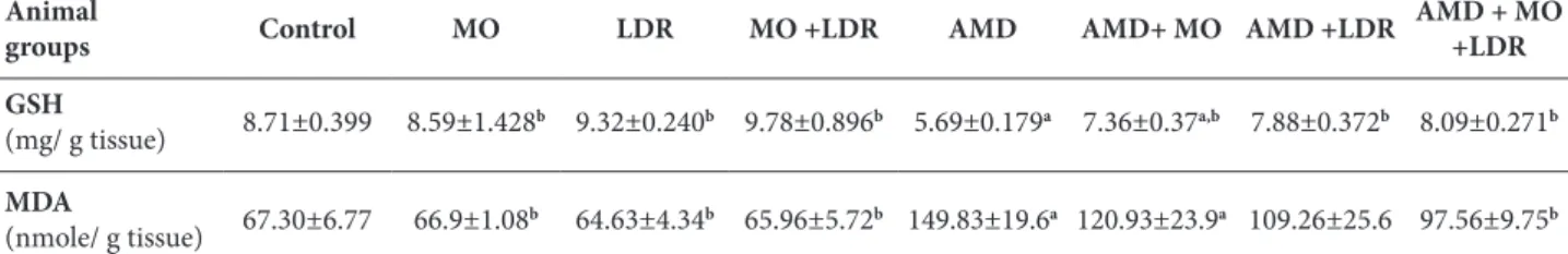
Table 1. GSH and MDA contents in different animal groups.
Animal
groups Control MO LDR MO +LDR AMD AMD+ MO AMD +LDR
AMD + MO +LDR
GSH
(mg/ g tissue) 8.71±0.399 8.59±1.428b 9.32±0.240b 9.78±0.896b 5.69±0.179a 7.36±0.37a,b 7.88±0.372b 8.09±0.271b
MDA
(nmole/ g tissue) 67.30±6.77 66.9±1.08b 64.63±4.34b 65.96±5.72b 149.83±19.6a 120.93±23.9a 109.26±25.6 97.56±9.75b
Data are expressed as means±SE (n=6). P<0.001; a: significant difference vs. control; b: significant difference vs control AMD treated group; Moringa oleifera (MO), low doses of gamma radiation (LDR), amiodarone (AMD), malondialdehyde (MDA), reduced glutathione (GSH)
Table 2. TNF-α and TGF-β levelsin different animal groups.
Animal
groups Control MO LDR MO+LDR AMD AMD+MO
AMD +LDR
AMD+ MO+LDR
TNFα (Pq/ml) 35.7±0.65 34.5±0.824b 33.7±0.72b 33.2±1.18b 57.36±1.94a 42.33±0.97a,b 39.1±1.008a,b 38.4±1.17b
TGF-β (Pq/ml) 26.5±0. 597 28.2±0. 91b 27.2±0.573b 25.7±0.901b 46.4±1.98a 35.03±0.842a,b 32.3±1.004a,b 29.03±0.98b
Data are expressed as means±SE (n=6). P < 0.001; a: significant difference vs control. b: significant difference vs control AMD treated group.
Table 3. LDH activity and HYP contents in different animal groups.
Animal
groups control MO LDR MO+LDR AMD AMD+MO
AMD +LDR
AMD+ MO+LDR
LDH
(U/L) 290.5±53.1 286.2±51.6b 250.2±28.02b 238.8±11.3b 398.5±60.5a 301.3±40.6b 293.06±41.1b 290.4±40.6b
HYP
(µg/ gm) tissue) 156.6±12.07 150.01±12.7b 145.89±15.2b 143.07±13.9b 259.2±21.9a 219.4±12.3a,b 209.59±9.1a,b 200.59±14.69a,b
current study, the administration of AMD to rats in-duced a marked elevation in the inflammatory cy-tokines (TNF-α and TGF-β), lactate dehydrogenase (LDH), malondialdehyde (MDA) and hydroxyproline (HYP) associated with a significant decrease in re-duced glutathione (GSH) as compared to the control rats (Tables 1-3).
These alterations could be attributed to AMD-induced pulmonary toxicity. Several conjunct mech-anisms involving the accumulation of iodine-rich AMD into pneumocytes have been suggested for this action of AMD. The potential mechanisms involved in the direct toxic effect that disrupts cellular mem-branes are through the activation of protein kinase C, release of toxic reactive oxygen species (ROS), de-creases in GSH, mitochondrial dysfunction, necrosis and eventually apoptosis (Mali et al., 2014).
AMD has been reported as an enhancing agent in free radical generation and also in mitochondrial hydrogen peroxide production (Chakraborty et al., 2014). It could be metabolized to an aryl radical that may give rise to other ROS (Nicolescu et al., 2007).
The powerful oxidizing capability of ROS can lead to the generation of advanced oxidation molecular products and induce damage to cellular and subcel-lular structures within the lung (Kinnula et al., 2005). Lipid peroxidation (LPO), measured by MDA, is one of the adverse effects of AMD therapy that occurs due to free radical-mediated chain oxidation result-ing in damage to the pulmonary endothelium. The released free radicals steal electrons from lipids of the cell membrane, leading to degradation of lipids and increased MDA content, which indicates intoxication and the generation of oxidative stress in lung tissue (Kaushik et al., 2001, Chakraborty et al., 2014). Oxi-dative stress, an imbalance between free radicals and the antioxidant defense system, is an important factor in the pathogenesis system involving polyunsaturated membrane lipid (Acar et al., 2012). The antioxidant GSH, which plays an important role in a variety of detoxification processes, was decreased significantly in AMD-treated rats, suggesting the role of oxidative stress mechanisms in AMD-induced lung toxicity (Gado and Aldahmash, 2013).
Fig. 1. Tissue micrographs lung tissues. A – Lung of rat from control group showing no histopathological changes (-). B – Lung of rat from MO group showing no histopathological changes. C – Lung of rat from LDR group showing no histopathological changes (-). D – Lung of rat from MO+LDR group showing no histopathological changes (-). All plates: (H&E, X 400). Degree of change from normal histological structure: +++ severe, ++ moderate, + mild, - nil.
Another mechanism for pulmonary toxicity by AMD is the immune-mediated mechanism involving an imbalance in helper T cells and overproduction of cytokines (Mali et al., 2014). The reported signifi-cant increase in serum TGF-β and TNF-α (Table 2) after AMD administration has been shown to be the major etiological factor in pulmonary injury (Pu-nithavathi et al., 2003). Free radicals can stimulate the expression and secretion of TGF-β and medi-ate TGF-β-induced differentiation of fibroblasts and myofibroblasts (Cucoranu et al., 2005). Also, the al-veolar macrophages isolated from AMD-treated rats released large amounts of TNF-α (Punithavathi et al., 2003). Moreover, the significant increase in HYP content (Table 3) after AMD administration could be attributed to the development of oxidative stress and increased inflammatory cytokines (TNF-α and TGF-β) (Table 1 and 2). The formation of HYP resi-dues is essential for triple helix formation and stabi-lization of collagen and its expression (Nokelainen et al., 2001, Stephens and Grande-Allen, 2007). Free radicals can stimulate platelet-derived growth factor receptors, which in turn results in increased synthesis of extracellular matrix and collagen production via the Ras/Erk pathway (Hecker et al., 2009). Further-more, TGF-β could stimulate collagen synthesis by inducing the differentiation of fibroblasts into my-ofibroblasts; it also lowers collagenase activity (Lasky and Brody, 2000). The accumulation of TNF-α might boost collagen synthesis via the modulation of fibrob-last functions (Punithavathi et al., 2003).
In addition, AMD-induced lung injury could be contributed to by the observed increase in serum lactate dehydrogenase (LDH) activity (Table 3). The extracellular appearance of LDH is used to detect cell damage or death and is abnormal in a host of dis-orders. The total serum LDH activity is elevated in several pulmonary disorders associated with fibrosis, and has been suggested to be a useful monitor of dis-ease activity (Cobben et al., 1997). Punithavathi et al. (2003) demonstrated that biochemical alterations such as increases in bronchoalveolar fluid (BALF) to-tal protein (a marker of damage to the alveolar-cap-illary barrier) and LDH activity were associated with lung injury induced after the administration of AMD,
which induced oxidative stress that led to impaired cell viability, also exerting toxic effects manifested by a decrease in GSH level and increase in LDH leakage and MDA (Krasteva et al., 2007).
The histological examination of AMD-treated rats’ lung sections revealed tissue damage represent-ed by focal interstitial pneumonia (Fig. 2E). AMD induced direct cellular damage, induction of phos-pholipidosis and immune-mediated mechanisms by activation of natural killer cell activity (Taylor et al., 2003). Madkour and Ahmed (2013) reported that AMD provoked histological changes in the lung tissue characterized by a thickening of interalveolar septa, cellular infiltration, vacuolar degeneration, conges-tion, inflammatory infiltration and focal necrosis.
Earlier studies reported that coagulation abnor-malities with oxidative metabolic reactions in lung injury are compelling reasons to use antioxidants in suppressing AMD-induced pulmonary toxicity (Gado and Aldahmash, 2013).
The experimental results revealed that rats receiv-ing oral administration of the MO methanolic extract and/or exposure to low doses of gamma irradiation showed no significant changes in inflammatory cy-tokines (TNF-α and TGF-β), oxidative indices (MDA and GSH), HYP lung content and LDH activity as compared to control rats. Also, the histological ex-amination of lung tissue revealed no alterations in the tissue architecture of rats treated with MO and/or LDR (Tables 1-3 and Fig. 1). Furthermore, the admin-istration of MO and/or exposure to LDR of AMD-treated rats led to a significant improvement in the biochemistry and histology of lung tissue (Tables 1-3 and Fig. 1).
cer-ebroprotective effect of MO against ischemic stroke to the antioxidant activity and decreased oxidative stress. Also, the antioxidant enzyme activities and GSH content increased significantly after exposure to whole-body LDR (0.5 Gy). Kojima et al. (2004) and Fahmy et al. (2013) specified the increased total GSH in liver, pancreas and brain.
The decrease in TNF-α and TGF-β levels could be attributed to the antioxidant activities of MO (which was responsible for the decrease in the levels of free radicals in the lungs), and it can inhibit the inflam-matory response manifested by decrease in TNF-α and TGF-β activation pathways. The immunomod-ulatory activity of MO could be exerted through downregulation and reduction of TNF-α expression (Oyewo et al., 2013b; Gupta et al., 2013) and inhibi-tion of nuclear factor kappa-B (NF-κB) (Wihastuti et al., 2007). Also, Kooltheat et al. (2014) illustrated that MO ethyl acetate fraction inhibited inflammatory cy-tokine including TNF-α production in the lungs of rats exposed to cigarette smoke. The improvement in serum TGF-β could be attributed to the MO-me-diated inhibition of TGF-β phosphorylation, which regulates the expression of fibronectin, type I colla-gen and plasminocolla-gen activator inhibitor-1 (Su-Hyun and Young-Chae, 2012). Furthermore, the significant decrease in serum TGF-β and TNF-α levels might be due to the various effects of LDR, including radio-adaptive response, modulation of immune function and an enhancement of resistance to high doses of radiation. These phenomena have generally been called radiation hormesis (Kojima et al., 2004). The obtained results coincide with the results obtained by Yu et al. (2013), who elucidated the pneumoprotec-tive efficiency of LDR (75 mGy) against bleomycin-induced lung injury through a reduction of TNF-α and TGF-β levels in the BALF. Also, the decrease in lung cell content of HYP (Table 3) in AMD rats treated with MO and/or LDR can be interpreted in the view of antioxidant and anti-inflammatory prop-erties of MO and LDR observed in the present study (Table 1-2). These results are in agreement with those obtained by Hamza (2010), who reported that MO decreased HYP content and collagen deposition in the liver of injured rats.
MO and/or LDR could afford protection to lung cells through the decreased production of free radi-cals, as was evident from the apparent antioxidant capacity and decreased LPO in the lungs of AMD-treated rats (Table 1). Babu et al. (2011) stated that MO has membrane-stabilizing activity. Thus, the amelioration of serum LDH activity (Table 3) could be due to the antioxidant capacity of MO and/or LDR, which stabilizes the membrane against the ROS re-leased and subsequently opposes the efflux of LDH enzymes into the blood stream from tissue.
The histological examination of lung tissue of AMD rats treated with MO showed a marked amel-ioration of the lung damage as depicted by preserved lung tissue with subtle perivascular inflammatory cell infiltration and slight thickening of interstitial tissue (Figs. 2 F and H). These results are in agreement with those obtained by Owolabi et al. (2013), who reported that the alcoholic extract of MO possessed prophy-lactic and regenerative effects against lung toxicity. This effect is mediated through its inhibitory effect on the expression of RelA, a gene implicated in NF-κβ p65 signaling, as well as reduction of inflammatory cytokines, which promotes neutrophil infiltration and lung tissue damage (Kooltheat et al., 2014). In the current experiment, LDR exhibited lung protection against AMD-induced damage as indicated by the re-duced degree of infiltrated inflammatory cells, and it exerted superior protection when combined with MO by retaining the normal architecture of the lung tissue (Figs. 2 G and H). The afforded protective effect of LDR is a result of enhanced endogenous antioxidant activities with special emphasis on GSH, which coun-teract excessive formation of ROS (Kawakita et al., 2003, Fahmy and Gharib, 2014). In the same context, Yu et al. (2013) ascribed the preventive efficiency of LDR against lung injury to a reduction in TNF-α and TGF-β levels in the BALF. It could be postulated that MO and/or LDR reversed AMD-induced lung toxic-ity via an adjustment of cellular redox tone and in-hibition of certain inflammatory response pathways.
Veterinary Medicine, Cairo University) for his examination of lung sections in the current study.
Authors’ contributions: All authors participated equally in this work.
Conflict of interest disclosure: The authors report no declaration of interest. The authors alone are responsible for the content and writing of the paper.
REFERENCES
Acar, N., Berdeaux, O., Grégoire, S., Cabaret, S., Martine, L., Gain, P., Thuret, G., Creuzot- Garcher, C.P., Bron, A.M. and L. Bretillon (2012). Lipid composition of human eye: Are red blood cells a good mirror of retinal and optic nerve fatty acid? PLoS ONE. 7, e35102.
Adedapo, A.A., Mogbojuri, O.M. and B.O.Emikpe (2009). Safety evaluations of the aqueous extract of the leaves of Moringa oleifera in rats. J. Med. Plants Res. 3, 586-591.
Babu, B.P., Kumar, P.R., Bharavi, K., Venkateswarlu, U., Devi, V.R.
and C. Srilatha (2011). Protective effect of Moringa olief-era lam leaf extract in paracetamol induced hepatotoxic rat model. J. Pharmacol. Toxicol. 1, 24-34.
Bergman, I. and R.Loxley (1963). Two improved and simplified methods for the spectrophotometric determination of hydroxyproline. Anal. Chem. 35, 1961–1965.
Berkovich, L., Earon, G., Ron, I., Rimmon, A., Vexler, A. and S. Lev-Ari (2013). MoringaOleifera aqueous leaf extract down-regulates nuclear factor-kappaB and increases cyto-toxic effect of chemotherapy in pancreatic cancer cells.
BMC Complem. Altern. Med. 13, 212.
Chakraborty, A., Mondal, C., Sinha, S., Mandal, J. and A.K. Chan-dra (2014). Amiodarone induced oxidative stress in stress vulnerable organs of adult male rats. Asian J. Pharm. Clin. Res. 7,177-183.
Cobben, N.A.M., Drent, M., Schols, A.M.W. J, Lamers, R.J. S., Wouters, E.F.M. and M.P.Van Dieijen-Visser (1997). Serum lactate dehydrogenase and its isoenzyme pattern in ex-coalminers. Respir. Med. 91, 616-623.
Corti, A., Fassino, J., Marcucci, F., Barbanti, E. and G.Cassani
(1992). Oligometric tumor necrosis factor slowly converts into the reactive forms at bioactive levels. Biochem. J. 284, 905-910.
Cucoranu, I., Clempus, R., Dikalova, A., Phelan, P.J., Ariyan, S., Dikalov, S. and D.Sorescu (2005). NAD (P) H oxidase 4 mediates transforming growth factor-β 1-induced differ-entiation of cardiac fibroblasts into myofibroblasts. Circul. Res. 97, 900-907.
Ellman, G.L. (1959). Tissue sulfhydryl groups. Arch. Biochem. Biophys. 82, 70-77.
Fahmy, H.A., Abd El-Azime, A.S. and O.A. Gharib (2013). Possible ameliorative role of low dose radiation against cisplatin
induced oxidative stress and tissue damage in male rats.
Euro. J. Biol. and Med. Sci. Res. 1, 10-18.
Fahmy, H.A. and O.A.Gharib (2014). Effect of low radiation dose on cisplatin induced hepato- testicular damage in male rats.
Br. J. Pharm. Res. 4, 1053- 1066.
Farooque, A., Mathur, R., Verma, A., Kaul, V., Bhatt, A.N., Adhi-kari, J.S., Afrin, F., Singh, S. and B.S.Dwarakanath (2011). Low-dose radiation therapy of cancer: role of immune enhancement Expert Rev. Anticancer Ther. 11, 791-802.
Gado, A.M. and B.A.Aldahmash, 2013). Protective Effect of L-Carnitine against Amiodarone-Induced Lung Toxicity in Rats. Internet. Toxicol. 10, 1.
Gunjal, M.A., Shah, A.S., Wakade, A.S. and A.R.Juvekar (2010). Protective effect of aqueous extract of Moringaoleifera
Lam. stem bark on serum lipids, marker enzymes and heart antioxidants parameters in isoproterenol-induced cardiotoxicity in Wistar rats. Indian J. Nat. Prod. Resour.
1, 485-492.
Gupta, S.K., Kumar, B., Srinivasan, B.P., Nag, T.C., Srivastava, S., Saxena, R. and A.Aggarwal (2013). Retinoprotective effects of Moringaoleifera via antioxidant, anti-inflammatory, and anti-angiogenic mechanisms in streptozotocin-induced diabetic rats. J Occul. Pharmacol. Ther. 29, 419-426.
Hamza, A.A. (2010). Ameliorative effects of Moringaoleifera Lam seed extract on liver fibrosis in rats. Food Chem. Toxicol.
48, 345-355.
Hecker, L., Vittal, R., Jones, T., Jagirdar, R., Luckhardt, T.R., Horow-itz, J.C., Pennathur, S., Martinez, F.J. and V.J. Thannickal
(2009). NADPH oxidase-4 mediates myofibroblast acti-vation and fibrogenic responses to lung injury. Nat. Med.
15,1077-1081.
Jamshidzadeh, A., Baghban, M., Azarpira, N., Bardbori, A.M. and
H.Niknahad (2008). Effects of tomato extract on oxida-tive stress induced toxicity in different organs of rats. Food Chem. Toxicol. 46, 3612-3615.
Kaushik, S., Hussain, A., Clarke, P. and H.L.Lazar (2001). Acute pulmonary toxicity after low-dose amiodarone therapy.
Ann. Thorac. Surg. 72, 1760-1761.
Kawakita, Y., Ikekita, M., Kurozumi, R. and S.Kojima (2003). Increase of intra cellular glutathione by low dose gamma-ray irradiation is mediated by transcription factor API in RAW 264.7. Cell Biol. Pharm. Bull. 26, 19-25.
Kim, S.j., Romeo, D., Yoo, Y.d. and K.Park (1994). Transforming growth factor-beta; Expression in normal and pathological conditions. Horm. Res. 42, 5-8.
Kinnula, V.L., Fattman, C.L., Tan, R.J. and T.D.Oury (2005). Oxidative stress in pulmonary fibrosis: a possible role for redox modulatory therapy. Am. J. Respir. Crit. Care Med.
172, 417-422.
Kojima, S., Nakayama, K. and H. Ishida (2004). Low dose γ-rays activate immune functions via induction of glutathione and delay tumor growth. J. Radiat. Res. 45, 33-39.
Kooltheat, N., Sranujit, R.P., Chumark, P., Potup, P., Laytragoon-Lewin, N. and K.Usuwanthim (2014). An ethyl acetate frac-tion of Moringaoleifera Lam. inhibits human macrophage cytokine production induced by cigarette smoke. Nutrien.
6, 697-710.
Krasteva, A.Z., Mitcheva, M.K., Kondeva-Burdina, M.S. and V.A. Descatoire (2007). In vitro study of lovastatin interactions with amiodarone and with carbon tetrachloride in isolated rat hepatocytes. World J. Gastroenterol. 13, 2198-2204.
Lasky, J.A. and A.R. Brody (2000). Interstitial fibrosis and growth factors. Environ. Health Perspect. 108, 751-762.
Luckey, T.D. (1982). Physiological benefits from small levels of ionizing radiation. Health Phys. 43, 771-789.
Madkour, N.K. and M. Ahmed (2013). Amelioration of amioda-rone-induced lung fibrosis by grape seed extract. J. Appl. Sci. Res. 9, 3698-3707.
Mali, P., Salzman, M.M.H., Vidaillet, H.J. and S.H.Rezkalla
(2014). Amiodarone therapy for cardiac arrhythmias: Is it associated with the development of cancers? World J. Car-diovasc. Dis. 4, 109-118.
Nanjappaiah, H.M. and S. Hugar (2012). Prophylactic and cura-tive effects of Moringaoleifera Lam. pods in CCL4 dam-aged rat liver. Indian J. Nat. Prod. Resour. 3, 541-546.
Nicolescu, A.C., Comeau, J.L., Hill, B.C., Bedard, L.L., Takahashi, T., Brien, J.F., Racz, W.J. and T.E.Massey (2007). Aryl radi-cal involvement in amiodarone-induced pulmonary toxic-ity: investigation of protection by spin trapping nitrones.
Toxicol. Appl. Pharmacol. 220, 60-71.
Nokelainen, M., Nissi, R., Kukkola, L., Helaakoski, T. and J. Myl-lyharju (2001). Characterization of the human and mouse genes for the alpha subunit of type II prolyl 4-hydroxylase: identification of a previously unknown alternatively spliced exon and its expression in various tissues. Eur. J. Biochem.
268, 5300-5309.
Owolabi, J., Ghazal, O., Dare, M., Olanrewaju, J., Caxton-Martins, A. and F. William (2013). Prophylactic and regenerative effects of alcoholic extract of Moringaoleifera on rat lung tissue following lead-induced damage. Eur. J. Anat. 17, 115-122.
Oyewo, E.B., Adeleke, E.G., Fakunle, B.P. and M.O. Iniaghe
(2013a). Blood glucose and lipid reducing activities of the oral administration of aqueous leaf extract of Moringa oleif-era in Wistar rats. J. Nat. Sci. Res. 3, 92-99.
Oyewo, E.B., Adetutu, A., Ayoade, A., Adesokan, A. and M.A. Kanji (2013b). Repeated oral administration of aqueous leaf extract of Moringaoleifera modulated immunoactivi-ties in Wistar rats. J. Nat. Sci. Res. 3, 100-109.
Punithavathi, D., Venkatesan, N. and M.Babu (2003). Protective effects of curcumin against amiodarone-induced pulmo-nary fibrosis in rats. Br. J. Pharmacol. 139, 1342-1350.
Rakesh, S. and V.J.Singh (2010). In vivo antioxidant activity of
Moringaoleifera leaf and pod extracts against carbon tetra chloride induced liver damage in albino mice. J. Chem. Pharm. Res. 2, 275-283.
Sinha, M., Das, D.K., Datta, S., Ghosh, S. and S.Dey (2012). Ame-lioration of ionizing radiation induced lipid peroxidation in mouse liver by Moringaoleifera Lam. leaf extract. Indian Ј. exp. Biol. 50, 209-215.
Smith, A.C. and M.R. Boyd (1983). Drug-induced pulmonary toxicity. Trends Pharmacol. Sci. 4, 275-278.
Stephens, E.H. and K.J. Grande-Allen (2007). Age-related changes in collagen synthesis and turnover in porcine heart valves.
J. Heart Valve Dis. 16, 672-682.
Stevens, A., Bancroft, J. and A.Stevens (1982) Theory and practice of histological Techniques, 2nd ed. Churchill Livingstone,
London.
Su-Hyun, P. and C.Young-Chae (2012). Anti-Fibrotic effects by
Moringa root extract in rat kidney Fibroblast. J. Life Sci.
22, 1371-1377.
Taylor, M., Antonini, J., Roberts, J., Leonard, S., Shi, X., Gannett, P., Hubbs, A. and M.Reasor (2003). Intratracheal amio-darone administration to F344 rats directly damages lung airway and parenchymal cells. Toxicol. Appl. Pharmacol.
188, 92-103.
Vassault, A. (1983). Lactate dehydrogenase. UV-method with pyruvate and NADH. In: Methods of enzymatic analysis. (Eds. J. Bergmeyer , and M. Grabl). Verlag-Chemie, Deer-field Beach, Florida.
Wihastuti, T.A., Sargowo, D. and M.S.Rohman (2007). The Effect of Moringa Oleifera Leaf Extract in Inhibition of NFκB Activation, TNF-α and ICAM-1 Expression in Oxidized LDL treated HUVECS. J. Cardiol. Indonesia.28, 181-188.
Yoshioka, T., Kawada, K., Shimada, T. and M.Mori (1979). Lipid peroxidation in maternal and cord blood and protective mechanism against activated-oxygen toxicity in the blood.
Am. J. Obstet. Gynecol. 135, 372-376.