Article
A Novel Cytosolic Isoform of Mitochondrial
Trans-2-Enoyl-CoA Reductase Enhances Peroxisome
Proliferator-Activated Receptor α Activity
Dong-Gyu Kim1, Jae Cheal Yoo1, Eunju Kim2, Young-Sun Lee1, Oleg V. Yarishkin2, Da Yong Lee2, Kun Ho Lee3, Seong-Geun Hong1, Eun Mi Hwang2, Jae-Yong Park1,2
1Department of Physiology, Institute of Health Science, Medical Research Center for Neural Dysfunction, Gyeongsang National University School of Medicine, Jinju; 2
Center for Functional Connectomics, Korea Institute of Science and Technology, Seoul; 3
Department of Marine Life Science, Chosun University College of Natural Sciences, Gwangju, Korea
Background: Mitochondrial trans-2-enoyl-CoA reductase (MECR) is involved in mitochondrial synthesis of fatty acids and is highly expressed in mitochondria. MECR is also known as nuclear receptor binding factor-1, which was originally reported with yeast two-hybrid screening as a binding protein of the nuclear hormone receptor peroxisome proliferator-activated receptor α (PPARα). However, MECR and PPARα are localized at different compartment, mitochondria, and the nucleus, respectively. There-fore, the presence of a cytosolic or nuclear isoform of MECR is necessary for functional interaction between MECR and PPARα.
Methods: To identify the expression pattern of MECR and the cytosolic form of MECR (cMECR), we performed reverse tran-scription polymerase chain reaction (RT-PCR) with various tissue samples from Sprague-Dawley rats. To confirm the interaction between cMECR and PPARα, we performed several binding assays such as yeast two-hybrid, coimmunoprecipitation, and bimo-lecular fluorescence complementation. To observe subcellular localization of these proteins, immunocytochemistry was per-formed. A luciferase assay was used to measure PPARα activity.
Results: We provide evidence of an alternatively spliced variant of the rat MECR gene that yields cMECR. The cMECR lacks the N-terminal 76 amino acids of MECR and shows uniform distribution in the cytoplasm and nucleus of HeLa cells. cMECR di-rectly bound PPARα in the nucleus and increased PPARα-dependent luciferase activity in HeLa cells.
Conclusion: We found the cytosolic form of MECR (cMECR) was expressed in the cytosolic and/or nuclear region, directly binds with PPARα, and enhances PPARα activity.
Keywords: Trans-2-enoyl-CoA reductase (NADPH); Cytosolic mitochondrial trans-2-enoyl-CoA reductase; PPAR alpha; Alter-native splicing; Mitochondrial targeting sequences
INTRODUCTION
Mammalian mitochondria perform nicotinamide adenine
di-nucleotide phosphate (NADPH)-dependent de novo fatty acid
synthesis (FAS) [1]. This mitochondrial FAS pathway resem-bles the well-understood bacterial FAS II pathway [2].
Mito-Received: 13 August 2013, Accepted: 11 October 2013 Corresponding author: Jae-Yong Park
Department of Physiology, Gyeongsang National University School of Medicine, 15 Jinju-daero 816beon-gil, Jinju 660-751, Korea
Tel: +82-55-772-8043, Fax: +82-55-772-8049, E-mail: jaeyong@gnu.ac.kr
Copyright © 2014 Korean Endocrine Society
chondrial trans-2-enoyl-CoA reductase (MECR) is a compo-nent of the mitochondrial FAS II pathway and catalyzes the fat-ty acid elongation cycle in the last step, which is the NADPH-dependent reduction of the enoyl-acyl carrier protein substrate [3-5]. MECR is primarily localized in mitochondria [6-8], and the N-terminal amino acids of this protein are important for its mitochondrial localization [9]. MECR is also known as nucle-ar receptor binding factor-1 (NRBF-1) because, using yeast two-hybrid screening, it was identified as a binding protein of the nuclear hormone receptor peroxisome proliferator-activat-ed receptor α (PPARα) [10]. However, the biological meaning of the interaction between MECR and PPARα has not been determined.
PPARα is a ligand-activated transcription factor that is one of three different PPAR subtypes: PPARα, PPARβ/δ, and PPARγ. The PPARs play important roles in nutrient homeostasis [11-13] and are localized in the nucleus. Although MECR was previous-ly reported as a binding protein of PPARα [10], interaction be-tween MECR and PPARα seems not to occur in mammalian cells due to their different subcellular localizations, mitochon-dria and the nucleus, respectively. Therefore, the presence of a cytosolic or nuclear isoform of MECR is necessary for function-al interaction between MECR and PPARα in the nuclei of cells. Here, we analyzed the expression pattern of MECR in sev-eral rat tissues and found a novel splice variant of MECR in which an additional exon was inserted between exon 1 and exon 2. The protein generated from this splicing variant has an N-terminal region that does not contain the mitochondrial tar-geting signal peptide and thus is not localized in mitochondria. Moreover, this MECR variant bound PPARα in the nucleus and enhanced PPARα transcriptional activity. Based on these results, we propose that this novel variant of MECR, cytosolic MECR (cMECR), plays a role in intracellular signal pathways as an interacting partner of PPARα.
METHODS
RNA extraction and reverse transcription polymerase chain reaction
Total RNA was extracted from tissues of male Sprague-Daw-ley rats (14 weeks old) using TRIzol reagent (Invitrogen, Carlsbad, CA, USA) according to the manufacturer’s protocol. Total RNA (1 μg) from each sample was reverse transcribed using random primers (50 pmol), SuperScript III Reverse Transcriptase (Invitrogen), and dNTPs (1 mM) at 42°C for 1 hour. The forward primer
(5′-ATGTTGGTCAGCCGGC-GACT-3′) and reverse primer (5′-TCACATAGTGAGAATCT-GCT-3′) were designed to amplify full-length MECR. Addi-tional primers were used for specific detection of MECR and cMECR cDNA: forward primer (5′-GTGCTGGAAGCG-GCATGTTG-3′) and reverse primer (5′-TGAGCTCCAG-GTTCTTCAGT-3′). cMECR and PPARα fragments were am-plified under the following cycle conditions: denaturation at 94°C for 30 seconds, annealing at 55°C for 30 seconds, and extension at 72°C for 30 seconds. This cycle was repeated 35 times. Glyceraldehyde 3-phosphate dehydrogenase fragments were also amplified under the same conditions except that 25 cycles were run. Polymerase chain reaction (PCR) products were analyzed with 2.0% agarose gel electrophoresis, purified, and ligated into the pGEM-T Easy vector (Promega, Madison, WI, USA). The recombinant plasmids were sequenced.
Construction of plasmids and antibodies
The rat full-length MECR (GenBank accession no. AB015724), cytosolic variant of MECR, and PPARα (GenBank accession no. NM_001001928) were cloned using the Gateway Cloning System (Invitrogen) as previously described [14]. After ampli-fication of the three genes, PCR products were cloned into the pDONR207 vector and then subcloned into self-constructed destination vectors such as pDS_XB-HA, pDs_XB-Flag, pDS_ XB-enhanced green fluorescent protein (EGFP), and pDS_XB-mCherry expression vectors. Anti-Flag (F6531) antibody was purchased from Sigma (St. Louis, MO, USA). Anti-EGFP (B-2) and antihemagglutinin (anti-HA) (F-7) antibodies were ob-tained from Santa Cruz Biotechnology (Santa Cruz, CA, USA).
Cell culture and transfection
HeLa cells were maintained in Dulbecco’s modified Eagle’s medium supplemented with 10% fetal bovine serum (Invitro-gen) and 1% penicillin-streptomycin in a humid atmosphere containing 5% CO2 at 37°C. When needed, cells were seeded onto cover slips for imaging analysis or 60-mm dishes for preparation of lysates for Western blot analysis. Transfection of expression vectors was performed with lipofectamine 2000 reagent (Invitrogen) according to the manufacturer’s protocol. Transfected cells were cultured for an additional 24 hours in growth medium and then used for further analysis.
Coimmunoprecipitation and Western blot analysis
Transfected cells were lysed with radioimmunoprecipitation assay buffer (50 mM Tris-HCl, pH 7.4, 150 mM NaCl, 5 mM
phenylmethanesulfo-nyl fluoride, and 1% NP-40) containing a protease inhibitor
cocktail (Sigma). Whole-cell lysates were incubated on ice for 30 minutes and then cleared at 13,000 rpm for 20 minutes at 4°C. The immune complexes were incubated for 1 hour at 4°C with gentle rotation with 20 μL protein A/G PLUS-agarose beads (Santa Cruz Biotechnology) that had been prewashed and suspended in 100 mL cold lysis buffer. The samples were washed three times in 1 mL cold lysis buffer and eluted in 20 μL sodium dodecyl sulfate polyacrylamide gel electrophoresis (SDS-PAGE) sample buffer. The proteins were separated with 10% SDS-PAGE and blotted onto polyvinylidene fluoride membranes. The blots were blocked with 5% skim milk in Tris-buffered saline with Tween 20 (20 mM Tris-buffered sa-line and 0.05% Tween 20, pH 7.5) at room temperature for 20 minutes and incubated overnight at 4°C with GFP anti-body (1:1,000), anti-HA antianti-body (1:1,000), or anti-FLAG an-tibody (1:1,000). Blots were then washed and incubated with horseradish peroxidase-conjugated secondary antibody (1: 3,000), followed by washing and detection of immunoreactiv-ity with enhanced chemiluminescence (Amersham, Piscat-away, NJ, USA).
Imaging analysis
Subcellular distribution of MECR, cMECR, and PPARα were confirmed according to the EGFP and mCherry fluorescence detection method with a confocal microscope (Olympus Flu- oview FV1000, Olympus, Tokyo, Japan). For imaging analysis, cells were plated onto glass cover slips. After transfection, the cells were grown for 24 hours and treated with the mitochon-drion-selective fluorescent dye mito-tracker red CMXRos (Mo- lecular Probes Europe BV, Leiden, Netherlands) according to the manufacturer’s instructions.
Yeast two-hybrid assay
cMECR was cloned into pGADT7 encoding the activation do-main, and PPARα was ligated into pGBKT7 encoding the GAL4 DNA binding domain. To evaluate the protein-protein interaction between cMECR and PPARα, both pGAD-cMECR and pGBK-PPARα were cotransformed into the yeast strain AH109. This strain is unable to synthesize histidine. However, interaction between cMECR and PPARα enables the yeast to make the His3 enzyme, thereby permitting histidine biosyn-thesis and growth on His minimal medium.
Bimolecular fluorescence complementation assays
C- and N-terminal venus plasmids were purchased from
Add-gene and modified to contain cMECR or PPARα with sub-cloning. Cells were transfected with the lipofectamine 2000 reagent (Invitrogen) with venus plasmids containing cMECR and PPARα. After 24 hours, cells were imaged with confocal microscopy as described above.
Luciferase assay
HeLa cells were plated at a density of 1.0 to 1.5×10⁵
cells per well onto six-well dishes. Twenty-four hours later, cells were cotransfected with 1 μg of PPAR responsive element (PPRE)-3x-TK-Luc, the firefly luciferase reporter gene containing PPRE; 0.2 μg of the PPARα expression vector; and either the MECR expression vector or cMECR expression vector. Trans-fections were accomplished with lipofectamine 2,000 reagent according to the manufacturer’s recommendations. The PPRE-3x-TK-Luc plasmid includes three PPREs upstream of an in-ducible thymidine kinase promoter controlling transcription of the firefly luciferase gene. Cells were harvested 24 hours after the start of exposure to vehicle or inducing agent. Luciferase assays were performed using the Luciferase Assay System (Promega) according to the manufacturer’s protocol.
RESULTS
Identification of a novel alternative transcript variant of MECR
MECR mRNA
cMECR
mRNA A
cMECR
cMECR
cMECR
cMECR
cMECR
cMECR MECR
MECR
MECR
MECR
MECR
MECR
B
GAPDH cMECR
MECR
Br He Ki Li Lu Pa Sp Th
C
Fig. 1. Identification of a novel variant of mitochondrial trans-2-enoyl-CoA reductase (MECR; cytosolic form of MECR [cMECR]) cDNA. (A) The genomic structure of MECR and cMECR and their coding regions are shown. Inverted triangles indicate the start and stop codons. The dotted-line box indicates the inserted nucleotide sequence. (B) Alignment of the MECR and cMECR sequences. The posi-tions of the upper and lower primers are marked with arrows. (C) Expression patterns of MECR and cMECR in various tissues. The upper polymerase chain reaction band is cMECR, and the lower band is MECR. Glyceraldehyde 3-phosphate dehydrogenase (GAPDH) was used as a positive control. Br, brain; He, heart; Ki, kidney; Li, liver; Lu, lung; Pa, pancreas; Sp, spleen; Th, thymus. aEach start codon.
a
named it cMECR.
Next, to compare expression of cMECR and MECR, RT-PCR analyses were done using RNA samples from eight differ-ent tissues with a specific primer set as shown in Fig. 1B. As shown in Fig. 1C, cMECR (306 bp) and MECR (210 bp) were ubiquitously expressed in most tissues examined, although the expression level of cMECR was lower than that of MECR. Furthermore, we found amino acid sequences of human MECR isoform 2 (GenBank accession no. NP001019903.1) were well matched with that of rat cMECR (Supplementary Fig. 1B). These data raise the possibility that cMECR can be produced in other species.
cMECR localized in the cytoplasm and nucleus of HeLa cells
To visualize the intracellular distribution of MECR in HEK cells, we generated two constructs containing EGFP-tagged full-length rat MECR. As shown in Supplementary Fig. 2, when EGFP was tagged to the C-terminal end of MECR (MECR-EGFP), this protein was distinctly expressed in
mito-chondria, as previously reported [3]. However, the N-terminal EGFP-tagged MECR (EGFP-MECR) was diffusely expressed in the cytoplasm of HEK cells. N-terminal tagging of the pro-tein with EGFP might have disrupted the proper mitochondrial localization of MECR, perhaps because the N-terminal mito-chondrial targeting sequence of MECR was masked by the EGFP. These results suggest that N-terminal mitochondrial targeting sequences are critical for mitochondrial localization of MECR proteins.
Because cMECR lacks the N-terminal mitochondrial target-ing sequences, the intracellular distribution of this protein was unclear. The intracellular distribution of MECR and cMECR was thus compared by tagging both cDNAs with EGFP at their C-terminal ends and then transiently expressing them in HeLa cells (Fig. 2). MECR-EGFP showed a well-defined punctate pattern that clearly colocalized with mito tracker, a marker of mitochondria (Fig. 2B). However, cMECR-EGFP was expressed in cytosolic and nuclear regions and did not co-localize with Mito tracker (Fig. 2C).
A
B
SupplementaryFig. 1. (A) Nucleotide sequence showing exon/intron junctions for the generation of cytosolic form of mitochondrial trans-2-enoyl-CoA reductase (cMECR): all exon-intron boundaries conform to consensus splice-donor and splice-acceptor sites (GT-AG). (B) Amino acid alignment of rat cMECR with human MECR isoform 2: the amino acid sequences of both proteins were aligned using the Uniprot alignment program. Sequence identity is 87.2%. Asterisks mean same amino acid.
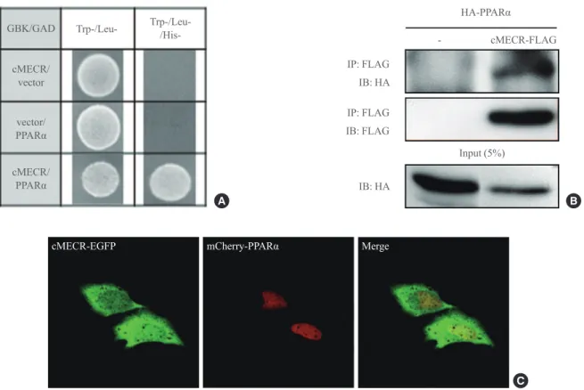
cMECR directly interacts with PPARα
MECR, also known as NRBF-1, was previously identified with yeast two-hybrid screening as a PPARα-interacting pro-tein [10], although explaining how mitochondrial-targeted MECR interacted with the nuclear hormone receptor PPARα was difficult. In contrast to MECR, cMECR was localized in the cytoplasm and nucleus of HeLa cells, and its amino acid sequence is exactly the same as that of MECR, except for the N-terminal 76 amino acids that compose the mitochondrial targeting sequence. Thus, cMECR, rather than mitochondrial MECR, could interact with PPARα in the nucleus. Indeed, the yeast two-hybrid assay showed a direct interaction between cMECR and PPARα (Fig. 3A). Immunoprecipitation and co-localization analyses also showed that cMECR interacted with PPARα in HeLa cells (Fig. 3B). Furthermore, cMECR-EGFP colocalized with mCherry-tagged PPARα in the nucleus of HeLa cells (Fig. 3C). In contrast, wild-type MECR was not colocalized with PPARα in HeLa cells (Supplementary Fig. 3). Taken together, these data strongly indicate that cMECR, but not MECR, is a bona fide binding partner of PPARα.
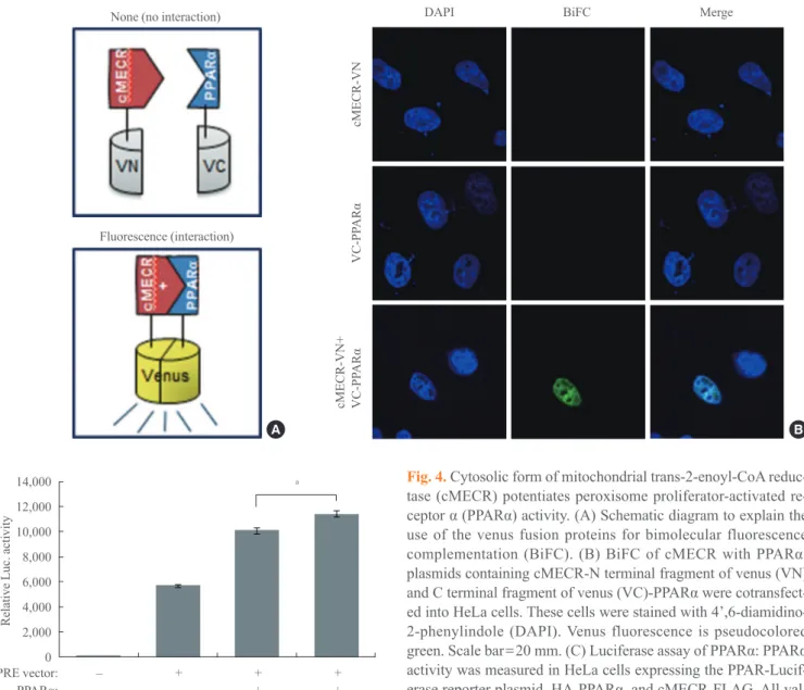
cMECR binds with PPARα in the nucleus and potentiates PPARα activity
To more clearly investigate whether the interaction between cMECR and PPARα can occur in the nucleus of HeLa cells, we performed a bimolecular fluorescence complementation (BiFC) experiment. The BiFC assay provides a direct ap-proach for the visualization of molecular interactions in living
MECR-EGFP
cMECR-EGFP
77 53 1
373 373
EGFP
EGFP
A
MECR-EGFP Mito tracker Merge
C
EGFP-MECR Mito tracker Merge
B
Fig. 2. Subcellular localization of mitochondrial
trans-2-enoyl-CoA reductase (MECR) and cytosolic form of MERC (cMECR) in HeLa cells. (A) The two constructs used in this study: MECR and cMECR were tagged at the C-terminus with enhanced green fluorescent protein (EGFP; green oval). The black box in MECR indicates the mitochondrial targeting domain. HeLa cells were transiently transfected with (B) MECR-EGFP or (C) cMECR-EGFP and then stained with Mito Tracker, a mitochondrial marker (red). Scale bar=20 μm.
EGFP-MECR MECR-EGFP
A
EGFP-MECR
MECR-EGFP EGFP
EGFP
B
EGFP-MECR Mito tracker Merge
MECR-EGFP Mito tracker Merge
Supplementary Fig. 2. (A) Schematic drawing of the effect of enhanced green fluorescent protein (EGFP) tags on the localization of
GBK/GAD
cMECR/ vector
vector/
PPARα
cMECR/
PPARα
Trp-/Leu- Trp-/Leu-
/His-A
HA-PPARα
Input (5%)
- cMECR-FLAG
IP: FLAG
IP: FLAG
IB: FLAG IB: HA
IB: HA
B
cMECR-EGFP mCherry-PPARα Merge
C
Fig. 3. Cytosolic form of mitochondrial trans-2-enoyl-CoA reductase (cMECR) directly interacts with peroxisome proliferator-activated receptor α (PPARα) in vitro. (A) Yeast two-hybrid assay between cMECR and PPARα: on a Trp−/Leu−/His− plate, colonies indicate an interaction between the two genes. (B) Coimmunoprecipitation assays with cMECR and PPARα: cMECR-FLAG was cotransfected with hemagglutinin (HA)-PPARα in HeLa cells. The lysates were immunoprecipitated with anti-FLAG antibody and then immunoblotted with anti-HAantibody. (C) Colocalization of cMECR and PPARα: HeLa cells were transiently transfected with cMECR-enhanced green fluorescent protein (EGFP; green) and mCherry-PPARα (red). Scale bar=20 μm.
calization analyses, the BiFC experiment also indicated pro-tein-protein interaction between cMECR and PPARα (Fig. 4B). Only when both constructs were cotransfected was the BiFC signal seen in the nucleus of HeLa cells. Individual
con-MECR-EGFP mCherry-PPARα Merge
Supplementary Fig. 3. Localization of mitochondrial trans-2-enoyl-CoA reductase (MECR) and peroxisome proliferator-activated re -ceptor α (PPARα): wild-type MECR is localized in cytosolic regions, whereas PPARα is localized in nuclear regions. HeLa cells were transiently transfected with MECR-enhanced green fluorescent protein (EGFP; green) and mCherry-PPARα (red). Scale bar=20 μm.
colo-DISCUSSION
MECR has been reported to be a mitochondrial protein [7] that catalyzes NADPH-dependent reduction of trans-2-enoyl thioesters to the corresponding saturated acyl thioesters [5]. However, NRBF-1 (also known as MECR) was originally identified as a binding protein of the nuclear receptor PPARα [10]. In that paper, the authors did not perform any assessment of endogenous binding in cells or report the biological mean-ing of this protein–protein interaction, although NRBF-1 binds with PPARα in the yeast two-hybrid system.
Here, we report a cMECR that is generated by alternative splicing. cMECR is an N-terminal-76-amino-acid truncated
14,000
12,000
10,000
8,000
6,000
4,000
2,000
0
Relative Luc. activity
PPRE vector: – + + +
– – + +
PPARα:
– – – + cMECR:
a
C
B
DAPI
cMECR-VN
VC-PP
ARα
cMECR-VN+ VC-PP
ARα
BiFC Merge
A
None (no interaction)
Fluorescence (interaction)
Fig. 4. Cytosolic form of mitochondrial trans-2-enoyl-CoA reduc-tase (cMECR) potentiates peroxisome proliferator-activated re-ceptor α (PPARα) activity. (A) Schematic diagram to explain the use of the venus fusion proteins for bimolecular fluorescence complementation (BiFC). (B) BiFC of cMECR with PPARα: plasmids containing cMECR-N terminal fragment of venus (VN) and C terminal fragment of venus (VC)-PPARα were cotransfect -ed into HeLa cells. These cells were stain-ed with 4’,6-diamidino-2-phenylindole (DAPI). Venus fluorescence is pseudocolored green. Scale bar=20 mm. (C) Luciferase assay of PPARα: PPARα
activity was measured in HeLa cells expressing the PPAR-Lucif-erase reporter plasmid, HA-PPARα, and cMECR-FLAG. All val -ues are mean±s.e.m. P values were obtained with Student’s t test.
aP<0.01.
structs (cMECR-VN or VC-PPARα) did not produce fluores-cence alone (Fig. 4B). These results imply that cMECR inter-acts with PPARα in the nucleus of HeLa cells.
isoform of MECR. Because mitochondrial targeting signal se-quences are contained between amino acids 1 and 53 of MECR [8], cMECR lacks this mitochondrial targeting signal sequence and was preferentially localizes to cytosolic and nuclear re-gions and was not present in mitochondria (Fig. 2C). In addi-tion, our data show that N-terminal EGFP-tagged MECR also lost its proper intracellular localization (Supplementary Fig. 2). Therefore, the N-terminal region of MECR is critical for mito-chondrial localization of the MECR protein.
Different intracellular distribution of cMECR strongly im-plies that its biological functions differ from the functions of MECR. In addition, cMECR mRNA was ubiquitously ex-pressed in eight tested tissues (Fig. 1C), implying that cMECR
could perform common essential functions in the various tis-sues. Although further studies should be performed to eluci-date the function(s) of cMECR, we present its first function in this study. Various nuclear receptors activate transcription of their target genes through interactions with coactivators or co-repressors [17,18]. Like other coactivators, cMECR directly bound PPARα in the nucleus and potentiated PPARα activity (Fig. 4).
Generally, PPARα is found in tissues where fatty acid me-tabolism is important and regulates genes involved in lipid and lipoprotein metabolism as well as glucose homeostasis [19-21]. Because cMECR potentiates PPARα activity in HeLa cells, cMECR could be involved in these biological phenome-na. An earlier study used the yeast two-hybrid assay and dem-onstrated that MECR (NRBF-1) also interacts with various nuclear hormone receptors such as TRβ, RARα, RXRα, and HNF-4 [10]. Because our data strongly support the idea that cMECR is a bona fide binding partner of PPARα and its amino acid sequence is the same as that of MECR except that it lacks the N-terminal 76 amino acids, cMECR might also bind with other nuclear hormone receptors. This possibility should be tested in future studies.
In summary, our results suggest that cMECR, a novel alter-natively spliced variant of MECR, directly interacts with PPARα in the nucleus and could be a positive regulator in the physiological regulation of PPARα.
CONFLICTS OF INTEREST
No potential conflict of interest relevant to this article was re-ported.
ACKNOWLEDGMENTS
This research was supported by the Basic Research Promotion Fund (NRF-2008-313-C00758).
REFERENCES
1. Chen Z, Leskinen H, Liimatta E, Sormunen RT,
Miinalain-en IJ, HassinMiinalain-en IE, HiltunMiinalain-en JK. Myocardial overexpres-sion of Mecr, a gene of mitochondrial FAS II leads to car-diac dysfunction in mouse. PLoS One 2009;4:e5589. 2. White SW, Zheng J, Zhang YM, Rock. The structural
biol-ogy of type II fatty acid biosynthesis. Annu Rev Biochem 2005;74:791-831.
3. Torkko JM, Koivuranta KT, Miinalainen IJ, Yagi AI, Schmitz W, Kastaniotis AJ, Airenne TT, Gurvitz A, Hil-tunen KJ. Candida tropicalis Etr1p and Saccharomyces cerevisiae Ybr026p (Mrf1’p), 2-enoyl thioester reductases essential for mitochondrial respiratory competence. Mol Cell Biol 2001;21:6243-53.
4. Chen ZJ, Pudas R, Sharma S, Smart OS, Juffer AH,
Hil-tunen JK, Wierenga RK, Haapalainen AM. Structural en-zymological studies of 2-enoyl thioester reductase of the human mitochondrial FAS II pathway: new insights into its substrate recognition properties. J Mol Biol 2008;379: 830-44.
5. Hoffmeister M, Piotrowski M, Nowitzki U, Martin W.
Mi-tochondrial trans-2-enoyl-CoA reductase of wax ester fer-mentation from Euglena gracilis defines a new family of enzymes involved in lipid synthesis. J Biol Chem 2005; 280:4329-38.
6. Pfanner N. Protein sorting: recognizing mitochondrial
pre-sequences. Curr Biol 2000;10:R412-5.
7. Emanuelsson O, Nielsen H, Brunak S, von Heijne G. Pre-dicting subcellular localization of proteins based on their N-terminal amino acid sequence. J Mol Biol 2000;300: 1005-16.
8. Claros MG, Vincens P. Computational method to predict
mitochondrially imported proteins and their targeting se-quences. Eur J Biochem 1996;241:779-86.
9. Chen JQ, Delannoy M, Cooke C, Yager JD. Mitochondrial localization of ERalpha and ERbeta in human MCF7 cells. Am J Physiol Endocrinol Metab 2004;286:E1011-22. 10. Masuda N, Yasumo H, Furusawa T, Tsukamoto T, Sadano
hor-fluorescent protein fragments for bimolecular fluorescence complementation analysis under physiological conditions. Biotechniques 2006;40:61-6.
17. Kallenberger BC, Love JD, Chatterjee VK, Schwabe JW. A
dynamic mechanism of nuclear receptor activation and its perturbation in a human disease. Nat Struct Biol 2003; 10:136-40.
18. McKenna NJ, Lanz RB, O’Malley BW. Nuclear receptor
coregulators: cellular and molecular biology. Endocr Rev 1999;20:321-44.
19. Plutzky J. The PPAR-RXR transcriptional complex in the
vasculature: energy in the balance. Circ Res 2011;108: 1002-16.
20. Ziouzenkova O, Plutzky J. Lipolytic PPAR activation: new
insights into the intersection of triglycerides and inflam-mation? Curr Opin Clin Nutr Metab Care 2004;7:369-75. 21. Han SH, Quon MJ, Koh KK. Beneficial vascular and
met-abolic effects of peroxisome proliferator-activated recep-tor-alpha activators. Hypertension 2005;46:1086-92. mone receptors. Gene 1998;221:225-33.
11. Desvergne B, Wahli W. Peroxisome proliferator-activated
receptors: nuclear control of metabolism. Endocr Rev 1999; 20:649-88.
12. Kersten S, Desvergne B, Wahli W. Roles of PPARs in
health and disease. Nature 2000;405:421-4.
13. Evans RM, Barish GD, Wang YX. PPARs and the complex
journey to obesity. Nat Med 2004;10:355-61.
14. Park JY, Hwang EM, Yarishkin O, Seo JH, Kim E, Yoo J,
Yi GS, Kim DG, Park N, Ha CM, La JH, Kang D, Han J, Oh U, Hong SG. TRPM4b channel suppresses store-oper-ated Ca2+ entry by a novel protein-protein interaction with the TRPC3 channel. Biochem Biophys Res Commun 2008; 368:677-83.
15. Hu CD, Chinenov Y, Kerppola TK. Visualization of
inter-actions among bZIP and Rel family proteins in living cells using bimolecular fluorescence complementation. Mol Cell 2002;9:789-98.
![Fig. 1. Identification of a novel variant of mitochondrial trans-2-enoyl-CoA reductase (MECR; cytosolic form of MECR [cMECR]) cDNA](https://thumb-eu.123doks.com/thumbv2/123dok_br/18153497.327864/4.892.92.840.127.986/identification-novel-variant-mitochondrial-trans-enoyl-reductase-cytosolic.webp)