the Rhodopsin-Like Thyrotropin Receptor
Gunnar Kleinau1, Holger Jaeschke2, Catherine L. Worth1, Sandra Mueller2, Jorge Gonzalez2, Ralf Paschke2, Gerd Krause1*
1Leibniz-Institut fu¨r Molekulare Pharmakologie (FMP), Berlin, Germany,2Department for Internal Medicine, Neurology and Dermatology, University of Leipzig, Leipzig, Germany
Abstract
In this study we wanted to gain insights into selectivity mechanisms between G-protein-coupled receptors (GPCR) and different subtypes of G-proteins. The thyrotropin receptor (TSHR) binds G-proteins promiscuously and activates both Gs (cAMP) and Gq (IP). Our goal was to dissect selectivity patterns for both pathways in the intracellular region of this receptor. We were particularly interested in the participation of poorly investigated receptor parts. We systematically investigated the amino acids of intracellular loop (ICL) 1 and helix 8 using site-directed mutagenesis alongside characterization of cAMP and IP accumulation. This approach was guided by a homology model of activated TSHR in complex with heterotrimeric Gq, using the X-ray structure of opsin with a bound protein peptide as a structural template. We provide evidence that ICL1 is significantly involved in G-protein activation and our model suggests potential interactions with subunits Ga as well as Gbc. Several amino acid substitutions impaired both IP and cAMP accumulation. Moreover, we found a few residues in ICL1 (L440, T441, H443) and helix 8 (R687) that are sensitive for Gq but not for Gs activation. Conversely, not even one residue was found that selectively affects cAMP accumulation only. Together with our previous mutagenesis data on ICL2 and ICL3 we provide here the first systematically completed map of potential interfaces between TSHR and heterotrimeric G-protein. The TSHR/Gq-heterotrimer complex is characterized by more selective interactions than the TSHR/Gs complex. In fact the receptor interface for binding Gs is a subset of that for Gq and we postulate that this may be true for other GPCRs coupling these G-proteins. Our findings support that G-protein coupling and preference is dominated by specific structural features at the intracellular region of the activated GPCR but is completed by additional complementary recognition patterns between receptor and G-protein subtypes.
Citation:Kleinau G, Jaeschke H, Worth CL, Mueller S, Gonzalez J, et al. (2010) Principles and Determinants of G-Protein Coupling by the Rhodopsin-Like Thyrotropin Receptor. PLoS ONE 5(3): e9745. doi:10.1371/journal.pone.0009745
Editor:Immo A. Hansen, New Mexico State University, United States of America
ReceivedJanuary 15, 2010;AcceptedFebruary 19, 2010;PublishedMarch 18, 2010
Copyright:ß2010 Kleinau et al. This is an open-access article distributed under the terms of the Creative Commons Attribution License, which permits unrestricted use, distribution, and reproduction in any medium, provided the original author and source are credited.
Funding:This work was supported by the Deutsche Forschungsgemeinschaft (KL2334/2-1; PA423/14-1; KR1273/2-1). The funders had no role in study design, data collection and analysis, decision to publish, or preparation of the manuscript.
Competing Interests:The authors have declared that no competing interests exist.
* E-mail: [email protected]
Introduction
G-protein coupled receptors (GPCRs) constitute the largest group of transmembrane-spanning receptors, conveying the extracellular signal into the intracellular region. They can be activated by a wide variety of endogenous stimuli such as amino acids, light photons, peptides, ions and (pher-)hormones (reviewed in [1–4]). In humans around 850 GPCRs are known [5,6]. The signaling process of these receptors is of high physiological importance and several diseases are caused by GPCR malfunction (reviewed in [4,7–10]). The relevance of the GPCRs is due to their role as signal transducers and regulators. Several crystal structures of family A GPCRs are available (reviewed in [11–13]).
At their intracellular region GPCRs bind to heterotrimeric guanine nucleotide-binding proteins (G-proteins), which play a crucial role in signal transduction towards second messenger cascades. G-proteins can be found in plants, fungi, bacteria, animals and protozoa (reviewed in [14–16]). The subunits are called alpha (a), beta (b) and gamma (c) and several subspecies of each subunit are known. G-protein activation induced by the receptor includes structural shifts, an exchange of GDP for GTP in thea-subunit, followed by separation of the Gafrom the Gbc-subunits. Confor-mational changes in the G-protein are thought to be sequential,
whereby receptor contacts induce a defined shift of G-protein regions relative to one another, mainly between the C-terminala5 helix (movement and rotation), thea2/3 region and the a4/b6 loops. Since the opposite ends ofa5, theb-strands and loops participate in forming the binding pocket for GDP, these conformational changes subsequently initiate specific structural modifications in the GDP binding pocket (reviewed in [16]). Furthermore the subunits Ga/ Gbc separate from each other, which opens interfaces to other contact partners like phospho-diesterase [17]. The complexed Gbc -subunits are required to stabilize the receptor-Gainterface.
Formerly the ‘‘collision coupling’’ theory was proposed for the receptor/G-protein interaction, however more recently an alter-native pre-coupled scenario has been suggested based on FRET results for particular receptors (reviewed in [16]). Knowledge concerning the mechanism and regulation of receptor/G-protein interaction is growing including processes like receptor/G-protein coupling [18,19], (selective) interaction patterns [20,21], structural movement(s) of receptor and G-protein relative to one another [19,22,23] and kinetics of interaction [1,16,19].
investigated as yet unknown details of (selective) interaction patterns at the intracellular receptor regions, with focus on intracellular loop (ICL) 1, that was, to our knowledge, had hardly been investigated or reported to be involved in the regulation of G-protein activation of GPCRs (reviewed in [27]). Together with the luteinizing hormone/chorionic gonadotropin receptor (LHCGR) and the follicle-stimulating hormone receptor (FSHR), the TSHR belongs to the glycoprotein-hormone receptor (GPHR) subfamily of family A G-protein-coupled receptors. The TSHR is an important key-player in endocrine signaling cascades and was recently demonstrated to be of high physiological importance for thyroid function by causing stimulation of phospholipase C via Gq/11-activation through a secondary pathway [28,29]. There is also evidence of a secondary pathway of phospholipase C activation for the homologous LHCGR [30] and FSHR [31,32]. Interestingly, it was shown that the LHCGR couples to both Gs and Gi, with bc-subunits released from either G-protein contributing to the stimulation of phospholipase C-beta isoforms [33,34].
Utilizing the active opsin structure in complex with a transducin peptide [35,36] and the consequential orientation between receptor and G-protein, we initially built a model of activated
TSHR that is bound with heterotrimeric Gq. Several new amino acid contacts between the TSHR and G-protein are suggested by this model, especially at ICL1 and helix 8. We performed model-driven site-directed mutagenesis of this loop and flanking transitions (the parts of transmembrane helices (TMHs) that extend outside of the membrane) to the TMH 1 and 2 and characterized functional properties of the mutated receptors. In light of the activated opsin structure bound with transducin, integration of our ICL1 results with our previous data for ICL2 and ICL3 [37,38] has allowed us to provide and discuss for the first time a completed map of potential intracellular interfaces between TSHR and heterotrimeric G-protein. The map encom-passes intermolecular recognition and mechanisms of selectivity comprised by patterns of selective interactions and specific structural properties.
Results
Molecular homology models of the active TSHR conformation in complex with the Gq heterotrimer
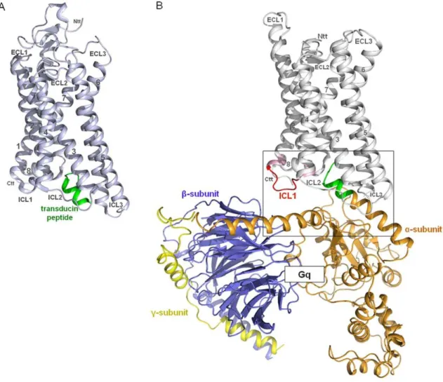
The crystal structure of opsin in complex with the C-terminal helical peptide of transducin (Figure 1A) was used as a structural
Figure 1. Opsin in complex with a transducin peptide. A) The crystal structure of Opsin (light blue) in complex with a synthetic peptide (green) of the C-terminal region of transducin (G-protein) (PDB entry 3DQB) was used as a structural template to buildB) a homology model of the active TSHR conformation (without the N-terminal extracellular region) coupled with Gq heterotrimer (Gaqbc). The superimposition of the C-terminal
residues (green) of the Gaq-protein model with the helical fragment of transducin from the X-ray structure allows a reliable orientation of the
complex.
template to build a homology model of the active TSHR conformation (without N-terminal extracellular domains) coupled to the Gq heterotrimer (Figure 1B). The structural template, rhodopsin/opsin, couples to transducin and Gq as well [35,36,39]. Due to the fact that TSHR is also known to couple to Gq [24,25,40] and as Gq shows higher sequence similarity to transducin than to Gs, we restricted our modeling study to the TSHR/Gq heterotrimer complex. In agreement with previous models [41] the C-terminal tail of the a5-helix of Gaq points directly into an intracellular cleft of the TSHR between helices 3, 5, 6 and 7, but for the first time the superimposition of the C-terminal residues of Gaq-protein with the helical transducin fragment can serve as a fixation point for orientation of the receptor to the G-protein. This allows a reliable orientation of the complex between TSHR and heterotrimeric Gq to be made (Figure 1B). Subsequent predictions about selective interactions, such as between helix 8 of TSHR and Gaq but not with Gas, were experimentally confirmed. The model is also generally in accordance with the recently suggested movement of an activating switch at the rhodopsin/transducin interface regarding the R*-Gt
-GDP complex [19]. Furthermore our model is consistent with GPCRs that have a large third intracellular loop like the dopamine receptors, which allows spatial extension of ICL3 alongside the Ga subunit without steric hindrance of G-protein coupling.
Functional characterization of mutations within the intracellular regions of TSHR
Amino acids of intracellular loop 1 and the transitions between this loop and transmembrane helix 1 and 2 were systematically mutated to alanine (region I438-F451, Table 1). Amino acid substitutions of H443 and R450 decreased the IP accumulation and were suggested by the homology model to interact directly
with Gaq and Gb, respectively (Figure 2). Furthermore, at position 450 a naturally occurring loss-of-function mutation R450H has been reported [42]. Therefore, we investigated biophysical properties of H443 and R450 by additional side-chain substitu-tions (Table 2). In addition, our molecular homology model of the TSHR/Gq complex predicted the involvement of R687 in the intermolecular interaction between helix 8 of the receptor and amino acid D313 in thea4-b6 loop of Gaq. We constructed the R687A mutation and tested it functionally.
Cell surface expression. FACS analyses revealed that the mutations showed a cell surface expression in the range of 49 to 108% of wt TSHR (Table 1).
The expression rate of mutants L446A, N447A and F451A was less than 60% of the wild type. Our molecular homology model suggests conformational functions for these amino acids whereby they form stabilizing intramolecular interactions. The side-chains of L446 and N447 were involved in stabilizing the loop conformation, while F451 is located on the junction between ICL1 and TMH2 and interacts with two hydrophobic residues of TMH4. We suggest that the observed decreased cAMP or IP accumulation for these residues might be caused indirectly by a structural misfold, which causes decreased receptor transport to the cell-surface, rather than based on interruption of direct G-protein contacts.
cAMP accumulation. Mutation L439A in TMH1 was characterized by an increased basal Gas mediated cAMP signaling compared to wt (Table 1). In contrast to this newly identified constitutively activating mutation (CAM) the mutants L440A, T441A, S442A, Y444A, V448A and R450A displayed decreased basal cAMP accumulation. For most of the mutants TSH-mediated cAMP accumulation was comparable to wild type or slightly decreased (not under 50% compared to maximum of
Table 1.Alanine mutagenesis and functional characterization of amino acids in the ICL1 and transitions to the helices 1 and 2.
Construct Cell surface expression cAMP accumulation IP accumulation [IPs (%IP/IPs+PI)]
FACS % of wt TSHR basal 100 mU/ml TSH constitutive activity (slope) basal 100 mU/ml TSH
wt TSHR 100 1 16.660.8 1 3.060.3 25.161.0
pcDNA 361 0.260.1 0.360.1 – 3.760.4 3.760.4
I438A 10866 0.860.3 9.361.3b – 2.5
60.9 4.161.1c
L439A 9565 2.960.5a 16.960.7 3.760.6c 2.660.5 22.360.5
L440A 8365b 0.660.1 15.661.3 – 2.860.7 14.160.9c
T441A 8267a 0.5
60.1 14.161.3 – 3.560.7 8.660.2c
S442A 8964 0.560.1 10.261.2b – 3.260.6 9.760.5c
H443A 9465 0.760.1 13.561.4 – 3.060.8 11.760.3c
Y444A 10467 0.660.1 16.161.6 – 2.960.6 17.560.7c
K445A 9266 0.760.2 14.861.6 – 3.360.3 19.960.9c
L446A 5263a 0.560.1 15.161.2 – 2.960.5 12.361.2c
N447A 5764a 0.7
60.0 13.761.1 – 2.960.6 9.960.7c
V448A 71610b 0.660.1 14.960.8 –– 2.560.1 16.762.3c
P449A 9267a 0.960.1 18.661.8 – 3.360.3 26.863.1
R450A 8167b 0.4
60.1 11.761.5b – 3.4
60.4 4.160.5c
F451A 4966a 0.560.1 11.961.6c – 3.160.3 10.860.9c
COS-7 cells were transfected with wt TSHR or various mutant TSHRs. The vector pcDNA3.1(2) / hygromycin was used as a control. The TSHR is characterized by an elevated cAMP level compared to the control vector alone [76]. Therefore, cAMP accumulation is expressed relative to wt TSHR basal level. TSH-mediated levels of cAMP and IP accumulation were determined after treatment of cells with 100 mU/ml bTSH. Expression of wt and mutant TSHRs were quantified on a FACS flow cytometer. Data are given as mean6standard deviation (SD) of at least three independent experiments (n = 3), each carried out in duplicate. Constitutive activity by linear regression analyses was determined for mutant L439A.aP
wild type), except for mutants I438A, S442A and R450A,Q,E which have significantly impaired signaling activity.
Inositol phosphate (IP) accumulation. None of the characterized mutations had an increased basal IP level. TSH-stimulated IP production is markedly reduced by alanine substitutions of I438, L440, T441, S442, H443 and R450 (Tables 1 and 2).
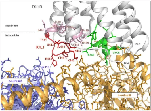
Side-chain variations of H443 and R450. The side-chain variations of H443 (Table 2) showed that a glutamate at this position leads to a decreased IP accumulation and that a phenylalanine side-chain impaired Gq mediated signaling. In contrast, an arginine mutation at position 443 showed signaling activity similar to the wild type. Upon variation of R450, the glutamate substitution impaired both signaling pathways, while Figure 2. Homology model of the complex of TSHR/Gq with focus on the interface between ICL1 and Gq heterotrimer.The TSHR model suggests that in ICL1 (red) and in the transitions with the adjacent transmembrane helices (pale pink) the signaling sensitive amino acids (H443, R450) directly contact Gbc(blue) and Gaq (C-term a5-helix: green), respectively. Dashed lines represent potential H-bonds. Others may indirectly affect Gq coupling (e.g. T441) via conformational changes of ICL1.
doi:10.1371/journal.pone.0009745.g002
Table 2.Side-chain variations and functional characterization of H443 and R450 in ICL1 and transition to helix 2 and R687 in helix 8.
Construct Cell surface expression cAMP accumulation IP accumulation [IPs (%IP/IPs+PI)]
FACS % of wt TSHR basal 100mU/ml TSH basal 100mU/ml TSH
wt TSHR 100 1 13.761.1 1.960.2 23.862.6
pcDNA 460 0.260.1 0.260.1 2.160.2 2.260.2
H443F 7764b 0.560.1 10.561.1 1.960.3 3.160.3c
H443E 9964 0.560.1 10.960.4 1.960.3 12.861.9c
H443R 10362 0.960.1 14.360.8 2.060.2 22.962.5
R450Q 9261 0.460.1 9.161.2c 2.460.1 8.360.6c
R450E 8666 0.360.1 4.860.6c 2.4
60.2 2.560.2b
R450K 8065 0.560.1 12.560.3 2.260.2 7.361.1c
R450M 6165c 0.560.2 11.760.5 2.060.3 3.160.5b
R687A 6965c 1.0
60.2 10.960.8 2.361.0 7.860.6b
COS-7 cells were transfected with wt TSHR or various mutant TSHRs. The vector pcDNA3.1(2) /hygromycin was used as a control. The TSHR is characterized by an elevated cAMP level compared to the control vector alone [76]. Therefore, cAMP accumulation is expressed relative to wt TSHR basal level. TSH-mediated levels of cAMP and IP accumulation were determined after treatment of cells with 100 mU/ml bTSH. Expression of wt and mutant TSHRs were quantified on a FACS flow cytometer. Data are given as mean6standard deviation (SD) of at least three independent experiments (n = 3), each carried out in duplicate.aP
,0.001,bP = 0.001 to 0.01,cP = 0.01 to 0.05.
lysine and methionine caused strong impairment of Gq mediated signaling only. Interestingly, the R450M substitution showed cAMP accumulation of around 80% of the wild type. Our model predicted that R687 in helix 8 interacts with Gaq selectively. Indeed mutation R687A decreased the IP accumulation significantly to 25% of the wild type without affecting cAMP signaling (Table 2).
Amino acids of the ICL1 and transition to TMH2 potentially interact with both Gaq and Gbc
The signaling-sensitive amino acids identified here are observed in our homology models to interact either directly via H-bonds (for example R450 with E349 of the C-terminal helix of Gaq and H443 with Gbc) or (Figure 2) indirectly affect G-protein activation via conformational changes within intracellular loop 1. Our new results summarized together with our recently published data [41,43] of intracellular key-players for G-protein coupling with the TSHR (GPHR information resources: http://www.ssfa-gphr.de [44] and http://gris.ulb.ac.be [45]), suggest a multiple contact interface between the TSHR and the G-protein heterotrimer of Gq (Figure 3, Table 3). All three ICLs of the TSHR contribute to the G-protein coupling process. Amino acids of ICL1 and the transitions between ICL1/TMH2, TMH3/ICL2 and ICL3/ TMH6 as well as helix 8 potentially interact with Gaq, while the ICL1 also interacts with Gbc.
Discussion
Despite fast progress in the investigation of molecular mechanisms concerning contacts between receptor and G-protein [18,19,22] or mechanisms of G-protein activation, fundamental questions regarding these processes are still open such as: How and
where the G-protein selectivity of GPCRs is determined? Several hypotheses regarding this question are under discussion (reviewed in [27,46]), two important of them are: 1) Different conformational states of the receptor are responsible for selectivity for certain G-protein subtypes, since extracellular mutations and different small ligands can cause different G-protein-subtype preferences for one receptor [10,27,46–48]. 2) Distinctive selective interaction patterns in terms of particular intracellular residues exist, which are responsible for G-protein subtype specific interactions [20,21].
A definitive answer in favour of one of the hypotheses cannot be given yet, but a combination between both mechanisms may be more probable. We were interested in the identification of molecular determinants of as yet unknown contribution to coupling and activation mechanism between TSHR and G-proteins (Gs and Gq) to add new information to this field.
Distinct amino acid side-chains of the ICL1 and its spatial conformation are important for (selective) G-protein activation
We identified L439A in TMH1 as a new CAM with elevated basal CAMP activation. Alanine mutations of I438 and S442 in ICL1 and R450 at the junction with TMH2 decrease both cAMP and IP-mediated cascades, while L440A, T441A and H443A in ICL1 impair IP activation selectively. It is to mention that these functional data are derived in COS-cells as an overexpression system for the GPHRs. For the GPHRs [49] like for other GPCRs it is known that different levels of expression can modify signaling capabilities due to different properties of the systems [50]. However the general comparability between results determined in different cell-type systems has been shown for the TSHR recently [51]. Although the relevance ofin vitroforin vivosituation
is still under discussion, two examples indicate their direct
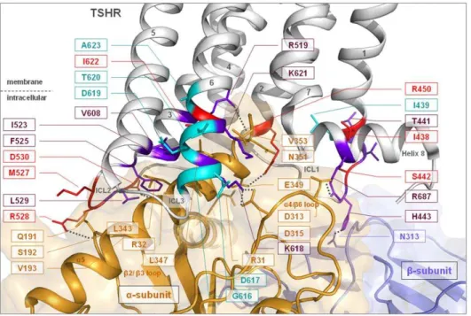
Figure 3. Homology complex model of the TSHR/Gq heterotrimer with focus on the interface between helix 8 and the transition of ICL3/transmembrane helix 6 with Gaq.Our new and recently published data of intracellular key-players for the TSHR and G-protein interaction
are summarized and mapped on to the 3D complex model. Several mutations in the intracellular region of the TSHR are known to prevent Gs and Gq signaling simultaneously. All mutants show decreased cAMP production by TSH in conjunction with decreased activation of the IP pathway. The following wild type amino acids should therefore be considered as commonly sensitive for regulation of the receptor/G-protein interplay: ICL1 - I438, S442, R450; ICL2 - M527, R528, D530; ICL3/TMH6 - I622. Colour codes: purple - selectively impaired Gq activation by mutation; red - inactivating mutants for Gs and Gq coupling and cyan - known constitutively activating mutants. Dashed lines indicate potential H-bonds.
relationship for GPHRs. First, especially the in former times questionablein vivorelevance of both cAMP and also IP signaling
pathways for the TSHR has now been clarified [28,29,40]. Second, overlap betweenin vitroandin vivostudies were recently
evidenced for signaling mechanisms at the LHCGR by a mouse-model [52] which confirmed previous insights fromin vitrostudies
about the significance of GPHR trans- activation [53–55]. Therefore we conclude that our experimental data are most likely relevant and that the investigated intracellular region is of importance. Noticing the remarkable high conservation of some amino acids and even of the conserved (short) length of ICL1 within GPCRs, our results about the G-protein sensitivity of ICL1 might also be important for other GPCRs.
One of our particular findings is that when mutated, R450, which is in the transition between ICL1 and TMH2, affects cAMP and IP accumulation (Table 1, 2). Our molecular homology model suggests direct interactions of R450 to Gaq, particularly to E349 at the C-terminus of thea5 helix (Figure 2, Table 3). Comparing the amino acid sequence in the C-terminal region of Gaq and Gas (Table 4) reveals that at the corresponding position of E349 in Gaq, the hydrophilic amino acid glutamine is found in Gas. If the interacting conformations of Gaq and Gas with TSHR were identical then an interaction of R450 with the H-bond accepting residue Q349 of Gas would be expected and indeed, the mutants R450A,Q,E significantly change the biophysical properties and impair both Gs and Gq subtype mediated signaling cascades. However, since mutation R450M selectively impairs IP but not cAMP accumulation (Table 2), we conclude that R450 of TSHR does not interact with a hydrophilic residue such as Q349 in the C-terminal region of Gas. Moreover, since the R450K mutant also impairs IP (Gq) mediated signaling selectively, it is assumed that it is not the positive charge but rather the full H-donator function and/or size of the arginine in position 450 which is necessary for establishing the Gaq specific interaction. Thus it follows that the interacting conformation of Gas with the TSHR might be different from that of Gaq.
Furthermore, histidine 443 is an important signaling sensitive residue in ICL1, of which aromatic or hydrophobic amino acid
substitutions impair IP but not cAMP accumulation. In contrast, H443 can be substituted by a positively charged arginine residue without any effect, even a negatively charged glutamic acid shows moderate (around 50%) influence compared to wild type function. Altogether, we are able to dissect fairly precisely the potential counterpart of side-chain H443 as being a hydrophilic and uncharged residue at heterotrimeric Gq. Our new opsin-based homology model of the TSHR/Gq complex (Figure 3) orientates this particular part of ICL1 towards Gb. An asparagine (N313 in Figure 2) located at the exterior of a ‘propeller-blade’ of the Gb -subunit is therefore suggested as a potential interaction partner. One has to take into account that by inducing a slight spatial tilt a few conserved hydrophilic amino acids such as N313, N268 and N293, which are found within a tight spatial neighbourhood in this area of the propeller blades of Gb, are also potential interaction partners of TSHR H443.
Determinants of the interfaces between the Thyrotropin receptor and G-protein heterotrimer
In combination with known mutational data of the TSHR (information resources of GPHR data: http://www.ssfa-gphr.de [44] and http://gris.ulb.ac.be [45]) our new findings for the ICL1 and molecular model of the TSHR/heterotrimeric Gq complex allow, for the first time, a systematically completed overview of potential intermolecular contact interfaces at the ICLs (Figure 3). It suggests that all three intracellular loops (and also helix 8) might establish direct side-chain contacts with the a-subunit, but that interaction between the ICL1 and the Gb-subunit also probably exist in the coupled state (Table 3):
& the ICL3/TMH6 transition (TSHR) contacts thea4/b6 loop (Gaq),
& ICL2 (TSHR) contacts theb2/3 loop (Gaq),
& components of the transitions ICL1/TMH2, TMH3/ICL2, and ICL3/TMH6 of the receptor interact with the C-terminal region of thea5 helix (Gaq),
& helix 8 (TSHR) provides charged interactions with thea4/b6 region (Gaq),
& interactions from the ICL1 to the Gb-subunit.
E/DRW motif (TMH3). Similar to observations in the crystal structure of opsin the arginine of the DRY motif in TMH3 (in the TSHR an ERW motif) forms an H-bond interaction with the Gaq backbone at Y350 in the a5
C-Table 3.Potential direct intermolecular interaction partners between TSHR and Gq.
G-protein TSHR
Localization
G-protein Gaq residue Localization
a5 E349 R450 ICL1/TMH2
a5 L343/L347 I523/F525 ICL2
b2-b3 S192 R528 ICL2
b2-b3 V193 L529 ICL2
aN R32 D530 ICL2
a4-b6 D315 K618/K621 ICL3/TMH6
a5 L347/V353 I622 TMH6
b2-b3 D313 R687 Helix 8
Gb
N313 H443 ICL1
The identification of potential interaction partners between TSHR and Gq was carried out using the molecular homology model of the entire receptor/Gq complex (Figure 3) in combination with functional data (GPHR information resources: http://www.ssfa-gphr.de [44] and http://gris.ulb.ac.be [45]). doi:10.1371/journal.pone.0009745.t003
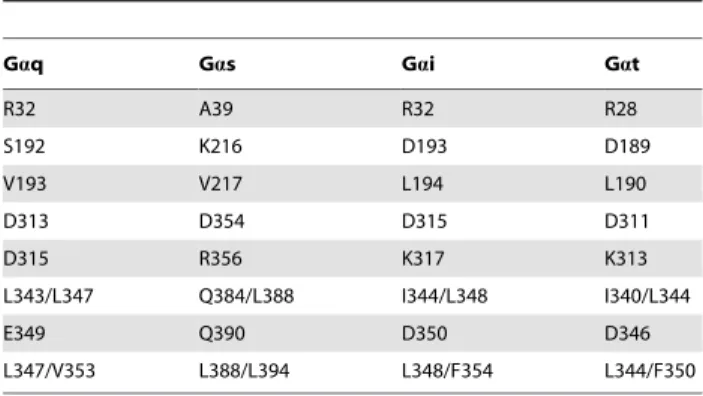
Table 4.Comparison between Gaq, Gas, Gai and Gat.
Gaq Gas Gai Gat
R32 A39 R32 R28
S192 K216 D193 D189
V193 V217 L194 L190
D313 D354 D315 D311
D315 R356 K317 K313
L343/L347 Q384/L388 I344/L348 I340/L344
E349 Q390 D350 D346
L347/V353 L388/L394 L348/F354 L344/F350
Corresponding residues of Gas, Gai and Gat at positions where Gaq is suggested to interact with the TSHR in our homology model. These residues were revealed by a sequence alignment of the alpha subunits.
terminal tail. The general importance of this conserved arginine in the GPCRs is well reported (reviewed in [56]).
ICL2. Furthermore, it was previously demonstrated by mutagenesis studies that particular parts of ICL2 and ICL3 contribute to G-protein activation in the TSHR [41,43], as well as in the homologous LHCGR [49,57–59] and the FSHR [60,61]. Within the ICL2 of the TSHR the residues M527, R528, D530 appeared to be critical for both Gs- and Gq-signaling mediated by TSH, whereas alanine mutation of I523, F525 and L529 led to selectively impaired Gq activation [43].
ICL3. Studies of ICL3 in TSHR comprised systematic mutagenesis and the first model of the complex between TSHR and Gq [41]. In this and in our new refined TSHR/Gq model the middle region of ICL3 is not involved in direct G-protein interaction. However, the junctions of TMH5/ICL3 and ICL3/ TMH6 of TSHR are known to be strongly involved in G-protein activation. In detail, mutation K618A located in the transition between ICL3 and TMH6 was reported to decrease Gq-mediated IP only and not Gs-related cAMP accumulation [41]. Besides being in very close proximity to the C-terminal region ofa5 helix (Gaq), we also suggest for K618 an ionic interaction with the charged partner D315 in thea4/b6 loop of Gaq (Figure 3), which is not present in Gas (R356, Table 4).
ICL1 and helix 8. Mutagenesis studies of ICL1 in TSHR were performed in the early ’90s [37,38]. Through multiple substitutions these studies have given the first hint that sensitivity for G-protein activation, including Gq at the TSHR, can be found in this loop. Here for the first time we systematically investigated each amino acid in ICL1, including those in the flanking peripheries of this loop, by alanine mutations and deciphered their particular influence on intermolecular signal transduction from the receptor to G-proteins. From this work we complete the gaps in our knowledge about determinants that form the TSHR interfaces for G-proteins. This includes the residues R450 and H443 that probably interact with Gaq and Gbc, respectively (see details above and Figure 2), but also the here suggested interaction between helix 8 of the TSHR (R687) anda4/b6 loop (D313) of Gaq (Figure 3).
Implications for selective G-protein activation by TSHR There is a large body of functional data for TSHR mutants in the intracellular loops that simultaneously affect both G-protein subtypes Gs and Gq [41,43]. However, within the entire intracellular portion several additional mutations have been identified that selectively decrease IP-mediated secondary path-ways only and not TSH-induced cAMP production. In contrast, no single mutation that only affects cAMP (Gs) accumulation induced by TSH has been yet observed in all three intracellular loops of the receptor. What can be learnt from these findings? Our results lead to the following conclusions:
& At first, the binding modes between TSHR and the
heterotrimeric G-protein subtypes Gs and Gq overlap partially according to mutants affecting the pathways of both G-protein subtypes.
& Secondly, regarding our identified selective IP mutants and the absence of selective cAMP mutants, it needs more and specific interaction points to achieve the full signaling activity in the receptor/Gq- than in the receptor/Gs-heterotrimer complex. The intracellular interface and the number of receptor contacts for cAMP activation is almost a subset of that for IP activation. This is probably due to a smaller number of interaction points that are spatially accessible and sufficient for Gs.
& Third, Gq specific TSHR residues do not seem to interfere with the interaction of TSHR with Gs. In other words, they are not selective in terms of excluding or inhibiting other G-protein subtypes. However, Gq specific residues are also located in close spatial neighbourhood to, or even overlap with Gs interacting residues (in proximity to C-terminala5 helix). This might be an indication for a likely, albeit small, but different structural arrangement between complexes of TSHR/Gs and TSHR/Gq to each other.
We therefore assume, in accordance with others [27,46,48], that a specific preference of a GPCR for a particular G-protein subtype is controlled by two major events: 1. particular structural features of the activated receptor such as an accessible intracellular conformation (e.g. an ‘‘open’’ or ‘‘widely open’’ surface) is mandatory for subtype preference; 2. characteristic complemen-tary biophysical/biochemical properties of particular interacting residues (intermolecular interaction patterns between receptor and G-protein) complete the G-protein subtype preference. These two mechanisms act together, however, the particular conformation of the intracellular region confers a specific recognition pattern.
There is experimental evidence that the intracellular confor-mation is significant for binding distinct G-protein subtypes and can comprise various locations distributed over the entire GPCR. In the transmembrane region different types of agonists can trigger diverse intracellular conformations, as shown for beta(2)-adrener-gic receptor [62] as well as different signaling types [10]. In contrast to the dual hormone related signal (Gs and Gq [30]), the stimulation of the LHCGR by a small agonistic molecule only induces activation of Gs [63]. For the TSHR it is reported that the naturally occurring loss-of-function mutation, L653V [29], in extracellular loop 3 leads to a selective impairment of IP but not of cAMP accumulation after TSH treatment. Several mutations identified by mutagenesis studies (L417V, TMH1 [64]; S562A, ECL2 [64]; Y605A, TMH5 [41]; N658A, ECL3 [65]) of the TSHR are characterized by the same functional finding. By simultaneous combination of CAMs in the TSHR it was recently shown that the transmembrane helices are characterized by different preferences for cooperative amplification of Gs and Gq mediated signaling pathways [66]. These examples indicate that for full and multiple GPHR activation in terms of dual Gs and Gq coupling, highly specific structural conformations of the intracel-lular region must be induced by the entire receptor protein. Subsequently, it is feasible that the structures of Gs and Gq adjust slightly differently to the receptor conformation to allow in each case optimal complementary intermolecular side-chain interactions.
is involved in IP-mediated intracellular signaling [17,33,67] and our suggested intermolecular contacts of TSHR/Gb might be necessary for separation of the Gaand Gbcsubunits. Thereafter Gb acts to directly regulate downstream signaling by inducing second messengers like IP.
Taken together, GPCRs with promiscuous binding of G-protein subtypes (like the TSHR) are promising targets for investigating G-protein selectivity by studying determinants responsible for differentiated G-protein activation. Utilizing molecular models based on the crystal complex between Opsin/Gt-peptide and functional data by site-directed mutagen-esis, we identified intracellular interfaces between TSHR and different G-proteins. We provide evidence that residues of ICL1 and adjacent transition to TMH2 are involved in Gq pre-coupling and interact with Ga (C-terminal helix a5) and potentially with Gbc as well. Apart from the identification of residues that are commonly sensitive for Gs and Gq signaling, we dissected new residues (in ICL1 and helix 8) that are selectively involved in the regulation of IP (Gq) and not in the cAMP pathway (Gs). In contrast, no single residue has yet been found in the entire intracellular TSHR region that selectively affects cAMP accumulation (Gs) only. Together with our previous data on ICL2 and ICL3 we are able to provide a completed overview of potential intermolecular contact interfaces between TSHR and heterotrimeric G-protein. Based on this, we postulate that binding modes and orientations between GPCR and Gs- and Gq- heterotrimers partially overlap, however, in addition more selective interactions are established in the receptor/Gq-hetero-trimer complex compared to TSHR/Gs. Our findings support that on the one hand G-protein preference is determined specifically by structural features of the entire intracellular region of the activated GPCR, but on the other hand is also completed by complementary recognition patterns between receptor and G-protein subtypes.
Materials and Methods
Structural Bioinformatics and Molecular Modeling We used as a structural template the X-ray structure of opsin (PDB code 3CAP [36]). Until recently, the available GPCR structures for generation of homology models wereb2-adrenergic receptor (b2-AR), rhodopsin, and adenosine-receptor (reviewed in [11,12,68,69]). These structures contain inverse agonists as ligands, some of them are modified by silencing mutations or proteins such as lysozyme are fused to keep the receptors in a more rigid conformation [12,70].
In contrast, the structure of opsin lacks the inverse agonistic ligand retinal, and represents structural features of an active receptor conformation. Furthermore, in 2008 an opsin structure in complex with a synthetic C-terminal transducin-peptide was published (PDB code: 3DQB, [22]). This structure (Figure 1A) was used to suggest a model of G-protein activation by rhodopsin, including recognition, binding and activation of transducin [19].
Firstly, several TSHR-specific corrections were made in the homology model of active TSHR based on opsin. In opsin interactions of the side-chains of three consecutive threonines (positions 2.59–2.61) with the helical backbone of the preceding residues cause a structural bulge in TMH2. In the TSHR no consecutive threonines exist in TMH2, which suggests the presence of a regular a-helix. In TMH5, a minor change of orientation (a twist of 10 to 15 degrees) of the N-terminal half of the helix was generated due to the lack of a proline compared to opsin/rhodopsin (position 5.50). Gaps of missing residues in the loops of the template structure were closed by the ‘Loop Search’
tool implemented in Sybyl 8.1 (Tripos Inc., St. Louis, Missouri, 63144, USA).
The heterotrimeric Gq-protein model was generated using the crystal structure of Gai (PDB entry 1GP2) as a template, which has high sequence similarity to Gi. The very last C-terminal residues of Gaq (343LQLNLKEYNAV), which are missing in the Gai structure, were built using a nuclear magnetic resonance (NMR) structure of an 11-residue C-terminal peptide (340IKENLK-DCGLF) with mainly helical conformation (PDB entry 1AQG [71]). The C-terminal helixa5 of Gaq was extended by the latter helical fragment. This conformation is also supported by the helical conformation of the last 11 C-terminal residues of Gas (384QRMHLRQYELL, PDB entry 1AZS).
The complex between Gq coupled to activated TSHR were built by spatial superimposition of the C-terminala-helix fragment of Gaq with the corresponding synthetic a-helical C-terminal peptide of transducin in the crystal structure. Side-chains and loops of each homology model were subjected to conjugate gradient minimization (until converging at a termination gradient of 0.05 kcal/(mol*A˚ )) and molecular dynamics simulation (2ns) by fixing the backbone of the transmembrane helices and beta-strands. Finally the models were minimized without constraints. All structure images were produced using PyMOL (DeLano WL, version 0.99, San Carlos, CA, USA).
Site-directed Mutagenesis
TSHR mutants were constructed by PCR mutagenesis using the human TSHR-pcDNA3.1(-)/hygro as a template as previously described [72]. Mutated TSHR sequences were verified by dideoxy sequencing with dRhodamine Terminator Cycle Se-quencing chemistry (ABI Advanced Biotechnologies, Inc., Colum-bia, MD).
Cell culture and transient expression of mutant TSHRs COS-7 cells [73] were grown in Dulbecco’s modified Eagle’s medium (DMEM) supplemented with 10% FCS, 100 U/ml penicillin and 100mg/ml streptomycin (Gibco Life technologies, Paisley, UK) at 37uC in a humidified 5% CO2 incubator. Cells
were transiently transfected using the GeneJammerHTransfection Reagent (Stratagene, Amsterdam, NL). 12-well plates (16105 cells/well) were transfected with 1mg DNA per well for
determination of cell surface expression and inositol phosphates. 24-well plates (0.56105cells per well) with 500 ng DNA per well were used for linear regression analysis and measuring of intracellular cAMP accumulation.
FACS Analyses
cAMP Accumulation Assay
Forty eight hours after transfection, cells were incubated in the absence or presence of 100 mU/ml bTSH (Sigma Chemical Co.) in serum free medium supplemented with 1mM IBMX (Sigma) for one hour. Reactions were terminated by aspiration of the medium. The cells were washed once with ice-cold PBS and then lysed by addition of 0.1 N HCl. Supernatants were collected and dried. cAMP content of the cell extracts was determined using the cAMP AlphaScreenTMAssay (PerkinElmerTMLife Sciences, Zaventem, Belgium) according to the manufacturer’s instructions.
Linear regression analysisof constitutive activity as a function of TSHR expression (slopes)
The constitutive activity is expressed as basal cAMP formation as a function of receptor expression determined by FACS. COS-7 cells were transiently transfected in 24-well plates (0.56105cells per well) with increasing concentrations of wt or mutant TSHR plasmid DNA (50; 100; 200; 300; 400 and 500 ng per well). The total DNA amount was kept constant by cotransfection with empty vector to the amount of the highest DNA concentration of 500 ng per well. For determination of cell surface expression and basal cAMP production see ‘‘FACS Analyses’’ and ‘‘cAMP Accumula-tion Assay’’, respectively. Basal cAMP formaAccumula-tion as a funcAccumula-tion of receptor expression was analyzed according to Ballesteros et al [74] using the linear regression module of GraphPad Prism 4 for Windows.
Activation of Inositol Phosphate Formation
Transfected COS-7 cells were incubated with 2mCi [myo-3
-H]inositol (Amersham Biosciences, Braunschweig, Germany) for 6
h. Thereafter, cells were incubated with serum-free DMEM containing 10 mM LiCl and 100 mU/ml TSH for the accumu-lation of intracellular IPs. Evaluation of basal and TSH-induced increases in intracellular IP levels was performed by anion exchange chromatography as previously described [75]. IP-values were expressed as the percentage of radioactivity incorporated from [3H] IP-1 to -3 over the sum of radioactivity incorporated in IPs and phophatidylinositol.
Statistics
Statistical analysis was carried out using the Mann-Whitney nonparametric t test using GraphPad Prism 4 for Windows (a p-value,0.001 extremely significant; bp-value 0.001 to 0.01 very significant;cp-value 0.01 to 0.05 significant; p-value. 0.05 not significant).
Acknowledgments
We thank Saskia Fiedler and Paul Grzesik for their technical assistance.
Author Contributions
Conceived and designed the experiments: GK GK. Performed the experiments: HJ SM JG. Analyzed the data: GK HJ RP. Contributed reagents/materials/analysis tools: CLW. Wrote the paper: GK HJ GK. Supervised the project: G. Krause. Built the receptor/G-protein complex model: G. Kleinau. Built the TSHR homology model: CLW. Supervised HJ, SM, JG: RP.
References
1. Deupi X, Kobilka B (2007) Activation of G protein-coupled receptors. Adv Protein Chem 74: 137–166.
2. Kristiansen K (2004) Molecular mechanisms of ligand binding, signaling, and regulation within the superfamily of G-protein-coupled receptors: molecular modeling and mutagenesis approaches to receptor structure and function. Pharmacol Ther 103: 21–80.
3. Schoneberg T, Schulz A, Gudermann T (2002) The structural basis of G-protein-coupled receptor function and dysfunction in human diseases. Rev Physiol Biochem Pharmacol 144: 143–227.
4. Smit MJ, Vischer HF, Bakker RA, Jongejan A, Timmerman H, et al. (2007) Pharmacogenomic and structural analysis of constitutive g protein-coupled receptor activity. Annu Rev Pharmacol Toxicol 47: 53–87.
5. Joost P, Methner A (2002) Phylogenetic analysis of 277 human G-protein-coupled receptors as a tool for the prediction of orphan receptor ligands. Genome Biol 3: RESEARCH0063.
6. Bjarnadottir TK, Gloriam DE, Hellstrand SH, Kristiansson H, Fredriksson R, et al. (2006) Comprehensive repertoire and phylogenetic analysis of the G protein-coupled receptors in human and mouse. Genomics 88: 263–273. 7. Dorsam RT, Gutkind JS (2007) G-protein-coupled receptors and cancer. Nat
Rev Cancer 7: 79–94.
8. Schoneberg T, Schulz A, Biebermann H, Hermsdorf T, Rompler H, et al. (2004) Mutant G-protein-coupled receptors as a cause of human diseases. Pharmacol Ther 104: 173–206.
9. Seifert R, Wenzel-Seifert K (2002) Constitutive activity of G-protein-coupled receptors: cause of disease and common property of wild-type receptors. Naunyn Schmiedebergs Arch Pharmacol 366: 381–416.
10. Wenzel-Seifert K, Seifert R (2000) Molecular analysis of beta(2)-adrenoceptor coupling to G(s)-, G(i)-, and G(q)-proteins. Mol Pharmacol 58: 954–966. 11. Hanson MA, Stevens RC (2009) Discovery of new GPCR biology: one receptor
structure at a time. Structure 17: 8–14.
12. Kobilka B, Schertler GF (2008) New G-protein-coupled receptor crystal structures: insights and limitations. Trends Pharmacol Sci 29: 79–83. 13. Lodowski DT, Angel TE, Palczewski K (2009) Comparative Analysis of GPCR
Crystal Structures. Photochem Photobiol 85: 425–430.
14. Assmann SM (2002) Heterotrimeric and unconventional GTP binding proteins in plant cell signaling. Plant Cell 14 Suppl: S355–S373.
15. Pandey S, Nelson DC, Assmann SM (2009) Two novel GPCR-type G proteins are abscisic acid receptors in Arabidopsis. Cell 136: 136–148.
16. Oldham WM, Hamm HE (2008) Heterotrimeric G protein activation by G-protein-coupled receptors. Nat Rev Mol Cell Biol 9: 60–71.
17. Smrcka AV (2008) G protein betagamma subunits: central mediators of G protein-coupled receptor signaling. Cell Mol Life Sci 65: 2191–2214. 18. Oldham WM, Van Eps N, Preininger AM, Hubbell WL, Hamm HE (2006)
Mechanism of the receptor-catalyzed activation of heterotrimeric G proteins. Nat Struct Mol Biol 13: 772–777.
19. Scheerer P, Heck M, Goede A, Park JH, Choe HW, et al. (2009) Structural and kinetic modeling of an activating helix switch in the rhodopsin-transducin interface. Proc Natl Acad Sci U S A 106: 10660–10665.
20. Horn F, van der Wenden EM, Oliveira L, Ijzerman AP, Vriend G (2000) Receptors coupling to G proteins: is there a signal behind the sequence? Proteins 41: 448–459.
21. Moller S, Vilo J, Croning MD (2001) Prediction of the coupling specificity of G protein coupled receptors to their G proteins. Bioinformatics 17 Suppl 1: S174–S181.
22. Scheerer P, Park JH, Hildebrand PW, Kim YJ, Krauss N, et al. (2008) Crystal structure of opsin in its G-protein-interacting conformation. Nature 455: 497–502.
23. Van Eps N, Oldham WM, Hamm HE, Hubbell WL (2006) Structural and dynamical changes in an alpha-subunit of a heterotrimeric G protein along the activation pathway. Proc Natl Acad Sci U S A 103: 16194–16199.
24. Allgeier A, Laugwitz KL, Van Sande J, Schultz G, Dumont JE (1997) Multiple G-protein coupling of the dog thyrotropin receptor. Mol Cell Endocrinol 127: 81–90.
25. Laugwitz KL, Allgeier A, Offermanns S, Spicher K, Van Sande J, et al. (1996) The human thyrotropin receptor: a heptahelical receptor capable of stimulating members of all four G protein families. Proc Natl Acad Sci U S A 93: 116–120. 26. Laurent E, Mockel J, Van Sande J, Graff I, Dumont JE (1987) Dual activation by thyrotropin of the phospholipase C and cyclic AMP cascades in human thyroid. Mol Cell Endocrinol 52: 273–278.
27. Wess J (1998) Molecular basis of receptor/G-protein-coupling selectivity. Pharmacol Ther 80: 231–264.
28. Kero J, Ahmed K, Wettschureck N, Tunaru S, Wintermantel T, et al. (2007) Thyrocyte-specific Gq/G11 deficiency impairs thyroid function and prevents goiter development. J Clin Invest 117: 2399–2407.
29. Grasberger H, Van Sande J, Hag-Dahood MA, Tenenbaum-Rakover Y, Refetoff S (2007) A familial thyrotropin (TSH) receptor mutation provides in vivo evidence that the inositol phosphates/Ca2+cascade mediates TSH action on thyroid hormone synthesis. J Clin Endocrinol Metab 92: 2816–2820. 30. Gudermann T, Birnbaumer M, Birnbaumer L (1992) Evidence for dual coupling
phosphoinositide breakdown and Ca2+mobilization. Studies with the cloned murine luteinizing hormone receptor expressed in L cells. J Biol Chem 267: 4479–4488.
31. Minegishi T, Tano M, Shinozaki H, Nakamura K, Abe Y, et al. (1997) Dual coupling and down regulation of human FSH receptor in CHO cells. Life Sci 60: 2043–2050.
32. Quintana J, Hipkin RW, Sanchez-Yague J, Ascoli M (1994) Follitropin (FSH) and a phorbol ester stimulate the phosphorylation of the FSH receptor in intact cells. J Biol Chem 269: 8772–8779.
33. Kuhn B, Gudermann T (1999) The luteinizing hormone receptor activates phospholipase C via preferential coupling to Gi2. Biochemistry 38: 12490–12498.
34. Herrlich A, Kuhn B, Grosse R, Schmid A, Schultz G, Gudermann T (1996) Involvement of Gs and Gi proteins in dual coupling of the luteinizing hormone receptor to adenylyl cyclase and phospholipase C. J Biol Chem 271: 16764–16772.
35. Murakami M, Kouyama T (2008) Crystal structure of squid rhodopsin. Nature 453: 363–367.
36. Park JH, Scheerer P, Hofmann KP, Choe HW, Ernst OP (2008) Crystal structure of the ligand-free G-protein-coupled receptor opsin. Nature 454: 183–187.
37. Kosugi S, Mori T (1994) The first cytoplasmic loop of the thyrotropin receptor is important for phosphoinositide signaling but not for agonist-induced adenylate cyclase activation. FEBS Lett 341: 162–166.
38. Chazenbalk GD, Nagayama Y, Russo D, Wadsworth HL, Rapoport B (1990) Functional analysis of the cytoplasmic domains of the human thyrotropin receptor by site-directed mutagenesis. J Biol Chem 265: 20970–20975. 39. Bhatia J, Davies A, Gaudoin JB, Saibil HR (1996) Rhodopsin, Gq and
phospholipase C activation in cephalopod photoreceptors. J Photochem Photobiol B 35: 19–23.
40. Van Sande J, Dequanter D, Lothaire P, Massart C, Dumont JE, et al. (2006) Thyrotropin stimulates the generation of inositol 1,4,5-trisphosphate in human thyroid cells. J Clin Endocrinol Metab 91: 1099–1107.
41. Claus M, Neumann S, Kleinau G, Krause G, Paschke R (2006) Structural determinants for G-protein activation and specificity in the third intracellular loop of the thyroid-stimulating hormone receptor. J Mol Med 84: 943–954. 42. Nagashima T, Murakami M, Onigata K, Morimura T, Nagashima K, et al.
(2001) Novel inactivating missense mutations in the thyrotropin receptor gene in Japanese children with resistance to thyrotropin. Thyroid 11: 551–559. 43. Neumann S, Krause G, Claus M, Paschke R (2005) Structural determinants for
g protein activation and selectivity in the second intracellular loop of the thyrotropin receptor. Endocrinology 146: 477–485.
44. Kleinau G, Brehm M, Wiedemann U, Labudde D, Leser U, et al. (2007) Implications for molecular mechanisms of glycoprotein hormone receptors using a new sequence-structure-function analysis resource. Mol Endocrinol 21: 574–580.
45. Van Durme J, Horn F, Costagliola S, Vriend G, Vassart G (2006) GRIS: glycoprotein-hormone receptor information system. Mol Endocrinol 20: 2247–2255.
46. Raymond JR (1995) Multiple mechanisms of receptor-G protein signaling specificity. Am J Physiol 269: F141–F158.
47. Evans PD, Robb S, Cheek TR, Reale V, Hannan FL, et al. (1995) Agonist-specific coupling of G-protein-coupled receptors to second-messenger systems. Prog Brain Res 106: 259–268.
48. Perez DM, Karnik SS (2005) Multiple signaling states of G-protein-coupled receptors. Pharmacol Rev 57: 147–161.
49. Feng X, Muller T, Mizrachi D, Fanelli F, Segaloff DL (2008) An intracellular loop (IL2) residue confers different basal constitutive activities to the human lutropin receptor and human thyrotropin receptor through structural commu-nication between IL2 and helix 6, via helix 3. Endocrinology 149: 1705–1717. 50. Ritter SL, Hall RA (2009) Fine-tuning of GPCR activity by receptor-interacting
proteins. Nat Rev Mol Cell Biol 10: 819–830.
51. Mueller S, Gozu HI, Bircan R, Jaeschke H, Eszlinger M, et al. (2009) Cases of borderline in vitro constitutive thyrotropin receptor activity: how to decide whether a thyrotropin receptor mutation is constitutively active or not? Thyroid 19: 765–773.
52. Rivero-Muller A, Chou YY, Ji I, Lajic S, Hanyaloglu AC, et al. (2010) Rescue of defective G protein-coupled receptor function in vivo by intermolecular cooperation. Proc Natl Acad Sci U S A 107: 2319–2324.
53. Jeoung M, Lee C, Ji I, Ji TH (2007) Trans-activation, cis-activation and signal selection of gonadotropin receptors. Mol Cell Endocrinol 260-262: 137–143.
54. Osuga Y, Hayashi M, Kudo M, Conti M, Kobilka B, et al. (1997) Co-expression of defective luteinizing hormone receptor fragments partially reconstitutes ligand-induced signal generation. J Biol Chem 272: 25006–25012.
55. Urizar E, Montanelli L, Loy T, Bonomi M, Swillens S, et al. (2005) Glycoprotein hormone receptors: link between receptor homodimerization and negative cooperativity. EMBO J 24: 1954–1964.
56. Schwartz TW, Frimurer TM, Holst B, Rosenkilde MM, Elling CE (2006) Molecular mechanism of 7TM receptor activation–a global toggle switch model. Annu Rev Pharmacol Toxicol 46: 481–519.
57. Wang Z, Wang H, Ascoli M (1993) Mutation of a highly conserved acidic residue present in the second intracellular loop of G-protein-coupled receptors does not impair hormone binding or signal transduction of the luteinizing hormone/chorionic gonadotropin receptor. Mol Endocrinol 7: 85–93. 58. Fernandez LM, Puett D (1997) Evidence for an important functional role of
intracellular loop II of the lutropin receptor. Mol Cell Endocrinol 128: 161–169. 59. Angelova K, Fanelli F, Puett D (2008) Contributions of intracellular loops 2 and
3 of the lutropin receptor in Gs coupling. Mol Endocrinol 22: 126–138. 60. Ulloa-Aguirre A, Uribe A, Zarinan T, Bustos-Jaimes I, Perez-Solis MA, et al.
(2007) Role of the intracellular domains of the human FSH receptor in G(alphaS) protein coupling and receptor expression. Mol Cell Endocrinol 260-262: 153–162.
61. Timossi C, Maldonado D, Vizcaino A, Lindau-Shepard B, Conn PM, et al. (2002) Structural determinants in the second intracellular loop of the human follicle-stimulating hormone receptor are involved in G(s) protein activation. Mol Cell Endocrinol 189: 157–168.
62. Zurn A, Zabel U, Vilardaga JP, Schindelin H, Lohse MJ, et al. (2009) Fluorescence resonance energy transfer analysis of alpha 2a-adrenergic receptor activation reveals distinct agonist-specific conformational changes. Mol Phar-macol 75: 534–541.
63. van Koppen CJ, Zaman GJ, Timmers CM, Kelder J, Mosselman S, et al. (2008) A signaling-selective, nanomolar potent allosteric low molecular weight agonist for the human luteinizing hormone receptor. Naunyn Schmiedebergs Arch Pharmacol 378: 503–514.
64. Kleinau G, Claus M, Jaeschke H, Mueller S, Neumann S, et al. (2007) Contacts between extracellular loop two and transmembrane helix six determine basal activity of the thyroid-stimulating hormone receptor. J Biol Chem 282: 518–525. 65. Claus M, Jaeschke H, Kleinau G, Neumann S, Krause G, et al. (2005) A hydrophobic cluster in the center of the third extracellular loop is important for thyrotropin receptor signaling. Endocrinology 146: 5197–5203.
66. Jaeschke H, Kleinau G, Sontheimer J, Mueller S, Krause G, et al. (2008) Preferences of transmembrane helices for cooperative amplification of G(alpha)s and G (alpha)q signaling of the thyrotropin receptor. Cell Mol Life Sci 65: 4028–4038.
67. Zhu X, Birnbaumer L (1996) G protein subunits and the stimulation of phospholipase C by Gs-and Gi-coupled receptors: Lack of receptor selectivity of Galpha(16) and evidence for a synergic interaction between Gbeta gamma and the alpha subunit of a receptor activated G protein. Proc Natl Acad Sci U S A 93: 2827–2831.
68. Schertler GF (2008) Signal transduction: the rhodopsin story continued. Nature 453: 292–293.
69. Worth CL, Kleinau G, Krause G (2009) Comparative sequence and structural analyses of G-protein-coupled receptor crystal structures and implications for molecular models. PloS One 4: e7011.
70. Tate CG, Schertler GF (2009) Engineering G protein-coupled receptors to facilitate their structure determination. Curr Opin Struct Biol 19: 386–395. 71. Kisselev OG, Kao J, Ponder JW, Fann YC, Gautam N, et al. (1998)
Light-activated rhodopsin induces structural binding motif in G protein alpha subunit. Proc Natl Acad Sci U S A 95: 4270–4275.
72. Jaeschke H, Neumann S, Kleinau G, Mueller S, Claus M, et al. (2006) An aromatic environment in the vicinity of serine 281 is a structural requirement for thyrotropin receptor function. Endocrinology 147: 1753–1760.
73. Gluzman Y (1981) SV40-transformed simian cells support the replication of early SV40 mutants. Cell 23: 175–182.
74. Ballesteros JA, Jensen AD, Liapakis G, Rasmussen SG, Shi L, et al. (2001) Activation of the beta 2-adrenergic receptor involves disruption of an ionic lock between the cytoplasmic ends of transmembrane segments 3 and 6. J Biol Chem 276: 29171–29177.
75. Berridge MJ (1983) Rapid accumulation of inositol trisphosphate reveals that agonists hydrolyse polyphosphoinositides instead of phosphatidylinositol. Biochem J 212: 849–858.