Research Article Open Access
Breast cancer prevention with Morinda citrifolia (noni) at the initiation
stage
Mian-Ying Wang, Lin Peng, Gary Anderson, Diane Nowicki
Department of Pathology, University of Illinois College of Medicine, Rockford, IL 61107, USA
Corresponding author: Mian-Ying Wang, MD & MS, Research Associate Professor,
Department of Pathology, University of Illinois College of Medicine, Rockford, IL 61107, USA
Submission date: January 7, 2013; Acceptance Date: May 6, 2013; Publication date: June 21, 2013.
ABSTRACT
Background: It has been reported that noni has multiple health benefits for over 2000 years. In this study, the cancer preventive effects of Tahitian noni® juice (TNJ) at the initiation stage on DMBA-induced mammary tumorigenesis in female SD rats was investigated.
Objective: We took advantage of the DMBA-induced mammary carcinogenic model to study the preventive effects of TNJ at the initiation stage of mammary carcinogenesis in female SD rats by using clinical observation, pathological examination, and 32P-postlabeling assay.
Methods: One hundred and sixty female SD rats were divided into eight groups with 20 rats in each group. Three doses of TNJ or placebo was given to the animals at the age of 35 days until the end of the experiment. When the animals were 55 days old, 25 mg/kg DMBA was fed to the animals in the DMBA group, placebo, and TNJ groups. The 20 rats were kept at age-matched controls. Palpable tumors were examined twice a week after DMBA administration in each group by an experienced professional. The size of tumor was measured by a graduated caliper. A piece of tumor, vascularization area, and mammary glands in the thoracic and abdomen areas of each rat were dissected respectively and fixed in 10% neutral buffered formalin for light microscope examination. The DMBA-DNA adduct formation in mammary tissues was detected by 32P-postlabeling assay.
different time points. Histological examination showed that the malignancy of lesions in TNJ groups did not show a significant change when compared with that in positive and placebo groups.
Conclusion: In conclusion, this is the first study which indicates that TNJ possesses a cancer preventive effect at the initiation stage of chemical carcinogenesis induced by DMBA in female SD rates.
Key words: Morinda citrifolia, Tahitian noni® juice, breast cancer, cancer prevention
BACKGROUND:
The species Morinda citrifolia L. (noni) belongs to the genus Morinda and the family Rubiaceae.
Morinda includes about 80 species in which noni is considered the “queen” of all the species (1).
The noni plant is an evergreen that can range from a small bush to a 30-foot tall tree. The tree produces a fragrant white flower, blooming year round. Noni fruit has a lumpy texture,
resembling a ‘hand grenade’, with a rancid taste and smell when fully ripened. The fruit is covered with reddish-brown pits that contain seeds. Each seed has an attached air sac, allowing it to float for months in the ocean. Historians believe that noni originated in Southeast Asia, and then migrated to colonize new lands such as Micronesia and Polynesia (2). The Polynesian
culture has a rich healing heritage and knowledge about noni’s medicinal uses that have been
handed down over many generations (3). Scientific literature about Noni is limited with only about 100 papers published to date. The majority of these publications are from South East Asia. Noni, named Ba Ji Tian in China, has been used since the Han Dynasty as a major Chinese herb for over 3000 years (4). Noni is the Hawaiian name for Morinda citrifolia, which is also known
as “Indian mulberry” or “Och” in India, “Mengkudo” in Malaysia, “Nhau” in Southeast Asia, “painkiller bush” in the Caribbean, “Cheesfruit” in Australia, and “Nono” in Tahiti (5). Noni has
recently received increased attention from modern herbalists, medical physicians, and high-tech
biochemists. Scientific studies within the last few decades support the Polynesians’ claim of its
from Noni roots, is an inhibitor of Ras function. The Ras oncogene is believed to be associated with signal transduction function in several human cancers including lung, colon, pancreas, and leukemia (10). Hiwasa and coworkers demonstrated that damnacanthal, an anthraquinone compound, isolated from the Noni root, had a potent inhibitory activity on tyrosine kinases such as: Lck, Src, Lyn, and EGF receptor (11). These compounds also blocked phosphorylation of c-Jun, a substrate of JNKs, suggesting that JNKs are a critical target for the compounds mediating AP-1 activity and cell transformation (12).
A number of major components have been identified in noni plant such as scopoletin, terpenoids, anthraquinone glycoside, -sitosterol, flavone glycosides, Alizarin, acubin, L. asperuloside, caproic acid, caprylic acid, ursolic acid, rutin, and a putative proxeronine (13-17). A research group led by Chi-tang Ho at Rutgers University has successfully identified several new flavonol glycosides, an iridoid glycoside from the Noni leaves, a trisacharide fatty acid ester, rutin, and an asperulosidic acid from the fruit. Two novel glycosides and a new unusual iridoid named citrifolinoside from Noni fruit have shown an inhibitory effect on AP-1 transactivation, and cell transformation, in the mouse epidermal JB6 cell line (18-20). James Duke listed 23 different phytochemicals found in Noni as well as 5 vitamins and 3 minerals in an authoritative CRC handbook (21).
Our hypothesis is that Noni juice possesses a preventive agent(s) that works at the initiation stage of mammary chemical carcinogenesis.
Breast cancer is the most common cancer and the second leading cause of cancer death in U.S. women (22). The etiology of breast cancer remains unknown and a preventive means for breast cancer is unavailable (23). Although Tamoxifen is able to reduce the risk of recurrence in localized breast cancer, there is still no definitive way to prevent breast cancer (24). Epidemiological studies indicate that environmental factors play an important role in breast cancer etiology and a diet rich in fruits and vegetables is associated with a reduced breast cancer risk (25-26). The last several decades have witnessed an incredible increase in the values placed on fruits, vegetables, medicinal herbs, and other botanical products in cancer prevention and
treatment (27). In fact, phytochemists have identified hundreds of ‘phytochemicals’ that are
The DMBA induced rat mammary carcinoma model, developed 40 years ago by Dr. Charles Brenton Huggins, has become the standard laboratory model of mammary carcinogenesis (32). This model illustrates basic principles of initiation, promotion, and progression stages of mammary carcinogenesis (33). This is an ideal model to test the hypotheses relating the role of specific oncological events to mammary cancer development, as well as to examine the strategies for the prevention of human breast cancer (34). This model has a number of advantages that make it particularly attractive to the experimental oncologist. They are as follows: (a) tumor induction is easy and reliable, a single feeding of DMBA to 40-60 day-old female SD rats results in mammary tumor yields of 100% induction only a few months after DMBA treatment; (b) tumor induction is target specific, primarily involving mammary glands; (c) tumor origin is from ducts, TEB (terminal end bud) is the target site that is similar to human breast cancer; (d) tumors induced by DMBA are predominantly carcinomatous histopathological, which mimics human breast cancer; (e) tumors induced by DMBA are responsive to growth factors and/or hormones; and (f) this model has the potential to examine a single stage of initiation, promotion, or progression in mammary carcinogenesis. Therefore, mammary tumorigenesis in female SD rats induced by DMBA is one of the most utilized models to study and understand the pathogenesis of mammary carcinogenesis and to screen for effective preventive agents (35).
In this study, we took the advantage of the fore mentioned attributes of the DMBA-induced mammary tumor model to evaluate the preventive effects of TNJ. Specifically we looked at the initiation stage of mammary carcinogenesis in female SD rats using clinical observation and pathological examination.
MATERIALS AND METHODS:
Noni juice and placebo: The TNJ used in this study was donated by Tahitian Noni International Inc., which is a formulated form of Tahitian Noni puree mixed with blueberry and grape juice (36). The placebo was prepared by the R & D department of Tahitian Noni International Inc. by following the same procedure of Noni juice preparation, the only difference between Noni juice and placebo is the Noni puree is replaced in the placebo with cheese flavor. Three doses of 3%, 5%, and 10% TNJ and three doses of 3%, 5%, and 10% placebo were freshly prepared in drinking water on a daily basis and supplied in the drinking water from the age of 35 days until the end of the study.
Chemicals: 7,12-Dimethylbenz(a)anthrancene (7,12-DMBA) was purchased from Sigma Chemical (St. Louis, MO, USA) and dissolved in DMSO. This was then diluted with corn oil to 1% DMSO in corn oil before use. All materials and enzymes used in 32P-postlabeling assay were described in a previous study (37).
Animals: Weanling female Spargue–Dawley rats were purchased from Charles River Laboratories, Inc. (251 Ballardvale Street, Wilmington, MA, USA). They were housed in stainless steel cages with sawdust bedding with four animals per cage, standard animal husbandry environment of controlled lighting (lights on 06.00–20.00 h), and temperature
Rat chow and tap water were provided ad libitum. The experimental protocol was approved by the animal ethical committee in accordance with the university guideline for the care and use of laboratory animals.
Experimental design: One hundred and sixty, 35 day old, virgin female SD rats weredivided into eight groups with 20 animals each. Animals in the age-matched control group were maintained on the regular animal chow and water to monitor the development of normal mammary glands and the incidence of spontaneous tumors during the period of the experiment. Animals in the DMBA group were positive controls to document the efficacy of the carcinogenic model. Three doses of 3%, 5%, and 10% placebo and three doses of 3%, 5%, and 10% TNJ groups were supplied in drinking water two weeks before DMBA administration until the end of the protocol. A single dose of 25 mg/kg DMBA was fed to all animals (except the age-matched control group) at the age of 55 days. Please see the details in Scheme 1.
Scheme 1. Experimental design to examine the cancer preventive effect of TNJ on mammary carcinogenesis induced by DMBA in female rats at the initiation stage
25 mg/kg DMBA dissolved in 1% DMSO in corn oil, p.o Animals were sacrificed
|___|______|______|______|______|______|______|______|______|____________ ______ ______ -15 0 30 60 90 100 120 150 180 210 240 270 300 330
Days
_______________________________________________________________________________
3%, 5%, and 10% of placebo or 3%, 5%, and 10% TNJ in drinking water was supplied 15 days before DMBA administration and continuously supplied until the end of the experiment.
Group 1 Age-matched control supplied with water only.
Group 2 Positive control group supplied with 25 mg/kg DMBA only. Group 3 3% placebo in drinking water with 25 mg/kg DMBA. Group 4 5% placebo in drinking water with 25 mg/kg DMBA. Group 5 10% placebo in drinking water with 25 mg/kg DMBA. Group 6 3% TNJ in drinking water with 25 mg/kg DMBA. Group 7 5% TNJ in drinking water with 25 mg/kg DMBA. Group 8 0% TNJ in drinking water with 25 mg/kg DMBA.
week. The surviving animals and the animals without tumors in each group were also verified at the same time points as those animals with tumors.
Histological examination: At 330 days post DMBA administration, all surviving animals were sacrificed in CO2 chamber, followed by a midline incision from the pubis to the sumaxillary
area. The skin was dissected to expose six pairs of mammary glands. Any gross modification of the mammary fat pad by vascularization or any tumors present were removed. A piece of tumor, vascularization area, and mammary glands in the thoracic and abdomen areas of each rat was dissected respectively and fixed in 10% neutral buffered formalin for light microscope examination. The paraffin blocks were serially sectioned at a thickness of 5 (38). Consecutive sections were stained with H & E and examined under the light microscope by experienced pathologists. The pathologists were blinded by code numbers.
The earliest pathogenic changes in mammary gland tissues were epithelial hypertrophy and
hyperplasia, called “intraductal proliferation” in the terminal end bud (TEB). TEB is the site of origin of most rat and human mammary adenocarcinomas. These pathogenic changes were easily
distinguished from the gland’s normal intraductal structures by the presence of a multi-layered
epithelium. The “initiated” intraductal proliferation remained unchanged during the entire
post-carcinogen observation period. The “promoted” intraductal proliferation could progress to “carcinoma in-situ” and then to “invasive adenocarcinoma”. The categories of histological examination were diagnosed by the defined stages of mammary carcinogenesis. TNJ might suppress tumor initiation and blunt the progression of initiated cells to full malignancy (39).
DMBA-DNA adduct detection in mammary gland tissue: Twelve female SD rats were divided into three groups consisting of 4 each (age 35 days): Control, 10% placebo, and 10% TNJ. Animals were pretreated with 10% placebo or 10% TNJ in drinking water for two weeks. Control animals were maintained on a regular diet and water. One dose of 25 mg/kg DMBA was fed to the animals when they were age 55 days old. Animals were sacrificed at the 24th hour post DMBA administration and the mammary gland tissues were collected for the DMBA-induced DNA adducts analysis. Mammary gland DNA was isolated by a classical phenol-chloroform extraction and ethanol precipitation method (40). The DMBA-DNA adduct formation was detected by nuclear P1 procedure using a 32P-postlabeling assay. The relative DNA adduct levels in the mammary gland tissue were calculated (41).
Statistical analysis and data interpretation
:
In order to assess the preventive effect of TNJ on mammary tumor models induced by DMBA, tumor incidence, latency, size, multiplicity, as well as body weight were evaluated at different time points in different groups after DMBA administration. The differences between these parameters in various groups were analyzed usingStudent’s T test (42).
RESULTS:
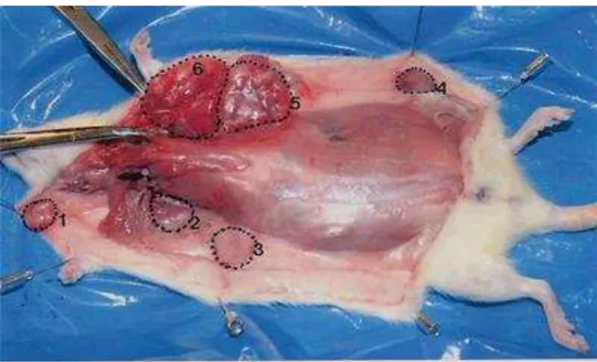
were at day 90 in 3% TNJ group, 120 days in 5% and 10% TNJ groups after DMBA administration (Figure 1).
Figure 1. Multi-tumors (six) detected in a female SD rat at 120 days after DMBA administration in positive control. Tumors are circled by a dotted line and numbered 1 through 6. Most tumors are located in the chest area.
These findings indicated that the latency of tumor appearance in 3% TNJ group was delayed by 30 days, and 60 days in 5% and 10% TNJ groups. Thus, the tumorigenesis was significantly suppressed by TNJ at the initiation stage of carcinogenesis. The earliest tumor detected in the three placebo groups was on day 120, similar to the three groups treated with TNJ.
0 10 20 30 40 50 60 70 80 90
30 60 90 120 150 180 210 240 270 300 330
N u mb er o f tu mo rs in each g ro u p Control DMBA 3% Placebo 5% Placebo 10% Placebo 3% Noni 5% Noni 10% Noni
Figure 2. The palpable tumors were detected in each group at different time points and the number of tumor detected in each group was increased with time after DMBA administration. A spontaneous tumor was also detected in a control animal.
Table 1A. Mean number of tumors at 210, 270, or 330 days by group
Days Mean Control Mean DMBA Mean 3% Placebo Mean 5% Placebo Mean 10% Placebo Mean 3% Noni Mean 5% Noni Mean 10% Noni
210 0.00 3.00 1.60 1.00 1.00 0.60 0.10 0.10
270 0.00 4.00 1.70 1.90 2.10 0.80 0.90 0.10
330 0.05 4.00 2.80 2.90 3.60 1.10 1.10 0.80
Table 1B: Group Comparisons: Non-parametric test of differences in mean number of tumors at 210, 270, or 330 days.
Comparison between groups 210 days 270 days 330 days p value p value p value
Control vs DMBA 0.000 0.000 0.000
DMBA vs 3% NJ 0.000 0.000 0.000
DMBA vs 5% NJ 0.000 0.000 0.000
DMBA vs 10% NJ 0.000 0.000 0.000
Notes: All comparisons show differences in the mean number of tumors at p < 0.01. Because variances were not equal between groups and n was 20 per group a nonparametric test, Mann-Whitney test, was used to compare group differences (50).
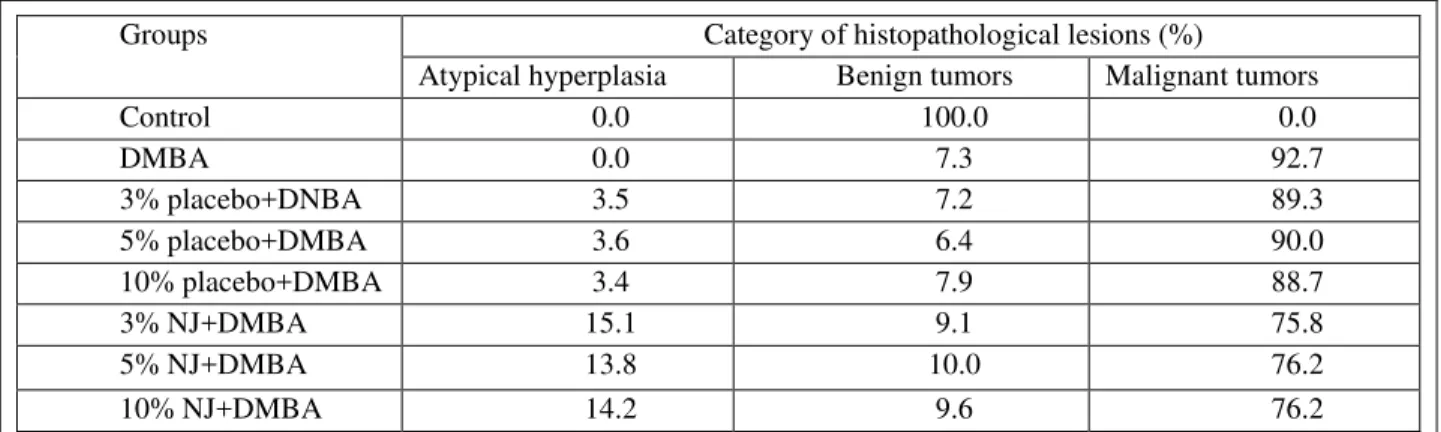
Table 2A. Distribution of the malignancy of tumor based on the histopathological report
Table 2B. Group Comparisons: Non parametric Test of Differences in category of
histopathological lesions (%) of percentage of atypical hyperplasia, benign, and malignant tumor
Comparisons between groups Atypical hyperplasia Benign tumors Malignant tumors
p value p value p value
Control vs DMBA > 0.05 < 0.05 < 0.05
3% placebo+ DNBA vs 3% NJ+DMBA < 0.05 > 0.05 > 0.05 5% placebo+ DMBA vs 5% NJ+DMBA < 0.05 > 0.05 > 0.05 10% placebo+ DMBA vs 10% NJ+DMBA < 0.05 > 0.05 > 0.05
DMBA vs 3% NJ < 0.05 > 0.05 > 0.05
DMBA vs 5% NJ < 0.05 > 0.05 > 0.05
DMBA vs 10% NJ < 0.05 > 0.05 > 0.05
Groups Category of histopathological lesions (%)
Atypical hyperplasia Benign tumors Malignant tumors
Control 0.0 100.0 0.0
DMBA 0.0 7.3 92.7
3% placebo+DNBA 3.5 7.2 89.3
5% placebo+DMBA 3.6 6.4 90.0
10% placebo+DMBA 3.4 7.9 88.7
3% NJ+DMBA 15.1 9.1 75.8
5% NJ+DMBA 13.8 10.0 76.2
10% NJ+DMBA 14.2 9.6 76.2
Our data indicated that NJ may prevent mammary tumorigenesis at the initiation stage. Less significance was observed on benign and malignant tumor categories although the absolute data of malignancy of tumor looks lower in three doses of NJ than corresponding placebo. It indicates that early prevention is the key to stop mammary tumorigenesis and NJ may be not able to revise the malignancy of tumor if NJ was given after tumor induction. NJ may be able to reduce the size of tumor, but not change the characterization of tumor.
Multiplicity: The tumor multiplicity was calculated based on the number of tumors detected, divided by number of animals in each group at day 210, 270, and 330 post DMBA administration. The data were shown in Table 3. The multiplicity of tumors in each of the TNJ groups was much lower than any of the positive control or placebo groups.
Table 3. Multiplicity of tumor
_____________________________________________________________
--- Groups 210 days 270 days 330 days ---
Control 0 0 0.05 DMBA 3 3.8 4.0 3% placebo 1.6 1.7 2.8 5% placebo 1.0 1.9 2.9 10% placebo 1.0 2.1 3.6 3% Noni 0.6 0.8 1.1 5% Noni 0.1 0.9 1.1 10% Noni 0.1 0.1 0.8 ________________________________________________________
Malignancy: The malignancy of tumors was examined by histological examination of each of the 12 pairs of mammary gland tissues per rat under the light microscope by experienced pathologists. The result was shown in Table 2A and Figure 3. The malignancy of tumors in TNJ group was shifted to typical hyperplasia and benign lesions compared with the malignancy of tumors in the positive control and placebo groups.
Table 2A. Distribution of the malignancy of tumor based on the histopathological report
Groups Category of histopathological lesions (%) Typical hyperplasia Begnin tumors Malignant tumors
Control 0.0 100.0 0.0
DMBA 0.0 7.3 92.7
3% placebo+DNBA 3.5 7.2 89.3
5% placebo+DMBA 3.6 6.4 90.0
10% placebo+DMBA 3.4 7.9 88.7
3% TNJ+DMBA 15.1 9.1 75.8
5% TNJ+DMBA 13.8 10.0 76.2
Figure 3. Histopathological examination of the mammary gland.a. Normal mammary tissue showing fibrous and fatty stroma containing benign ducts and lobules. b. Mild hyperplasia of ductal epithelium is noted.
c. Ducts lined by benign epithelium are distorted in shape in this example of fibroadenoma- like change. d. Ductular proliferation of monotumous cells with nuclear enlargement and atypia characterizes this adenocarcinoma.
Survival animals per group at different time point after DMBA administration: The number of animals which survived in each group overtime was recorded and the data were shown in Figure 4. The age-matched controls and animals in TNJ groups showed a higher survival rate than the positive control and placebo groups.
Figure 4. The surviving rats in the age-matched control and TNJ groups were higher survival rates than positive control and placebo groups.
0 3 6 9 12 15 18 21
-15 30 60 90 120 150 180 210 240 270 300 330
Days
N
u
mb
er
o
f
an
imal
s
su
rvi
ved
Growth curve
:
The trends of growth curves in different groups were similar, except for the positive controls. The animals in the positive control showed body weight lost after 270 days post DMBA administration due to bearing a larger amount of bigtumors (Figure 5).0 50 100 150 200 250 300 350 400 450
-15 0 30 60 90 120 150 180 210 240 270 330
Days
Bo
d
y weig
h
t (
g
)
Control
DMBA
3% placebo
5% placebo
10% placebo
3% Noni
5% Noni
10% Noni
Figure 5. The growth curve in each group showed a similar trend except animals in the positive control group had decreased body weight at day 270 (post DMBA administration) due to their tumor burden.
DNA adduct analysis: A typical DMBA-DNA adduct pattern was observed in the mammary gland tissues in female SD rats (Figure 6). The DMBA-DNA adduct level was significantly decreased after drinking 10% TNJ for two week before DMBA administration. There was no detectable DNA adducts observed in mammary gland tissues of control animals (Table 4).
Table 4. TNJ reduced DMBA-DNA adducts level in mammary gland tissues
Groups DMBA-DNA adduct level x 106
Mean ± SD Reduction (%) P value
DMBA 3.55 ± 0.94
10% placebo + DMBA 3.28 ± 0.94 7.6 > 0.05
10%TNJ + DMBA 1.30 ± 0.82 63.4 < 0.01
DISCUSSION:
Several interesting findings were discovered in this study. Mammary tumors were successfully induced by feeding one dose of 25 mg/kg DMBA (at age 55 days) in female SD rats. The number of palpable tumors rapidly increased with time in the positive control group, and a solitary spontaneous tumor was also observed in one of the 20 age-matched control animals (at day 325). TNJ continuously suppressed the tumor incidence and tumor growth in a dose dependent manner until day 270 (after DMBA administration). While placebo suppressed palpable tumor incidence was only at the early stage of tumorigenesis, the number of palpable tumors rapidly increased with time after day 210 (post DMBA administration). The number of tumors in the placebo groups approached the positive control group by the 330 days (post DMBA administration). Therefore, the lowest number of tumors observed in different TNJ groups indicated that TNJ indeed had a significant preventive effect on DMBA-induced mammary carcinogenesis in female SD rats (p < 0.01).
tumorigenesis by drinking 5% or 10% TNJ in drinking water. If we group the average tumor number in each animal of these different groups, the animals in the 3%, 5%, and 10% TNJ groups have much fewer tumors. There is a 73%, 73%, and 80% reduction of tumors respectively compared with that in DMBA positive control animals at day 330 (after DMBA administration). Meanwhile, there was only a 30% reduction in the 3% placebo, 27.5% reduction in the 5% placebo, and 10% reduction in the 10% placebo groups respectively. Thus, the significant reduction of tumor number per animal was observed in TNJ groups (p < 0.01).
Figure 7. A big tumor was detected in positive control group at day120 post DMBA administration. Abundant blood supply was clearly shown surrounding the tumor. This tumor was adenocarcinoma confirmed by histopathological examination.
Histopathological examinations showed the neoplastic growth in the TMJ group to be shifted to hyperplasia and benign tumors, rather than the malignant tumors detected in the positive control and placebo groups.
The growth curve in each group showed the same trend except the positive controls. The body weight in positive control group was decreased after 270 days post DMBA administration because of their tumor burden. The animals in this group were skinny, were less active, and had dry hair. Animals in TNJ group looked much healthier, were more energetic, highly active, and had shinny hair. There were no adverse effects of TNJ observed in this long-term experiment.
enzymes, remains to be determined in the future (43). In addition, the oxidant property, anti-inflammatory, anti-proliferative activities, and modification of immune function of TNJ may also contribute to the mechanisms of the preventive effect of TNJ at the initiation stage of chemical carcinogenesis (44-47). The data from previous studies has indicated that TNJ is able to
scavenge superoxide free radicals (SAR) and quench lipid peroxides (LPO) in vitro and in vivo (48), reduce inflammatory reaction in CCl4-induced liver injury model (49), inhibit partially COX-2 in vitro (50),and to prevent mammary gland tumors at the initiation stage of DMBA-induced mammary gland tumor (51). All of this data supports our hypothesis. Most experts agree that the avoidance of DNA adduct formation and/or removal of carcinogen-DNA adducts is, at least, a possible avenue for cancer prevention since carcinogen-induced DNA adduct formation has long been recognized as the earliest and most critical step of chemical carcinogenesis (52). Previous studies have demonstrated that aromatic DNA adducts have been detected in the adjacent normal tissues of cancer patients (53). The level of these DNA adducts in cancer patients is significantly higher than those of non-cancer control patients. The detection of aromatic adducts in adjacent normal breast tissues of breast cancer patients suggests that exogenous carcinogens may be involved in human breast carcinogenesis (54).
Further studies will be focused on the mechanisms of the mammary tumor preventive effect of TNJ, as well as the illustration of the major preventive components from Noni and their actions. Based on our data, it may be possible to develop a botanical product which is preventive for the people who are on the high risk of breast cancer (55).
CONCLUSION:
This is the first study indicated that TNJ possesses a cancer preventive effect at the initiation stage of chemical carcinogenesis induced by DMBA in female SD rates.
The nutrition enhancement, reduction of carcinogen (DMBA)-induced DNA adduct formation, anti-angiogenesis, delay the patency of tumor appearance may play the key roles in the preventative mechanisms of TNJ at the initiation stage of DMBA-induced breast carcinogenesis.
Abbreviations: Breast cancer, DMBA, initiation stage, carcinogenesis, DNA adducts, cancer prevention, Morinda citrifolia (noni), Tahitian Noni juice (TNJ).
Acknowledgements and Funding: This study was supported by NCI/NIH grant CA121682-01A2. We thank Tahitian noni International Inc. for providing Tahitian noni® juice and placebo for this study. We sincerely thank Dr. Martin MacDowell for his assistant on the data analysis and Mr. John Javaherian for his excellent job on the long-term animal care.
Competing Interests: We do not have competing interest to report.
Author’s contribution: All authors contributed to this study.
REFERENCES:
2. Solomon, N. The tropical fruit with 101 medical uses NONI juice. Second editior Woodland Publishing, 1999.
3. Heinicke, R.M. The pharmacologically active ingredient of Noni. Bulletin of the National tropical Botanical Garden. 1985.
4. Wang MY, West BJ, Jensen CJ, Nowicki D, Su C, Palu AK, Anderson G. Morinda citrifolia 9Noni): a literature review and recent advances in Noni research. Acta Pharmacol Sin. 2002 Dec;23(12):1127-41. Review.
5. McClatchey W. From Polynesian healers to health food stores: changing perspectives of Morinda citrifolia (Rubiaceae). Integr Cancer Ther. 2002 Jun;1(2):110-20; discussion 120. Review.
6. Solomon, N. The Noni Phenomenon. Direct Source Publ., Orem, UT; 1999.
7. Hirazumi A, Furusawa E, Chou SC, Hokama Y. Anticancer activity of Morinda citrifolia (noni) on intraperitoneally implanted Lewis lung carcinoma in syngeneic mice. Proc West Pharmacol Soc. 1994;37:145-6.
8. Furusawa E, Hirazumi A, Story S, Jensen J. Antitumour potential of a polysaccharide-rich substance from the fruit juice of Morinda citrifolia (Noni) on sarcoma 180 ascites tumour in mice. Phytother Res. 2003 Dec;17(10):1158-64.
9. Hiramatsu, T. Induction of normal phenotypes in ras –transformed cells by damnacanthal from Morinda citrifolia. Cancer Lett. 73(2-3):161-6, 1993.
10.Hiwasa, T., Arase, Y., Chen, Z., Kita, K., Umezawa, K., Ito, H., Suzuki, N. Stimulation of ultraviolet-induced apoptosis of human fibroblast UVr-1 cells by tyrosine kinase inhibitors. FEBS Lett 444(2-3):173-6,1999.
11.Liu G, Bode A, Ma WY, Sang S, Ho CT, Dong Z. Two novel glycosides from the fruits of Morinda citrifolia (noni) inhibit AP-1 transactivation and cell transformation in the mouse epidermal JB6 cell line. Cancer Res. 2001 Aug 1;61(15):5749-56.
12.Arpornsuwan T, Punjanon T. Tumor cell-selective antiproliferative effect of the extract from Morinda citrifolia fruits. Phytother Res. 2006 Jun;20(6):515-7
13.Chang P, Lee KH, Shingu T, Hirayama T, Hall IH, Huang HC. Antitumor agents 50. 1 Morindaparvin-A, a new antileukemic anthraquinone, and alizarin-1-methyl ether from Morinda parvifolia, and the antileukemic activity of the related derivatives. J Nat Prod.1982 Mar-Apr;45(2):206-10.
14.Moorthy, N.K. and Reddy, G.S. Preliminary phytochemical and pharmacological study of Morinda citrifolia, Linn. The Antiseptic 67(3): 167-171, 1970.
15.Tona L, Cimanga RK, Mesia K, Musuamba CT, De Bruyne T, Apers S, Hernans N, Van Miert S, Pieters L, Totté J, Vlietinck AJ. In vitro antiplasmodial activity of extracts and fractions from seven medicinal plants used in the Democratic Republic of Congo. J Ethnopharmacol. 2004 Jul;93(1):27-32.
16.Potterat O, Hamburger M. Morinda citrifolia (Noni) fruit--phytochemistry, pharmacology, safety. Planta Med. 2007 Mar;73(3):191-9. Epub 2007 Feb 7. Review. 17.Kanchanapoom T, Kasai R, Yamasaki K. Iridoid and phenolic glycosides from Morinda
18.Wang M, Kikuzaki H, Jin Y, Nakatani N, Zhu N, Csiszar K, Boyd C, Rosen RT, Ghai G, Ho CT. Novel glycosides from noni (Morinda citrifolia). J Nat Prod. 2000 Aug;63(8):1182-3.
19.Sang, S., Cheng, X., Zhu, N., Stark, R.E., Badmaev, V., Ghai, G., Rosen, R.T., Ho,C.T. Flavonol glycosides and novel iridoid glycoside from the leaves of Morinda citrifolia. J Agric Food Chem 49(9):4478-81, 2001.
20.Duke, J. A. Handbook of phytochemicals. CRC Publishing. Boca Raton, FL
21.HK, Thun MJ, Hankey BF, Ries LA, Howe HL, Wingo PA, Jemal A, Ward E, Anderson RN, Edwards BK. Annual report to the nation on the status of cancer, 1975-2000, featuring the uses of surveillance data for cancer prevention and control. J Natl Cancer Inst. 2003 Sep 3;95(17):1276-99. Review.
22.Salehi F, Turner MC, Phillips KP, Wigle DT, Krewski D, Aronson KJ. Review of the etiology of breast cancer with special attention to organochlorines as potential endocrine disruptors. J Toxicol Environ Health B Crit Rev. 2008 Mar;11(3-4):276-300. Review. 23.Wiebe VJ, Osborne CK, Fuqua SA, DeGregorio MW. Tamoxifen resistance in breast
cancer. Crit Rev Oncol Hematol. 1993 Jun;14(3):173-88. Review.
24.Li, D.H., wang, M.Y., Dhingra, K. And Hittelman, W.N. Aromatic DNA adducts in adjacent tissues of breast cancer patients: clues to breast cancer etiology. Cancer Res 56: 287-293, 1996.
25.CRF and AICR. Food, nutrition and prevention of cancer: a global perspective. Page: 252-287, 1997.
26.Maizes V. Reducing the risk of breast cancer: Nutritional strategies. Explore (NY). 2005 Mar;1(2):130-2. Review. No abstract available.
27.Bradford PG, Awad AB. Phytosterols as anticancer compounds. Mol Nutr Food Res. 2007 Feb;51(2):161-70. Review.
28.Le Marchand L. Cancer preventive effects of flavonoids--a review. Biomed Pharmacother. 2002 Aug;56(6):296-301. Review.
29.Clayson DB. Nutrition and experimental carcinogenesis: a review. Cancer Res. 1975 Nov;35(11 Pt. 2):3292-300.
30.Clapp RW, Jacobs MM, Loechler EL. Environmental and occupational causes of cancer: new evidence 2005-2007. Rev Environ Health. 2008 Jan-Mar;23(1):1-37.
31.Weisch, C.W. Host factors affecting the growth of carcinogen-induced rat mammary carcinomas: A review and tribute to Charles Brenton Huggins. Cancer Res 45:3415-3443, 1985.
32.Russo, J., and Russo, I.H. Experimentally induced mammary tumors in rats. Breast 33.cancer Res and Treat. 39:7-20, 1996.
34.Russo, J. and Russo, I.H. Biology of disease, biological and molecular bases of mammary carcinogenesis. Laboratory Investigation 57(2): 112-137, 1987.
36.36. European Commission.. Commission decision 0f 5 June 2003 authorizing the placing
on the market of “Nonie juice” as a novel food ingredient under regulation (EC ) Nr.
258/97 of the European parliament and of the council. Official J. of the European Union.200 12(6):144
37.Wang MY., and Liehr JG. “Detection of DNA adducts of unstaurated fatty acid hydroperoxides by 32P-postlabeling analysis.” In “Eicosanoids and other bioactive lipids
in cancer, inflammation and radiation injury.” Edited by Nigam, S., Marmett, L.L.,
Kluwer, K.V. and Walden, Jr., T.L. Kluwer Academic Publishers. 1993, Chapter 89; 453-455.
38.Hirose M, Masuda A, Ito N, Kamano K, Okuyama H. Effects of dietary perilla oil, soybean oil and safflower oil on 7,12-dimethylbenz[a]anthracene (DMBA) and 1,2-dimethyl-hydrazine (DMH)-induced mammary gland and colon carcinogenesis in female SD rats. Carcinogenesis. 1990 May;11(5):731-5.
39.Russo J, Russo IH. Atlas and histologic classification of tumors of the rat mammary gland. J Mammary Gland Biol Neoplasia. 2000 Apr;5(2):187-200. Review.
40.Wang, M-Y.. And Liehr, J.G. “Induction by estrogens of lipid peroxidation and lipid peroxide derived malonaldehyde-DNA adducts in male syrian hamsters: role of lipid perodidation in estrogen-induced kidney carcinogenesis.” Carcinogenesis, 1995, 16(8):1941-1945.
41.Wang, M-Y., and Liehr, J.G. Lipid hydroperoxide-induced endogenous DNA adducts in hamsters: possible mechanism of lipid hydroperoxide-mediated carcinogenesis. Archives Biochemistry Biophysics, 1995, 316(1):38-46.
42.Schapira DV, Kumar NB, Lyman GH. Variation in body fat distribution and breast cancer risk in the families of patients with breast cancer and control families. Cancer. 1993 May 1;71(9):2764-8.
43.Akihisa T, Matsumoto K, Tokuda H, Yasukawa K, Seino K, Nakamoto K, Kuninaga H, Suzuki T, Kimura Y. Anti-inflammatory and potential cancer chemopreventive constituents
44.of the fruits of Morinda citrifolia (Noni). J Nat Prod. 2007 May;70(5):754-7.
45.Wang, M.Y., and Su, Chen “Cancer Preventive Effect of Morinda citrifolia (Noni)”
Annals New York Academy of Sciences, 2002, Vol 952;161-168.
46.Akihisa T, Matsumoto K, Tokuda H, Yasukawa K, Seino K, Nakamoto K, Kuninaga H, 47.Suzuki T, Kimura Y. Anti-inflammatory and potential cancer chemopreventive constituents of the fruits of Morinda citrifolia (Noni). J Nat Prod. 2007 May;70(5):754-7. 2007 May 5.
48.Wong DK. Are immune responses pivotal to cancer patient's long term survival? Two clinical case-study reports on the effects of Morinda citrifolia (Noni). Hawaii Med J. 2004 Jun;63(6):182-4.
NCI Chemoprevention Drug Development Program. J Cell Biochem Suppl. 1994;20:32-54. Review.
50.Wang MY, Cheerva C, Su C, Jensen J, Nowicki D, Anderson G, Jensen S, Fritz JW. Protective effects of Morinda citrifolia (Noni) on plasma SAR and LPO in current smokers. XI Biennial Meeting of the Society for Free radical Research International.
International Proceeding Division, MONDUZZI EDITORE s.p.A. 2002. P729-734.
51.Wang, MY, Nowicki D, Anderson G, Jensen J, West B. Liver Protective Effects of Morinda citrifolia (Noni). Plant Foods Hum Nutr. 2008 Jun;63(2):59-63.
52.50. Su, C., Wang, M-Y., Nowicki, D., Jensen, J., and Anderson, G. Selective COXII inhibition of Morinda citrifolia (Noni) in vitro. The proceeding of Eicosanoids and Other Bioactive Lipids in Cancer, Inflammation and Related Diseases. The 7th Annual Conference, Loews Vanderbilt Plaza, Nashville, Tennessee, U.S.A. Poster #107 on page 127.
53.Wang, MY., Anderson, G., Nowicki, D. “Synergistic effect of Morinda citrifolia
methylsulfonymethane (MSM) on mammary breast cancer prevention at the initiation stage of chemical carcinogenesis induced by DMBA in female Sprague-Dawley (SD)
rats.” American Association for Cancer Research, Cancer Epidemiology Biomarkers and
Prevention. ISSN 1055-9965, Nov. 2003, Vol. 12 (11), Part 2: 1277s-1388s.
54.Ken-Dror G. DNA adducts as biological markers for human exposure to polycyclic aromatic compounds. Harefuah. 2005 Aug;144(8):583-7, 596. Review.
55.Li D, Wang MY, Firoz PF, Chang P, Zhang W, Moorthy B, Vulimiri SV, Goth-Goldstein R, Weyand EH, and DiGiovanni, J. “Characterization of a major aromatic DNA adduct
detected in human breast cancer.” Environ Mol Mutagen. 39(2-3):193-200. 2002.
56.Vineis P, Husgafvel-Pursiainen K. Air pollution and cancer: biomarker studies in human 57.populations. Carcinogenesis. 2005 Nov;26(11):1846-55. Epub 2005 Aug 25. Review. 58.Prasain JK, Barnes S. Metabolism and bioavailability of flavonoids in chemoprevention: