Strana 322 VOJNOSANITETSKI PREGLED Vojnosanit Pregl 2013; 70(3): 322–325.
Correspondence to: Žaklina Mijoviý, Vase þarapiýa 85, 18 000 Niš, Serbia; Phone: +381 18 4234 092, +381 64 6625346. E-mail: zaklinamijovic@gmail.com
C A S E R E P O R T UDC: 616.833-006.38::617.553
DOI: 10.2298/VSP1303322M
A rare case of retroperitoneal malignant Triton tumor invading renal
vein and small intestine
Redak slu
þ
aj retroperitonealnog malignog tumora Triton sa invazijom renalne
vene i tankog creva
Žaklina Mijoviü*, Dragan Mihailoviü*, Nikola Živkoviü*, Miloš Kostov†, Sladjana Živkoviü‡, Nebojša Stojanoviü‡
*Institute of Pathology, Faculty of Medicine, University of Niš, Niš, Serbia;
†
Department of Pathology, ‡Department of Urology, Military Hospital, Niš, Serbia
Abstract
Introduction. Malignant Triton tumor is a very rare malig-nant peripheral nerve sheath tumor with rhabdomyosarco-matous differentiation. Most of those tumors occur in pa-tients with von Recklinghausen’s disease or as a late compli-cation of irradiation and commonly seen in the head, neck, extremities and trunk. Case report. We reported retroperito-neal malignant Triton tumor in a 57-year-old female patient. Skin lesions were not present, and there was no family history of neurofibromatosis or previous irradiation. The presented case is one of a few recorded in the specialized literature that occurs in the retroperitoneal space in sporadic form. In this case, tumor consisted of a multilobular mass was in close re-lation with the abdominal aorta and inferior vena cava and involved the renal vein with gross invasion of the small intes-tine. The patient underwent total resection of the tumor and left nefrectomy was performed. The small intestine 10 cm in length was also resected and end-to-end anastomosis was conducted. The postoperative course was uneventful and the patient was discharged from the hospital ten days after the surgery. Conclusion. Diagnostically, it is crucial to recognize this uncommon histological variant because malignant Triton tumor has a worse prognosis than classic malignant periph-eral nerve sheath tumor does. The use of the immunohisto-chemistry is essential in making the correct diagnosis. Only appropriate pathological evaluation supported by immu-nostaining with S-100 protein and desmin confirmed the di-agnosis. Aggressive surgical management treatment improves the prognosis of such cases with adjuvant radiotherapy.
Key words:
peripheral nervous system neoplasms; diagnosis; immunohistochemistry; surgical procedures, operative; treatment outcome.
Apstrakt
Uvod. Maligni tumor Triton je veoma redak maligni tu-mor omotaÿa perifernih nerava sa rabdomiosarkomatoz-nom diferencijacijom. Veýina ovih tumora javlja se kod bolesnika sa Recklinghausen-ovom bolešýu ili kao kasna komplikacija zraÿenja i najÿešýe se viĀa na glavi, vratu, ek-stremitetima i trupu. Prikaz bolesnika. Prikazan je retro-peritonealni maligni Triton tumor kod bolesnice starosti 57 godina. Kožne lezije nisu bile prisutne i nije bilo poro-diÿne anamneze neurofibromatoze ili prethodnog zraÿenja. Prikazana bolesnica jedna je od retkih koji su zabeleženi u specijalizovanoj literaturi sa tumorom u retroperitoneal-nom prostoru u sporadiÿnoj formi. Kod ove bolesnice tu-mor se sastojao od multilobularne mase, bio je u tesnoj vezi sa abdominalnom aortom i donjom šupljom venom sa makroskopski vidljivom invazijom tankog creva. UraĀena je totalna resekcija i leva nefrektomija. TakoĀe, uraĀena je i resekcija tankog creva dužine 10 cm i anastomoza end-to-end. Postoperativni tok je protekao regularno i bolesnik je otpušten deset dana posle hirurške intervencije. Zaklju-ÿak. Dijagnostiÿki, prepoznavanje ove retke histološke va-rijante ima poseban znaÿaj, jer maligni tumor Triton ima goru prognozu od klasiÿnog malignog tumora omotaÿa pe-rifernih nerava. Korišýenje imunohistohemije važno je za postavljanje taÿne dijagnoze. Jedino adekvatna patološka procena sa imunohistohemijskim bojenjem S-100 proteina i desmina potvrĀuje dijagnozu. Agresivno hirurško leÿenje sa adjuvantnom radioterapijom može poboljšati prognozu ovakvih sluÿajeva.
Kljuÿne reÿi:
živci, periferni, neoplazme; dijagnoza;
Volumen 70, Broj 3 VOJNOSANITETSKI PREGLED Strana 323
Mijoviý Ž, et al. Vojnosanit Pregl 2013; 70(3): 322–325. Introduction
Malignant Triton tumor (MTT) is a malignant peripheral nerve sheath tumor (MPNST) with rhabdomyosarcomatous differentiation. MTT constitutes about 5% of all MPNSTs. MTT arises in two principal forms: sporadic or in association with neurofibromatosis type 1 [von Recklinghausen’s disease (NF-1)]. Slightly more than half of the cases of MTT have been reported in conjunction with NF-1. MTT is commonly seen in the head, neck, extremities and trunk 1. The fact that the presence of this unusual tumor in the retroperitoneal space is extremely rare has prompted the authors to report this case. We presented a 57-year-old female patient in whom a retro-peritoneal paravertebral mass was postoperatively diagnosed as MTT and describeed the histomorphological and immuno-histochemical features of this uncommon tumor. In this case, tumor developed outside the setting of NF-1.
Case report
A 57-year-old female patient presented with a 2-month history of abdominal pain radiating to the back. Physical ex-amination revealed a painful abdomen in the region of umbili-cus. Full blood cell count, serum urea levels, and electrolyte levels were within normal limits. Chest X-ray was normal. At laparotomy, a multilobular paravertebral mass the size of two male fists was found occupying the left abdominal region. Tumor was in close relation with the abdominal aorta and infe-rior vena cava and appeared to involve the left renal vessels (artery and vein) with gross invasion of the small intestine. The patient underwent total resection of the tumor and left ne-frectomy was performed. Upon resection, thrombosis was pre-sent on part of a renal artery and there was no need for further pathological analysis. The small intestine 10 cm in length was also resected and end-to-end anastomosis was conducted. The postoperative course was uneventful and the patient was dis-charged from the hospital ten days after the surgery.
On the basis of histological findings and immunohisto-chemistry, a malignant Triton tumor, an uncommon subtype of peripheral nerve sheath tumor with rhabdomyosarcoma-tous elements, was diagnosed. Thereafter, the patient was re-ferred to the Oncology Department for Radiotherapy.
During the 8-month follow-up, the patient showed no evidence of recurrence or metastasis.
Macroscopically, the tumor was received in three frag-ments measuring 7, 5 and 4.5 cm. The cut sections showed solid, firm and yellow whitish tissue with areas of necrosis (Figure 1). The left kidney measured 11 x 6 x 5 cm. Cut sec-tion showed no gross abnormalities. Samples of left renal vein measured 0.6 and 0.7 cm, and part of small intestine was 10 cm in length.
Microscopically, the tumor was composed of spindle cells arranged in a fasciculated pattern and whorls (Figure 2). Spindle cells showed wavy, hyperchromatic nuclei with in-distinct light staining cytoplasm. Hypercellular and hypo-cellular zones with areas of palisading necrosis and myxoid stroma were also present. Scattered large round cells with eosinophilic cytoplasm and hyperchromatic nuclei were seen admixed with neoplastic spindle cells.
Fig. 1 – A fragment of primary malignant Triton tumor. The cut surface was solid, firm and yellow whitish with the areas
of necrosis
Fig. 2 – Malignant Triton tumor (spindle cells arranged in interlacing fascicles, sheaths and whorls; H&E, ×100)
On histology, the kidney showed no significant mor-phological abnormalities. The lesion involved renal vein and a part of the small intestine were similar, displaying poorly differentiated spindle cells.
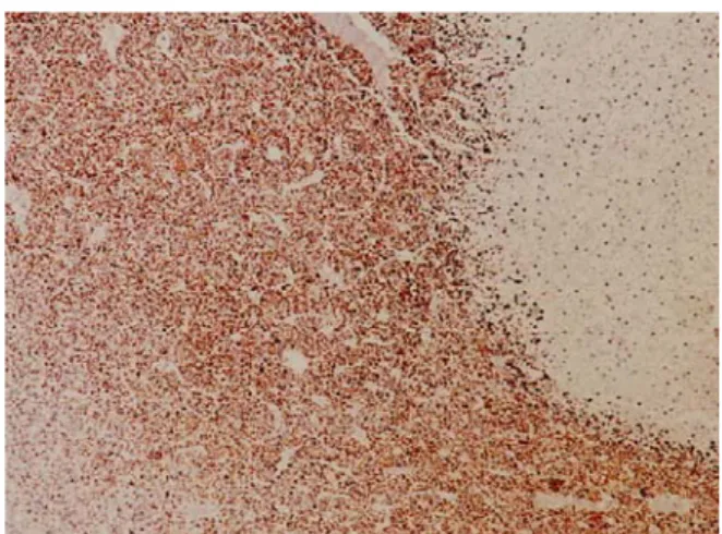
Immunohistochemically, the spindle cells were focally positive for S-100 (Figure 3), strongly positive for vimentin, and negative for cytokeratin 7, EMA, HMB-45, and CK AE1/AE3. The large, round eosinophilic pleomorphic cells
Strana 324 VOJNOSANITETSKI PREGLED Volumen 70, Broj 3
Mijoviý Ž, et al. Vojnosanit Pregl 2013; 70(3): 322–325. were immunoreactive for muscle-specific actin and desmin
(Figure 4). Immunohistochemical staining with S-100 and desmin indicated that the tumor cells originated from Schwann cells and showed rhabdomyosarcomatous differentiation. Ki-67 was expressed in a large number of cells (30%).
Fig. 4 – Staining for desmin was strongly positive in the area of malignant Triton tumor (peroxidase-antiperoxidase
technique, ×100)
Based on these findings, the histopathological diagnosis was malignant peripheral nerve sheath tumor with rhabdo-myosarcomatous differentiation, namely malignant Triton tumor.
Discussion
MPNSTs are neoplasms derived from the cellular con-stituents of the peripheral nerve sheath. This term replaces the earlier terms malignant schwannoma, neurofibrosarcoma, and neurogenic sarcoma 1. The majority arise from Schwann cells, but some could develop from fibroblasts and support-ing cells known as perineural cells. The capacity of MPNSTs to undergo focal divergent differentiation to rhabdomyosar-coma, chondrosarrhabdomyosar-coma, osteosarrhabdomyosar-coma, angiosarrhabdomyosar-coma, epithelial elements, or a combination thereof is well known 2–6. MTT is a variety of this type of tumors which presents a rhabdomyoblastic differentiation. This composite neoplasm was first described in 1938 by Masson and Martin, who suggested that the neural elements in the tumor induced differentiation of skeletal muscle in much the same fashion as normal nerve was believed to induce the regeneration of skeletal muscle in the Triton salamander 1. This tailed am-phibian displays the ability to regenerate limbs after the cut end of the sciatic nerve is implanted into the soft tissue of its back. Although Masson believed that one cell line induced the other, it seems more likely that both cell lines originate from less well-differentiated neural crest cells 1. The term “malignant Triton tumor” was first introduced by Woodruff et al. 7 in 1973.
MTTs are extremely rare, with less than 100 cases documented world-wide to date 8–10. Regarding location, MTT occurs predominantly in the head, neck, trunk regions
and lower extremities 1, 3. To the best of our knowledge, there are only a few reports of these tumors developing in the ret-roperitoneal space 11–14.
MTT shows marked male predominance with more predilections for younger age groups. The sporadic forms mostly occurring in females of older age groups 15, as the case reported herein. These tumors may also arise in sites of previous radiation therapy. We reportɟd the case of paraver-tebral MTT occurring in thɟ patient without clinical setting of NF-1 or previous irradiation.
MPNST is one of the most histologically variable soft tissue tumors, and use of immunohistochemistry is essential in making a correct diagnosis 1, 3, 16, 17. The fasciculated, spin-dle cell growth pattern may cause confusion with leiomyo-sarcoma, fibroleiomyo-sarcoma, or monophasic synovial sarcoma. In addition, MPNST must be distinguished from melanoma malignum. In the present case, histology and immunohisto-chemical staining revealed a typical pattern of MPNST with the additional features of rhabdomyoblastic differentiation supported by positive staining with desmin and muscle-specific actin 3, 4, 8.
Retroperitoneal localization MTT has the most unfa-vorable prognosis due to the delayed diagnosis but also due to the relation to adjacent organs 13. This case of MTT was presented as a large abdominal mass with invasion of the left renal vein and the small intestine since these retroperitoneal tumors are often asymptomatic in the earlier stages 12, 14. The natural history of MTT is much more aggressive than MPNST 3, 15, 18. The tumor has a high propensity for early lo-cal recurrence, rather than metastatic disease. The prognosis is poor with a 5-year survival rate around 12% 15. Location has been correlated with survival 11.Tumors occurring in the head and neck, upper and lower extremities have a better prognosis than tumors located in the retroperitoneum, but-tocks or trunk. Cytogenetic studies have revealed a break-point in 11p15, considered a region of myogenic differentia-tion. Amplification of c-myc oncogene is probably responsi-ble for aggressive biologic behavior of MTT 3. As MTT is a very aggressive tumor, behaving like a high grade sarcoma, it is believed that to obtain the best outcome a full surgical resection with as wide a margin as possible is vital followed by adjuvant radiotherapy 19.
Conclusion
Retroperitoneal malignant Triton tumor is extremely rare, but an important pathological condition. This uncom-mon histological variant has the worse prognosis than classic malignant peripheral nerve sheath tumor does. Immunohisto-chemistry is an essential tool for ruling out differential diag-nostic considerations. Radical surgical excision of the tumor followed by radiation therapy is the treatment of choice.
Acknowledgement
Volumen 70, Broj 3 VOJNOSANITETSKI PREGLED Strana 325
Mijoviý Ž, et al. Vojnosanit Pregl 2013; 70(3): 322–325.
R E F E R E N C E S
1. Weiss SW, Goldblum JR. Malignant tumors of peripheral nerves. In: Weiss SW, Goldblum JR, editors. Enzinger and Weiss’s Soft Tissue Tumors. 5th ed. St. Louis, Mo: Mosby; 2008. p. 903–44. 2. Ducatman BS, Scheithauer BW. Malignant peripheral nerve sheath tumors with divergent differentiation. Cancer 1984; 54(6): 1049–57.
3. Stasik CJ, Tawfik O. Malignant peripheral nerve sheath tumor with rhabdomyosarcomatous differentiation (malignant Triton tumor). Arch Pathol Lab Med 2006; 130(1): 1878–81.
4. Huang L, Espinoza C, Welsh R. Malignant peripheral nerve sheath tumor with divergent differentiation. Arch Pathol Lab Med 2003; 127(3): e147–50.
5. Malerba M, Garofalo A. A rare case of nerve-sheath sarcoma with rhabdomyoblastic differentiation (malignant triton tu-mor). Tumori 2003; 89(Suppl 4): 246–50. (Italian)
6. Ballas K, Kontoulis T, Papavasiliou A, Pissas D, Pavlidis T, Katsiki E, et al. A rare case of malignant Triton tumor with pluridi-rectional differentiation. South Medic J 2009; 102(4): 435–7. 7. Woodruff JM, Chernik NL, Smith MC, Millet WB, Foote FW Jr.
Peripheral nerve tumors with rhabdomyosarcomatous differ-entiation (malignant "Triton" tumors). Cancer 1973; 32(2): 426–39.
8. Lang-Lazdunski L, Pons F, Jancovici R. Malignant “Triton“ tumor of the posterior mediastinum: prolonged survival after staged resection. Ann Thorac Surg 2003; 75(5): 1645–8.
9. Cano JR, Algar FJ, Alvarez A, Salvatierra A. A Triton tumour of the left sympathetic nerve. Interact Cardio Vasc Thorac Surg 2006; 5(6): 790–1.
10.Dartnell J, Pilling J, Ferner R, Cane P, Lang-Lazdunski L. Malig-nant Triton tumor of the brachial plexus invading the left tho-racic inlet: a rare differential diagnosis of pancoast tumor. J Thorac Oncol 2009; 4(1): 135–7.
11.Yakulis R, Manack L, Murphy AI Jr. Postradiation malignant triton tumor. A case report and review of the literature. Arch Pathol Lab Med 1996; 120(6): 541î8.
12.Murtaza B, Gondal ZI, Mehmood A, Shah SS, Abbasi MH, Tamimy MS, et al. A huge malignant triton tumour. J Coll Physicians Surg Pak 2005; 15(11): 728î30.
13.Radovanovic D, Vukotic-Maletic V, Stojanovic D, Lalosevic Dj, Likic I, Stojsic Z,et al. Retroperitoneal ”Triton“ tumor. Hepatogas-troenterology 2008; 55(82î3): 527î30.
14.Hoshimoto S, Morise Z, Takeura C, Ikeda M, Kagawa T, Tanahashi Y, et al. Malignant Triton tumor in the retroperitoneal space associated with neurofibromatosis type 1: a case study. Rare Tumors 2009; 1(2): e27.
15.Brooks JS, Freeman M, Enterline HT. Malignant “Triton” tumor: natural history and immunohistochemistry of nine new cases with literature review. Cancer 1985; 55(11): 2543–9.
16.Stojanoviý M, Radojkoviý M, Jeremiý Lj, Zlatiý A, Stanojeviý G, Jova-noviý M, et al. Malignant schwannoma of the pancreas involv-ing transversal colon treated with en-bloc resection. World J Gastroenterol 2010; 16(1): 119î22.
17.Kostov M, Mijovic Z, Visnjic M, Mihailovic D, Stojanovic M, Zdravk-ovic M. Malignant peripheral nerve sheath tumour in a dog pre-senting as a pseudo aneurysm of the left jugular vein: a case report. Vet Med 2008; 53: 685î9.
18.Aldlyami E, Dramis A, Grimer RJ, Abudu A, Carter SR, Tillman RM. Malignant triton tumor of the thigh –a retrospective analysis of nine cases. Eur J Surg Oncol 2006; 32(7): 808–10. 19.Grobmyer SR, Reith JD, Shahlaee A, Bush CH, Hochwald SN.
Ma-lignant Peripheral Nerve Sheath Tumor: molecular pathogene-sis and current management considerations. J Surg Oncol 2008; 97(4): 340–9.