Role of Areca Nut Induced TGF-
β
and
Epithelial-Mesenchymal Interaction in the
Pathogenesis of Oral Submucous Fibrosis
Ila Pant
1, Neeraj Kumar
1, Imran Khan
1, Somanahalli Girish Rao
2, Paturu Kondaiah
1*
1Department of Molecular Reproduction, Development and Genetics, Indian Institute of Science, Bangalore, Karnataka, India,2Department of Oral and Maxillofacial Surgery, D.A. Pandu Memorial—R.V. Dental
College and Hospital, Bangalore, Karnataka, India
Abstract
Areca nut consumption has been implicated in the progression of Oral Submucous fibrosis
(OSF); an inflammatory precancerous fibrotic condition. Our previous studies have
demon-strated the activation of TGF-
β
signaling in epithelial cells by areca nut components and
also propose a role for epithelial expressed TGF-
β
in the pathogenesis of OSF. Although
the importance of epithelial cells in the manifestation of OSF has been proposed, the actual
effectors are fibroblast cells. However, the role of areca nut and TGF-
β
in the context of
fibroblast response has not been elucidated. Therefore, to understand their role in the
con-text of fibroblast response in OSF pathogenesis, human gingival fibroblasts (hGF) were
treated with areca nut and/or TGF-
β
followed by transcriptome profiling. The gene
expres-sion profile obtained was compared with the previously published transcriptome profiles of
OSF tissues and areca nut treated epithelial cells. The analysis revealed regulation of 4666
and 1214 genes by areca nut and TGF-
β
treatment respectively. The expression of 413
genes in hGF cells was potentiated by areca nut and TGF-
β
together. Further, the
differen-tially expressed genes of OSF tissues compared to normal tissues overlapped significantly
with areca nut and TGF-
β
induced genes in epithelial and hGF cells. Several positively
enriched pathways were found to be common between OSF tissues and areca nut +TGF-
β
treated hGF cells. In concordance, areca nut along with TGF-
β
enhanced fibroblast
activa-tion as demonstrated by potentiaactiva-tion of
α
SMA,
γ
SMA and collagen gel contraction by hGF
cells. Furthermore, TGF-
β
secreted by areca nut treated epithelial cells influenced fibroblast
activation and other genes implicated in fibrosis. These data establish a role for areca nut
influenced epithelial cells in OSF progression by activation of fibroblasts and emphasizes
the importance of epithelial-mesenchymal interaction in OSF.
OPEN ACCESS
Citation:Pant I, Kumar N, Khan I, Rao SG, Kondaiah P (2015) Role of Areca Nut Induced TGF-βand Epithelial-Mesenchymal Interaction in the Pathogenesis of Oral Submucous Fibrosis. PLoS ONE 10(6): e0129252. doi:10.1371/journal. pone.0129252
Editor:Philip C. Trackman, Boston University Goldman School of Dental Medicine, UNITED STATES
Received:February 7, 2015
Accepted:May 6, 2015
Published:June 24, 2015
Copyright:© 2015 Pant et al. This is an open access article distributed under the terms of the Creative Commons Attribution License, which permits unrestricted use, distribution, and reproduction in any medium, provided the original author and source are credited.
Data Availability Statement:Data are available at GEO database, accession No. GSE59414.
Introduction
Oral submucous fibrosis is prevalent in South and South East Asia [
1
]. It is a pre-cancerous
condition characterized by inflammation, epithelial atrophy and trismus of the oral cavity due
to excessive extracellular matrix deposition [
2
,
3
]. Extracellular matrix remodeling including
deregulation of synthesis and degradation of collagen, up regulation of pro-fibrotic
Transform-ing growth factor-
β
(TGF-
β
) and down regulation of Bone Morphogenic Protein 7 (BMP7) are
characteristic features of OSF [
4
,
5
,
6
]. Habit of areca nut chewing is considered as the most
probable etiological factor in OSF manifestation [
7
,
8
], which is supported by a mouse model
[
9
]. The alkaloid and polyphenol components of areca nut were found to induce and activate
TGF-
β
in epithelial cells [
10
]. Earlier studies documented increased collagen content in OSF
derived fibroblasts [
11
] and arecoline treated mucosal fibroblasts [
12
]. A recent report
high-lights activation of mucosal fibroblasts by areca nut extract suggesting involvement of the PLC/
IP3/Ca2+/Calmodulin and Rho signaling pathways along with actin filament polymerization
[
13
]. However, the response of fibroblasts to areca nut together with TGF-
β
representing OSF
pathology is not well studied. Therefore, to gain further insights we studied the effects of areca
nut with or without TGF-
β
on human gingival fibroblasts by transcriptome profiling. The
pro-file obtained was further compared with the transcriptome of OSF tissues and areca nut
induced transcriptome in epithelial cells [
6
,
10
]. These data demonstrate the involvement of
both areca nut and epithelium derived TGF-
β
in altering fibroblast phenotype, highlighting the
importance of epithelial mesenchymal interaction in OSF.
Materials and Methods
This study has been approved by the Institutional Ethics Committee of DA Pandu Memorial
RV Dental College and Hospital. Informed written consent of the participants has been
obtained.
The study is designed to understand the role of areca nut on the modulation of fibroblasts
that is essential in the manifestation of oral submucous fibrosis. This has been accomplished by
treating the human gingival fibroblasts (hGF) with areca nut extract (with or without TGF-
β
)
and subsequent transcriptome profiling and qPCR. The expression profiles were compared to
the transcriptome profile of OSF tissues to arrive at possible genes/pathways that may be
essen-tial to drive OSF progression. Details of the protocols are as follows:
Areca nut extract preparation
Previously described protocols were followed for areca nut water extract preparation
[
10
,
14
,
15
]. Dried and de-husked areca nut was ground to powder and extracted using constant
stirring in 100 ml de-ionized water at 4°C for 4 hours. This was filtered through a sintered glass
funnel followed by lyophilization. The lyophilized form was re-dissolved in de- ionized water.
The extract was filtered through 0.2 micron filter, lyophilized again and stored at 4°C. The
powder obtained was weighed and dissolved in filtered de-ionized water for treatment
pur-poses and was stored at -20°C. To avoid repeated freeze thaw cycles once dissolved; extract was
stored in aliquots.
Cell lines and treatments
Primary human gingival fibroblast cells (hGF) [
16
] and HaCaT cells [
17
] were cultured as
described [
10
]
.For the microarray experiments and validations; hGF cells were serum deprived
for 24 hours followed by treatment with sub-cytotoxic dose of 5
μ
g/ml areca nut water extract
with or without 5 ng/ml of TGF-
β
(R&D systems, USA) for 72 hours. To study the epithelial
data collection and analysis, decision to publish, orpreparation of the manuscript.
mesenchymal interaction, conditioned media from HaCaT cells was collected as follows.
Con-fluent cultures of HaCaT cells were serum starved for 24 hours followed by 10
μ
M ALK5
inhib-itor (TGF
β
RI inhibitor, SB 431542, Sigma-Aldrich, USA) [
18
] treatment 2 hours prior to areca
nut treatment (5
μ
g/ml). Meanwhile hGF cells were serum deprived for 24 hours such that the
treatment time point coincided with completion of 48 hour treatment on HaCaT cells. At this
time point, the condition medium of areca nut (with or without ALK5 inhibitor; SB 431542)
treated HaCaT cells was transferred to hGF cells and simultaneously direct treatment of areca
nut with or without ALK5 inhibitor; SB 431542 was also performed and both were maintained
for 48 hours.
Tissue Samples, RNA isolation and real time RT-PCR
Tissue samples of OSF and normal subjects were procured from D.A Pandu Memorial R.V
Dental College and Hospital, Bangalore. This study has been approved by the Institutional
Eth-ics Committee of DA Pandu Memorial RV Dental College and Hospital. Informed written
con-sent of the participants has been obtained. Normal oral tissues were obtained from non-OSF
patients who underwent third molar extraction. All the tissues were evaluated by a pathologist.
Total RNA was isolated from homogenates of tissues, hGF and HaCaT cells using TRI
reagent (Sigma-Aldrich, St. Louis, USA) as per manufacturer
’
s protocol. For RT-PCR, 2
micro-grams of RNA was converted to cDNA using high capacity cDNA synthesis kit (Applied
Bio-systems, Foster City, USA). Semi-quantitative PCR was performed using DreamTaq Green
PCR 2X master mix (Thermo Scientific). 20 ng of cDNA per 20
μ
L reaction containing gene
specific expression primers was used. Real time PCR was performed using ABI Prism 7900HT
sequence detection system. 20 ng of cDNA per 10
μ
l reaction was used for real time PCR
analy-sis using Dynamo SYBERgreen 2X mix (Finnzymes, Finland) along with the specific primer
pair.[
10
]. The sequences of the primers used are enlisted in
Table 1
. RPL35A expression was
used for normalization [
19
]. Differential expression of genes was determined using the formula
d
Ct
¼
Ct gene
Ct RPL
35
A
dd
Ct
¼
d
Ct treated
d
Ct untreated
Fold Change
ð
FC
Þ ¼
2
ddCtMicroarray and data analysis
Whole human genome (4X44 K) oligonucleotide arrays (Agilent Technologies, Santa Clara,
USA) were used for microarray experiments. Briefly, 200 ng of RNA from untreated and
treated biological duplicate samples (5H; 5
μ
g/ml areca nut, T; 5 ng/ml TGF-
β
and 5H+T; 5
μ
g/
For hierarchical cluster, Pearson correlation matrix was used as a distance matrix and
aver-aged method for linkage [
21
]. Gene Set Enrichment Analysis (GSEA) was done as described
[
22
]. Venn diagrams were made using Venny (
http://bioinfogp.cnb.csic.es/tools/venny/index.
html
). The data is submitted to GEO database with accession number GSE59414.
Collagen contraction assay
The collagen contraction assay was performed with 1.5 x 10
5serum deprived hGF cells
embed-ded in collagen gels which were prepared as described [
13
,
23
]. Cells were pelleted down and
re-suspended in fresh serum free DMEM. Total number of cells was counted and 1.5x10
6cells
per 600
μ
l were aliquoted in fresh tubes. Chilled 330
μ
l of collagen (dissolved in 0.2% acetic
acid to a final concentration of 3 mg/ml) was added to the cells and pH was neutralized
imme-diately by the addition of 1M NaOH. This solution was mixed gently and plated into 12 well
plates. They were left undisturbed for 2 hours at 37°C to allow gelation of collagen populated
with hGF cells. The gels were subsequently detached slowly from the plate using P1000 tip. 1
ml of serum free media was added to each well such that the gels were free floating. Each free
floating hGF populated collagen gel was treated with 5
μ
g/ml of areca nut with or without 5 ng/
Table 1. List of primers used for PCR.
S. No. GENE FORWARD PRIMER 5’-3’sequence REVERSE PRIMER 5’-3’sequence DETAILS
1 αSMA CAGCCAAGCACTGTCAGG CAATGGATGGGAAAACAGC 150 bp, 59.5°C
2 γSMA CCTCAGTCACTGGGAGAAGAA ATCATCTCCTGCGAAGCCT 150 bp, 59.5°C
3 BMP1 ACAGCACAGGCAACTTCTCC GGGACGTGAAGTTCAGGATG 117 bp, 59.5°C
4 CD248 TGCCAACGTGTGTCTTTTTG GTTCTGTTGGGCTCTTGCTC 141 bp, 59.5°C
5 COL15A1 CAGTGCTGGTGTCTGCTGAT GACAAAGGATACGGACGAGG 150 bp, 59.5°C
6 CTGF CAGCATGGACGTTCGTCTG CCAACCACGGTTTGGTCCTT 117 bp, 59.5°C
7 EDN1 CGAGCACGTTGTTCCGTATG CAGCCCTGAGTTCTTTTCCTG 164 bp,55°C
8 EGR2 GTGACCATCTTTCCCAATGC AGCAAAGCTGCTGGGATATG 135 bp, 62°C
9 ELN GTCCTCCTGCTCCTGCTGT CTCCTCCTCCAAGGGCTC 127 bp, 62°C
10 FN1 AAACCAATTCTTGGAGCAGG CCATAAAGGGCAACCAAGAG 142 bp, 50°C
11 GATA6 TGCAGCAAAAATACTTCCCC TGTAGAGCCCATCTTGACCC 133 bp, 62°C
12 IGF2 GCTTCCAGACACCAATGGGAATCC TCATATTGGAAGAACTTGCCCACG 364 bp, 60°C
13 IGFBP3 AGAGCACAGATACCCAGAACT TGAGGAACTTCAGGTGATTCAGT 105 bp, 59.5°C
14 INHBB GCGTTTCCGAAATCATCAG TTTCAGGTAAAGCCACAGGC 134 bp, 59.5°C
15 LIMK1 GGAGAGGAAGGAAGCGAGTT TAGTACTGGTGCGACAGGGA 146 bp, 59.5°C
16 PLOD2 GGACTCGGAGAAGCCCTC CCTTGACCAAGGACCTTCAC 138 bp, 59.5°C
17 PTN TGCAACAAAGGCAGACTGAG TCCCTGCTTCAGCAGTATCC 148 bp, 59.5°C
18 RPL35A GAACCAAAGGGAGCACACAG CAATGGCCTTAGCAGGAAGA 236 bp, 58°C
19 TAGLN GCTCTACTGTCTGTTGCCCC CCTCCAGCTCCTCGTCATAC 135 bp, 62°C
20 TGFβ2 AGTGCCTGAACAACGGAT GTACAAAAGTGCAGCAGG 218 bp, 55°C
22 TGM2 TGACCTCCGCAAAGACAAAG CCAAGTTGCGGAAGCAGTA 241 bp,50°C
23 THBS1 CCGGCGTGAAGTGTACTAGCTA TGCACTTGGCGTTCTTGTT 317 bp, 59°C
24 TGFBI TGTGTGCTGAAGCCATCGTTG CCGGCTTGTCTGAAAAGGTCA 313 bp,50°C
25 TMEPAI TTCATTCCCTGTCCTCATTGG GCACAACAGCCATGGAATCA 228 bp, 50°C
26 TGFβ1 TCCGAGAAGCGGTACCTGAA TGCTGTCACAGGAGCAGTGG 266 bp,63.7°C
27 TGFβ3 GCGTGAGTGGCTGTTGAGA CCAAGTTGCGGAAGCAGTA 306 bp,52.7°C
28 TGFβRI TACAGCTTTGCCTGAACTCT CACGACAGAGTTACCTAAAG 311 bp,52.7°C
29 TGFβRII AGTGTTGGGTTATTGCTAAT AGTGACTTCACAATGTAAAC 240 bp,54.3°C
30 TGFβRIII ATTCTTTTCAGGCCAGTGGC TGGAACCTGTATCACAATGGAG 182 bp,63.2°C
ml of TGF-
β
. Decrease in collagen gel diameter was recorded after 24 hours using a ruler along
four axis and images were taken under white light in gel documentation system (UviPro
plati-num, Uvitech UK). Results were plotted as change in total surface area.
Immunocytochemistry
For immunocytochemistry, 50,000 hGF cells were seeded on cover slips in 12 well plate, serum
deprived and treated with 10
μ
M ALK5 inhibitor (TGF
β
RI inhibitor, SB 431542,
Sigma-Aldrich, USA) two hours prior to 5
μ
g/ml of areca nut treatment. The treatments with HaCaT
condition media are as described earlier. After completion of 48 hours of treatment, hGF cells
were fixed and permeabilized using chilled methanol. Residual methanol was washed off with
phosphate buffered saline (1X PBS). The protocol for ICC is essentially as described [
24
]. To
block nonspecific staining, cells were incubated with 10% serum for one hour.
α
SMA antibody
was then added at a dilution of 1:150 (ab32575, Abcam, USA) and kept overnight at 4°C. Cells
were then washed twice with 1X PBS and incubated with secondary antibody (anti-mouse
Alexafluor 488, 1:200 dilution; Invitrogen, USA) for 1 hour at room temperature. Excess
sec-ondary antibody was washed off using 1X PBS and the nucleus was stained with propidium
iodide (1 mg/ml) for 1
–
2 minutes. Cover slips were then washed again and mounted in antifade
(Invitrogen, Life Technologies, USA).
α
SMA expression was detected using confocal laser
scan-ning microscope (Ziess, LSM550, apocromat).
Direct red 80 staining for collagen
Collagen protein was detected by Direct Red 80 dye (Sigma- Aldrich, USA) using previously
described protocol [
25
]. hGF cells were washed with PBS and 1 ml of Bouin
’
s solution (15:5:1
of aqueous picric acid: 35% formaldehyde: glacial acetic acid) was used as fixative for an hour.
Cells were washed with PBS and air dried for 15 minutes followed by addition of 1 ml Direct
Red 80 dye (100 mg/ml Direct Red 80 dye prepared in saturated aqueous picric acid solution).
Staining was done for 1 hour with mild shaking. After removal of the dye, the excess dye was
washed off using 1 ml of 0.01N HCl for 10 minutes. The stained cells were photographed and
the dye from cells was extracted using 0.1N NaOH whose optical density (OD) was measured
at 550 nm using spectrophotometer (Bio-Rad, SmartSpec-3000 Spectrophotometer). 0.1N
NaOH was used as blank and results were plotted as OD/10
5cells.
Statistical analysis
One way analysis of variance (ANOVA) and Benferroni
’
s Multiple Comparison test was
employed to test for significance while making multiple comparisons. Wilcoxon signed rank
test was used to compare the significance of median expression values of genes in normal and
OSF tissue samples. P value
0.05 was considered as significant with P value
0.01,
0.001
and
0.0001 represented as
,
and
respectively.
Results
Areca nut and/or TGF-
β
induced gene expression profile in human
gingival fibroblast (hGF) cells
transcriptome profiling was performed on hGF cells treated with areca nut (5H), TGF-
β
(T)
and areca nut along with TGF-
β
(5H+T). Data analysis identified 4666 and 1214 differentially
regulated genes by areca nut and TGF-
β
, respectively while areca nut and TGF-
β
together
regu-lated 5752 genes (Tables
2
,
3
,
4
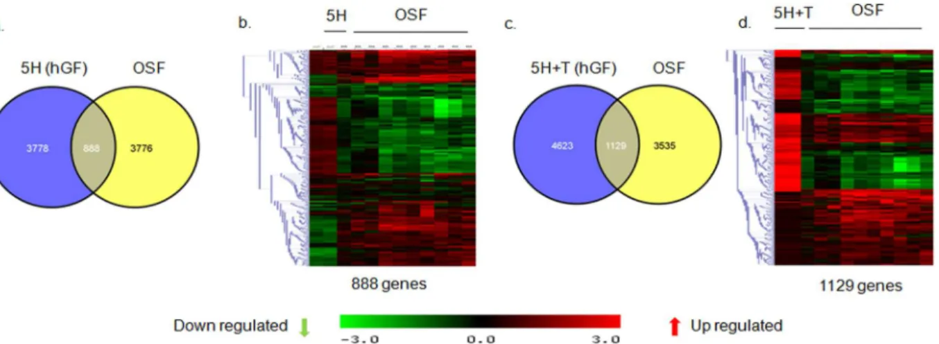
) as compared to control cells. Venn diagram identified 1040
genes exclusively regulated by 5H as they did not appear in the 5H+T list. Similarly, 247 genes
were regulated by T and not by 5H. 5H+T could induce 1692 genes, which were not regulated
by either 5H or T. Interestingly, 413 genes were commonly regulated in all the three treatments
(
Fig 1A
).
Hierarchal clustering of these 413 genes revealed that 5H+T profile clustered in between 5H
and T thereby signifying similarity in the expression of these genes by 5H+T treatment with
either 5H or T profiles. Moreover, the expression of most of these genes seemed to be enhanced
by the combined treatment with areca nut and TGF-
β
(5H+T) (
Fig 1B
). Intriguingly, there
were 60 genes which showed opposite regulation by 5H or T treatments and hence they did not
appear in the profile of 5H+T treatment (
Fig 1A and 1C
). These genes may not have any
impli-cation in OSF. The 3153 genes regulated by 5H but not by T (
Fig 1A
) could be exclusive targets
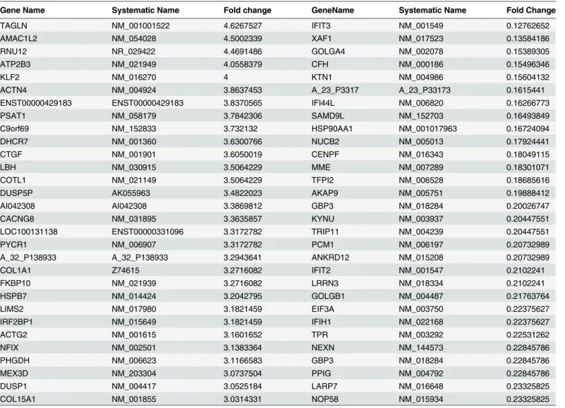
Table 2. List of top 30 up or down regulated genes in hGF cells by areca nut (5H).Gene Name Systematic Name Fold change GeneName Systematic Name Fold Change
TAGLN NM_001001522 4.6267527 IFIT3 NM_001549 0.12762652
AMAC1L2 NM_054028 4.5002339 XAF1 NM_017523 0.13584186
RNU12 NR_029422 4.4691486 GOLGA4 NM_002078 0.15389305
ATP2B3 NM_021949 4.0558379 CFH NM_000186 0.15496346
KLF2 NM_016270 4 KTN1 NM_004986 0.15604132
ACTN4 NM_004924 3.8637453 A_23_P3317 A_23_P33173 0.1615441
ENST00000429183 ENST00000429183 3.8370565 IFI44L NM_006820 0.16266773
PSAT1 NM_058179 3.7842306 SAMD9L NM_152703 0.16493849
C9orf69 NM_152833 3.732132 HSP90AA1 NM_001017963 0.16724094
DHCR7 NM_001360 3.6300766 NUCB2 NM_005013 0.17924441
CTGF NM_001901 3.6050019 CENPF NM_016343 0.18049115
LBH NM_030915 3.5064229 MME NM_007289 0.18301071
COTL1 NM_021149 3.5064229 TFPI2 NM_006528 0.18685616
DUSP5P AK055963 3.4822023 AKAP9 NM_005751 0.19888412
AI042308 AI042308 3.3869812 GBP3 NM_018284 0.20026747
CACNG8 NM_031895 3.3635857 KYNU NM_003937 0.20447551
LOC100131138 ENST00000331096 3.3172782 TRIP11 NM_004239 0.20447551
PYCR1 NM_006907 3.3172782 PCM1 NM_006197 0.20732989
A_32_P138933 A_32_P138933 3.2943641 ANKRD12 NM_015208 0.20732989
COL1A1 Z74615 3.2716082 IFIT2 NM_001547 0.2102241
FKBP10 NM_021939 3.2716082 LRRN3 NM_018334 0.2102241
HSPB7 NM_014424 3.2042795 GOLGB1 NM_004487 0.21763764
LIMS2 NM_017980 3.1821459 EIF3A NM_003750 0.22375627
IRF2BP1 NM_015649 3.1821459 IFIH1 NM_022168 0.22375627
ACTG2 NM_001615 3.1601652 TPR NM_003292 0.22531262
NFIX NM_002501 3.1383364 NEXN NM_144573 0.22845786
PHGDH NM_006623 3.1166583 GBP3 NM_018284 0.22845786
MEX3D NM_203304 3.0737504 PPIG NM_004792 0.22845786
DUSP1 NM_004417 3.0525184 LARP7 NM_016648 0.23325825
COL15A1 NM_001855 3.0314331 NOP58 NM_015934 0.23325825
of 5H as they were not potentiated by 5H+T (
Fig 1D
). Similarly, 494 genes which were TGF-
β
targets but not regulated by areca nut (
Fig 1A
) were not potentiated in 5H+T treatment
imply-ing that these were essentially TGF-
β
targets (
Fig 1E
).
Areca nut induces different gene expression profiles in fibroblast and
epithelial cells
We previously reported the transcriptome profile induced by areca nut in epithelial cells [
10
].
To further explore whether areca nut actions are similar on epithelial (HaCaT) and fibroblast
(hGF) cells; expression profiles of genes regulated by areca nut in these cell types were
com-pared. Analysis revealed 457 commonly regulated genes by areca nut in both HaCaT and hGF
cells while regulation of 1152 genes in HaCaT and 4209 genes in hGF was non-overlapping
(
Fig 2
). This indicates that areca nut induces differential transcriptome profiles in epithelial
and fibroblast cells.
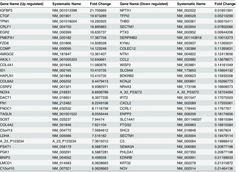
Table 3. List of top 30 up or down regulated genes in hGF cells by TGF-β(T).
Gene Name (Up regulated) Systematic Name Fold Change Gene Name (Down regulated) Systematic Name Fold Change
IGFBP3 NM_001013398 21.705669 NPTX1 NM_002522 0.01651591
CTGF NM_001901 19.973289 TFPI2 NM_006528 0.05219299
TPM1 NM_001018004 19.292925 THBD NM_000361 0.06515411
CRLF1 NM_004750 18.895883 SECTM1 NM_003004 0.07802066
EGR2 NM_000399 18.635737 PTX3 NM_002852 0.09944206
PMEPA1 NM_020182 17.387758 SERPINB2 NM_001143818 0.10013373
FZD8 NM_031866 14.928528 KYNU NM_003937 0.11265631
COMP NM_000095 14.123248 COLEC12 NM_130386 0.11265631
AMIGO2 NM_181847 13.361407 NTN1 NM_004822 0.12413656
AK3L1 NM_001005353 12.640661 CCL2 NM_002982 0.13678671
COL4A1 NM_001845 11.080876 WISP2 NM_003881 0.14161049
ID1 NM_002165 10.410735 SLC9A9 NM_173653 0.15604132
HAPLN1 NM_001884 10.410735 BDKRB2 NM_000623 0.15932008
COL8A2 NM_005202 9.4479413 KCNJ2 NM_000891 0.16266773
CSRP2 NM_001321 8.9382971 NR4A3 NM_173198 0.16608573
NOX4 NM_016931 8.6938789 A_32_P23272 A_32_P23272 0.16724094
DACT1 NM_016651 8.3977335 IFIT2 NM_001547 0.17075503
FN1 NM_212482 8.2249106 CXCL2 NM_002089 0.17555561
FNDC1 NM_032532 8.1116758 CCRL1 NM_178445 0.1767767
TAGLN NM_001001522 8.0556444 ENPP2 NM_006209 0.18174656
SOST NM_025237 7.94474 SLC14A1 NM_001146037 0.18815584
COL4A2 NM_001846 7.621104 PTGS2 NM_000963 0.18815584
C5orf13 NM_004772 7.5684612 SHC3 NM_016848 0.1907824
LDHA NM_005566 7.516182 SECTM1 NM_003004 0.19479114
A_23_P123234 A_23_P123234 7.3615012 C3 NM_000064 0.19888412
PSAT1 NM_058179 6.5887281 SEMA3A NM_006080 0.20877198
PGK1 NM_000291 6.5887281 PHLDA1 NM_007350 0.20877198
BNIP3 NM_004052 6.408559 EDNRB NM_003991 0.21168633
LMCD1 NM_014583 6.0628663 KRT32 NM_002278 0.21315872
C10orf10 NM_007021 6.0628663 NOV NM_002514 0.21464136
Transcriptome profile of OSF shares similarity with areca nut and TGF-
β
regulated profiles in HaCaT and hGF
Since our data indicated differential response of both cell types to areca nut, we hypothesized
that the transcriptome profile of OSF tissues is a combination of areca nut response of both
epi-thelial and fibroblast cells. Therefore, the previously published microarray data of OSF tissues
[
6
] was compared with that of areca nut and/ or TGF-
β
regulated gene expression in HaCaT
[
10
] and hGF cells. The data analysis suggests that majority of the genes regulated by areca nut
in HaCaT cells and common with OSF are TGF-
β
targets (
Fig 3A
–
3F
).
Among the 4666 genes differentially regulated by areca nut in hGF cells; 888 were common
with genes regulated in OSF compared to normal tissues (
Fig 4A and 4B
). Upon areca nut and
TGF-
β
treatment of hGF cells and comparison with differentially regulated genes in OSF, the
number of common genes increased to 1129 (
Fig 4C and 4D
). This suggests combined actions
of areca nut and TGF-
β
on fibroblasts are important in the disease process.
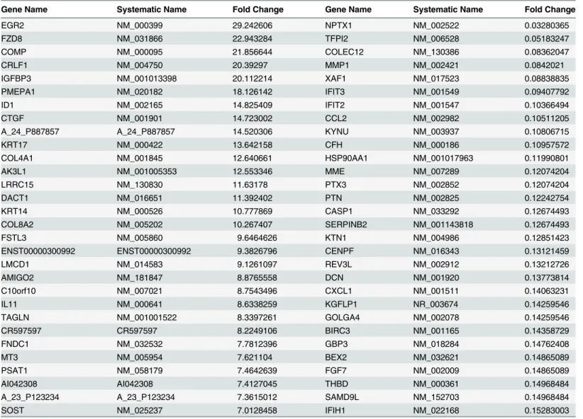
Table 4. List of top 30 up or down regulated genes in hGF cells by areca nut and TGF-β(5H+T).
Gene Name Systematic Name Fold Change Gene Name Systematic Name Fold Change
EGR2 NM_000399 29.242606 NPTX1 NM_002522 0.03280365
FZD8 NM_031866 22.943284 TFPI2 NM_006528 0.05183247
COMP NM_000095 21.856644 COLEC12 NM_130386 0.08362047
CRLF1 NM_004750 20.39297 MMP1 NM_002421 0.0842021
IGFBP3 NM_001013398 20.112214 XAF1 NM_017523 0.08838835
PMEPA1 NM_020182 18.126142 IFIT3 NM_001549 0.09407792
ID1 NM_002165 14.825409 IFIT2 NM_001547 0.10366494
CTGF NM_001901 14.723002 CCL2 NM_002982 0.10511205
A_24_P887857 A_24_P887857 14.520306 KYNU NM_003937 0.10806715
KRT17 NM_000422 13.642158 CFH NM_000186 0.10957572
COL4A1 NM_001845 12.640661 HSP90AA1 NM_001017963 0.11990801
AK3L1 NM_001005353 12.553346 MME NM_007289 0.12074204
LRRC15 NM_130830 11.63178 PTX3 NM_002852 0.12074204
DACT1 NM_016651 11.392402 PTN NM_002825 0.12242754
KRT14 NM_000526 10.777869 CASP1 NM_033292 0.12674493
COL8A2 NM_005202 10.267407 SERPINB2 NM_001143818 0.12674493
FSTL3 NM_005860 9.6464626 KTN1 NM_004986 0.12851423
ENST00000300992 ENST00000300992 9.3826796 CENPF NM_016343 0.13121459
LMCD1 NM_014583 9.1261097 REV3L NM_002912 0.13212726
AMIGO2 NM_181847 8.8765558 DCN NM_001920 0.13773814
C10orf10 NM_007021 8.7543496 CXCL1 NM_001511 0.14063231
IL11 NM_000641 8.6338259 KGFLP1 NR_003674 0.14259546
TAGLN NM_001001522 8.3397261 GOLGA4 NM_002078 0.14259546
CR597597 CR597597 8.2249106 BIRC3 NM_001165 0.14358729
FNDC1 NM_032532 7.7812396 GBP3 NM_018284 0.14762408
MT3 NM_005954 7.621104 BEX2 NM_032621 0.14865089
PSAT1 NM_058179 7.4642639 FGF7 NM_002009 0.14865089
AI042308 AI042308 7.4127045 THBD NM_000361 0.14968484
A_23_P123234 A_23_P123234 7.3615012 SAMD9L NM_152703 0.14968484
SOST NM_025237 7.0128458 IFIH1 NM_022168 0.15283003
Validation of areca nut and TGF-
β
regulated genes in hGF
Some of the differentially expressed genes in OSF [
10
] which were also found to be regulated in
hGF cells by areca nut and/or TGF-
β
were selected for validation by qPCR (
Fig 5
). Connective
tissue growth factor (CTGF), Endothelin (EDN1) and Fibronectin 1(FN1) are over expressed
and implicated in OSF pathogenesis [
4
,
6
,
26
]. These genes are significantly up regulated by
areca nut and TGF-
β
in hGF cells. Similarly, other genes like Early Growth response protein 2
(EGR2), GATA binding protein 6 (GATA6), Collagen 15A1 (COL15A1), Bone morphogenetic
protein 1 (BMP1), Procollagen-lysine,2-oxoglutarate 5-dioxygenase 2 (PLOD2), LIM domain
kinase 1 (LIMK1), Transgelin (TAGLN), Inhibin beta B (INHBB); Insulin growth factor 2
(IGF2), Insulin growth factor binding protein 3 (IGFBP3), Endosialin (CD248) and
Pleiotro-phin (PTN) were also validated as areca nut and TGF-
β
targets.
Fig 1. Areca nut and TGF-βinduced transcriptome profile in fibroblast (hGF) cells.a] Venn diagram representing differentially regulated genes by areca nut and/or TGF-βin hGF cells. b] Hierarchal cluster of the 413 genes commonly regulated in hGF by areca nut water extract (5H) and areca nut with TGF-β(5H+T). c] Hierarchal cluster of 60 genes oppositely regulated in hGF by areca nut (5H) and TGF-β(T). d] Hierarchal cluster of 3153 genes commonly regulated in hGF by areca nut (5H) and areca nut with TGF-β(5H+T) treatment. e] Hierarchal cluster of genes (494) whose regulation by TGF-β(T) is not influenced by the addition of areca nut (5H). In all hierarchal clusters; red, green and black colours represent up, down and un-regulated genes respectively. The rows represent genes and columns represent various treatments of areca nut on hGF cells (5H; 5μg/ml) and/or TGF-β(T; 5 ng/ml).
doi:10.1371/journal.pone.0129252.g001
Fig 2. Areca nut induces different gene expression profiles in fibroblast and epithelial cells.Venn diagram representation of genes regulated by areca nut in epithelial (HaCaT, GSE 38227) and fibroblast cells (hGF). 457 genes are commonly regulated by areca nut in both the cell types while bulk of the differentially regulated genes (1152 in HaCaT and 4209 in hGF) are mutually exclusive.
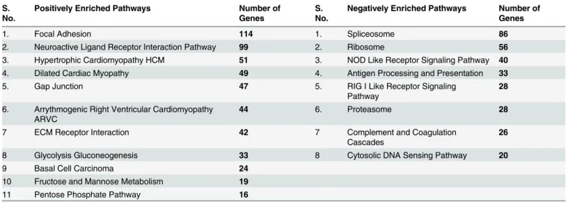
Gene Set Enrichment Analysis (GSEA) of differentially regulated genes
by areca nut and TGF-
β
revealed pathways that are common with OSF
GSEA was performed to further explore whether areca nut and TGF-
β
regulated pathways in
hGF were common with the previously reported differentially regulated pathways in OSF [
6
].
Interestingly, all the positively enriched pathways by areca nut and TGF-
β
in hGF cells were
differentially regulated in OSF also (
Table 5
). These pathways may have important role in OSF
manifestation. In contrast none of the negatively enriched pathways in hGF were differentially
regulated in OSF (
Table 5
) and therefore, may not contribute to OSF pathology. Hence, this
corroborated the role of areca nut and TGF-
β
in manifesting fibrotic phenotype.
Areca nut enhances TGF-
β
mediated fibroblast activation
Fibroblast activation is a hall mark of fibrotic disorders. We therefore confirmed the over
expression of myofibroblast markers Alpha smooth muscle actin (
α
SMA) and Gamma smooth
muscle actin (
γ
SMA) in OSF patients (
Fig 6A
). In light of the observation of areca nut and
TGF-
β
induced profile in hGF being similar to OSF, we studied the activation of hGF cells by a
combination of both areca nut and TGF-
β
. Both
α
SMA and
γ
SMA expression was more by
Fig 3. Genes regulated by areca nut in epithelial cells and OSF are via TGF-β.Previously published transcriptome profile of OSF tissues (GSE 20170) [6] was compared with that of areca nut and/ or TGF-βregulated transcriptome in HaCaT (GSE 38227) [10] and hGF cells. a & b] Venn diagram and hierarchal cluster representing distribution of differentially or commonly regulated genes by areca nut in HaCaT cells and OSF. c & d] Venn diagram and hierarchal cluster representing distribution of differentially or commonly regulated genes by TGF-βin HaCaT cells and OSF. e] Venn diagram represents the 252 genes common between the 362 (252+104) genes regulated by areca nut in HaCaT and OSF and the 731 (252+ 473) genes regulated by TGF-βin HaCaT cells and OSF. f] Out of the 252 genes discussed in 3e, 182 genes are not regulated by areca nut in presence of ALK5 inhibitor (SB 431542) in HaCaT cells. This indicates that these 182 genes are regulated by areca nut are via TGF-βin HaCaT cells and possibly in OSF. In all hierarchal clusters; red, green and black colours represent up, down and un-regulated genes respectively. The rows represent genes and columns represent OSF and various treatments of areca nut (5H; 5μg/ml) and/or TGF-β(T; 5 ng/ml).Fig 4. Areca nut and TGF-βinduced transcriptome profile in fibroblast (hGF) cells is similar to OSF.a & b] Venn diagram and hierarchal cluster representing genes commonly or differentially regulated by areca nut in hGF cells and OSF. c & d] Venn diagram and hierarchal cluster representing genes commonly or differentially regulated in hGF by areca nut with TGF-β(5H+T) and OSF. In all hierarchal clusters; red, green and black colours represent up, down and un-regulated genes respectively. The rows represent genes and columns represent OSF and various treatments of areca nut (5H; 5μg/ml) and/or TGF-β(T; 5 ng/ml).
doi:10.1371/journal.pone.0129252.g004
Fig 5. Validation of areca nut and TGF-βregulated genes in hGF cells.Serum starved hGF cells were treated with areca nut water extract (5H; 5μg/ml), TGF-β(T; 5 ng/ml) and both together (5H+T) for 72 hours (n = 3) followed by the study of gene expression changes using qPCR. Y-axis represents the fold changes of each treatment. P values0.0001,0.001,0.01 are depicted as***,**and*respectively.
areca nut and TGF-
β
treatment than with each of them alone (
Fig 6B
). Collagen contraction
assay was performed to confirm that this potentiation in the expression of myofibroblast
mark-ers by areca nut and TGF-
β
translated into a stronger contractile phenotype. Areca nut along
with TGF-
β
decreased the surface area of hGF populated collagen gels to a significantly greater
extent than areca nut or TGF-
β
alone (
Fig 6C
). Therefore, TGF-
β
and areca nut together could
enhance fibroblast activation thereby increasing contractility of the cells which is also
impli-cated in OSF [
13
].
Areca nut actions on fibroblasts are enhanced by epithelial
mesenchymal interaction via TGF-
β
Areca nut has been shown to induce TGF-
β
in epithelial cells [
10
] and also enhances fibroblast
activation in combination with TGF-
β
invoking a possible epithelial mesenchymal interaction
in the initiation of OSF. Therefore, to test this possibility
in-vitro
, HaCaT cells were treated
with areca nut with or without ALK5 inhibitor (TGF
β
RI inhibitor). The condition medium of
these cells was used to treat serum deprived hGF cells to study the effect of epithelial factors
induced by areca nut, in particular TGF-
β
.
Qunatitative RT-PCR was performed to confirm the induction of TGF-
β
by areca nut and
compromised by ALK5 inhibitor in HaCaT cells (
Fig 7A
). PCR studies were performed on
areca nut treated hGF cells to evaluate the expression of TGF-
β
ligands and receptors.
Treat-ment of areca nut on hGF cells did not induce any of the three TGF-
β
ligands nor influenced
the expression of TGF-
β
receptor isoforms (
Fig 7B
). This also corroborates with the microarray
data of hGF cells treated with areca nut wherein none of the TGF-
β
ligands or receptors were
found to be regulated by areca nut in hGF cells.
Treatment of hGF with conditioned medium of areca nut treated HaCaT cells induced
α
SMA,
γ
SMA, Thrombospondin 1 (THBS1); Transglutaminase 2 (TGM2); Transmembrane
prostrate androgen-induced protein (TMEPAI); Transforming growth factor
β
induced
(TGFBI); CTGF; PLOD2; BMP1; LIMK1; LOXL3 and EDN1 to a significantly higher extent
than control. This induction was compromised by ALK5 inhibitor highlighting the
involve-ment of TGF-
β
in the secretome (
Fig 8
). Interestingly, over expression of these genes has been
observed in OSF [
6
]. To corroborate this, staining for
α
SMA and total collagen was performed
Table 5. Gene Set Enrichment analysis of differentially regulated genes in hGF cells by areca nut and TGF-β.S. No.
Positively Enriched Pathways Number of Genes
S. No.
Negatively Enriched Pathways Number of Genes
1. Focal Adhesion 114 1. Spliceosome 86
2. Neuroactive Ligand Receptor Interaction Pathway 99 2. Ribosome 56
3. Hypertrophic Cardiomyopathy HCM 51 3. NOD Like Receptor Signaling Pathway 40
4. Dilated Cardiac Myopathy 49 4. Antigen Processing and Presentation 33
5. Gap Junction 47 5. RIG I Like Receptor Signaling
Pathway
28
6. Arrythmogenic Right Ventricular Cardiomyopathy ARVC
44 6. Proteasome 28
7 ECM Receptor Interaction 42 7 Complement and Coagulation
Cascades
26
8 Glycolysis Gluconeogenesis 33 8 Cytosolic DNA Sensing Pathway 20
9 Basal Cell Carcinoma 24
10 Fructose and Mannose Metabolism 19
11 Pentose Phosphate Pathway 16
on hGF cells following the same treatment protocol. Areca nut induced factors in HaCaT cells
increased the protein expression of
α
SMA and total collagen more than that of control and
direct treatment of areca nut or TGF-
β
on hGF cells. This increase was obliterated in the
pres-ence of ALK5 inhibitor (
Fig 9A, 9B and 9C
). This is in line with the mRNA expression data
dis-cussed earlier.
Taken together, these data suggest that areca nut induced secretory factors by HaCaT cells
are able to induce myofibroblast phenotype akin to OSF. Moreover, TGF-
β
is responsible for
this phenotype in the areca nut induced secretome.
Epithelial factors maintain basal expression of pro-fibrotic genes in
fibroblasts
Comparison of the basal expression of fibroblast activation markers in hGF cells cultured in
serum deprived medium and in conditioned medium of untreated HaCaT cells was also done.
Expression of pro fibrotic genes
α
SMA,
γ
SMA, TGM2, TGFBI, CTGF, PLOD2, BMP1, LIMK1,
Fig 6. Areca nut enhances TGF-βmediated fibroblast activation.a] Scatter plots of qPCR evaluation of myofibroblast markersαSMA andγSMA in normal and OSF tissues. Each square represents expression in one sample and horizontal line represents the median expression. Both the genes are up regulated in OSF as compared to normal tissues. P values calculated using Wilcoxon signed rank test are<0.0066 forγSMA and 0.03053 forαSMA. b] qPCR analysis ofγSMA andαSMA regulation by areca nut and/or TGF-βin hGF cells. c] Estimation of collagen contraction of hGF populated collagen gel upon areca nut and/or TGF-βtreatment. Areca nut (5H), TGF-β(T) and areca nut together with TGF-β(5H+T) decrease the collagen surface area by 233.599 mm2, 191.663 mm2and 136.633 mm2respectively as compared to control (290.941 mm2). Areca nut and TGF-βtogether (5H+T) decrease the surface areaof collagen gel significantly more than areca nut (5H) or TGF-β(T) alone. Representative images of the decrease in surface area by each of the treatments are given below the graph. P values0.0001,0.001,0.01 are depicted as***,**and*respectively. UN- untreated, areca nut- 5H; 5μg/ml, T–TGF-β;
5 ng/ml.
LOXL3 and EDN1 were found to be down regulated in hGF cells treated with condition
medium of untreated HaCaT cells (
Fig 10
). These observations suggest a role for epithelium in
maintaining the normal fibroblast phenotype.
Discussion
Oral submucous fibrosis (OSF) is a condition affecting habitual chewers of areca nut. Our
pre-vious work has indicated that areca nut extract as well as its alkaloid and polyphenol fractions
induce TGF-
β
in epithelial cells [
10
]. Hence, we hypothesized that fibroblasts may respond not
only to areca nut but also to TGF-
β
, to attain a phenotype similar to OSF. In tune with this,
transcriptome profiles suggested that areca nut and TGF-
β
together potentiate the regulation
of genes in human gingival fibroblast (hGF) cells.
Epithelial atrophy and increase in fibroblast population along with deposition of excess
extra cellular matrix are hallmarks of OSF [
2
]. In line with this, differential response of
epithe-lial and fibroblast cells to areca nut also implied that these cell types play different roles in the
disease process. Additionally, both epithelium and fibroblast cells can be implicated in OSF
manifestation as areca nut and TGF-
β
regulated transcriptome profiles of HaCaT and hGF
cells overlapped significantly with OSF profile. Moreover, areca nut and TGF-
β
were found to
enrich pathways in hGF cells which are differentially regulated in OSF; notably metabolic and
Fig 7. Areca nut induces TGF-βin HaCaT but not in hGF cells.a] Serum deprived HaCaT cells were treated with areca nut (5H; 5μg/ml) with or without ALK5 inhibitor (10μM of SB 431542) as described in material and methods. Expression of TGF-βand its activator THBS1 was analyzed by qPCR. Areca nut induced TGF-βand THBS1 which was compromised by ALK5inhibitor (SB 431542) P values0.0001,0.001,0.01 are depicted as*** **and*respectively. b] Serum deprived hGF cells were treated with areca nut and TGF-β(5H; 5μg/ml; 10H; 10μg/ml; TGF-β; T; 5 ng/ml). Expression of TGF-β
ligands and receptor isoforms were assessed by semi quantitative PCR. Areca nut treatment did not induce TGF-βligands and receptor isoforms. TGF-β
treatment induced TGF-β1 isoform in hGF cells.
matrix associated pathways. They also regulated the expression of pro-fibrotic growth factors;
CTGF, FN1, EDN1, collagen stabilizing and maturation genes; PLOD2, BMP1 and cytoskeletal
reorganizing genes; LIMK1 and TAGLN and transcription factors GATA6, EGR2 in
fibro-blasts. EGR2 is reported to mediate pro-fibrotic actions of TGF-
β
in pulmonary fibrosis [
27
].
Expression of all these genes may have important implications in the progression of OSF.
Our study revealed that areca nut and TGF-
β
can confer enhanced contractile phenotype
(hallmark of fibroblasts in various fibrotic disorders) as well as induce myofibroblast markers
α
SMA and
γ
SMA in hGF cells. The expression of
γ
SMA in OSF has not been reported and
TGF-
β
is known to induce
γ
SMA in prostrate myofibroblasts [
28
].
Our data also highlights that direct treatment of areca nut does not regulate TGF-
β
ligands
and receptors in hGF cells. This is in line with our previously published report that areca nut
does not induce pSMAD2 (read out of activated TGF-
β
signaling) in hGF cells [
10
]. In
addi-tion, we provide proof of epithelial- mesenchymal interaction which is mediated via TGF-
β
induced by areca nut in epithelial cells. This suggests that areca nut induced secretory factors
by HaCaT cells could activate fibroblasts and induce genes which play important roles in
mani-festation of OSF. Corroborating these data; areca nut induced secretome by HaCaT cells also
Fig 8. Areca nut actions on fibroblasts are enhanced by epithelial mesenchymal interaction via TGF-β.To study the epithelial mesenchymal interaction, confluent cultures of HaCaT cells were serum starved for 24 hours followed by 10μM ALK5 inhibitor (TGFβRI inhibitor, SB 431542, Sigma-Aldrich, USA) treatment 2 hours prior to areca nut treatment (5μg/ml). Meanwhile hGF cells were serum deprived for 24 hours such that the treatment time point coincided with completion of 48 hour treatment on HaCaT cells. At this time point, the condition medium of areca nut (with or without ALK5 inhibitor; SB 431542) treated HaCaT cells was transferred to hGF cells and simultaneously direct treatment of areca nut with or without ALK5 inhibitor; SB 431542 was also performed and both were maintained for 48 hours and gene expression was studied by qPCR. The bar diagrams represent regulation ofαSMA/ACTA2,γSMA/ACTG2, THBS1, TGM2, TMEPAI, TGFBI, CTGF, PLOD2, BMP1, LIMK1, LOXL3 and EDN1 in hGF cells upon treatment with 1- untreated; 2- areca nut, 3- ALK5 inhibitor(SB 431542), 4- areca nut with ALK5 inhibitor (SB 431542) (white bars), 5- condition media of untreated HaCaT cells, 6- condition media of areca nut treated HaCaT cells, 7- condition media of ALK5 inhibitor (SB 431542) treated HaCaT cells and 8- condition media of areca nut with ALK5 inhibitor (SB 431542)treated HaCaT cells (black bars). P values<0.0001,<0.001,<0.01 are depicted as***,**and*respectively.
increased protein expression of
α
SMA and collagen which was abrogated with ALK5 inhibitor
providing further evidence of TGF-
β
’
s contribution. This is similar to the role of injured
epithe-lium and epithelial cell- fibroblast interaction in the manifestation of pulmonary and liver
fibrosis [
29
,
30
]. Arecoline has also been shown to injure epithelial cells via ROS induction and
induce cell cycle arrest [
31
]. In light of our data and these studies, we propose that constant
injury inflicted by areca nut and its constituents to the epithelium may drive OSF.
Down-regulation of genes in the fibrosis pathway when hGF cells treated with condition
medium of untreated HaCaT cells is intriguing. This suggests that epithelial cells can suppress
an inherent capability of fibroblasts to activate the fibrotic/wound repair program. This is
cor-roborated by the report that mouse lung epithelial cell derived secretory factors can suppress
fibroblast growth whereas bleomycin mouse model derived epithelial cell factors promote
growth of fibroblasts
in vitro
[
32
]. Further studies are needed to identify the factor (s)
responsi-ble for the suppression of fibrosis related genes, which may lead to a potential therapeutic
target.
Fig 9. Epithelial secretome induces fibroblast activation and collagen via TGF-β.Condition medium of HaCaT cells treated with areca nut with or without ALK5 inhibitor; SB 431542 (-/UN; condition media of untreated HaCaT cells,-/5H, areca nut treated HaCaT cells,-/ALK5; ALK5 inhibitor treated HaCaT cells and-/5H+ALK5 inhibitor treated HaCaT cells was used to treat serum deprived hGF cells for 48 hours. Simultaneous direct treatment of areca nut with or without ALK5 inhibitor (SB 431542) (UN/-, 5H/-, ALK5/-, 5H+ALK5/-) was given to another set of serum deprived hGF cells for the same duration. Fibroblast activation and total collagen was assessed byαSMA stress fiber formation by immunocytochemistry and direct red 80 staining respectively. a] Condition media of areca nut treated HaCaT cells (-/5H) inducedαSMA stress fibres significantly more as compared to untreated (-/UN) and direct treatment of hGF cells with areca nut (5H/-). It got compromised with ALK5 inhibitor, SB 431542 (-/ALK5). Direct treatment of TGF-βwith (T+ALK/-) or without ALK5 inhibitor (T/-) was used as positive control for the experiment (image magnification 63X). b] Representative images for total collagen staining by direct red 80 (image magnification 10X) expressed in hGF cells upon respective treatments depicted above each panel. The treatments are as described in 4a. Note the significant increase in the collagen staining when hGF cells were treated directly with TGF-βor conditioned media of HaCaT cells treated with areca nut. Both these regulations were compromised in the presence of ALK5 inhibitor (SB 431542). c] Bar diagram showing quantitation of direct red staining for total collagen measured as O.D per 105cells of treatments described in 4b.
Fig 10. Epithelial factors maintain basal expression of pro-fibrotic genes in fibroblasts.Condition medium of untreated HaCaT cells (-/UN, white bars) was used to treat serum deprived hGF cells for 48 hours. Simultaneously, hGF cells were maintained in serum free medium as control (UN/-, black bars). Expression ofαSMA/ACTA2,γSMA/ACTG2, TGM2, TGFBI, CTGF, PLOD2, BMP1, LIMK1, LOXL3 and EDN1 genes significantly decreased upon treatment with condition medium of untreated HaCaT cells.
doi:10.1371/journal.pone.0129252.g010
Fig 11. A model on the role of areca nut and TGF-βin OSF progression.Areca nut can induce and activate TGF-βin epithelial cells which can act together on the fibroblast cells and induce expression of other pro-fibrotic cytokines (Endothelin and CTGF). These cytokines can further enhance the fibrotic response and aid in conversion of fibroblasts to myofibroblasts expressingγSMA andαSMA markers. Areca nut and TGF-βcan influence expression of cytoskeletal reorganizing protein LIMK1. The overall collagen production shall also increase. Collagen maturation and stabilizing enzymes (BMP1 and PLOD2 respectively) can also be induced by areca nut along with TGF-β. All these changes may lead to excessive deposition of extracellular matrix characteristic of OSF.
Conclusion
This study provides a comprehensive over view of fibroblast response to areca nut and TGF-
β
.
We propose an important role of epithelium in OSF progression. Areca nut insult to the
epithe-lium may injure the epitheepithe-lium as well as induce pro-fibrotic factors; primarily TGF-
β
which
along with areca nut alters the fibroblast phenotype by activation of a fibrogenic gene
expres-sion profile (
Fig 11
).
Acknowledgments
The authors would like to acknowledge University Grants Commission; DST-FIST and
Department of Biotechnology, Government of India for providing infrastructural support to
the department. This research is funded by the Departments of Science and Technology and
Biotechnolgy, Government of India. IP is recipient of a fellowship from Council of Scientific
and Industrial Research, New Delhi. The funders had no role in study design, data collection
and analysis, decision to publish, or preparation of the manuscript.
Author Contributions
Conceived and designed the experiments: IP PK. Performed the experiments: IP IK. Analyzed
the data: IP NK PK. Contributed reagents/materials/analysis tools: SGR PK. Wrote the paper:
IP PK.
References
1. Cox SC, Walker DM. Oral submucous fibrosis. A review. Aust Dent J. 1996; 41: 294–299. PMID:
8961601
2. Pindborg JJ, Sirsat SM. Oral submucous fibrosis. Oral Surg Oral Med Oral Pathol. 1996; 22: 764–779.
3. Pindborg JJ, Murti PR, Bhonsle RB, Gupta PC, Daftary DK, Mehta FS. Oral submucous fibrosis as a precancerous condition. Scand J Dent Res. 1984; 92: 224–229. PMID:6589738
4. Utsunomiya H, Tilakaratne WM, Oshiro K, Maruyama S, Suzuki M, Ida-Yonemochi H, et al. Extracellu-lar matrix remodeling in oral submucous fibrosis: its stage-specific modes revealed by immunohis-tochemistry and in situ hybridization. J Oral Pathol Med. 2005; 34: 498–507. PMID:16091118
5. Haque MF, Meghji S, Khitab U, Harris M. Oral submucous fibrosis patients have altered levels of cyto-kine production. J Oral Pathol Med. 2000; 29: 123–128. PMID:10738939
6. Khan I, Agarwal P, Thangjam GS, Radhesh R, Rao SG, Kondaiah P. Role of TGF-beta and BMP7 in the pathogenesis of oral submucous fibrosis. Growth Factors. 2011; 29: 119–127. doi:10.3109/
08977194.2011.582839PMID:21591998
7. Sinor PN, Gupta PC, Murti PR, Bhonsle RB, Daftary DK, Mehta FS, et al. A case-control study of oral submucous fibrosis with special reference to the etiologic role of areca nut. J Oral Pathol Med. 1990; 19: 94–98. PMID:2341977
8. Jacob BJ, Straif K, Thomas G, Ramadas K, Mathew B, Zhang ZF, et al. Betel quid without tobacco as a risk factor for oral precancers. Oral Oncol. 2004; 40: 697–704. PMID:15172639
9. Sumeth Perera MW, Gunasinghe D, Perera PA, Ranasinghe A, Amaratunga P, Warnakulasuriya S, et al. Development of an in vivo mouse model to study oral submucous fibrosis. J Oral Pathol Med. 2007; 36: 273–280. PMID:17448137
10. Khan I, Kumar N, Pant I, Narra S, Kondaiah P. Activation of TGF-beta Pathway by Areca Nut Constitu-ents: A Possible Cause of Oral Submucous Fibrosis. PLoS One. 2012; 7(12): e51806. doi:10.1371/ journal.pone.0051806PMID:23284772
11. Meghji S, Scutt A, Harvey W, Canniff JP. An in-vitro comparison of human fibroblasts from normal and oral submucous fibrosis tissue. Arch Oral Biol. 1987; 32: 213–215. PMID:3478024
12. Harvey W, Scutt A, Meghji S, Canniff JP. Stimulation of human buccal mucosa fibroblasts in vitro by betel-nut alkaloids. Arch Oral Biol. 1986; 31: 45–49. PMID:3458437
13. Chang MC, Lin LD, Wu HL, Ho YS, Hsien HC, Wang TM, et al. Areca nut induced buccal mucosa fibro-blast contraction and its signaling: a potential role in oral submucous fibrosis—a precancer condition.
14. Jeng JH, Kuo ML, Hahn LJ, Kuo MY. Genotoxic and non-genotoxic effects of betel quid ingredients on oral mucosal fibroblasts in vitro. J Dent Res. 1994; 73: 1043–1049. PMID:8006230
15. IARC. Betel-quid and areca-nut chewing and some areca-nut derived nitrosamines. IARC Monogr Eval Carcinog Risks Hum. 2004; 85: 1–334. PMID:15635762
16. Nigam M, Ranjan V, Srivastava S, Sharma R, Balapure AK. Centchroman induces G0/G1 arrest and caspase-dependent apoptosis involving mitochondrial membrane depolarization in MCF-7 and MDA MB-231 human breast cancer cells. Life Sci. 2008; 82: 577–590. doi:10.1016/j.lfs.2007.11.028PMID:
18279897
17. Boukamp P, Petrussevska RT, Breitkreutz D, Hornung J, Markham A, Fusenig NE. Normal keratiniza-tion in a spontaneously immortalized aneuploid human keratinocyte cell line. J Cell Biol. 1988; 106: 761–771. PMID:2450098
18. Inman GJ, Nicolas FJ, Callahan JF, Harling JD, Gaster LM, Reith AD, et al. SB-431542 is a potent and specific inhibitor of transforming growth factor-beta superfamily type I activin receptor-like kinase (ALK) receptors ALK4, ALK5, and ALK7. Mol Pharmacol. 2002; 62: 65–74. PMID:12065756
19. Ranganathan P, Agrawal A, Bhushan R, Chavalmane AK, Kalathur RK, Takahashi T, et al. Expression profiling of genes regulated by TGF-beta: differential regulation in normal and tumour cells. BMC Geno-mics. 2007; 8: 98. PMID:17425807
20. Smyth GK. Linear models and empirical bayes methods for assessing differential expression in micro-array experiments. Stat Appl Genet Mol Biol. 2004; 3: Article3.
21. Saeed AI, Sharov V, White J, Li J, Liang W, Bhagabati N, et al. TM4: a free, open-source system for microarray data management and analysis. Biotechniques. 2003; 34: 374–378. PMID:12613259
22. Subramanian A, Tamayo P, Mootha VK, Mukherjee S, Ebert BL, Gillette MA, et al. Gene set enrichment analysis: a knowledge-based approach for interpreting genome-wide expression profiles. Proc Natl Acad Sci U S A. 2005; 102: 15545–15550. PMID:16199517
23. Ngo P, Ramalingam P, Phillips JA, Furuta GT. Collagen gel contraction assay. Methods Mol Biol. 2006; 341: 103–109. PMID:16799192
24. Sehgal P, Kumar N, Praveen Kumar VR, Patil S, Bhattacharya A, Vijaya Kumar M, et al. Regulation of protumorigenic pathways by insulin like growth factor binding protein2 and its association along withβ -catenin in breast cancer lymph node metastasis. Molecular Cancer. 2013; 12:63. doi: 10.1186/1476-4598-12-63PMID:23767917
25. Tullberg-Reinert H, Jundt G. In situ measurement of collagen synthesis by human bone cells with a sir-ius red-based colorimetric microassay: effects of transforming growth factor beta2 and ascorbic acid 2-phosphate. Histochem Cell Biol. 1999; 112: 271–276. PMID:10550611
26. Xu C, Peng X, Liu S, Fang C. Quantitative and immunohistochemical analysis of endothelin-1 in oral submucous fibrosis. Hua Xi Kou Qiang Yi Xue Za Zhi. 2000; 18: 394–396, 418. PMID:12539469
27. Fang F, Ooka K, Bhattacharyya S, Wei J, Wu M, Du P, et al. The early growth response gene Egr2 (Alias Krox20) is a novel transcriptional target of transforming growth factor-beta that is up-regulated in systemic sclerosis and mediates profibrotic responses. Am J Pathol. 2011; 178: 2077–2090. doi:10.
1016/j.ajpath.2011.01.035PMID:21514423
28. Untergassera G, Gandera R, Lilga C, Lepperdingera G, Plasb E, Berger P, et al. Profiling molecular tar-gets of TGF-beta1 in prostate fibroblast-to-myofibroblast transdifferentiation. Mech Ageing Dev. 2005; 126: 59–69. PMID:15610763
29. Novo E, Cannito S, Paternostro C, Bocca C, Miglietta A, Parola M. Cellular and molecular mechanisms in liver fibrogenesis. Arch Biochem Biophys. 2014; 548: 20–37. doi:10.1016/j.abb.2014.02.015PMID:
24631571
30. Norihiko S, Tager AM. Fibrosis of two: Epithelial cell-fibroblast interactions in pulmonary fibrosis. Bio-chim Biophys Acta. 2013; 1832: 911–921. doi:10.1016/j.bbadis.2013.03.001PMID:23499992
31. Thangjam GS, Kondaiah P. Regulation of oxidative-stress responsive genes by arecoline in human keratinocytes. J Periodontal Res. 2009; 44: 673–682. doi:10.1111/j.1600-0765.2008.01176.xPMID:
19364390

![Fig 1. Areca nut and TGF-β induced transcriptome profile in fibroblast (hGF) cells. a] Venn diagram representing differentially regulated genes by areca nut and/or TGF-β in hGF cells](https://thumb-eu.123doks.com/thumbv2/123dok_br/17316766.249508/9.918.59.718.115.369/induced-transcriptome-profile-fibroblast-diagram-representing-differentially-regulated.webp)
![Fig 3. Genes regulated by areca nut in epithelial cells and OSF are via TGF-β. Previously published transcriptome profile of OSF tissues (GSE 20170) [6] was compared with that of areca nut and/ or TGF-β regulated transcriptome in HaCaT (GSE 38227) [10] and](https://thumb-eu.123doks.com/thumbv2/123dok_br/17316766.249508/10.918.59.744.114.555/regulated-epithelial-previously-published-transcriptome-compared-regulated-transcriptome.webp)
![Fig 6. Areca nut enhances TGF-β mediated fibroblast activation. a] Scatter plots of qPCR evaluation of myofibroblast markers αSMA and γSMA in normal and OSF tissues](https://thumb-eu.123doks.com/thumbv2/123dok_br/17316766.249508/13.918.65.711.117.596/enhances-mediated-fibroblast-activation-scatter-evaluation-myofibroblast-tissues.webp)
![Fig 7. Areca nut induces TGF-β in HaCaT but not in hGF cells. a] Serum deprived HaCaT cells were treated with areca nut (5H; 5 μg/ml) with or without ALK5 inhibitor (10 μM of SB 431542) as described in material and methods](https://thumb-eu.123doks.com/thumbv2/123dok_br/17316766.249508/14.918.60.727.119.574/induces-hacat-deprived-treated-inhibitor-described-material-methods.webp)
