Oxygen-Sensitive K
Channels Modulate
Human Chorionic Gonadotropin Secretion
from Human Placental Trophoblast
Paula Díaz1,2*, Colin P. Sibley1,2, Susan L. Greenwood1,2
1Maternal and Fetal Health Research Centre, Institute of Human Development, The University of Manchester, Manchester Academic Health Science Centre, Manchester, United Kingdom,2St. Mary's Hospital, Central Manchester University Hospitals NHS Foundation Trust, Manchester, United Kingdom
Abstract
Human chorionic gonadotropin (hCG) is a key autocrine/paracrine regulator of placental syncytiotrophoblast, the transport epithelium of the human placenta. Syncytiotrophoblast hCG secretion is modulated by the partial pressure of oxygen (pO2), reactive oxygen
spe-cies (ROS) and potassium (K+) channels. Here we test the hypothesis that K+channels mediate the effects ofpO2and ROS on hCG secretion. Placental villous explants from
nor-mal term pregnancies were cultured for 6 days at 6% (normoxia), 21% (hyperoxia) or 1% (hypoxia)pO2. On days 3–5, explants were treated with 5mM 4-aminopyridine (4-AP) or
tet-raethylammonium (TEA), blockers ofpO2-sensitive voltage-gated K+(KV) channels, or
ROS (10–1000μM H2O2). hCG secretion and lactate dehydrogenase (LDH) release, a
marker of necrosis, were determined daily. At day 6, hCG and LDH were measured in tissue lysate and86Rb (K+) efflux assessed to estimate syncytiotrophoblast K+permeability. hCG secretion and86Rb efflux were significantly greater in explants maintained in 21%pO2than
normoxia. 4-AP/TEA inhibited hCG secretion to a greater extent at 21% than 6% and 1%
pO2, and reduced86Rb efflux at 21% but not 6%pO2. LDH release and tissue LDH/hCG
were similar in 6%, 21% and 1%pO2and unaffected by 4-AP/TEA. H2O2stimulated86Rb
efflux and hCG secretion at normoxia but decreased86Rb efflux, without affecting hCG secretion, at 21%pO2. 4-AP/TEA-sensitive K+channels participate inpO2-sensitive hCG
secretion from syncytiotrophoblast. ROS effects on both hCG secretion and86Rb efflux are
pO2-dependent but causal links between the two remain to be established.
Introduction
The endocrine and nutrient transport functions of the human placenta depend on appropriate maintenance of syncytiotrophoblast, a highly specialised multinucleate epithelial cell. Syncytio-trophoblast has a short life span and is renewed during pregnancy by cellular turnover. Prolif-erative mononucleate cytotrophoblasts exit the cell cycle, differentiate and fuse into the overlying syncytiotrophoblast and then aged syncytial nuclei are removed, possibly by OPEN ACCESS
Citation:Díaz P, Sibley CP, Greenwood SL (2016) Oxygen-Sensitive K+Channels Modulate Human Chorionic Gonadotropin Secretion from Human Placental Trophoblast. PLoS ONE 11(2): e0149021. doi:10.1371/journal.pone.0149021
Editor:Manu Vatish, University of Oxford, UNITED KINGDOM
Received:October 27, 2015
Accepted:January 26, 2016
Published:February 10, 2016
Copyright:© 2016 Díaz et al. This is an open access article distributed under the terms of the Creative Commons Attribution License, which permits unrestricted use, distribution, and reproduction in any medium, provided the original author and source are credited.
Data Availability Statement:All relevant data are within the paper.
Funding:This work was supported by Tommy’s, the baby charity, CONICYT-Becas Chile 72090593 and Action Medical Research. The funders had no role in study design, data collection and analysis, decision to publish, or preparation of the manuscript.
apoptosis and autophagy, to complete turnover [1,2]. In normal pregnancy these processes are highly coordinated but in pregnancies complicated by pre-eclampsia [3,4], fetal growth restric-tion [4] and maternal obesity [5], an imbalance in cell turnover dysregulates syncytiotropho-blast renewal which compromises function and contributes to maternal and fetal mortality, and morbidity associated with these pregnancy complications.
Cell turnover to renew syncytiotrophoblast is maintained by several hormones including human chorionic gonadotrophin (hCG). hCG is an autocrine/paracrine regulator of syncytio-trophoblast renewal, acting via the G-protein coupled luteinizing hormone/hCG-receptor to elevate cAMP/protein kinase A and promote cytotrophoblast differentiation [6], gap junction communication and cellular fusion to form multinucleated syncytia [7]. hCG is synthesized and secreted by terminally differentiated syncytiotrophoblast and promotes continued tropho-blast renewal by positive feedback. Thus appropriate regulation of hCG synthesis and secretion is essential for maintenance of syncytiotrophoblast and successful pregnancy.
Syncytiotrophoblast hCG secretion is modulatedin vitroby oxygen tension (pO2) and
reac-tive oxygen species (ROS). LoweringpO2inhibited hCG secretion by villous explants [8] and
by primary cultures of cytotrophoblasts [9] from normal term placentas. Hydrogen peroxide (H2O2) treatment of cytotrophoblasts, to generate oxidative stress, inhibited hCG secretion at
high (>50μM) but markedly stimulated secretion at lower (1–50μM) concentrations [10].
Modulation of hCG secretion by these factors is likely to be of pathophysiological significance in pre-eclampsia and fetal growth restriction as altered placentalpO2and increased placental
oxidative stress are associated with these conditions [11–13]. Indeed, increased levels of mark-ers of oxidative stress are found in placental tissue from women with pre-eclampsia [14,15] as well as elevated serum levels of H2O2compared to normal pregnancies [16]. In addition to
modulating hCG secretion, alteredpO2and elevated ROS also dysregulate syncytiotrophoblast
turnoverin vitro[8,17,18], but the underlying mechanism/s are currently unexplored. hCG secretion by term placental trophoblast involves constitutive release [19] and Ca2
+-dependent exocytosis [20]. Accordingly, the regulated component of hCG secretion is
modu-lated by factors that influence intracellular Ca2+, including ion channels. We have previously shown that pharmacological blockade of Ca2+entry channels [21] and voltage-gated K+(KV)
channels, inhibit hCG secretion from placental villous explants and isolated cytotrophoblasts [22]. Blocking KVchannels also inhibits trophoblast fusion to form multinucleate syncytia [22]
suggesting that activity of these channels is required both for hCG secretion and syncytiotro-phoblast renewal.
The KVchannel family comprises 11 members [23], and the expression/activity of some KV
channel subunits is acutely and chronically modulated bypO2[24,25].pO2-sensitive KV
chan-nels close in response to loweredpO2, raising the possibility that the reduction in hCG
secre-tion from syncytiotrophoblast under hypoxic condisecre-tions is a result of blocking KVchannels.
Furthermore, long term exposure to oxidative stress (ROS) alters K+channel expression/activ-ity and acute exposure has direct effects on K+channel proteins to alter their activity [26,27]. The effects of H2O2are diverse and depend on tissue type; H2O2has been reported to both
close [28] and open [29,30] KVchannels. As KVchannels are modulated by ROS in
non-pla-cental tissue, it is plausible that ROS regulate syncytiotrophoblast hCG secretion through effects on KVchannels.
Here we test the hypothesis thatpO2and/or ROS regulate hCG secretion through an effect
on K+channels. Using placental villous tissue from normal term pregnancy we compared the effect of KVchannel blockers on hCG secretion and86Rb efflux (a marker of K+permeation
through ion channels) from villous explants maintained at placental normoxia (6%pO2), with
extreme hypoxia (1%pO2) and hyperoxia (21%pO2). We also investigated the effect of H2O2,
Materials and Methods
Materials
Unless otherwise stated, all chemicals were from Sigma-Aldrich (Poole, UK).
Ethics Statement
Human placentas used in this study were obtained from St. Mary’s Hospital Maternity Unit (Manchester, UK) following written informed consent as approved by the Local Research Eth-ics Committee (North West—Haydock Research Ethics Committee (Ref: 08/H1010/55), Cen-tral Manchester University Hospitals NHS Foundation Trust). Normal term placentas (37–42 weeks gestation) were obtained from uncomplicated pregnancies following vaginal delivery or Caesarean section. 3–14 placentas were collected depending on the type of experiment. The investigation conforms to the principles outlined in the Declaration of Helsinki.
Placental villous explant culture
Term placental villous tissue maintained in explant culture is a well characterised model [31] which has been used extensively to study the chronic effects of regulators on syncytiotropho-blast biology [8,17,18] and the method for culture of villous explants has been published pre-viously [22,31]. Briefly, chorionic villous sections (1.5cm3) were sampled, further dissected into explants (3–5mm3) and cultured at 37°C in explant culture medium (10% CMRL-1066, 100μg/ml streptomycin sulphate, 100IU/ml penicillin-G, 0.1μg/ml hydrocortisone, 0.1μg/ml
retinol acetate, 0.1μg/ml insulin, 5% fetal calf serum, pH 7.2). Explants were placed onto
Net-well permeable supports (70μM mesh; Corning Costar, Loughborough, UK) at the air/liquid
interface and cultured in humidified incubators at 6%pO2(with 5% CO2/balance N2; normoxic
for term placenta, 40–50mmHg; assuming 1atm = 760mmHg), 21%pO2(with 95% air/5%
CO2; hyperoxia for term placenta, 160mmHg) or 1%pO2(with 5% CO2/balance N2; hypoxia
for term placenta, 7.6mmHg) for 6 days. Culture medium was replaced daily and fresh medium was pre-equilibrated (24h in advance) at eachpO2before addition to explants. On days 3–5,
explants were untreated (control) or treated daily withpO2-sensitive K+channel blockers 5mM
4-AP or 5mM TEA (these concentrations have been previously reported to produce the maxi-mal inhibitory effect on hCG secretion without effecting tissue integrity [22]), or H2O2(10,
100μM or 1mM).
Explant culture medium was collected daily and stored at -20°C before measuring hCG secretion and lactate dehydrogenase (LDH; released from necrotic cells and used as marker of cellular viability).
On day 6 explants were dissolved in 0.3M NaOH at 37°C for 24h to measure protein con-tent. Otherwise explants were placed into water for 18h at room temperature to lyse for mea-surement of cellular hCG/LDH. The supernatant was collected and stored at -20°C, and explants were dissolved into 0.3M NaOH. These samples were used to measure protein content with Bio-Rad Protein Assay (Bio-Rad Laboratories, Hempstead, UK).
Measurement of hCG and LDH
hCG was assayed in the explant-conditioned culture medium and in villous explants lysed in water at day 6 of culture using an ELISA (DRG Diagnostics, Marburg, Germany) following the instructions of the manufacturer. hCG secretion was expressed as mIU/ml/h/mg protein.
dehydrogenase from rabbit muscle as an internal control. LDH release was expressed as absor-bance units/mg protein/h.
86
Rb efflux from placental villous explants
86Rb, a tracer of K+, permeates most K+-selective channels. It has been used to indirectly assess
K+permeability of the syncytiotrophoblast [31,32].86Rb efflux was measured in placental vil-lous explants using a technique previously described [31]. In principle, the tissue is incubated with86Rb to achieve a stable intracellular level of isotope and then the extracellular86Rb is removed by washing. Efflux of86Rb into86Rb-free buffer is measured over time and expressed either as a proportion of86Rb in the tissue (%86Rb efflux) or as the fall in intracellular86Rb (86Rb efflux rate constant). Specifically, fragments were incubated for 2h at 37°C in 1ml Tyr-ode’s buffer (135mM NaCl, 5mM KCl, 1.8mM CaCl2, 1mM MgCl2, 10mM HEPES, 5.6mM
glucose, pH 7.4; osmolality 300mOsm/kgH2O) containing 4μCi/ml86Rb (89.7μM;
PerkinEl-mer, Waltham, MA, USA). After incubation, fragments were washed in 15ml Tyrode’s buffer twice for 5min each. Basal86Rb efflux was then measured by changing and collecting 4ml Tyr-ode’s buffer every 2min for 10min at 37°C. Finally, villi were lysed in water for 18h to release intracellular non-membrane bound86Rb which was then measured in the supernatant to give a measure of total86Rb remaining in the tissue at the end of the experiment (86Rb in tissue). Effluxed and tissue86Rb was measured in a beta-counter (Packard 2000, CA, USA).
The time course of percentage (%)86Rb efflux was calculated as:
%efflux
2min¼
86Rb effluxed
86Rb in tissue
100
The efflux rate constant was also determined, making the assumption that, in control untreated explants,86Rb efflux at steady state reflects the loss of86Rb from a single compart-ment (syncytiotrophoblast) limited by the K+permeability of the microvillous membrane. Consequently, the loss of86Rb was measured by a first-order rate constant which was calcu-lated over 16min experimental period as:
ln
86Rb in tissue at time t
86Rb in tissue at start
Expression of Results and Statistics
Statistical analysis was performed using GraphPad Prism version 5 software. hCG secretion and LDH release from control untreated explants were expressed as mean ± SE (n = number of placentas). Due to variability in hCG secretion between placentas [33], hCG secretion in treated explants at days 4, 5 and 6 of culture was expressed as a percentage of control (estab-lished as a 100%) and analysed with a Wilcoxon signed-rank test. Apvalue less than 0.05 was considered statistically significant. Data are median ± interquartile range (IQR).
%86Rb efflux from placental villous explants was expressed as mean ± SE for each time point. For all86Rb efflux experiments, significant differences between86Rb rate constants were assessed using least squares linear regression. Apvalue less than 0.05 was considered statisti-cally significant.
Results
Effect of
p
O
2on hCG secretion from placental villous explants
The temporal changes in hCG secretion from term placental villous explants maintained at 21%
Fig 1. Effect ofpO2on hCG secretion from villous explants.A: time course of hCG secretion from explants maintained at 21%, 6% and 1%pO2during 6 days of culture. Values are mean±SE; n = 14 placentas (n = 3 placentas maintained at 1%pO2).B: hCG secretion in explants maintained at 21%pO2 expressed as a percentage of hCG secretion at 6%pO2(100%, dotted line); data are expressed as median±IQR; n = 14 placentas, Wilcoxon signed-rank test compared to 100%,*p<0.001.C: time course of LDH release from explants maintained at 6%, 21% and 1%pO2during 6 days of culture. Values are mean±SE; n = 14 placentas (n = 3 placentas maintained at 1%pO2).D: cellular LDH measured at day 6 of culture in explants maintained at 6%, 21% and 1%pO2. Scatter dot plot shows line at median; n = 10 placentas (n = 2 placentas maintained at 1%pO2).
At allpO2, hCG secretion was high at day 1 and fell markedly at day 2. Afterwards, hCG
secretion increased 4-fold by day 4 in 21%pO2, showed a slight gradual increase towards the
end of culture in 6%pO2but remained stable at low values at 1%pO2(Fig 1A). Compared to
hCG secretion at 6%pO2(considered to be placental normoxia), secretion was significantly
higher (4.1-fold) at 21%pO2(Fig 1B) but not different at 1%pO2(data not shown).
Fig 1Cshows that after the first day in culture, LDH release declined in explants maintained at 6%, 21% and 1%pO2, indicating that tissue viability and cellular integrity was maintained in
allpO2. However, the low LDH release at 1%pO2might be due to reduced production of the
enzyme in hypoxia as cellular LDH was ~3 times lower in 1% than either 21 or 6%pO2(Fig 1D).
Effect of
p
O
2-sensitive K
+channel blockers on hCG secretion from
placental villous explants
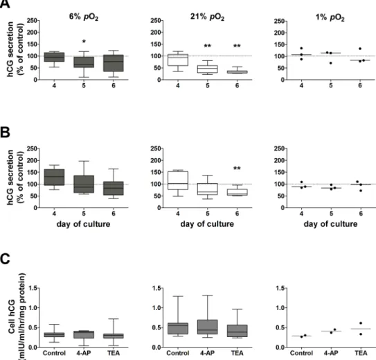
Fig 2Aand 2Bshow the effects ofpO2-sensitive K+channel blockers 4-AP and TEA
respec-tively on hCG secretion from placental villous explants maintained at 6%, 21% and 1%pO2.
Fig 2. Effect ofpO2-sensitive K+channel blockers on hCG secretion from placental villous explants maintained at 6%, 21% or 1%pO2.hCG secretion at days 4, 5 and 6 of culture was normalized as a percentage of hCG secretion in control untreated explants at the correspondingpO2(dotted line, 100%); assessed by Wilcoxon signed-rank test compared to 100%.A: 4-AP (*p= 0.04,**p= 0.008; n = 8 placentas;
In villous explants maintained at 6%pO2, 4-AP caused a transient decrease (35%) in hCG
secretion on day 5 compared to control untreated explants (Fig 2A). In contrast, explants main-tained at hyperoxia (21%pO2) showed a significant reduction in hCG secretion when treated
with 4-AP at days 5 (52%) and 6 (68%) of culture (Fig 2A). This effect was completely sup-pressed under hypoxia (1%pO2), where hCG secretion was unaffected by 4-AP (Fig 2A).
TEA did not affect hCG secretion by explants maintained in placental normoxia (Fig 2B). On the contrary, when explants were maintained at 21%pO2, treatment with TEA caused a
sig-nificant reduction in hCG secretion at day 6 of culture (41%;Fig 2B). TEA had no effect on explants maintained at 1%pO2(Fig 2B).
LDH release from placental villous explants was not affected by treatment with 4-AP or TEA compared to their corresponding controls at the samepO2(data not shown), indicating
that tissue viability was not compromised by treatment with thesepO2-sensitive K+channel
blockers.
Neither 4-AP nor TEA altered cellular hCG at any of thepO2tested (Fig 2C), implicating an
effect of these K+channel blockers on the secretory mechanism for hCG and not hCG production.
From these data it is evident that culture of term placental villous explants for 6 days in hyp-oxic (1%pO2) conditions reduced hCG secretion to a low level, inhibited the temporal recovery
in hCG secretion which is associated with syncytiotrophoblast regeneration/renewal at higher
pO2and reduced the cellular production of LDH and hCG. Furthermore, hCG secretion at 1%
pO2was unaffected by 4-AP and TEA. Therefore, experiments to evaluate the effect of these K+
channel blockers on86Rb efflux were not performed at 1%pO2.
Basal
86Rb (K
+) efflux from syncytiotrophoblast: chronic effect of
p
O
286Rb efflux was measured at day 6 of culture in placental villous explants maintained at 6% or
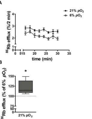
21%pO2.Fig 3Ashows the time course for basal %86Rb efflux at a steady state over a 16min
period in explants maintained at 6% and 21%pO2. %86Rb efflux is higher from explants
main-tained at 21% than 6%pO2.Fig 3Bshows the area under the curve for the total %86Rb efflux
over 16min from explants maintained at 21%pO2as a percent of efflux from explants at 6%
pO2(100%, dotted line). Basal %86Rb efflux was significantly higher in explants maintained for
6 days in hyperoxia (21%pO2) compared to normoxia.
The86Rb efflux rate constants calculated for untreated control explants maintained at 6% and 21%pO2is shown inTable 1. Rate constant analysis shows that the fall in intracellular 86Rb can be described by a single exponential decline indicating that efflux is predominantly
from a single tissue compartment, which we take to be the syncytiotrophoblast, in agreement with previous reports [31]. The mean rate constant for86Rb efflux was significantly lower in explants maintained at placental normoxia (6%pO2) than hyperoxia (21%pO2) (Table 1).
Long term effects of
p
O
2: effect of
p
O
2-sensitive K
+channel blockers on
syncytiotrophoblast K
+permeability
86Rb (K+) permeability was assayed at day 6 in villous explants cultured at 6% and 21%pO 2.
Explants were untreated (controls) or treated from day 3 onwards with 4-AP or TEA.
The effect of the K+channel blockers was assessed by analysing the differences between the rate constant of decline in intracellular86Rb for each treatment compared to controls (per-formed in the same placentas n = 4) at the samepO2(Table 1). The efflux rate constant was
sig-nificantly reduced by 4-AP and TEA in explants maintained in hyperoxia (21%pO2) but was
Fig 3. Effect ofpO2on syncytiotrophoblast K+permeability.A: Time course for the %86Rb efflux over 16min. Untreated (control) villous explants were cultured at 6% and 21%pO2and basal86Rb efflux was measured at day 6. Data are expressed as mean±SE (n = 10 placentas).B: The %86Rb efflux over 16min in explants maintained at 21% was expressed as a percentage of efflux in explants maintained at 6%pO2 (placental normoxia; dotted line). Data are median±IQR, Wilcoxon signed-rank test compared to 6%pO2,
*p= 0.002; n = 10 placentas. doi:10.1371/journal.pone.0149021.g003
Table 1. Rate constants of86Rb efflux in control and treated placental villous explants.
Condition 86Rb ef
flux rate constant(ln86Rb (t = x)/(t = 0)/min-1) r2 p value n
Control 6%pO2 -0.0111±0.0003 0.956 10
Control 21%pO2 -0.0136±0.0003* 0.954 <0.0001 10
Control for treatments 6%pO2 -0.0107±0.0005 0.948 4
5mM 4-AP 6%pO2 -0.0114±0.0009† 0.840 0.494 4
5mM TEA 6%pO2 -0.0111±0.0005† 0.944 0.630 4
100μM H2O26%pO2 -0.0119±0.0003† 0.976 0.048 4
1mM H2O26%pO2 -0.0118±0.0007† 0.916 0.002 4
Control for treatments 21%pO2 -0.0144±0.0006 0.953 4
5mM 4-AP 21%pO2 -0.0113±0.0006** 0.918 0.0007 4
5mM TEA 21%pO2 -0.0117±0.0002** 0.987 <0.0001 4
100μM H2O221%pO2 -0.0122±0.0004** 0.963 0.004 4
1mM H2O221%pO2 -0.0130±0.0005** 0.963 0.014 4
Rate constants of86Rb ef
flux in control and treated placental villous explants maintained at 6% and 21%pO2over 16 min.Data are mean±SE; n
is the number of placentas. In all conditions the r2values, determined by linear regression, were close to 1 and signi
ficant (p<0.001), indicating a single exponential decline in intracellular86Rb over 16 min. The
pvalue corresponds to the following differences in the rate constants between groups:*control 6%pO2vs control 21%pO2;†treatments at 6%pO2vs corresponding control at 6%pO2;**treatments at 21%pO2vs corresponding control at 21%pO2.
Effect of H
2O
2on basal
86Rb (K
+) permeability and hCG secretion from
placental villous explants
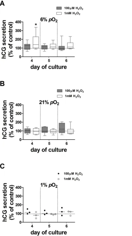
The effect of H2O2,used to generate oxidative stress, on hCG secretion was tested in explants
maintained at 6%, 21% and 1%pO2over days 3–5 of culture. There was no effect of 100μM
H2O2at 6%, 21% or 1%pO2(Fig 4). In contrast, 1mM H2O2transiently increased hCG
secre-tion by 40% in explants maintained at 6%pO2compared to controls (Fig 4A). 10μM H2O2had
no effect on hCG secretion (data not shown). Treatment with H2O2did not affect LDH release
from villous explants at any of the concentrations used (data not shown).
The effect of H2O2on86Rb efflux was measured at day 6 in explant cultures maintained at
6% and 21%pO2(Table 1). H2O2increased the86Rb efflux rate constant at 100μM and 1mM
compared to corresponding controls in explants maintained at 6%pO2. In contrast, treatment
of villous explants with 100μM and 1mM H2O2produced the opposite effect in 21%pO2, and
significantly reduced the86Rb rate constant (Table 1) compared to controls at the samepO2.
10μM H2O2had no effect on basal86Rb efflux from explants cultured at either 6% or 21%pO2
(data not shown).
Discussion
This study confirms and extends previous observations that hCG secretion from term placental trophoblast is sensitive topO2[8,18] and ROS [10]. In villous explants prepared from the
same placenta, hCG secretion was higher in 21%pO2, and lower in 1%pO2, than 6%pO2
(nor-moxic) culture conditions. Syncytiotrophoblast K+permeability, estimated by86Rb efflux, was higher in explants cultured in 21% than 6%pO2. In accordance with this, 4-AP and TEA,
blockers ofpO2-sensitive KVchannels, inhibited hCG secretion and86Rb efflux to a greater
extent in 21% than 6%pO2. H2O2, used to induce oxidative stress, stimulated86Rb efflux and
transiently increased hCG secretion from explants cultured at 6%pO2but inhibited86Rb efflux,
without affecting hCG secretion, at 21%pO2. Thus, effects of oxidative stress on hCG secretion
and syncytiotrophoblast K+permeability depend on thepO2.
hCG secretion from term placental syncytiotrophoblast is
p
O
2-dependent
The temporal pattern of hCG secretion from term placental explants maintained at 21%pO2
for 6 days was originally described by Simanet al. [31]. Similar studies to relate hCG secretion to syncytiotrophoblast regeneration over 6 days at placental normoxia (6%pO2), and extreme
hypoxia (1%pO2), have not been performed. However, in shorter term cultures (4 days), hCG
secretion was lower at 6% and 1% compared to 21%pO2and this was associated with
dysregu-lated syncytiotrophoblast turnover such as decreased cytotrophoblast proliferation and enhanced apoptosis [18]. In the present study, the time course of hCG secretion from explants maintained at 21%pO2coincided with previous observations [22,31]. Secretion was
signifi-cantly lower at 6%pO2, with only a small rise on day 4, and at 1%pO2low hCG secretion at
day 2 persisted for the duration of the culture. Cellular hCG was lower (1.7-fold) in explants maintained at 6%pO2compared to 21%pO2, suggesting thatpO2regulates hCG synthesis.
However, as hCG secretion at 6%pO2was 4.1-fold lower than at 21%pO2, the hCG secretory
mechanism is additionally regulated bypO2. In contrast, 1%pO2reduced cellular hCG to the
same proportion as secretion, indicating that the reduced secretion in hypoxia is predomi-nantly due to altered synthesis. LDH release was unaffected bypO2which could indicate that
pO2inhibited LDH synthesis and using LDH release alone as a marker of tissue viability in
hypoxia might not be reliable.
Fig 4. Effect of 100μM and 1mM H2O2on hCG secretion from villous explants.hCG secretion from H2O2-treated explants maintained at 6% (A), 21% (B) and 1%pO2(C) at days 4, 5 and 6 of culture was expressed as a percentage of control (100%, dotted line); data are expressed as median±IQR; n = 9
placentas for 100μM H2O2(except n = 3 placentas at 1%pO2); n = 8 placentas for 1mM H2O2(except n = 3 placentas in 1%pO2). Wilcoxon signed-rank test compared to 100%,*p= 0.04.
Inhibition of hCG secretion by K
Vchannel blockers is
p
O
2-sensitive
We previously demonstrated that chronic exposure to the KVchannel blockers 4-AP and TEA
induced a concentration-dependent inhibition of hCG secretion in explants and cytotropho-blasts maintained at 21%pO2[22]. In the current study, 4-AP and TEA inhibited hCG
secre-tion from villous explants to a greater extent in 21% than 6%pO2and had no effect on
secretion at 1%pO2. As the activity and expression of KVchannels can be down-regulated by
hypoxia [24,25], our results support the possibility that the lower hCG secretion at 6% com-pared to 21%pO2is mediated by closure of KVchannels.
Inhibition of
86Rb (K
+) efflux by K
Vchannel blockers is
p
O
2-sensitive
Direct study of ion channels in the syncytiotrophoblast of intact placental villi using patch clamp methods is technically challenging as seals are hard to achieve [34] and the multinucle-ate nature of the tissue precludes whole cell recording. In this study we used86Rb (K+) efflux to assess whether 4-AP and TEA inhibited K+conductance in the syncytiotrophoblast and whether the inhibition waspO2-sensitive. We have previously shown that basal86Rb efflux
from placental explants cultured at 21%pO2is inhibited by Ba2+, a broad spectrum K+channel
blocker [31], implicating K+conductances in the microvillous, maternal facing plasma mem-brane of the syncytiotrophoblast.
In the current study,86Rb efflux measured on day 6 of culture showed that basal K+ perme-ability was significantly higher in explants maintained in hyperoxia compared to placental nor-moxia, suggesting that chronic exposure to 21%pO2over a 6-day period increases the activity
and/or expression of syncytiotrophoblast K+channels. In support of this, treatment of villous explants with 4-AP and TEA significantly reduced86Rb efflux when the tissue was cultured at 21% but not 6%pO2, consistent with an inhibition ofpO2-sensitive KVchannels that are more
active/more highly expressed at 21% than at 6%pO2. In addition, the inhibition of both86Rb
efflux and syncytiotrophoblast hCG secretion by 4-AP and TEA at 21% but not 6%pO2,
sug-gests that 4-AP and TEA-sensitive KVchannels mediate the stimulatory effect of higherpO2on
hCG secretion.
Effect of H
2O
2on hCG secretion and
86Rb efflux
Placental oxidative stress and reduced antioxidant defences are key features of pre-eclampsia [35,36]. In this study we explored the effects of oxidative stress (H2O2) on syncytiotrophoblast
hCG secretion and whether they could be modulated through K+channels. Previous reports showed thatin vitrotreatment of placental villous tissue with 1mM H2O2caused oxidative
stress which was reversed by vitamins C and E [37].
H2O2(10μM-1mM) did not alter hCG secretion at 21%pO2but 1mM caused a transient
increase at 6%pO2. This contrasts with the concentrations of H2O2reported to affect hCG
secretion by cytotrophoblasts (inhibition at>50μM and stimulation at 1–50μM) perhaps due
to differences between thesein vitropreparations; in explants cellular interactions are main-tained and tissue antioxidant defences are available to scavenge ROS [38].
H2O2also hadpO2-dependent effects on86Rb efflux, with 100μM-1mM increasing
syncy-tiotrophoblast86Rb efflux at 6%pO2, but inhibiting efflux at 21%pO2. This is consistent with
the variable effects of ROS on K+channel activity reported on the literature [28,30] and raises the possibility that in placental normoxia, K+channels can be activated by H2O2. However, it is
possible that the effect of H2O2on86Rb efflux and hCG secretion can be independent events
and further work is required to determine which channels are activated by H2O2in normoxia
Mechanism of hCG secretion: Role of K
+channels
In contrast to hCG secretion in the first trimester of pregnancy [19], the mechanism of secre-tion by the syncytiotrophoblast at term is not fully elucidated. The present work proposes a role for 4-AP and TEA-sensitivepO2-sensitive K+channels in regulating hCG secretion.
According to the specificity and the concentration of 4-AP and TEA used, the targeted K+ channels belong mainly to the KVchannel family [23]. Indeed, K+channels belonging to other
families such as ATP-sensitive K+channels are not involved in hCG secretion [39].
KVchannel mRNA is expressed by whole placental homogenate [40,41] and
immunostain-ing for KV1.5 and 2.1 has localized protein expression to the syncytiotrophoblast [42]. KV1.5
and 2.1 arepO2-sensitive [43] and blocked by 4-AP and TEA [44] and thus closure of these
channels could underlie the lower hCG secretion from placentas maintained in 6% compared to 21%pO2.
In the normal placenta at term where villi are exposed to maternal blood at 6%pO2,pO2
-sensitive KVchannels could be down-regulated/closed. We have previously shown that hCG
secretion at 21%pO2is stimulated by Ca2+entry through non-selective cation channels
(NSCC; [45]). Therefore, a relatively depolarised membrane potential could minimise Ca2+ entry through NSCC and sustain basal levels of syncytiotrophoblast hCG secretion seen under normoxic conditions. Elevated K+permeability by ROS (H2O2) in normoxia could reflect
increased K+channel activity, membrane hyperpolarization, promotion of Ca2+entry and stimulation of hCG secretion.
In conditions such as pre-eclampsia, a pregnancy complication associated with alteredpO2
[13], whilst the range ofpO2in the placental bed is unlikely to be as wide as that usedin vitro
in this study, current data suggest the syncytiotrophoblast could be exposed to both hypoxia [46] and/or hyperoxia [47,48]. In the latter,pO2-sensitive KVchannels could be activated,
hyperpolarising the membrane potential, stimulating Ca2+entry and promoting syncytiotro-phoblast hCG secretion. In this regard it is interesting to note that maternal plasma hCG is higher in women with late onset pre-eclampsia compared to women having normal pregnancy [16], and that serum hCG levels vary depending on the severity of disease showing a several-fold increase in severe [49] but not in moderate pre-eclampsia. Consequently,pO2changes in
the placenta might differ related to the severity of disease and this could influence the regula-tion of hCG secreregula-tion by elevated ROS.
Several mechanistic links remain to be explored. For example, although hypoxia inhibits cytotrophoblast cell fusion and hCG secretion [9], there is insufficient evidence at present to confirm that these events are independentlypO2-sensitive. Using primary cultures of placental
trophoblastin vitro, Alsatet al. (1996) demonstrated that low oxygen (~9%pO2) reduced the
formation of multinucleate cells (syncytialisation) and this was associated with an increase in expression of desmoplakin and e-cadherin, and a reduction in hCG secretion. While these data illustrate a clear effect of oxygenation on morphological and biochemical differentiation of cytotrophoblasts, it is unclear whether the primary effect is to reduce hCG secretion, which then inhibits fusion, or whether the primary effect of lowpO2is to inhibit fusion which
pre-vents biochemical differentiation. Furthermore, it is also unknown whether the expression/ activity ofpO2-sensitive KVchannels is altered either by cytotrophoblast differentiation, or
pO2, per se.
The extent to whichpO2-sensitive K+channels play a role in hCG secretion in pregnancy
disease has yet to be explored; dysregulation of syncytiotrophoblast K+channel activity and/or expression through chronic exposure to alteredpO2and/or increased ROS could potentially
Acknowledgments
The authors wish to thank the midwives and patients at the Maternity Unit at St. Mary’s Hospi-tal, Manchester, UK, for their assistance.
Author Contributions
Conceived and designed the experiments: PD CPS SLG. Performed the experiments: PD. Ana-lyzed the data: PD. Contributed reagents/materials/analysis tools: PD SLG. Wrote the paper: PD CPS SLG.
References
1. Longtine MS, Barton A, Chen B, Nelson DM. Live-cell imaging shows apoptosis initiates locally and propagates as a wave throughout syncytiotrophoblasts in primary cultures of human placental villous trophoblasts. Placenta. 2012; 33(12):971–6. doi:http://dx.doi.org/10.1016/j.placenta.2012.09.013 PMID:23102999
2. Longtine MS, Chen B, Odibo AO, Zhong Y, Nelson DM. Caspase-mediated apoptosis of trophoblasts in term human placental villi is restricted to cytotrophoblasts and absent from the multinucleated syncy-tiotrophoblast. Reproduction (Cambridge, England). 2012; 143(1):107–21. Epub 2011/11/03. doi: REP-11-0340 [pii] doi:10.1530/REP-11-0340PMID:22046053.
3. Lim KH, Zhou Y, Janatpour M, McMaster M, Bass K, Chun SH, et al. Human cytotrophoblast differentia-tion/invasion is abnormal in pre-eclampsia. Am J Pathol. 1997; 151(6):1809–18. Epub 1997/12/24. PMID:9403732; PubMed Central PMCID: PMC1858365.
4. Crocker IP, Tansinda DM, Baker PN. Altered cell kinetics in cultured placental villous explants in preg-nancies complicated by pre-eclampsia and intrauterine growth restriction. The Journal of pathology. 2004; 204(1):11–8. PMID:15307133.
5. Higgins L, Mills TA, Greenwood SL, Cowley EJ, Sibley CP, Jones RL. Maternal obesity and its effect on placental cell turnover. J Matern Fetal Neonatal Med. 2012; 26(8):783–8. doi:10.3109/14767058.2012. 760539
6. Weedon-Fekjaer MS, Tasken K. Review: Spatiotemporal dynamics of hCG/cAMP signaling and regula-tion of placental funcregula-tion. Placenta. 2012; 33 Suppl:S87–91. doi:10.1016/j.placenta.2011.11.003 PMID:22103973.
7. Cronier L, Bastide B, Herve JC, Deleze J, Malassine A. Gap junctional communication during human trophoblast differentiation: influence of human chorionic gonadotropin. Endocrinology. 1994; 135 (1):402–8. Epub 1994/07/01. PMID:8013377.
8. Crocker IP, Tansinda DM, Jones CJ, Baker PN. The influence of oxygen and tumor necrosis factor-alpha on the cellular kinetics of term placental villous explants in culture. J Histochem Cytochem. 2004; 52(6):749–57. PMID:15150283.
9. Alsat E, Wyplosz P, Malassine A, Guibourdenche J, Porquet D, Nessmann C, et al. Hypoxia impairs cell fusion and differentiation process in human cytotrophoblast, in vitro. J Cell Physiol. 1996; 168 (2):346–53. Epub 1996/08/01. doi:10.1002/(SICI)1097-4652(199608)168:2<346::AID-JCP13>3.0. CO;2–1[pii]. PMID:8707870.
10. Kharfi Aris A, Leblanc S, Ouellet A, Moutquin JM. Dual action of H2O2 on placental hCG secretion: implications for oxidative stress in preeclampsia. Clinical biochemistry. 2007; 40(1–2):94–7. PMID: 17150203.
11. Poston L, Igosheva N, Mistry HD, Seed PT, Shennan AH, Rana S, et al. Role of oxidative stress and antioxidant supplementation in pregnancy disorders. Am J Clin Nutr. 2011; 94(6 Suppl):1980S–5S. doi: 10.3945/ajcn.110.001156PMID:21613560
12. Myatt L, Cui X. Oxidative stress in the placenta. Histochemistry and cell biology. 2004; 122(4):369–82. PMID:15248072.
13. Hung TH, Burton GJ. Hypoxia and reoxygenation: a possible mechanism for placental oxidative stress in preeclampsia. Taiwanese journal of obstetrics & gynecology. 2006; 45(3):189–200. PMID: 17175463.
15. Takagi Y, Nikaido T, Toki T, Kita N, Kanai M, Ashida T, et al. Levels of oxidative stress and redox-related molecules in the placenta in preeclampsia and fetal growth restriction. Virchows Arch. 2004; 444(1):49–55. Epub 2003/10/24. doi:10.1007/s00428-003-0903-2PMID:14574573.
16. Kharfi A, Giguere Y, De Grandpre P, Moutquin JM, Forest JC. Human chorionic gonadotropin (hCG) may be a marker of systemic oxidative stress in normotensive and preeclamptic term pregnancies. Clin-ical biochemistry. 2005; 38(8):717–21. PMID:15904911.
17. Moll SJ, Jones CJ, Crocker IP, Baker PN, Heazell AE. Epidermal growth factor rescues trophoblast apoptosis induced by reactive oxygen species. Apoptosis. 2007; 12(9):1611–22. PMID:17573555.
18. Heazell AE, Lacey HA, Jones CJ, Huppertz B, Baker PN, Crocker IP. Effects of oxygen on cell turnover and expression of regulators of apoptosis in human placental trophoblast. Placenta. 2008; 29(2):175– 86. PMID:18155142.
19. Yoshida Y. Secretion of human chorionic gonadotropin in early pregnancy. Medical molecular morphol-ogy. 2005; 38(2):104–11. PMID:15944817.
20. Meuris S, Polliotti B, Robyn C, Lebrun P. Ca2+ entry through L-type voltage-sensitive Ca2+ channels stimulates the release of human chorionic gonadotrophin and placental lactogen by placental explants. Biochimica et biophysica acta. 1994; 1220(2):101–6. PMID:7508753.
21. Long O, Clarson LH. The effect of Ca2+-permeable channel blockers on human chorionic gonadotro-phin (hCG) secretion by villous fragments from term placentas. The Journal of physiology. 2002; 539P:126P.
22. Williams JL, Fyfe GK, Sibley CP, Baker PN, Greenwood SL. K+ channel inhibition modulates the bio-chemical and morphological differentiation of human placental cytotrophoblast cells in vitro. American journal of physiology. 2008; 295(4):R1204–13. PMID:18703414. doi:10.1152/ajpregu.00193.2008
23. Gutman GA, Chandy KG, Grissmer S, Lazdunski M, McKinnon D, Pardo LA, et al. International Union of Pharmacology. LIII. Nomenclature and molecular relationships of voltage-gated potassium channels. Pharmacological reviews. 2005; 57(4):473–508. PMID:16382104.
24. Platoshyn O, Yu Y, Golovina VA, McDaniel SS, Krick S, Li L, et al. Chronic hypoxia decreases K(V) channel expression and function in pulmonary artery myocytes. Am J Physiol Lung Cell Mol Physiol. 2001; 280(4):L801–12. Epub 2001/03/10. PMID:11238022.
25. Lopez-Barneo J, del Toro R, Levitsky KL, Chiara MD, Ortega-Saenz P. Regulation of oxygen sensing by ion channels. J Appl Physiol. 2004; 96(3):1187–95; discussion 70–2. Epub 2004/02/10. doi:10. 1152/japplphysiol.00929.200396/3/1187 [pii]. PMID:14766769.
26. Kourie JI. Interaction of reactive oxygen species with ion transport mechanisms. The American journal of physiology. 1998; 275(1 Pt 1):C1–24. PMID:9688830.
27. Gutterman DD, Miura H, Liu Y. Redox modulation of vascular tone: focus of potassium channel mecha-nisms of dilation. Arterioscler Thromb Vasc Biol. 2005; 25(4):671–8. PMID:15705931.
28. Archer SL, Wu X-C, Thebaud B, Moudgil R, Hashimoto K, Michelakis ED. O2 sensing in the human ductus arteriosus: redox-sensitive K+ channels are regulated by mitochondria-derived hydrogen perox-ide. Biol Chem. 2004; 385(3–4):205–16. PMID:15134333
29. Rogers PA, Chilian WM, Bratz IN, Bryan RM Jr, Dick GM. H2O2 activates redox- and 4-aminopyridine-sensitive Kv channels in coronary vascular smooth muscle. Am J Physiol Heart Circ Physiol. 2007; 292 (3):H1404–11. doi:10.1152/ajpheart.00696.2006PMID:17071731
30. Appiah I, Milovanovic S, Radojicic R, Nikolic-Kokic A, Orescanin-Dusic Z, Slavic M, et al. Hydrogen per-oxide affects contractile activity and anti-oxidant enzymes in rat uterus. Br J Pharmacol. 2009; 158 (8):1932–41. Epub 2009/11/18. doi: BPH490 [pii] doi:10.1111/j.1476-5381.2009.00490.xPMID: 19917063.
31. Siman CM, Sibley CP, Jones CJ, Turner MA, Greenwood SL. The functional regeneration of syncytio-trophoblast in cultured explants of term placenta. American journal of physiology. 2001; 280(4):R1116– 22. PMID:11247834.
32. Boyd CA. Cotransport systems in the brush border membrane of the human placenta. Ciba Found Symp. 1983; 95:300–26. PMID:6303722
33. Turner MA, Roulstone CJ, Desforges M, Cretney M, Champion E, Lacey H, et al. The extent and vari-ability of effects of culture conditions on the secretion of human chorionic gonadotrophin and interleu-kin-6 by human, term placental explants in culture. Placenta. 2006; 27(1):98–102. PMID:16310043.
34. Brown PD, Greenwood SL, Robinson J, Boyd RD. Chloride channels of high conductance in the micro-villous membrane of term human placenta. Placenta. 1993; 14(1):103–15. Epub 1993/01/01. PMID: 7681209.
36. Wang Y, Walsh SW. Antioxidant activities and mRNA expression of superoxide dismutase, catalase, and glutathione peroxidase in normal and preeclamptic placentas. Journal of the Society for Gyneco-logic Investigation. 1996; 3(4):179–84. PMID:8796828.
37. Cindrova-Davies T. Gabor Than Award Lecture 2008: pre-eclampsia-from placental oxidative stress to maternal endothelial dysfunction. Placenta. 2009; 30 Suppl A:S55–65. PMID:19118896. doi:10.1016/ j.placenta.2008.11.020
38. Hempstock J, Bao YP, Bar-Issac M, Segaren N, Watson AL, Charnock-Jones DS, et al. Intralobular dif-ferences in antioxidant enzyme expression and activity reflect the pattern of maternal arterial bloodflow within the human placenta. Placenta. 2003; 24(5):517–23. PMID:12744928.
39. Lybaert P, Hoofd C, Guldner D, Vegh G, Delporte C, Meuris S, et al. Detection of KATP channels sub-units in human term placental explants and evaluation of their implication in human placental lactogen (hPL) and human chorionic gonadotropin (hCG) release. Placenta. 2013; 34(6):467–73. doi:http://dx. doi.org/10.1016/j.placenta.2013.03.006PMID:23587463
40. Lacey H, Glazier JD, Greenwood SL, Sibley CP. Potassium channel gene expression over gestation in human placenta. The Journal of physiology. 2005; 565P:PC173.
41. Corcoran J, Lacey H, Baker PN, Wareing M. Altered Potassium Channel Expression in the Human Pla-cental Vasculature of Pregnancies Complicated by Fetal Growth Restriction. Hypertens Pregnancy. 2008; 27(1):75–86. doi:10.1080/10641950701826158PMID:18293206
42. Williams JL, Jones RL, Sibley CP, Baker PN, Greenwood SL. Elevated expression of oxygen-sensitive K+ channels in human placental syncytiotrophoblast of growth restricted fetuses. Reprod Sci. 2009; 16 (No. 3 (Suppl)):346A.
43. Patel AJ, Honore E. Molecular physiology of oxygen-sensitive potassium channels. Eur Respir J. 2001; 18(1):221–7. PMID:11510795.
44. Burg ED, Remillard CV, Yuan JX. K+ channels in apoptosis. The Journal of membrane biology. 2006; 209(1):3–20. PMID:16685597.
45. Clarson LH, Roberts VH, Hamark B, Elliott AC, Powell T. Store-operated Ca2+ entry in first trimester and term human placenta. The Journal of physiology. 2003; 550(Pt 2):515–28. Epub 2003/05/27. doi: 10.1113/jphysiol.2003.044149[pii]. PMID:12766233; PubMed Central PMCID: PMC2343039.
46. Burton GJ. Oxygen, the Janus gas; its effects on human placental development and function. J Anat. 2009; 215(1):27–35. Epub 2009/01/30. doi: JOA978 [pii] doi:10.1111/j.1469-7580.2008.00978.x PMID:19175804.
47. Kingdom JCP, Kaufmann P. Oxygen and placental villous development: Origins of fetal hypoxia. Pla-centa. 1997; 18(8):613–21. doi:http://dx.doi.org/10.1016/S0143-4004(97)90000-XPMID:9364596
48. Huppertz B, Weiss G, Moser G. Trophoblast invasion and oxygenation of the placenta: measurements versus presumptions. Journal of Reproductive Immunology. 2014; 101–102:74–9. doi:http://dx.doi.org/ 10.1016/j.jri.2013.04.003PMID:23747129