Recovery of Cognitive Dysfunction via Orally
Administered Redox-Polymer
Nanotherapeutics in SAMP8 Mice
Pennapa Chonpathompikunlert1☯¤a, Toru Yoshitomi1☯¤b, Long Binh Vong1, Natsuka Imaizumi1, Yuki Ozaki1, Yukio Nagasaki1,2,3*
1Department of Materials Sciences, Graduate School of Pure and Applied Sciences, University of Tsukuba, Tennoudai 1-1-1, Tsukuba, Ibaraki 305–8573, Japan,2Master’s School of Medical Sciences, Graduate School of Comprehensive Human Sciences, University of Tsukuba, Tennoudai 1-1-1, Tsukuba, Ibaraki 305– 8573, Japan,3Satellite Laboratory, International Center for Materials Nanoarchitechtonics (WPI-MANA), National Institute for Materials Sciences (NIMS), University of Tsukuba, Tennoudai 1-1-1, Tsukuba, Ibaraki 305–8573, Japan
☯These authors contributed equally to this work.
¤a Current address: Department of Physiology, Faculty of Science, Prince of Songkla University, Hatyai, Songkhla 90112, Thailand
¤b Current address: Department of Chemistry, Graduate School of Science, The University of Tokyo, 7-3-1, Hongo, Bunkyo, Tokyo 113–0033, Japan
Abstract
Excessively generated reactive oxygen species are associated with age-related neurode-generative diseases. We investigated whether scavenging of reactive oxygen species in the brain by orally administered redox nanoparticles, prepared by self-assembly of redox polymers possessing antioxidant nitroxide radicals, facilitates the recovery of cognition in 17-week-old senescence-accelerated prone (SAMP8) mice. The redox polymer was deliv-ered to the brain after oral administration of redox nanoparticles via a disintegration of the nanoparticles in the stomach and absorption of the redox polymer at small intestine to the blood. After treatment for one month, levels of oxidative stress in the brain of SAMP8 mice were remarkably reduced by treatment with redox nanoparticles, compared to that observed with low-molecular-weight nitroxide radicals, resulting in the amelioration of cognitive im-pairment with increased numbers of surviving neurons. Additionally, treatment by redox nanoparticles did not show any detectable toxicity. These findings indicate the potential of redox polymer nanotherapeutics for treatment of the neurodegenerative diseases.
Introduction
Aging increases the risk of neurodegenerative diseases, such as Alzheimer’s disease (AD), which mostly affect quality of life in the elderly. Although the average human life span has increased because of progress in medical and health care, the socioeconomic burden of the elderly is a concern in developed countries. Oxidative stress caused by overproduction of reactive oxygen OPEN ACCESS
Citation:Chonpathompikunlert P, Yoshitomi T, Vong LB, Imaizumi N, Ozaki Y, Nagasaki Y (2015) Recovery of Cognitive Dysfunction via Orally Administered Redox-Polymer Nanotherapeutics in SAMP8 Mice. PLoS ONE 10(5): e0126013. doi:10.1371/journal.pone.0126013
Academic Editor:Masaya Yamamoto, Institute for Frontier Medical Sciences, Kyoto University, JAPAN
Received:December 21, 2014
Accepted:March 27, 2015
Published:May 8, 2015
Copyright:© 2015 Chonpathompikunlert et al. This is an open access article distributed under the terms of theCreative Commons Attribution License, which permits unrestricted use, distribution, and reproduction in any medium, provided the original author and source are credited.
Data Availability Statement:All relevant data are within the paper and its Supporting Information files.
Funding:Grant-in-Aid for Scientific Research S (No. 25220203) to Y.N. Grant-in-Aid for Exploratory Research (24659014) to T.Y.
species (ROS) is well known as one of the direct causes of aging. Under normal physiological conditions, ROS can be scavenged by endogenous antioxidant-defense systems including super-oxide dismutase (SOD), catalase (CAT), and glutathione peroxidase (GPx). With advancing age, however, the production of ROS dramatically increases, and endogenous antioxidants fail to completely scavenge all of the ROS, followed by production of oxidative components. An in-crease in the oxidative stress in the brain is reported to be involved in aging-related neural dys-function and/or learning and memory deficiency [1]. Previous studies have suggested that an increase in the expression of pro-inflammatory cytokines, such as interleukin (IL)-1β, IL-6, and tumor necrosis factor-α(TNF-α), in the brain is involved in aging-related neural dysfunction and/or learning and memory deficiency in animals. Although the promising low-molecular-weight (LMW) antioxidant, vitamin E, was reported to show slight efficacy such as slowing of functional decline, in clinical trials of AD, a complete recovery was not observed [2,3].
the blood uptake of the redox polymer and its delivery in the brain after oral administration of RNPNin wild-type ICR mice. Furthermore, we evaluated whether our redox polymer nanother-apeutics was effective in senescence-accelerated prone (SAMP8) mice, which are suitable models of the accelerated senescence with early learning and memory deficits [18,19].
Methods
Preparation of the RNP
NThe RNPNwas prepared by self-assembly of redox polymers (MW [PEG] = 5,500 Da; MW [PMNT] = 4,500 Da) by using the dialysis method reported previously [20]. Please see the sup-porting information for a comprehensive description of the methods.
Animals
Male ICR, male SAMP8 and SAMR1 mice were used in this study. They were housed in the ex-perimental animal center of University of Tsukuba under controlled temperature (23 ± 1°C), humidity (50 ± 5%) and lighting (12 h light/dark cycles). The animals had free access to food and water. All the experiments were carried out in accordance with the guidelines for animal care and use of Japan and were approved by the animal ethics committee of the Institutional Animal Experiment Committee of the University of Tsukuba and in accordance with the Regu-lation for Animal Experiments in our university and the Fundamental Guideline for Proper Conduct of Animal Experiments and Related Activities in Academic Research Institutions under the jurisdiction of the Ministry of Education, Culture, Sports, Science, and Technology.
Evaluation of the delivery of redox polymer to the brain of wild-type ICR
mice after oral administration of RNP
NSix-week-old male ICR mice (Charles River Japan, Inc., Kanagawa, Japan) were used for the evaluation of the delivery of the redox polymer to the brain. Please see detailed methods in the supporting information.
Evaluation of therapeutic effects of redox polymer nanotherapeutics
Seventeen-week-old male SAMP8 (body weight; 30 ± 2.0 g) and 17-week-old male SAMR1 mice (body weight; 35 ± 2.0 g) were purchased from Japan SLC, Inc. (Shizuoka, Japan) for this study. All of the SAMR1 and SAMP8 mice were trained using the Morris water maze for seven days and using an open field instrument for three days before the mice were randomly divided into five groups (10 mice/group). Two hundred microliters of RNPN(60 mg/mL) were orally administered to SAMP8 mice at a dose of 300 mg/(kgd) (nitroxide radical concentration: 42.5 mg/(kgd)) for four consecutive weeks. Blank micelles (nanoparticles without nitroxide radicalsin the core: 60 mg/mL) and 4-hydroxy-2,2,6,6-tetramethylpiperidine-1-oxyl (TEMPOL), a ROS-scavenging drug, were also orally administered to mice at doses of 300 mg/(kgd) and
as the liver, kidney, spleen, heart, lung, and testicle, were also fixed in 10% formalin solution for hematoxylin and eosin staining. Blood was then immediately centrifuged at 3,000 rpm for 10 min at 4°C to separate the sera. All the sera and brain tissues were stored at−80°C until analysis. Please see the supporting information for detailed methods of the Morris water maze test, object recognition test, measurement of the density of suviving neurons in the brain, anti-oxidant enzyme, ROS products, cytokine product, body weight, organ weight, blood pressure, hepatic function, acetylcholinesterase (AChE) activity assays, and histopathology.
Statistical analysis
All data are presented as the mean ± SEM values. The intergroup differences in the latency time in the Morris water maze test and exploration time in the object recognition test were ana-lyzed by two-way analysis of variance (ANOVA) with repeated measurements. The other data were analyzed by one-way ANOVA followed by the Tukey’s post hoc test. APvalue of less than 0.05 was considered significant. Statistical analysis was performed using the SPSS 17.0 software package for Windows.
Results
Delivery of the redox polymer to the brain by oral administration of RNP
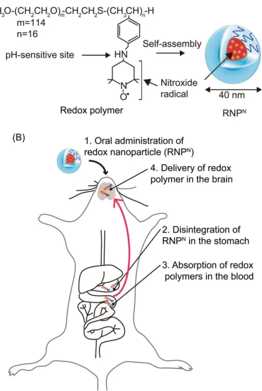
NFig 1. Concept of orally administered redox polymer nanotherapeutics for treatment of the
senescence-accelerated neurodegenerative diseases.(A) Structures of redox polymers and RNPNand (B) illustration of delivery of redox polymer to the brain after oral administration of RNPNare shown.
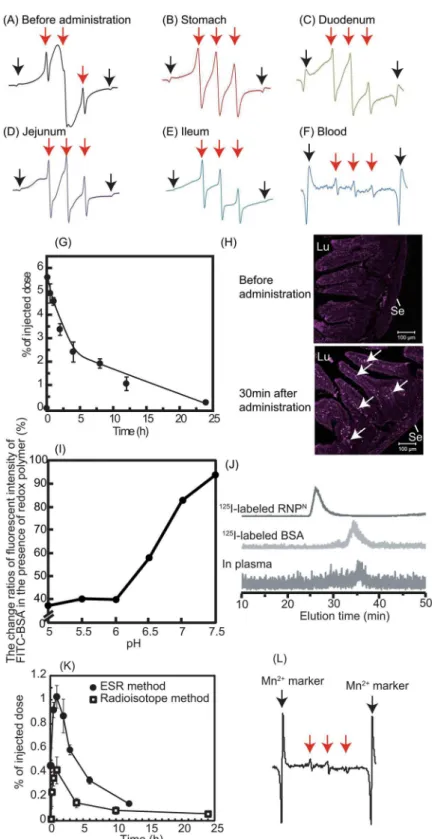
diffusion of RNPOacross the intestinal mucus layer to reach the epithelium is difficult, given its size of 40 nm [7,21]. Contrary to the pH-insensitive RNPO, the redox polymer after disinte-gration of RNPNwas internalized deeply in the villi across the intestinal epithelium. Consider-ing both the ESR spectra shown inFig 2Fand the fluoresceint signal of Cy5.5-labeled redox polymer in the villi shown inFig 2H, it seems that the redox polymers were absorbed in the bloodstream across the intestinal epithelium. It should be noted that cleaved LMW nitroxide radicals and/or Cy5.5 did not contribute to these results because they were covalently conjugat-ed to the polymer. After internalization of the rconjugat-edox polymer into the bloodstream, its cationic charge of the redox polymer might play an important role in interaction with blood proteins. In order to confirm the interaction between the redox polymer and albumin, which is one of the negatively charged proteins in the blood, model experiments were carried out using fluores-cent-labeled albumin.Fig 2Ishows the change in fluorescent intensity of fluorescein-labeled bovine serum albumin (BSA) in the presence of the redox polymers as a function of pH. Under neutral conditions, almost no change in fluorescent intensity based on the fluorescein-labeled BSA was observed in the presence of redox polymers, which form a core—shell-type RNPNat neutral pH, while a dramatic decrease in fluorescent intensity of fluorescein-labeled BSA was observed with decreasing pH. It is known that nitroxide radicals quench fluorescence when they are in proximity of each other [22]. The observed decrease in the fluorescent intensity means that fluorescent quenching occurred between the fluorescein on albumin and the nitrox-ide radicals of redox polymers by disintegration of RNPNat the acidic pH, indicating that the redox polymer itself interacts with albumin. The interaction between the blood proteins and the redox polymers also was confirmedin vivoby size exclusion chromatography using a radio-isotope detector. As shown inFig 2J,125I-labeled RNPNhad an elution time of 27 min, while the elution time of125I-labeled BSA was 35 min; the lowest chromatogram was obtained from mouse serum at 2 h after oral administration of125I-labeled RNPN. A low level, but definite peak, was observed at approximately 36 min, which provides some evidence that the absorbed redox polymers interact with blood proteins and circulate over a long period, although the mo-lecular weight of each polymer was 10 kDa. On the basis of these data, it was concluded that the long-term circulation of redox polymers was achieved by the formation of a complex be-tween the cationic redox polymers and blood proteins following gradual internalization of the redox polymers through the intestinal epithelium. Next, the uptake of orally administered RNPNto the brain of normal mice was investigated. As shown inFig 2K, the uptake of orally administered RNPNin the brain was measured, both by125I-labeled RNPNand by ESR analy-ses. Small, but definite signals, were observed (0.5–1.0% of injected dose) several hours after oral administration of the RNPN. In our previous study, we found that cationic large molecular weight compounds avoid P-glycoprotein pathway due to the large size and preferable
determined after oral administration of Cy5.5-labeled RNPN. Mice were sacrificed at 0.5 h after oral administration of 1 mL of Cy5.5-labeled RNPNat a dose of 2 mg/mL, and the duodenum section was cut circularly. The localization of Cy5.5-labeled redox polymer in the duodenum was analyzed by fluorescent confocal microscopy (Zeiss LSM 700 under oil immersion; Scale bars = 100μm). Lu and Se in the figure indicate lumen and serosa, respectively. Arrows indicate fluorescent signal of Cy5.5-labeled redox polymer. (I, J) Redox polymers interacted with serum proteins in the bloodstream after oral administration. (I) Interaction between redox polymers and FITC-BSAin vitro, determined by fluorescent quenching of FITC-BSA by nitroxide radical moieties in redox polymers (n = 1). (J) Interaction between redox polymers with serum proteins in the blood. Chromatogram of125I-labeled RNPN(upper chromatogram),125I-labeled BSA (middle chromatogram), and the blood sample after oral administration of125I-labeled RNPN(lower chromatogram). (K) The biodistribution of RNPNin the brain via oral administration using125I-labeled RNPN (white square) and ESR measurement (black circle). The data are expressed as mean±SEM, n = 5. (L) ESR spectrum of redox polymer in the brain at 30 min after oral administration of RNPN(500 mg/kg). Red arrows show the ESR signal of redox polymers. Black arrows show the ESR signal of Mn2+marker.
internalizes via endocytosis pathway [23]. Since MeO-PEG-b-PMNT also possesses cationic PMNT segment, it might avoid drug efflux by P-glycoprotein and be internalized in the brain via endocytosis pathway. In addition, the long-term access of our redox polymers coupled with serum protein to the brain vessel wall by extended blood circulation tendency might increase internalization tendency to the brain. Since redox catalytic species are covalently conjugated to redox polymers as stated above, they were internalized together with the polymers.Fig 2L
shows the triplet ESR signal of redox polymer in the normal brain after oral administration of RNPN, which is strong proof of the delivery of the redox polymer to the brain. Previous studies reported that SAMP8 mice was found to increase permeability of blood-brain-barrier (BBB) [24] and also show P-glycoprotein deficiency [25]. Thus, we assume that the redox catalytic species could be internalized into the brain of SAMP8 mice.
Effect of redox polymers in a mouse model of age-associated deficiency
in learning and memory
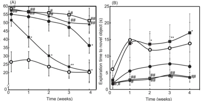
Here, SAMP8 mice were used as a model of age-associated deficiency in learning and memory [26]. The latency period in the Morris water maze test and the exploration time for obtaining novel objects in the object-recognition test were assessed once per week for one month, as shown in Fig3Aand3B, respectively. Compared with SAMR1 mice with normal aging charac-teristics (seeS1 Video), the SAMP8 mice required a longer latency period and showed lesser novel exploration time (P<0.01 for both) owing to the learning and memory deficits (seeS2
Video). After treatment for four weeks, the symptoms did not improve significantly, even after the administration of LMW TEMPOL. In contrast, oral administration of RNPNled to signifi-cant recovery of the symptoms (seeS3 Video). As can be seen inFig 3A, four weeks of
Fig 3. Therapeutic effect of RNPNon cognitive dysfunction.(A) The latency periods of saline-treated SAMR1 mice (open circle), saline-treated SAMP8 mice (open square), blank micelles-treated SAMP8 mice (closed triangle), TEMPOL-treated SAMP8 mice (closed circle), and RNPN-treated SAMP8 mice (closed square) were measured by the Morris water-maze test. The values are expressed as mean±SEM values (n = 10).#P<0.05,##P<0.01, compared with SAMR1 mice.*P<0.05,**P<0.01, compared with SAMP8 control mice. (B) The exploration times of treated SAMR1 mice (open circle), saline-treated SAMP8 mice (open square), blank micelles-saline-treated SAMP8 mice (closed triangle), TEMPOL-saline-treated SAMP8 mice (closed circle), and RNPN-treated SAMP8 mice (closed square) were measured by the object-recognition test. The values are expressed as mean±SEM values (n = 10).#P<0.05,##P<0.01,
compared with SAMR1 mice.*P<0.05,**P<0.01, compared with SAMP8 control mice.
treatment with RNPNdrastically decreased in escape latency time from 51.68 s to 20.96 s, which reached same level as SAMR1 mice (20.4 s at four weeks). In addition, surprisingly, after two weeks of treatment, the RNPN-treated group had evidently higher novel exploration times than those of the saline-treated group; exploration times of RNPN-treated SAMP8 and SAMR1 mice are 17 s and 13 s at four weeks, respectively (seeFig 3B). These results demonstrated that the redox polymers were effective in improving the learning ability of SAMP8 mice.
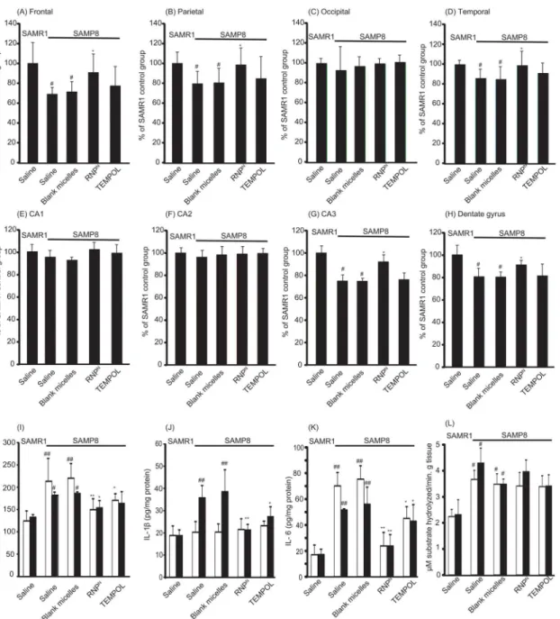
We determined the effect of RNPNon neuron density in various sub-regions of the cortex and the hippocampal areas, which play important roles in learning and memory. It was found that, in the brain of SAMP8 mice, neuron densities significantly decreased in the frontal, parie-tal, and temporal areas of the cortex and in the CA3 and dentate gyrus of the hippocampus (P<0.05 for all). It is interesting to note that the neuron densities of the RNPN-treated SAMP8 mice were almost the same as those of the SAMR1 mice, as shown in Fig4A–4H. The results clearly demonstrated that the redox polymer significantly attenuated neurodegeneration.
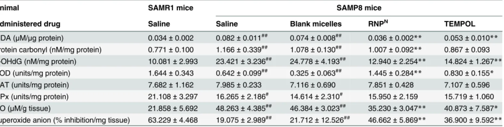
To evaluate the oxidative stress state in the brain of SAMP8 mice, we measured the levels of malondialdehyde (MDA), protein carbonyl, 8-hydroxy-20-deoxyguanosine (8-OHdG), and
ni-tric oxide (NO), as well as superoxide scavenging activities (% inhibition of superoxide anion) and the levels of SOD, GPx, and CAT, in the brain after treatment. As shown in Tables1and2, MDA, protein carbonyl, 8-OHdG, and NO were significantly higher in the brains of SAMP8 mice than those of SAMR1 mice (P<0.01 for all). Treatment with redox polymer significantly decreased levels of MDA, protein carbonyl, 8-OHdG, and NO, compared with SAMP8 control mice (P<0.01 for all). Compared with SAMR1 mice, SAMP8 mice showed a significant de-crease of 86.9% (P<0.01), 16.6% (P<0.05), and 21.3% (P<0.05) in the activities of SOD, CAT, and GPx in the cortex area of the brain, respectively. Treatment with redox polymer markedly increased only SOD activity in the brain of SAMP8 mice (P<0.01). In fact, the activ-ities of superoxide scavenging in the brain after treatment with redox polymers are higher than those of TEMPOL-treated mice. In addition, levels of inflammatory cytokines in the brain after one month of treatment were measured. Compared with SAMR1 mice, SAMP8 mice have higher levels of the pro-inflammatory cytokines, TNF-α, IL-1β, and IL-6, especially in the hip-pocampus area, as shown in Fig4I–4K, respectively. Treatment with redox polymer decreased the levels of those pro-inflammatory cytokines. Since changes within the cholinergic systems so far have been reported to be involved in cognitive and behavioral functions that are widely disturbed in AD [27], we evaluated the effects of redox polymers on AChE activity in SAMP8 mice. As shown inFig 4L, however, we did not find the amelioration of AChE activity in the cortex and hippocampus areas by treatment with redox polymer, indicating that the therapeu-tic effect of oral administration of RNPNis attributable to the suppression of oxidative stress, but not the AChE pathway.
Toxicity of orally administered RNP
Nfor treatment of
senescence-accelerated prone mice
redox polymer did not show this adverse effect. In addition, no deaths were observed throughout the experimental period, which is in sharp contrast to LMW TEMPOL. There were no significant differences in the weights of body and major organs (liver, spleen, kidney, lung, testicle and heart) of animals in all the treatment groups (see Table A inS1 File). Gross pathological changes due to administration of any of the substances were not observed in any of the organs. Histopathological examination of the organs did not show any abnormalities
Fig 4. The density of surviving neurons in SAMP8 mice was increased by oral administration of RNPN.The densities of surviving neurons in various subregions of (A) frontal, (B) parietal, (C) occipital, (D) temporal, (E) CA1, (F) CA2, (G) CA3, and (H) dentate gyrus in the brain of SAMP8 mice were assessed by cresyl violet staining. The values are expressed as the mean±SEM values (n = 5).#P<0.05, compared with SAMR1 mice.
*P<0.05, compared with SAMP8 control mice. (I-K) Levels of proinflammatory cytokines of (I) TNF-α, (J) IL-1βand (K) IL-6 in the cortex (white bar) and hippocampus (black bar) areas of the brain of SAMR1 and SAMP8 mice are shown. Values are expressed as mean±SEM (n = 10).#P<0.05,##P<0.01, compared with SAMR1 mice.*P<0.05, compared with SAMP8 control mice. (L) Effect of redox polymer nanotherapeutics on acetylcholinesterase (AChE) activity in the SAMP8 mice brain. Values are expressed as mean±SEM (n = 10).#P<0.05 compared with SAMR1 mice.
and conspicuous damages in tissues (see Fig B inS1 File). Regarding hepatic function, as shown inFig 5B, it was found that SAMP8 mice showed higher serum levels of both AST and ALT than those of SAMR1 mice (P<0.01 and P<0.05, respectively), which corresponds to a previous report [28]. When RNPNwas orally administered to SAMP8 mice, these levels tended to decrease (P<0.05 in the level of ALT), which is promising in terms of liver protec-tion. In our current study, we have confirmed that orally administered RNPNshows thera-peutic effect of non-alcoholic steatohepatitis because redox polymers are delivered to the liver (Eguchi et al., submitted for publication).
Table 1. The levels of MDA, protein carbonyl, 8-OHdG, NO, superoxide scavenging activity (% inhibition of superoxide anion), antioxidant enzyme activity of SOD, catalase and GPx in the cortex area of SAMP8 mice.
Animal SAMR1 mice SAMP8 mice
Administered drug Saline Saline Blank micelles RNPN TEMPOL
MDA (μM/μg protein) 0.037±0.06 0.114±0.029## 0.106±0.041## 0.040±0.020** 0.075±0.025*
protein carbonyl (nM/mg protein) 0.718±0.090 1.483±0.275## 1.648±0.256## 0.820±0.111
** 1.122±0.321*
8-OHdG (nM/mg protein) 6.950±1.614 15.441±2.567## 14.270±1.985## 11.284±3.775
** 10.856±1.366**
SOD (units/mg protein) 1.025±0.119 0.134±0.027## 0.113±0.04## 0.852±0.04** 0.335±0.06* CAT (units/mg protein) 7.583±0.960 6.325±0.823# 5.695±0.536# 6.115±0.378 7.116±1.659*
GPx (units/mg protein) 18.754±1.904 14.755±1.392# 13.924±1.730# 14.177±1.275 11.720±0.747 NO (μM/g tissue) 18.387±3.754 47.081±5.551## 45.121±3.483## 30.664±9.534
** 39.571±4.613*
superoxide anion (% inhibition/mg tissue) 63.878±4.61 17.827±6.427## 15.326±4.896## 54.021±6.751** 37.133±8.532**
(n = 5) #P<0.05 ##
P<0.01, compared with SAMR1 mice
*P<0.05
**P<0.01, compared with SAMP8 control mice
doi:10.1371/journal.pone.0126013.t001
Table 2. The levels of MDA, protein carbonyl, 8-OHdG, NO, superoxide scavenging activity (% inhibition of superoxide anion), antioxidant enzyme activity of SOD, catalase and GPx in the hippocampus area of SAMP8 mice.
Animal SAMR1 mice SAMP8 mice
Administered drug Saline Saline Blank micelles RNPN TEMPOL
MDA (μM/μg protein) 0.034±0.002 0.082±0.011## 0.074±0.008## 0.036±0.002
** 0.053±0.010**
protein carbonyl (nM/mg protein) 0.771±0.100 1.166±0.339## 1.078±0.130## 1.007±0.092** 0.867±0.093 8-OHdG (nM/mg protein) 10.081±2.993 23.421±3.236## 24.778±4.193## 12.940±2.254** 14.824±1.267**
SOD (units/mg protein) 1.644±0.343 0.642±0.099## 0.325±0.063## 1.445±0.284
** 0.830±0.155*
CAT (units/mg protein) 7.682±1.162 7.985±0.233 7.116±0.690 7.851±0.428 7.107±0.596 GPx (units/mg protein) 21.108±3.297 16.265±2.186# 14.614±2.310# 15.950±2.159 15.719±1.060 NO (μM/g tissue) 21.858±5.692 48.263±4.385## 46.384±3.023## 35.230±3.047** 40.873±7.587*
superoxide anion (% inhibition/mg tissue) 63.229±4.468 19.075±2.989## 21.712±12.526## 46.662±5.869
** 36.900±9.592**
(n = 5) #P<0.05
##P<0.01, compared with SAMR1 mice
*P<0.05
**P<0.01, compared with SAMP8 control mice
Discussion
In this study, we newly developed oral redox polymer nanotherapeutics for treatment of chron-ic diseases. It was confirmed that the redox polymer was absorbed into the blood after disinte-gration of the nanoparticle in the stomach through the intestinal epithelium as expected, followed by delivery of redox polymer to the brain. The oral administration of RNPNto the SAMP8 mice for one month exhibited an improvement of cognitive function without adverse effects by suppressing oxidative stress in the brain, unlike treatment with LMW TEMPOL.
One of the important characteristics of redox polymer nanotherapeutics is long-term circu-lation of redox polymer in the bloodstream after oral administration of RNPN. The brain deliv-ery of nanoparticle with ability of long-term blood circulation has been so far reported by several research groups using normal mice [29–32]. Similar to the EPR effect, the continuous access of the nanoparticles to the cerebral blood vessels due to long-term blood circulation might increase its uptake in the brain due to the fairly large access area of cerebral blood vessel walls. In fact, we could confirm internalization of the redox polymers even in brain of normal mice, which indicates that this continuous access tendency might work well (see Fig2Kand
2L). In addition, since it was reported that aged SAMP8 mice have vessels with a disrupted BBB and even IgG with 150 kDa are internalized in the brain, more redox polymers might be delivered to the brain of SAMP8 mice [24]. Although, for treatment of brain deseases, intrave-nously [33] and intranasally [34] administered nanomedicines have been reported, there are no reports of orally administered nanomedicine. Moreover, the LMW anti-oxidative therapies failed due to its easily internalize in healthy cells and disruption of normal redox reaction such as electron transport chain. This limits application using enough amount of LMW drug admin-istration. Here, we emphasize that this redox polymer nanotherapeutics is the first concept of orally injectable nanomedicine using the pH-responsiveness of redox polymers. In addition,
Fig 5. Measurement of adverse effects.(A) Effects of RNPNon blood pressure of SAMP8 mice in tail-cuff blood pressure method. Blood pressures before administration (white bar), after single administration (black bar), one week after starting treatment (red bar), two weeks after starting treatment (green bar), three weeks after starting treatment (yellow bar), and four weeks after starting treatment (blue bar) are shown. Values are expressed as mean±SEM (n = 10).*P<0.05 compared with SAMP8 control mice. (B) Effects of redox polymer nanotherapeutics on AST (white bar) and ALT (black bar) levels of SAMP8 mice. Values are expressed as mean±SEM (n = 10).#P<0.05,##P<0.01 compared with SAMR1 mice.
*P<0.05 compared with SAMP8 control mice.
since redox polymers suppress the systemic oxidative stress after oral administration of RNPN, it must possess the potential to decrease the risk of various oxidative stress-related diseases.
Conclusions
In conclusion, we demonstrate that orally administered pH-sensitive redox nanoparticles al-most completely revived the cognition in 17-week-old SAMP8 mice (Fig3Aand3B). Small, but evident, amounts of the redox polymer were internalized in the brain of normal mice (as shown in Fig2Kand2L). After treatment with redox nanoparticles, ROS levels were decreased significantly in the brain of SAMP8 mice (as shown in Tables1and2), which is probably due to the long access of redox polymers to blood vessel in brain. The scavenging of ROS in the brain prevented oxidative stress and resulted in recovery of endogenous anti-oxidative enzymes (Tables1and2), thus protecting neuronal cells effectively (Fig4A–4H). In addition, orally ad-ministered redox polymers did not show any detectable toxicity to main organs (Fig5Aand5B
and Fig B and Table A inS1 File).
Supporting Information
S1 File. Text of further experimental procedures, Fig A and B, and Table A. (DOCX)
S1 Video. The Morris water-maze test of SAMR1 mice. (MOV)
S2 Video. The Morris water-maze test of SAMP8 mice. (MOV)
S3 Video. The Morris water-maze test of RNPN-treated SAMP8 mice. (MOV)
Author Contributions
Conceived and designed the experiments: YN PC TY. Performed the experiments: PC TY LBV NI YO. Analyzed the data: PC TY LBV YN. Contributed reagents/materials/analysis tools: PC TY LBV NI YO. Wrote the paper: PC TY YN.
References
1. Okatani Y, Wakatsuki A, Reiter RJ, Miyahara Y. Melatonin reduces oxidative damage of neural lipids and proteins in senescence-accelerated mouse. Neurobiol Aging. 2002; 23(4):639–44. Epub 2002/05/ 16. S0197458002000052 [pii]. PMID:12009513.
2. Dysken MW, Sano M, Asthana S, Vertrees JE, Pallaki M, Llorente M, et al. Effect of vitamin E and mem-antine on functional decline in Alzheimer disease: the TEAM-AD VA cooperative randomized trial. Jama. 2014; 311(1):33–44. Epub 2014/01/02. doi:10.1001/jama.2013.282834PMID:24381967; PubMed Central PMCID: PMC4109898.
3. Grundman M. Vitamin E and Alzheimer disease: the basis for additional clinical trials. The American journal of clinical nutrition. 2000; 71(2):630S–6S. Epub 2000/02/19. PMID:10681271.
4. Kim BYS, Rutka JT, Chan WCW. Nanomedicine. New England Journal of Medicine. 2010; 363 (25):2434–43. doi:10.1056/NEJMra0912273PMID:21158659.
5. Matsumura Y, Maeda H. A new concept for macromolecular therapeutics in cancer chemotherapy: mechanism of tumoritropic accumulation of proteins and the antitumor agent smancs. Cancer research. 1986; 46(12 Pt 1):6387–92. Epub 1986/12/01. PMID:2946403.
7. Vong LB, Tomita T, Yoshitomi T, Matsui H, Nagasaki Y. An orally administered redox nanoparticle that accumulates in the colonic mucosa and reduces colitis in mice. Gastroenterology. 2012; 143(4):1027– 36. doi:10.1053/j.gastro.2012.06.043PMID:22771506
8. Nagasaki Y. Nitroxide radicals and nanoparticles: a partnership for nanomedicine radical delivery. Ther Deliv. 2012; 3(2):165–79. PMID:22834195
9. Yoshitomi T, Miyamoto D, Nagasaki Y. Design of core—shell-type nanoparticles carrying stable radi-cals in the core. Biomacromolecules. 2009; 10(3):596–601. doi:10.1021/bm801278nPMID:19191564 10. Yoshitomi T, Nagasaki Y. Nitroxyl radical-containing nanoparticles for novel nanomedicine against oxi-dative stress injury. Nanomedicine (Lond). 2011; 6(3):509–18. Epub 2011/05/06. doi:10.2217/nnm.11. 13PMID:21542688.
11. Yoshitomi T, Hirayama A, Nagasaki Y. The ROS scavenging and renal protective effects of pH-respon-sive nitroxide radical-containing nanoparticles. Biomaterials. 2011; 32(31):8021–8. doi:10.1016/j. biomaterials.2011.07.014PMID:21816462
12. Marushima A, Suzuki K, Nagasaki Y, Yoshitomi T, Toh K, Tsurushima H, et al. Newly synthesized radi-cal-containing nanoparticles enhance neuroprotection after cerebral ischemia-reperfusion injury. Neu-rosurgery. 2011; 68(5):1418–25. doi:10.1227/NEU.0b013e31820c02d9PMID:21273921
13. Chonpathompikunlert P, Fan CH, Ozaki Y, Yoshitomi T, Yeh CK, Nagasaki Y. Redox nanoparticle treat-ment protects against neurological deficit in focused ultrasound-induced intracerebral hemorrhage. Nanomedicine. 2012; 7(7):1029–43. doi:10.2217/nnm.12.2PMID:22394184
14. Monti E, Supino R, Colleoni M, Costa B, Ravizza R, Gariboldi MB. Nitroxide TEMPOL impairs mito-chondrial function and induces apoptosis in HL60 cells. J Cell Biochem. 2001; 82(2):271–6. PMID: 11527152
15. Shimizu M, Yoshitomi T, Nagasaki Y. The Behavior of ROS-Scavenging Nanoparticles in Blood. Jour-nal of Clinical Biochemistry and Nutrition. 2014; 54(3):166–73. doi:10.3164/jcbn.13-85PMID: 24895479
16. Vong LB, Yoshitomi T, Matsui H, Nagasaki Y. Development of an oral nanotherapeutics using redox nanoparticles for treatment of colitis-associated colon cancer. Biomaterials. 2015:in press.
17. Chonpathompikunlert P, Yoshitomi T, Han J, Isoda H, Nagasaki Y. The use of nitroxide radical-contain-ing nanoparticles coupled with piperine to protect neuroblastoma SH-SY5Y cells from Abeta-induced oxidative stress. Biomaterials. 2011; 32(33):8605–12. doi:10.1016/j.biomaterials.2011.07.024PMID: 21855995
18. Morley JE. The SAMP8 mouse: a model of Alzheimer disease? Biogerontology. 2002; 3(1–2):57–60. Epub 2002/05/17. PMID:12014843.
19. Morley JE, Armbrecht HJ, Farr SA, Kumar VB. The senescence accelerated mouse (SAMP8) as a model for oxidative stress and Alzheimer's disease. Bba-Mol Basis Dis. 2012; 1822(5):650–6. doi:10. 1016/J.Bbadis.2011.11.015. WOS:000302486400006. PMID:22142563
20. Yoshitomi T, Suzuki R, Mamiya T, Matsui H, Hirayama A, Nagasaki Y. pH-sensitive radical-containing-nanoparticle (RNP) for the L-band-EPR imaging of low pH circumstances. Bioconjug Chem. 2009; 20 (9):1792–8. Epub 2009/08/19. doi:10.1021/bc900214fPMID:19685867.
21. Sha S, Vong LB, Chonpathompikunlert P, Yoshitomi T, Matsui H, Nagasaki Y. Suppression of NSAID-induced small intestinal inflammation by orally administered redox nanoparticles. Biomaterials. 2013; 34(33):8393–400. Epub 2013/07/31. doi:10.1016/j.biomaterials.2013.06.032PMID:23896000. 22. Zhu P, Clamme JP, Deniz AA. Fluorescence quenching by TEMPO: a sub-30 A single-molecule ruler.
Biophysical journal. 2005; 89(5):L37–9. Epub 2005/10/04. doi:10.1529/biophysj.105.071027PMID: 16199509; PubMed Central PMCID: PMC1366861.
23. Oishi M, Hayashi H, Iijima M, Nagasaki Y. Endosomal release and intracellular delivery of anticancer drugs using pH-sensitive PEGylated nanogels. J Mater Chem. 2007; 17:3720–5.
24. Pelegri C, Canudas AM, del Valle J, Casadesus G, Smith MA, Camins A, et al. Increased permeability of blood-brain barrier on the hippocampus of a murine model of senescence. Mech Ageing Dev. 2007; 128(9):522–8. Epub 2007/08/19. doi:10.1016/j.mad.2007.07.002PMID:17697702.
25. Zhang G, Zhang B, Fu X, Tomozawa H, Matsumoto K, Higuchi K, et al. Senescence-Accelerated Mouse (SAM) strains have a spontaneous mutation in the Abcb1a gene. Experimental Animals. 2008; 57(4):413–7. doi:10.1538/expanim.57.413PMID:18633165
26. Takeda T. Senescence-accelerated mouse (SAM) with special references to neurodegeneration mod-els, SAMP8 and SAMP10 mice. Neurochem Res. 2009; 34(4):639–59. Epub 2009/02/10. doi:10.1007/ s11064-009-9922-yPMID:19199030.
28. Ye X, Meeker HC, Kozlowski PB, Wegiel J, Wang KC, Imaki H, et al. Pathological changes in the liver of a senescence accelerated mouse strain (SAMP8): a mouse model for the study of liver diseases. Histol Histopathol. 2004; 19(4):1141–51. Epub 2004/09/18. PMID:15375757.
29. Han LM, Guo J, Zhang LJ, Wang QS, Fang XL. Pharmacokinetics and biodistribution of polymeric mi-celles of paclitaxel with Pluronic P123. Acta pharmacologica Sinica. 2006; 27(6):747–53. Epub 2006/ 05/26. doi:10.1111/j.1745-7254.2006.00340.xPMID:16723095.
30. Wang Y, Li Y, Wang Q, Wu J, Fang X. Pharmacokinetics and biodistribution of paclitaxel-loaded pluro-nic P105/L101 mixed polymeric micelles. Yakugaku zasshi: Journal of the Pharmaceutical Society of Japan. 2008; 128(6):941–50. Epub 2008/06/04. PMID:18520140.
31. Jose S, Anju SS, Cinu TA, Aleykutty NA, Thomas S, Souto EB. In vivo pharmacokinetics and biodistri-bution of resveratrol-loaded solid lipid nanoparticles for brain delivery. International journal of pharma-ceutics. 2014; 474(1–2):6–13. Epub 2014/08/08. doi:10.1016/j.ijpharm.2014.08.003PMID:25102112. 32. Md S, Ali M, Baboota S, Sahni JK, Bhatnagar A, Ali J. Preparation, characterization, in vivo
biodistribu-tion and pharmacokinetic studies of donepezil-loaded PLGA nanoparticles for brain targeting. Drug de-velopment and industrial pharmacy. 2014; 40(2):278–87. Epub 2013/02/02. doi:10.3109/03639045. 2012.758130PMID:23369094.
33. Estevez AY, Pritchard S, Harper K, Aston JW, Lynch A, Lucky JJ, et al. Neuroprotective mechanisms of cerium oxide nanoparticles in a mouse hippocampal brain slice model of ischemia. Free radical biology & medicine. 2011; 51(6):1155–63. Epub 2011/06/28. doi:10.1016/j.freeradbiomed.2011.06.006PMID: 21704154.