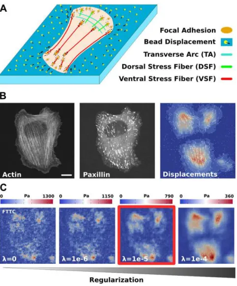
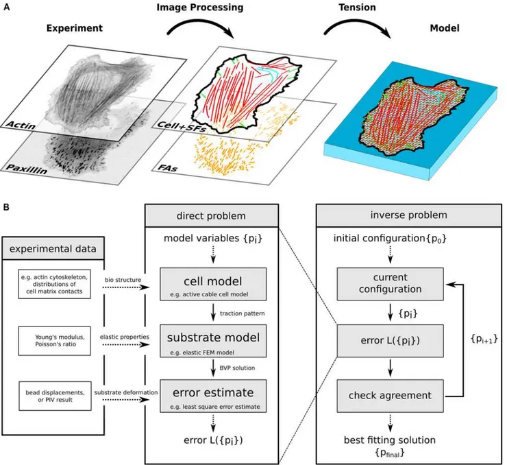
Model-based traction force microscopy reveals differential tension in cellular actin bundles.
Texto
Imagem


Documentos relacionados
This research paper was developed with the purpose to represent theoretical and practically the way of governance of an open-privately owned corporation, innovation being found
However, the agglomeration force deriving from spatial economies of scope (leading to a single-plant pattern) might be more than offset by the opposing forces of dispersion,
Based on the estimated value for the initial straining force at the hy - draulic jack (table 2) and on the tendon tension loss shown in table 3, the remaining force at the
In the case of tension dominant states without activation of dam- age processes in previous compression, the original version of the damage model is recovered.. It can be veriied
Time course studies of dry cell biomass formation, surfactin production, surface tension reduction and substrate utilization by the culture media of Bacillus subtilis MTCC
At lower mole fractions of CTAB, the mixed solution interface is excessively covered by DDPO molecules than CTAB molecules, as shown from the surface tension values of the
Figure 17 - LDH enzymatic activity in human prostate PNT1A epithelial cells after exposure to 20 μg/mL of saco (July crop) cherry for 72 hours, determined by spectrophotometric
dF Elemento de fora interfacial no domnio lagrangiano df Elemento de fora interfacial no domnio euleriano F Interface tension force in the Lagrangian domain f Interface tension force
